Contents
Page
List .of Tables………... I
List of Figures……….. II
List of Mathematical equations……..…...………….……… V
I) INTRODUCTION………...………... 1
1. Human African trypanosomiasis (HAT)………. 1
2. Life Cycle of Trypanosoma brucei. Early and Late Symptoms…... 2
3. Epidemiological Studies………. 3
4. Drugs and Target………. 4
5. Structure of 6- phosphogluconate dehydrogenase (6PGDH)……...……….. 10
5.a Primary structure………. 10
5.b The 3-dimensional struc……….. 11
5.c The T brucei monom……… 12
5.d Coenzyme binding-site……… 13
5.e The dimer interface………. 16
5.f Substrate binding site……….. 17
6. Action mechanism of 6-phosphogluconate dehydrogenase….………... 19
6.a The two key residues………... 19
6.b.1 Asymmetry ……….…………... 20
6.b.2 Other 6PG-induced effects………... 21
6.b.3 Isomerization and allostery………... 22
6.b.4 Other significant effects along the reaction……... 23
II) AIM OF THE THESIS……….……….….. 24
1. Structural Studies ………..…... 24
2. Functional Studies………...…….……… 24
III) MATHERIALS AND METHODS………..….. 26
1. Site-Directed Mutagenesis ……….……….... 26
2. Overexpression of the T. brucei enzyme in E. coli……….... 29
3. Purification of 6-PGDH wt / mutant of T. brucei by bacteria E. coli ..……….. 29
4. Determination of protein6PGDH wt/mutand concentration….……….………. 30
5. Enzyme activity. Assay of activity for 6PGDH………..………... 31
6. Cysteines reactivity……….…….………... 31
7. Polyacrylamide gel electrophoresis (SDS PAGE)………….….……… 31
8. Gel Filtration (SEPHACRYL S 200 and AcA 34)………...……….….. 34
9. Chemical cross-linking of 6PGDH……….…….……... 35
10. Dynamic Light scattering (DLS) measurements………...………... 38
11. Sucrose-density-gradient centrifugation………...………. 41
12. Isothermal titration calorimetry (ITC) studies…………...….………. 42
12.a Preparation of the sample and the ligand……….….. 44
12. a.1 enzyme preparation………. 44
12.a.2 ligand preparation……… 45
12.c Data processing using software provided by the OriginTM.……. 46
12.c.1 Changes in heat capacity ( ΔCp°)... 48
12.c.2 Effect of pH on dissociation constant ………. 49
12.c.3 ) Effect of pH on binding enthalpy……….………… 49
IV) RESULTS, ANALYSIS AND CONCLUSIONS
Part I. Structural studies.
Results and Discussion……….….……… 52
a) Gel-filtration of 6PGDH……….……… 52
b) Velocity Sedimentation of 6PGDH……….…... 53
c) Glutaraldehyde cross-linking of 6PGDH……….... 54
d) Dynamic light scattering………. 57
e) Calculation of heat capacity change (ΔCp ) by microcalorimetry……….... 57
f) Dimer-tetramer equilibrium in 6PGDH mutants……….… 60
Conclusion……….……… 61
Part II. Functional studies.
Results and Discussio……….………... 63
a) Effect of mutation on the enzyme activity………. 64
a.1) E192Q………..……… 64 a.2) K185H……….. 64 a.3) H188L ………. 65 b) Cysteine reactivity………. 65 c) 6PG binding to 6PGDH……….…… 67 c.1) The binding of 6PG to 6PGDH WT……….….... 67
c.2) The binding of 6PG to E192Q mutant……….……… 69
c.3) The binding of 6PG to K185H mutant………. 70
c.4) The binding of 6PG to H188L mutant………. 70
Conclusion ………... 75
V) REFERENCES ….………..………. 76
I
List of Tables
Pag
INTRODUCTION
Table 1: HAT drugs in clinical use……….. 5
Table 2: 6PGDH inhibition constants of compounds……...……….…….. 10
MATERIALS AND METHODS Table 3. Primer oligonucleotides synthetized ……….……….... 27
Table 4: The protocol Mutagenesis reaction……….…... 27
Table 5: The temperature cycling. Mutagenesis reaction……… 27
Table 6: the stacking gel solution 5% (w/v). SDS PAGE……….……….. 33
Table 7: Coomassie blue C250 without methanol for SDS-PAGE……..………... 34
Table 8: pK and ΔH ionization of buffers used for Isothermal titration calorimetry (ITC) studies……….…. 46
RESULTS ANALYSIS AND CONCLUSIONS I Table 9: DLS measurements of mean hydrodynamic radius. ……… 57
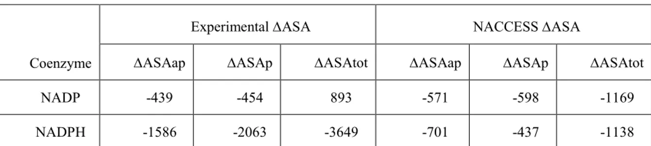
Table 10: Comparison between the changes in the subunit polar and apolar solvent exposed surface area (ΔASAp and ΔASAap in Å2)….……….. 59
Table 11: Binding parameters of NADP to 6PGDH from Trypanosoma brucei ……….………….. 61
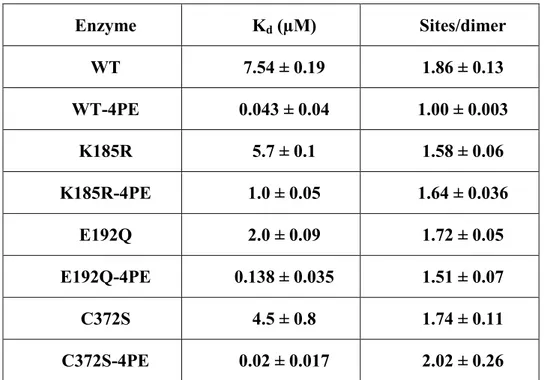
RESULTS ANALYSIS AND CONCLUSIONS II Table 12. Intrinsic Kd, pKa and number of proton exchanged at the 6PG binding……….. 67
Table 13. Buffer-independent enthalpy change and number of hydrogen ions exchanged at different pH in the mutant E192Q at the 6PG binding………... 69
II
List of Figure
Pag INTRODUCCTION
Figure 1: WHO Report on Global Surveillance of Epidemic –prone Infectious
Diseases - African trypanosomiasis ………. 4
Figure 2: Pentose Phosphate Pathway……….………… 7
.. Figure 3: Reaction catalyzed by 6-PGDH and the two main amino acid residues involved ………... 8
Figure 4: Structures of some substrate analogues of 6PGDH………... 9
Figure 5: Multiple alignment of 6PGDH from several species ………...…... 12
Figure 6: Monomer of the T. brucei 6PGDH ………... 13
Figure 7: Coenzyme binding site of the T. brucei 6PGDH ………... 14
Figure 8: Dimer of the T. brucei 6PGDH ………. 16
Figure 9: Substrate binding site of the T. brucei 6PGDH ……… 17
Figure 10: T. brucei 6PGDH active site with the position of the two residues, discussed in the introduction …………... 19
Figure 11:. Half of the site mechanism. ………... 20
III MATERIALS AND METHODS
Figure 13: Scheme of the QuickChange II site-directed mutagenesis method ……… 26 Figure 14: Reaction of the amino groups of proteins with glutaraldehyde……… 36 Figure 15: Formation of pyridine rings after the reaction of glutaraldehyde
with amino groups of proteins……….. 37
Figure 16: Summary of the possible forms of glutaraldehyde in aqueous
solution……….……….. 37
Figure 17 : Collecting of the fractions after the density-gradient centrifugation... 42 Figures 18 :Schematic representation of a VP-ITC microcalorimeter and the
syringe……… 43
RESULTS ANALYSIS AND CONCLUSIONS I
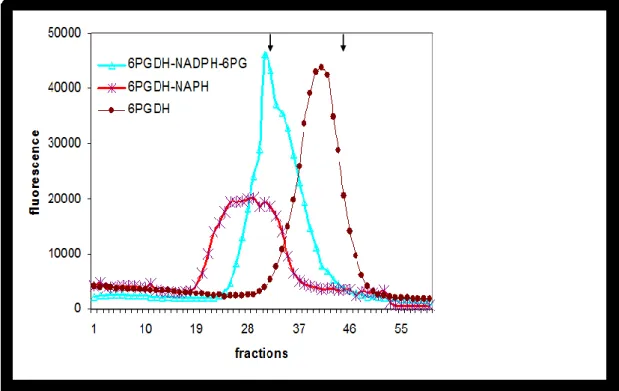
Figure 19: Gel-filtration profileofT. brucei 6PGDHwith and without either
NADPH or other ligands………..……. 52
Figure 20. Sedimentation of T. brucei 6PGDH with and without 0.02 mM NADPH
or with both NADPH and 0.16 mM 6PG ……….. 53
Figure 21. Enzyme concentration dependence of the association reaction between
dimers to form tetramer for T. brucei 6PGDH, in absence or presence
of 0.02 mM NADPH……… 54
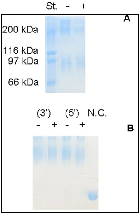
Figure 22. SDS-PAGE at 7,5 % acrylamide in the resolving gel, of chemically
cross-linked T. brucei 6PGDH, in the absence (A) and presence (B)
of 40 µM NADPH, with glutaraldehyde……… 55
Figure 23. SDS-PAGE of T. brucei 6PGDH, treated for different times with
IV
Figure: 24. T. brucei 6PGDH titration with NADP by ITC, in
Hepes buffer, pH 7.5 ... 58
Figure 25. Buffer-independent enthalpy change (ΔHo) dependence
from the temperature for the binding to T. brucei 6PGDH of NADP
and NADPH………..………. 59
RESULTS ANALYSIS AND CONCLUSIONS II
Figure 26. pKm dependence from pH for 6PG in T. brucei 6PGDH ……… 63
Figure 27. Kinetics of the mutant K185H.………... 65
.
Figure 28. The cysteines reactivity in absence and in presence of 6PG for
WT and H188L mutant………..……… 66
Figure 29. pH dependence for 6PG apparent pKd………. 68
Figure 30.Scheme with three residues involved in the change of the protonation
state of the enzyme at the substrate binding………...………. 68
Figure 31: Kinetic mechanism of 6PGDH……… 71 Figure 32: Scheme for the pKa shifts for ionizable residues in the
V
List of Mathematical Equations
MATERIALS AND METHODS Pag
Dinamic Light Scatering Data processing
Equation 1: The wave vector……….…………... 38
Equation 2: Correlation function. of the scattering signal………. 39
Equation 3: equation for single exponential decay for the correlation function……….. 39
Equation 4: The Stokes-Einstein relation……….. 40
ITC Data processing Equation 5: the Gibbs free energy……… 46
Equation 6: the Gibbs free energy ……….. 47
Equation 7: Observed enthalpy equation (Hobs )………. 47
Equation 8: Number of hydrogen ions released or taken………. 47
ITC Data processing. Changes in heat capacity ( ΔCp°) Equation 9: Changes in heat capacity (ΔCp) from exposed surfaces variations………..……….…. 48
Equation 10: ΔH reference value obtained at the temperature (60 °C)…..…… 48
ITC Data processing . Effect of pH on dissociation constant Equation 11: The intrinsic dissociation constant (Kapp) ……….. 49
ITC Data processing Effect of pH on binding enthalpy Equation 12: The capture/release of hydrogen ions (nH) depending on the pH and pKa………. 49
Equation 13: enthalpy changes as a function of the protonation / deprotonation and the intrinsic enthalpy binding ……… 50
VI
Equation 14: HO transformed………..………... 50
1
INTRODUCTION
1) Human African trypanosomiasis
The Kinetoplastida are a protozoan class belonging to the Excavata supergroup, encompassing numerous medically and agriculturally important pathogens, as well as free-living representatives that have huge ecological impact.4.
Within the kinetoplastida, the order Trypanosomatida contains many pathogens, including Trypanosoma brucei, T. cruzi and Leishmania spp., the causative agents of African trypanosomiasis, Chagas‘ disease and leishmaniasis respectively. These parasites have evolved diverse immune evasion strategies; while Leishmania spp. and T. cruzi exploit intracellular lifestyles by invasion of host cells, T. brucei persists within the host bloodstream and lymphatic system, and is therefore continually exposed to both innate and adaptive immune mechanisms. The surface of mammalian infective T. brucei is dominated by approximately 2 x 10⁷ molecules of a single GPI-anchored variant surface glycoprotein (VSG), shielding invariant surface antigens from antibody recognition4.
Trypanosoma brucei brucei causes the veterinary disease Nagana, but it is unable to establish infections in humans. Human resistance to T. brucei brucei infection is due to the presence of a trypanolytic component of human serum, which provides innate immunity against infection. This component is a minor subfraction of high-density lipoproteins (HDLs) called the trypanosome lytic factor 1 (TLF-1). This toxic class of HDLs is internalized in T. brucei brucei via receptor-mediated endocytosis and is ultimately targeted to the lysosome, where it initiates low-pH-dependent killing. However, T. b. rhodesiense and T. b. gambiense, managed to escape this immunity system, enabling them to grow in humans where they cause sleeping sickness, as Human African Trypanosomiasis (HAT) is called.5
2 The mechanism of resistance to TLF-1 remains to be fully elucidated; however, it is well established that the resistance phenotype of T. brucei gambiense (chronic form of the disease in West Africa- Congo) and T. brucei rhodesiense (acute form in Est Africa- Zimbabwe, Tanzania, Zambia, Angola (Figure 1) is due to the expression of the serum resistance associated (SRA) protein (a member of the VSG gene family). 1.
2) Life Cycle of Trypanosoma brucei. Early and Late Symptoms
Both trypanosomes are morphologically identical and are transmitted to human hosts by bites of infected tsetse flies (Glossina palpalis transmits T. brucei gambiense and Glossina morsitans transmits T. brucei rhodesiense), which are found only in Africa. 2.
Life cycle of the parasites starts when the trypanosomes are ingested during a blood meal by the tsetse fly from a human reservoir in West African trypanosomiasis or an animal reservoir in the East African form. The trypanosomes multiply over a period of 2-3 weeks in the fly midgut; then, the trypanosomes migrate to the salivary gland, where they develop into epimastigotes. The metacyclic trypomastigotes infect humans, following a fly bite, which occasionally causes a skin canker at the site 54.
In the first stage, the trypanosomes multiply in subcutaneous tissues, blood and lymph. This is known as a haemolymphatic phase, which entails bouts of fever, headaches, joint pains and itching.
In the second stage the parasites cross the blood-brain barrier to infect the central nervous system. This is known as the neurological phase. In general this is when more obvious signs and symptoms of the disease appear: changes of behaviour, confusion, sensory disturbances and poor coordination. Disturbance of the sleep cycle, which gives the disease its name, is an important feature of the second stage of the disease. Without treatment,
sleeping sickness is considered fatal. The first stage of T. brucei gambiense sleeping
3
3) Epidemiological studies
Epidemiological studies indicate that this disease threatens millions of people in 36 countries in sub-Saharan Africa. Many of the affected populations live in remote areas with limited access to adequate health services, which hampers the surveillance and therefore the diagnosis and treatment of cases. In addition, displacement of populations, war and poverty are important factors leading to increased transmission and this alters the distribution of the disease due to weakened or non-existent health systems. 8
In 2009, after continued control efforts, the number of cases reported has dropped below 10 000 for the first time in 50 years. This trend has been maintained in 2010 with 7139 new cases reported (WHO). Despite these efforts, the problematic of internal conflicts by armed groups has impaired the work of sanitary control of active outbreaks of disease as well as promoted the mobilization of citizens to areas where they are more exposed to the vector and access to health centre to treat the disease is less or naught.
Additionally cases of African trypanosomiasis have been reported in recent years in travellers coming from various parts of Africa, particularly from the natural parks of Tanzania 6;7
This means that although African trypanosomiasis is endemic in an area of Africa, there is a high risk factor for travellers.
4
Figure 1: WHO Report on Global Surveillance of Epidemic -prone Infectious
Diseases African trypanosomiasis
4) Drugs and target
Currently development of effective vaccines against these parasites remains an unrealized goal and clinical management is based on chemotherapeutics. Cost, toxicity and resistance problems of conventional drugs result in an urgent need to identify and develop new therapeutic alternatives. Traditionally, neglected tropical diseases have not been the focus of robust efforts to identify new drugs due to lack of a profitable market and effective strategies to implement control programmes 9.
The global effort to control the infection is based on a combination strategy of vector control and drug treatment of infected patients. Currently there is no drug that is effective against both stages of the disease or both subspecies, and all therapies require parenteral
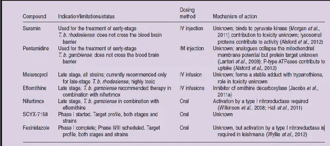
5 administration (Table 1). Early-stage disease is treated with pentamidine (T. b. gambiense) or suramin (T. b. rhodesiense) 9], but both compounds show some toxicity.
Suramin has been reported to inhibit a number of glycolytic enzymes 10, including a recent report that it inhibits pyruvate kinase by binding the ATP site as shown by X-ray structure analysis 11.
Treatment of late-stage disease is more problematic. Historically the highly toxic arsenical compound melarsoprol was used to treat both subspecies of the disease, causing 5–10% fatality in treated patients. However in 2009 a new nifurtimox/eflornithine combination therapy (NECT) was advanced for the treatment of late-stage T. b. gambiense after showing equivalent to better efficacy than eflornithine (DFMO) alone in clinical trials. NECT has not yet been tested against T. b. rhodesiense, and despite improvements over eflornithine alone, administration still requires 7 days of twice daily i.v. infusions of eflornithine along with oral nifurtimox administration. 9.
Table 1: HAT drugs in clinical use.
The organization Drugs for Neglected Diseases Initiative (DNDi) is emphasizing development of new compounds, such as oxaborole SCYX-7158 and fexinidazole for the treatment of both early and late-stage disease and they are currently undergoing clinical trials 9.
Eflornithine target has been shown to be ornithine decarboxylase, an essential enzyme in the biosynthesis of polyamines 12. Regarding nifurtimox, there is good evidence that activation by a type I nitroreductase leading to production of intracellular free radicals, is
6 key to its efficacy 13;16. Analogues of pentamidine have been shown to collapse the mitochondrial membrane potential but the protein targets that mediate these effects are unknown 14. Recent efforts to utilize genome-wide RNAi approaches have led to the identification of genes involved in suramin uptake, and to the identification of several other lysosomal proteins that contribute to its action 15. However, for pentamidine, suramin and melarsoprol, a number of genes were identified that modulate their function, suggesting that clear identification of their molecular targets will not be straightforward 9.
Others proteins such as trypanothione reductase, which are unique for trypanosomes, have been proposed as ideal target [9]. However also other enzymes, which are present both in parasites and in the guests, may be effective targets, if they have structural characteristics that may be subject to selective inhibitors, such the case of ornithine decarboxylase. This was clearly demonstrated also in the case of the enzyme of glycolysis glyceraldehyde-3-phosphate dehydrogenase (GAPDH). Differences in the binding site of the coenzyme (NAD+) were identified using a comparative analysis by X-ray crystallography [10]. Structures adenosine-based, as analogues of the co-factor, have been shown to selectively inhibit the enzymes of trypanosome and also to kill both T. brucei and T. cruzi. [19].
The bloodstream-form of T. brucei has the blood glucose as the sole source of nutrition. For this reason, the glycolysis that is responsible for substrate-level ATP production in the cell, was considered a good target for the development of new drugs. The absence of glucose or incubation of the parasite with inhibitors of certain glycolytic enzymes, leads to a rapid lysis and death of the parasite present in the bloodstream of the host. Also demonstrations based on mathematical models and experiments of gene knock-out revealed that glycolysis is essential and validated this pathway as drug target. [18, 52] .
Glycosomes are a specialized form of peroxisomes (microbodies) present in unicellular eukaryotes that belong to the Kinetoplastea order, such as Trypanosoma and Leishmania species. The organelles harbour most enzymes of the glycolytic pathway 52. Some drug targets may be found in the glycosomes compartments, and in the metabolism of lipids and of purines too [6].
7 The pentose phosphate pathway also plays a crucial role in the metabolism of the parasite and in the host-parasite relationship, since its main function is the production of NADPH, which is necessary for the defense against oxidative stress and for different reductive biosynthetic reactions. (Figure 2).
Figure 2: Pentose Phosphate Pathway. 6PGDH is the third enzyme of
the Pentose Phosphate Pathway (Gupta S et al 2011)
The decrease of the availability of this reduced coenzyme increases susceptibility to oxidative stress and hampers the reductive biosyntheses of the parasite. [20].
6-phosphogluconate dehydrogenase (6PGDH) is the third enzyme of the pentose phosphate pathway, it catalyzes the oxidative decarboxylation of 6-phosphogluconate (6PG) to ribulose-5-phosphate (RU5P), with redox reaction preceding decarboxylation , via 3-keto 6PG and a probable 1, 2-enediol as intermediates (Figure 3)
8 Figure 3: reaction catalyzed by 6-PGDH and the two main amino acid
residues involved
The inhibition of 6PGDH causes an accumulation of 6PG in the cell, which acts as an inhibitor of 6-phosphoglucose isomerase, the enzyme that converts glucose-6-phosphate to fructose-6-phosphate during glycolysis. The accumulation of glucose-6-phosphate in the cell addresses this to enter the pentose phosphate pathway by triggering a positive feedback loop that feeds on itself with fatal consequences for the parasite. [21].
Enzyme inhibition studies have shown that there are strong inhibitors against 6PGDH, which show some selectivity versus the parasite enzyme compared the mammalian one. T. brucei 6PGDH shows only a 33% amino acid identity with the mammalian 6PGDH even if their structures have a similar overall fold and many residues nearest neighbours to the substrate are conserved 22
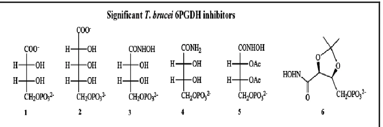
Three reviews have dealt with T. brucei 6PGDH inhibitors as lead compounds for new drugs against African trypanosomes [21,23,53]. Inhibitors have been found, which mimic the transition-state and high-energy intermediates of the enzymatic reaction of 6PGDH [24]. Hydrophobic analogues of these also revealed some anti-parasite activity [25]. A
9 number of phosphorylated carboxylic acids derived from aldose sugars were tested against 6PGDH, two particularly notable inhibitors were identified. Both 4-phospho-D-erythronate (4PE) and 5-phospho-D-ribonate (5PR) (Fig. 4, compounds 1 and 2) were competitive with respect to substrate, with Ki values for the T. brucei 6PGDH equal to 130 and 950 nM, respectively.
Figure 4: Structures of some substrate analogues (Ac=Acetyl group). . 4-phospho-D-erythronate
(compounds 1); 5-phospho-D-ribonate (compounds 2) ; 4-phospho-D-erythronohydroxamate (compound 3), erythronamide (compound 4) and two protected analogues of 4-phospho-D-erythronohydroxamate (compounds 5 and 6)
Their selectivities for the T. brucei 6PGDH over the sheep liver one (ratio Ki sheep/ Ki T. brucei) were measured at 83-fold and 70-fold respectively. Ki values for both are under the Km for 6PG (= 3.5 μM), indicating that they mimic high energy reaction intermediates ( Figure 3) rather than the substrate per se [21,26, 19].
Another potent and selective T. brucei 6PGDH inhibitor is 4-phospho-D-erythronohydroxamate (Figure 4, compound 3), synthesized specifically to mimic the high-energy intermediates produced following the second (decarboxylation) step of the catalyzed reaction, shown in Figure 3 . This hydroxamate, with a Ki = 10 nM and selectivity of 254-fold for the parasite enzyme over the sheep liver enzyme, is the compound with the highest affinity for the T. brucei 6PGDH reported to date and it also shows the highest selectivity for the parasite over the sheep liver enzyme [24]. In Table 2, inhibition constants versus 6PG (Ki) at pH 7.5 (which is the enzyme optimum pH) for all T. brucei 6PGDH inhibitors, which are shown in the figures included in this text, are
10 reported, together with selectivity values over the sheep liver enzyme (ratio sheep liver 6PGDH Ki / T. brucei 6PGDH Ki).
Table 2: 6PGDH inhibition constants of compounds in figure 4.. 4-phospho-D-erythronate
(compounds 1); 5-phospho-D- (compounds 2) ; 4-phospho-Derythronohydroxamate(compound 3) ; erythronamide (compound 4) and two protected analogues of 4-phospho-D-erythronohydroxamate (compounds 5 and 6)
Genetic studies and RNA interference, which leads to the selective suppression of the expression of a gene, are today highly used, also for their extreme speed, to verify the effect of inhibition of an enzyme in the parasite [21]. These studies have indicated that in T. brucei 6PGDH can be a drug target since expression of the gene encoding 6PGDH has been shown essential for growth of T. brucei bloodstream form.
5) Structure of 6-phosphogluconate dehydrogenase
5.a) Primary structure
The comparative study of the amino acid sequence, using alignment, has revealed a 33% identity between the amino acid sequence of T. brucei 6PGDH and mammalian enzyme
11 43 while identity is somewhat higher (37.3%) both with the chloroplast and the cytosolic 6PGDH from spinach [42].
This may reflect the evolutionary history of the kinetoplastids, the phylogenetic order to which trypanosomes belong, which have been proposed to have derived from an ancestor common with the primitive plants Euglenoid algae [40].
Furthermore the closer relationship between the genes of Trypanosoma and those of cyanobacteria and plastids as well as algal and plant genes rather than with those of other eukaryotic lineages can be explained by the fact that some ancestors of trypanosomes housed in endosymbiont prokaryotes from which a number of genes was then acquired by the nucleus of the trypanosome. Some enzymes of the glycolytic pathway and other pathways seem very close to those found in cyanobacteria and plastids [21].
The sequence of 6PGDH from different species, have been aligned using the programs MultAlin [36] e ESPript [37] to show the conserved amino-acids among species. Of the 482 amino-acids that belong to one subunit, 88 are conserved in all species (Figure 5) [46].
5.b) The 3-dimensional structure
The 3-dimensional structure of the T. brucei 6-phosphogluconate dehydrogenase has been solved at 2.8 Å resolution [22]. The sheep liver enzyme, which has 97% sequence identity to the human enzyme, has been described at 2 Å [47] resolution and enzyme-coenzyme and enzyme-substrate complexes have been reported at 2.3 Å -2.5 Å resolution [39]. The structures of Lactococcus lactis 6PGDH in ternary complex with NADP and the product Ru5P or the inhibitor 4-phospho-D-erythronohydroxamic acid (PEX) were also reported [48].
12 Figure 5. Multiple alignment of 6PGDH from several species.
5.c) The T.brucei monomer
Each monomeric subunit weighs 52 kDa and it is formed by 482 amino acid residues. The T. brucei monomer is comprised of three domains or regions (Figure 6):
An N-terminal domain or coenzyme binding (residues 1-178), characterized by a Rossmann fold typical for binding to dinucleotides (residues 1-130, sheets from βA to βF alternating with intervening helices) and a unit α-β-α (residues 132 to 161, αf-βG-αg) with a section βG antiparallel to the first six strands. The coenzyme binding site is at the carboxyl ends of the strands of the parallel sheet, with the two ribose moieties and the bis-phosphate straddling the sheet. The dinucleotide binding fingerprint, at the tight turn following A, is GxGxxG in the T. brucei enzyme while in the ovine enzyme it is
13 GxAxxG as in almost all known 6PGDHs. As the two subunits of the T. brucei dimer were not crystallographically equivalent, slightly different conformations of two loops in the coenzyme domain could be seen [39].
Figure 6: Monomer of the T. brucei 6PGDH
.
Central helical domain "all helix" (residues 179-441) that includes most of the protein, formed only by alpha helices; it forms a part of the interface of the dimer and the binding site to the substrate.
C-terminal "tail" (442-478), of small dimensions which penetrates the central domain of the second subunit, thus completing the binding site to the substrate. (Figure 6).
5.d) Coenzyme binding-site
The coenzyme binding domain of 6PGDH has 70 residues identical in the sheep and T. brucei enzymes (Figure 7), 35 of which are totally conserved.
14 Figure 7. Coenzyme binding site
In the oxidized coenzyme binding site, there are 17 residues within 4 Å from the oxidised coenzyme analogue, nicotinamide-8-bromo-adenine dinucleotide phosphate (Nbr8ADP+) 6 of which are totally conserved and 12 are identical in T. brucei and sheep.
The Km for NADP+ for T. brucei 6PGDH is 1 µM while that for sheep enzyme is several times higher ranging from 5.7-8.9 µM depending on pH and ionic strength [49]. Sequence differences at the coenzyme binding site may affect the affinity for NADP+ between species.
Thus, although most of the binding site is highly conserved, however, some small difference exists. Between these, the most important are the transformation of Ala 11 (sheep) in Gly 10 (T. brucei), of Lys 75 (sheep) in Gln 77 (T. brucei); and Phe 83 (sheep) in Thr 85 (T. brucei) [21]. The mean main-chain movement for the 17 residues of the sheep enzyme on binding the analogue is 0.31 Å; only Lys 75s moves significantly (0.77 Å).
Among the hydrogen bond interactions of the sheep enzyme with oxidised coenzyme, that of the 2‘-phosphate and adenine ribose to triplet Asn 32s, Arg 33s, Thr 34s directly following βB (Asn 31, Arg 32, Thr 33 in T. brucei) is predominant. The additional hydrogen bonds are to the nicotinamide amide function from a conserved methionine of the fingerprint (Met 13s) and from a glutamate of αf (Glu 131s) conserved in 68 of 70 species. All residues with side-chain hydrogen bonds are conserved between sheep and T.
15 brucei, though it should be noted that, in two other species, a tyrosine replaces the arginine 32, (33s), which interacts with the 2‘- phosphate. The most important changes in sequence between the sheep and T. brucei enzymes, which may affect binding are Ala 11s to Gly 10, Lys75s to Gln 77 and Phe 83s to Thr 85. The Cβ of Ala 11s in sheep 6PGDH protrudes into the bis-phosphate binding site. This residue corresponds to the central glycine of the generic fingerprint; the very small number of direct interactions of the bis-phosphate to the protein is almost certainly a consequence of this alanine. The substitution of glycine for alanine in the T. brucei enzyme should allow direct interactions between the enzyme and the NADP+ bis-phosphate and has a further consequence in that the highly conserved valine 11 (Val 12s) faces towards the putative nicotinamide site in T. brucei 6PGDH and away from it in the sheep enzyme. Val 11 would provide further Van der Waals contacts for the nicotinamide ring. The substitution of Gly 10 in the T. brucei enzyme would suggest a tighter binding of coenzyme as reflected in the higher affinity.
All links between the 6PGDH side chains and the coenzyme are highly conserved in different species.
Site-directed mutagenesis experiments in which the residue Arg 33 was mutated to tyrosine have revealed the importance of this residue in the binding of 2'-phosphate, which differs in the NADP compared to NAD [51].
As the two subunits of the T. brucei dimer were not crystallographically equivalent, slightly different conformations of two loops in the coenzyme domain could be seen [39]. Crystallographic symmetry precludes such observations in the sheep liver enzyme.
Structural studies on the ternary complex of the E. coli enzyme have shown that significant conformational changes of this site are required to open the binding pocket at the entrance or release of the coenzyme. Comparing the conformations of the catalytic sites of the two subunits of the enzyme, it seems that while one is in the conformation "open", the other is in the conformation "closed", suggesting that the two subunits are working one at a time, consistent with the mechanism of "half -of-the-site reactivity "of the enzyme induced by the substrate [50]. Other conformational changes at the level of this domain occur following the reduction of the coenzyme.
16
5.e) The dimer interface
Although the interface of the dimer is highly conserved, with 106 of the 134 residues involved, which in the enzyme of the parasite enzyme are structurally equivalent to those of sheep, only nine of them have a major role in the formation of the dimer. Three of these residues are part of the domain of the tail. In the enzyme of T. brucei the monomer-monomer contact area is of 6210 Ǻ while in that of sheep is of 5488 Ǻ and the interface of the dimer in T. brucei presents a greater number of hydrophobic side chains (66 of 134 compared to 49 of 115 of the sheep enzyme).
However, the most notable difference occurs at the point where the tail of a monomer crosses the other helping to form the binding site to the substrate. The tail of the enzyme of T. brucei, unlike the sheep one, is full of charges, positive and negative: 13 out of 37 residues are aspartate, glutamate, histidine, lysine and arginine in T. brucei, while only 7 of 48 are charged in sheep. Of these 13 charged residues present in T. brucei, 5 form inter-subunit salt bridges: two at the coenzyme binding domain and three in the helical domain. None of these salt bridges are conserved among different species. Instead two charged residues, Arg 453 and His 459, highly conserved, interact with sulphate ions firmly bound to the active site [22]. These differences at the level of the queues might be susceptible to drug-targeting [21].
17
5.f) Substrate binding site
The binding site to the substrate consists of 19 residues within a distance of 4 Å from the substrate 6PG: Asn 104 -102s (the sheep numbering is denoted by a lower case s), Ile 129 - Val 127s, Ser 130 - 128s, Gly 131 - 129s, Gly132- 130s, Lys185 - 183s, His 188-186s, Asn 189 - 187s, Glu 192 - 190s, Tyr 193-191s, Ser 261- Gln 259s, Lys 262 -260s, Gly 263-261s, Thr 264- 262s, Arg 289-287s, Ile 373- 366s, Arg 453-446s, Phe 456- 449s, His 459- 452s (figure 9).
Of these, 14 are conserved residues in all species studied: five residues from the domain of the coenzyme, eleven from the helical domain and three from the tail of the second subunit. Of particular importance is the triad S128 - H186 - N187, which turns out to have a multifunctional role in 6PGDH. Site-directed mutagenesis experiments have in fact indicated that these residues are involved in precatalytic conformational changes, helping to keep under control the balance between the open and closed conformation of the enzyme in the binding to 6PG (only S128 and H186) and to NAPDH (all three).
The conserved lysine (Lys 183s, Lys 185) predicted to be the base in the reaction is on h; the five residues in this helix, which interact with substrate, have moved less than 0.5 Å in the T. brucei 6PGDH compared to the sheep structure.
18 Of particular importance is the Arg 446 (equivalent to Arg 453 of T. brucei) that binds the 6-phosphate of the substrate.
The N 1 of Arg 446s (Arg 453) is a ligand to the 6-phosphate of 6-phosphogluconate. In the sheep enzyme Arg 446s is oriented by a hydrogen bond from its N to the O1 of Gln 443s (equivalent to Ser 450 in T. brucei), while Gln 443s N2 interacts with the carbonyl of Gly 258s (equivalent to Gly 260 in T. brucei).
Residue 443s is therefore important in constraining the movement of Arg446s and defining the orientation of the phosphate of 6PG. In T. brucei 6PGDH, the hydroxyl of Ser 450 hydrogen bonds to the main-chain carboxyl oxygen of Arg 258, but there is no interaction with Arg 453. Arg 458 interacts with the highly conserved Glu 133 (Glu 131s), which should have further implications for interaction with substrate since the carboxyl oxygens of 6PG interact with residues of the F-f turn (130- 132, 128s- 130s). These residues have moved almost 1 Å from their position in the sheep enzyme; the movement is correlated with the differing inter-domain hinge angles.
Despite the conservation of first neighbours to 6PG, these changes provide means by which the affinity for substrate and substrate analogues may vary between species, and suggest possible targets for substrate analogue and potential drug interaction.
Additionally, in relation to the Arg 447 of L. lactis (equivalent to Arg 446 of sheep) site specific mutagenesis experiments of this residue mutated into Lys (with conservation of charge but size reduction), into Ala (with loss of charge), into Asp (with inversion of the charge) and Trp (with addition of an aromatic group) have confirmed that this residue plays a key role in the activity of the enzyme [52].
Analysis of the structure revealed that, despite the structures of sheep and T. brucei 6PGDHs are overall similar, alterations in specific residues involved in binding to the coenzyme, and structural differences that affect the way with which the enzyme binds the substrate, can be used for the development of selective inhibitors for the 6PGDH of T. brucei [21].
19
6) Action mechanism of 6-phosphogluconate dehydrogenase
6.a) The two key residues
Two residues, one acting as an acid and the other as a base are postulated to assist all the three catalytic steps of the reaction: dehydrogenation, decarboxylation and keto-enol tautomerization. These residues, which in the T. brucei enzyme are Glu-192 and Lys-185, have been identified on the basis of crystallographic evidence and site-directed mutagenesis [27-28].
The lysine residue is thought to be protonated in the free enzyme and unprotonated in the enzyme-substrate complex, where it has to receive a proton from the 3-OH of 6PG as a hydride is transferred from C-3 of 6PG to NADP (Figure 3).
The resulting 3-keto-6PG intermediate is then decarboxylated to form the enediol of 5-phospho-ribulose. At this stage an acid, which is thought to be the same Lys-185, is required to donate a proton to the C-3 carbonyl group of the keto intermediate to facilitate decarboxylation. Both a base and an acid are needed in the tautomerization of the enediol intermediate to yield the ketone ribulose 5-phosphate product, with the acid (Glu-192) required to donate a proton to the C-1 of the enediol intermediate and the base (the same Lys-185) accepting a proton from its 2-hydroxyl (figure 3, and figure 10). At the end of the reaction, the protonation state of the two catalytic groups is the opposite to that at the beginning of the reaction; thus an intramolecular proton transfer is required for another cycle of enzyme activity.
Figure 10: T. brucei 6PGDH active site with the position of the two residues, discussed in the introduction.
20
6.b) Allosteric modulation by the substrate
6.b.1 Asymmetry
6PGDH is a homodimer, but, in many species, it shows functional and structural asymmetry [29- 30]. For instance, both the yeast and sheep liver enzyme bind covalently two molecules of periodate-oxidized NADP, but, in the presence of 6PG, a half-site reactivity is acquired with only one subunit binding the NADP analogue (Figure 11). The T. brucei 6PGDH also binds only one 3-amino-pyridine adenine dinucleotide phosphate (aPyADP) per dimer, in the presence of the substrate. NADP inhibition, at low 6PG concentrations, can also be explained by the fact that at low substrate concentration, equilibrium is shifted to the enzyme-(NADP)2 inactive form, incapable of binding the substrate, while at high substrate concentrations the equilibrium is shifted towards the enzyme-substrate active form [31]. Also 6PGDH from human erythrocytes shows a half-of-the-sites reactivity, indeed it is able to bind two molecules of NADP, but only one of NADPH, and together with rat 6PGDH presents negative cooperativity for NADP. Furthermore, stopped-flow experiments with the sheep enzyme have indicated in the first turnover the formation of only one NADPH molecule per enzyme dimer [32].
Figure 11: Half of the site mechanism. In the presence of the substrate, modification of one of the
two NADP binding sites precludes the capability of binding NADP to the corresponding site belonging to the other subunit. These experiments suggest that when one subunit is involved in the ternary complex enzyme-6PG-NADP, the other subunit is unable to bind NADP and is thus inactive
21 6.b.2 Other 6PG-induced effects
An other significant 6PG effect is the activation of the decarboxylation reaction of 3-keto 2-deoxy 6PG, an analogue of the reaction intermediate 3-keto 6PG, consistent with asymmetry and better with an alternating site co-operativity model (Figure 12), since each subunit has only one substrate binding site. This model foresees that the two enzyme subunits have an alternating role in the oxidative decarboxylation: while one of the two equal subunits catalyses the redox reaction, the other subunit, with a different conformation, catalyses the decarboxylation and tautomerization reactions. Then the two subunits alternate their conformation and role. Both subunits are simultaneously active, participating in different steps of the reaction cycle and inverting the roles. According to this hypothesis while one of the two subunit binds the substrate, the other is involved in the decarboxylation of the intermediate 3-keto-6PG, which is accelerated by conformational changes induced by the binding of the substrate on both subunits. An alternative, less likely, hypothesis foresees the dimer with one permanently catalytic subunit and the other with only a regulatory role; in this case the turnover number would be lower [37].
The substrate binding site is made up of residues from both subunits, allowing the communication between the two active sites [38].
In the presence of either 6PG or phosphate or sulphate, the activity of 6PGDH is much more stable against inactivation by seven proteolytic enzymes, acid, heat, cystamine, DTNB, urea, SDS [33], and different inactivating chemical reagents [34,35-36]. In presence of phosphate buffer the order of binding of 6PG and NADP to the enzyme is different than in triethanolamine buffer [31] and in the presence of 6PG the reactivity of several 6PGDH thiol groups with DTNB is reduced [45]. Again, 6PG increases the reactivity with periodate-oxidized NADP [37,29].
22 Figure 12: Alternating site co-operativity model for the enzyme 6PGDH. "INT" is the reaction intermediate after the redox reaction while "Ru5P" represents both the enolic and the ketonic form of ribulose-5-phosphate. According this hypothesis, 6PG binds to one (B) of the two structurally equal subunits, inducing in both a different conformational change (step 1). Also NADP binds to the same subunit. Subunit B catalyzes (step 2) the redox reaction producing the intermediate and NADPH. Now 6PG binds to subunit A (step 3) inducing in both subunits a conformational change which promotes subunit B to catalyze the decarboxylation of the intermediate. The reaction goes on, till the products release, then at the binding of NADP to subunit A (step 4), in the A subunit an other cycle begins, with the redox reaction (step 5) producing NADPH and the intermediate. The steps 6 and 7 are a repetition of phase 3 and 4, but the subunits have the roles reversed.
6.b.3 Isomerization and allostery
2H and 13C isotope effects have shown the both dehydrogenation and decarboxylation steps are partially rate limiting, whereas solvent isotope effects have highlighted a kinetically significant isomerization step preceding dehydrogenation, which is also partially rate-limiting [ 56; 57; 58].
Even in the reverse reaction an isomerisation step preceding the chemical steps is partially rate-limiting but 6PG is able to allosterically activate the reaction, by increasing the affinity for the Ru5P [74]
6.b.4 Other significant effects along the reaction
Binding of 4-phospho-D-erythronate (4PE) decreases the dissociation constant of the coenzymes bytwo orders of magnitude. In a similar manner, the Kd value of 4PE in the
23 presence of the coenzymes decreases by two orders of magnitude till 18 nM. The results suggest that 4PE mimics the transition state of dehydrogenation and that 6PGDH undergoes other significant conformational changes along this step of the reaction [19]. However crystallographic data do not show any significant conformational change upon binding of substrate or coenzyme. Instead, all data before described indicate that the binding of the substrate and intermediates modifies in part the conformation of the enzyme in solution, making it, perhaps, more rigid.
All the crystallographic data were obtained with crystals prepared in ammonium sulphate. In the crystals each enzyme subunit has firmly bound three sulphate ions[39]; each of these ions bridges different segments of the protein chain in the same or in a different subunit stabilizing the conformation of the enzyme. Two of these sulphates bind to the active site and one is displaced by 6PG. The finding that the enzyme in the crystals does not show conformational change in presence of 6PG could be due to the fact that these changes were already induced by bound sulphates.
24
Aim of the Thesis
The aim of the thesis is to acquire functional and structural information suitable for a more efficient design of inhibitors for 6PGDH from T. brucei.
The thesis is divided into two parts, which concern, the first, about structural studies on T. brucei 6PGDH, and, the second, about functional studies on the enzyme.
1) Structural studies
Much discrepancy exists between the fixed crystallographic picture of 6PGDH and the ligand-induced significant properties of the enzyme. For instance, overlays between the X-ray structures of 6PGDH alone and in binary complexes with either 6PG or coenzymes result in a r.m.s.d. of 0.22 Å for C and 0.6-0.7 Å for all atoms, suggesting that ligand binding does not modify enzyme conformation. Nevertheless, compared to free enzyme, the enzyme-6PG complex shows lower reactivity toward chemicals, denaturing agents and proteolysis, and many other evidences exist on significant conformational changes induced by 6PG (see introduction). Some studies of the past years suggested that the T. brucei 6PGDH might undergo an oligomerization process on particular conditions. One objective of the thesis is to study in depth this phenomenon by means of several experimental approaches:
* gel filtration
* crosslinking with glutaraldehyde * dynamic light scattering (DLS)
* sucrose density gradient centrifugation * isothermal titration calorimetry (ITC) studies.
2) Functional studies
One important information that cannot be obtained from X-ray structures is the protonation state of the ionisable residues present in a specific site. After a catalytic cycle, T. brucei 6PGDH requires an intramolecular transfer of a proton. In fact at the end of the reaction Lys 185 should become protonated and Glu 192 unprotonated; but for a new catalytic cycle
25 is necessary that lysine is unprotonated and glutamate protonated. Hence an other objective of the thesis is to explore the ionisation state and the pKa of Lys 185 and Glu 192 in the free enzyme and in the enzyme-substrate complex, by the combined use of site-directed mutants and ITC studies. We further explored the ionisation state of His 188 which is a conserved residue at 4-5 Å from Glu 192.
26
MATERIALS AND METHODS
1) Site-Directed MutagenesisWe used the technique of site-specific mutagenesis to obtain mutants of interest. (K185H; E192Q and H188L) by changing a single nucleotides triplet in the gene of T. brucei 6PGDH, cloned in the vector of expression pET3a (double-stranded). The complete plasmid is called pT7gnd. The plasmids extraction was from bacterial stock JM109 (genotype: F '[traD36, proAB +, laclq, lacZD (M15)], recA1 edna1 gyrA96 thi hsdR17 supE44 relA1 D (lacproAB).
The stock JM109 has been used for cloning of the gene of the wild type enzyme. Quick Change II site-directed mutagenesis kits were used (Stratagene) (Figure 13).
Figure 13: Scheme of the QuickChange II site-directed mutagenesis method.
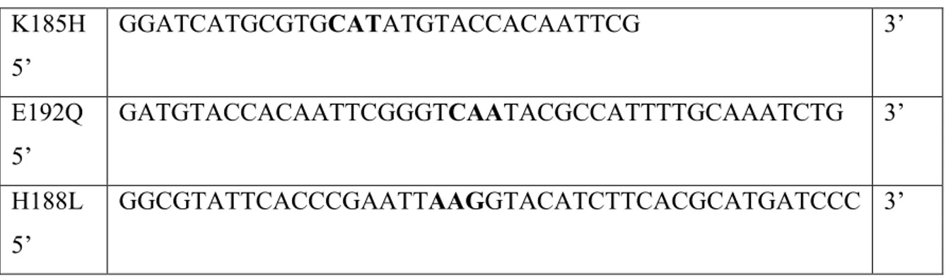
Mutagenesis was conducted in a double-stranded vector DNA, which was denatured to allow the pairing of oligonucleotides with small sequences of the 6PGDH gene of T. brucei containing the mutation of interest. (Table 3).
27 K185H 5‘ GGATCATGCGTGCATATGTACCACAATTCG 3‘ E192Q 5‘ GATGTACCACAATTCGGGTCAATACGCCATTTTGCAAATCTG 3‘ H188L 5‘ GGCGTATTCACCCGAATTAAGGTACATCTTCACGCATGATCCC 3‘
Table 3. Primer oligonucleotides synthetized by MWG-biotech AG.
For each mutation two primers were used, complementary to opposite strands of the vector, both containing the desired mutation. With dNTP and a PfuUltra ™ High Fidelity (HF) DNA polymerase two new chains are synthesized, generating plasmids bearing the mutation, containing "nicks" arranged at intervals (Table 4). The temperature cycling used is indicated in Table 5.
Table 4: The protocol reaction
Table 5: The temperature cycling
reaction buffer (10X) 5μl dsDNA template (5 -50 ng) 2 μl
Forward Oligonucleotide (125 ng) 12.5 μl Reverse Oligonucleotide (125 ng) 12.5 μl dNTP mix 1 μl
ddH2O to a final volume of a 50 μl
28 After primer extension, the reaction mixture was placed on ice for 2 minutes to cool to ≤ 37°C.
The vector filaments moulds were removed using Dpn I endonuclease (target sequence: 5'-Gm6ATC-3 ') which is able to digest the methylated and hemimethylated DNA (the DNA isolated from almost all strains of E. coli is methylated). In this way only the newly synthesized DNA containing the mutation was selected. Dpn I endonuclease (10U/ μl), was mixed with the reaction solution gently by pipetting the solution. The reaction mixtures are then spun down in a microcentrifuge for 1 minute and immediately incubated at 37°C for 2 hours to digest the parental supercoiled dsDNA.
Finally mutated carriers must be repaired by removing the "nicks" and amplified. Therefore, the vectors containing the desired mutations were transformed into XL1-Blue supercompetent cells. The supercompetent cells are gently thawed on ice, for each sample reaction to be transformed an aliquot of 50 μl were put in a tube and 1 μl of the Dpn I-treated DNA is transferred. The transformation reactions were gently swirled and incubated on ice for 30 minutes, then heat pulsed for 45 seconds at 42°C and after placed on ice for 2 minutes. 0.5 ml of NZY+ broth preheated to 42°C was added (NZY+ broth: 10g of NZ amine, 5g of yeast extract, 5g of NaCl, 12.5 ml of 1 MgCl2, 12.5 ml of 1 M MgSO4 and 20ml of 20% (w/v) glucose per liter, adjusted to pH 7.5).
The transformation reactions were then incubated at 37°C for 1 hour with shaking at 225-250 rpm. 225-250 µl of each transformation reaction was plated on agar /antibiotic, then put at 37 ° C for > 16 hours.
The following steps were the extraction of plasmidic DNA from a single colony and sequencing of the entire coding region of mutants T. brucei 6PGDH to check that only the single mutation was present, using the dideoxynucleotide chain-termination method. Once mutated sequence was obtained, it was introduced into E. coli strain BL21 (DE3) for inducible expression.
29
2) Overexpression of the T. brucei enzyme in E. coli.
PT7gnd is a plasmid obtained from cloning of the gene gnd (encoding 6-phosphogluconate dehydrogenase) into the vector pET3a, with the ATG initiation codon oriented adjacent to the bacteriophage T7 RNA polymerase promoter. BL21 (DE3) are E. coli cells with the phage DE3 encoding T7 RNA polymerase under the control of the lacUV5 promoter.
15 ul of transformed bacteria (stored at -80 °C in glycerol) were added to a Petri dish with agar, then left overnight at 37 °C. The next day a single colony was taken and added to 16 ml LB/16 ul amp (Luria Bertani broth plus ampicillin: 10g tryptone, 5 g yeast extract, 10 g NaCl water until 1 liter) and stirred overnight at 37 °C.
The 16 ml of the preculture were added to 400 ml of LB/amp, and incubated at 28 °C with shaking until the culture reached an optical density of 0.6 at 600nm. At this time the inducer IPTG (the lac operon inducer isopropylthiogalactopyranoside) was added to the culture to a final concentration of 0.4 mM. After adding IPTG , cultivation was left at 28 °C under shaking for 4 hours.
Then cells were centrifuged at 4000 rpm for 10 minutes at 4 °C. Pelleted cells were resuspended in buffer TEA (50mM TEA; 0.1 mM EDTA; 1 mM mercaptoethanol, pH 7.5) to a final volume of 20 ml and stored at -80 °C.
3) Purification of 6-PGDH wt / mutant of T. brucei by bacteria E. coli
The recombinant T. brucei 6PGDH, overexpressed in Escherichia coli, was purified according to a technique that was slightly modified compared to the original of Barrett [2] After keeping at -80 °C the bacteria were sonicated (XL2020 model sonicator, Heat Systems, power level to 4, 5 short bursts of 10 sec, followed by intervals of 20 sec for cooling, keeping the suspension at all times on ice).
Cell debris and insoluble material were then spun down (40,000 rpm, 30min). The supernatant was applied to a 15 ml DEAE-Sepharose column equilibrated with TEA
30 buffer, which was then washed with the same buffer and the flow through material absorbing at 280 nm was loaded directly onto a 5 ml 2‘,5‘-ADP-Sepharose column, equilibrated with TEA buffer diluted 10 x.
After washing with the same buffer, the enzyme was eluted with natrium pyrophosphate (Na4P2O7) 150 mM containing 1mM EDTA pH 7.2 and the specific activity assayed in buffer containing 0.6 mM 6PG and 0.26 mM NADP+.
The whole purification lasted less than one day and was monitored both by SDS-PAGE and activity assays. Enzyme was stored in the presence of 50% glycerol at -20°C.
4) Determination of protein and 6PGDH wt/mutant concentration
Spectrophotometric measurements were made throughout with a Jenway 6715 or a Kontron Uvikon 930 spectrophotometer or a Tecan infinite 200 microplate reader. Protein fluorescence was measured with the same microplate reader.
The protein purification fractions were measured spectrophotometrically at a wavelength of 280 nm for determination of the protein concentration assuming that a solution containing 1mg/ml of protein have an absorbance of 1 O.D. A solution containing 1 mg/ml of pure 6PGDH has instead at 280 nm an absorbance of 1.023. Knowing that each monomer unit of the T. brucei 6PGDH weights 52 kDa we can calculate that 1 mg of protein contains 19.2 nmoles. Therefore 1 mg/ml of the enzyme corresponds to a 19,2 microM concentration having an absorbance at 280 nm, 1.023 O.D.
To facilitate the storage of samples, the fractions were concentrated by the technique of ultrafiltration using Amicon Ultra-15 Centrifugal Filter Devices (Millipore) at 4000 rpm. The concentrated samples are assayed for the concentration and stored in 50% glycerol at -20 ° C.
31
5) Enzyme activity. Assay of activity for 6PGDH
The assay is based on the measurement of kinetics of a reaction mixture containing the 6PG substrate (0.6 mM), the NADP cofactor (0.26 mM) and our enzyme at a proper dilution; in 50 mM TEA buffer with 0.1 mM EDTA at pH 7.5, we proceeded to measure at a wavelength of 340 nm the amount of NADPH produced (product of the reduction of the cofactor NADP.) The absorbance value is divided by the molar extinction coefficient of NADPH which is 6,220.
The international unit of enzyme activity (IU) is the amount of enzyme which catalyzes in one minute, the formation of one micromol NADPH in standard conditions (25 °C and pH optimum). The specific activity of the enzyme has been calculated as the number of UI divided by the number of mg of protein.
6) Cysteines reactivity
The method G. Ellman reaction, 59 is based on the capacity of sulphidrilic groups of cysteines to react with 5,5-ditiobis-2-nitro benzoic acid (DTNB), developing a spectrophotometrically measurable complex at the wavelength of 412 nm. The method is rapid and the stoichiometry is 1:1, coloured product : thiol. Before the reaction, enzyme is freed from glycerol, by gel-filtration or dialysis, and used at a concentration 6 microM. DTNB is used at a concentration of 4 mg/ml and prepared in natrium phosphate buffer 0.2 M. Labelling has been measured for 45 min. The product molar extinction coefficient is 13,600.
7) Polyacrylamide gel electrophoresis (SDS-PAGE)
The polyacrylamide electrophoresis was performed following the method described by Laemmli [60 ] using a 8 x 10 cm gel in a socket for vertical electrophoresis.
The electrophoretic technique involves the preparation of two mixtures: one for the stacking gel which is at the top of the complete gel, in which the concentration of
32 acrylamide is 5% (w/v) and pH 6.8, and the second for the running gel which is under the stacking gel, in which the acrylamide concentration varies depending on the chosen pore size and the pH is 8,8. The running gel is the gel where protein separation is performed. In our case the acrylamide concentration used was 10% to verify the purification of the enzyme while both 10 and 7.5% for cross-linking experiments.
Running Gel (10%)
1.5 ml of 40% acrylamide buffer (39.3% acrylamide, 0.7% methylenbisacrylamide) 1.5 ml of TrisHCl (Tris-hydroxymethyl-aminomethane) 1.5 M pH 8.8,
15 µl of 6.6 M TEMED (N, N, N ‗, N‘-tetramethyethylenediamine) 2.9 ml of water
60 µl of SDS (sodium dodecyl ulphate) 10% w/v, 30 µl of ammonium persulphate 10% w/v.
Running Gel (7,5%)
1.125 ml of 40% acrylamide buffer (39.3% acrylamide, 0.7% methylenbisacrylamide) 1.5 ml of TrisHCl 1.5 M pH 8.8,
15 µl of 6.6 M TEMED 3.26 ml of water
60 µl of SDS 10% w/v,
30 µl of ammonium persulphate 10% w/v.
The solution of the running gel was poured between the two glass panes and left to solidify.
33 500 µ l acrylamide buffer 20% (19.6% acrylamide, 0.4% methylenbisacrylamide)
500 µ l 0.5 M TrisHCl pH 6.8 8 µ l 6.6 M di TEMED 930 ul of water
20 ul SDS 10% w/v
20 µ l ammonium persulphate 10% w/v. Table 6: the stacking gel solution 5% (w/v)
The solution of the stacking gel was poured over the polymerized running gel and the spacer comb for the wells was fitted between the two panes. Then the gel was left to polymerize. After, spacer was removed and the gel was installed in the device for electrophoresis, the tray containing the electrodes was filled with running buffer (25 mM Tris, 192 mm glycine, 0.1% SDS, at pH 8.3).
The samples were denatured before to be loaded into the wells. Buffer of denaturation (12.5% v/v 0.5 M Tris pH 6.8, 2% w/v SDS, 5% v/v β-mercaptoethanol, 10% v/v glycerol) containing a small amount of bromophenol blue as a tracer, was added to samples and then these were brought to 90 °C for 4 min. After denaturation 20μl of each sample was applied to the wells of the gel.
The electrophoresis was conducted by applying a current of 25 mA until the tracer had reached the lower limit of the gel, (power supply parameters: 0.46 KV, 25mA, 35 W, 45-50 min).
Since the run was done in the presence of SDS, the samples acquired the same negative charge and the migration is according the relative molecular weight of the sample.
After the run was completed the gel was extracted from the support and stained with Comassie blue C250 coloration without methanol (Table 7).
34 In 1 liter of bidistilled water
80 mg of Coomassie Brilliant Blue G-250 HCl in 40 mM final concentration.
Table 7: Coomassie blue C250 without methanol.
The gel in the dye solution was put in a microwave oven for 2-3 cycles of 15 seconds at maximum power. Then it is left in slow shaking for about 15 minutes at room temperature. Finally, the dye was removed and gel is soaked in distilled water at room temperature, rinsed several times and left in water in gentle agitation. Densitometric analysis of the gel was done with the program Quantity One (Biorad).
8) Gel Filtration (Sephacryl S-200 HR and AcA 34)
In gel filtration chromatography – also known as size exclusion chromatography – separation is based on differences in the size and/or shape of the analyte molecules, which governs the analytes‘ access to the pore volume inside the column packing particles. The exclusion limit of a size exclusion packing indicates the molecular weight, for a particular polymer type, above which analytes are fully excluded from entering the pores and thus will not be separated. According to their size, smaller analytes have partial to complete access to the pore volume. Larger molecules with less access to the pore volume elute first, while the smallest molecules elute last. The fractionation range means that molecules within that molecular weight range can be separated.
In the study, two gel filtration matrices were used: Sephacryl S-200 from GE Healthcare and ACA-34 Ultrogel from LKB. Sephacryl High Resolution is a cross-linked copolymer of allyl dextran and N,N‘-methylene bisacrylamide, which can separate proteins in the molecular weight range 5 × 10³ – 2.5 × 10⁵ Da. The diameter of the Sephacryl column was 0.7 cm and height 16.5 cm. The flow rate was of 3.3 microl/sec.
35 ACA-34 Ultrogel is polyacrylamide/agarose with a linear fractionation range of 20-350 kDa and an exclusion limit of 750 kDa for globular proteins. The diameter of the column was 0.8 cm and height 20 cm (7.6 ml of resin). The flow rate was of 210 microl/sec. Calibrations were performed with aldolase (160 kDa), bovine serum albumin (66 kDa), beta-lactoglobulin (35 kDa).
Columns were equilibrated with 50 mM triethanolamine, pH 7.5, 0.1 mM EDTA and 1.0 mM 2-mercaptoethanol. 20-40 microl of protein at a subunit concentration of 50 microM (about 3 mg/ml) were loaded onto the columns. The effect of ligands was determined by equilibrating the column with 70 microM NADP(H) or/and 0.16 mM 6PG. 4PE was used at 0.16 mM. Ligands were added to the enzyme sample before column loading to the same final concentrations used for the columns or higher. Eluted fractions were collected in
opaque-walled plate, with 100 microl/well and a total of 24 fractions. Protein detection was
by measure of the protein fluorescence, due essentially totryptophans, using an excitation
wavelength of 280 nm and an emission wavelength of 340 nm. The emission at 340 nm is proportional to the concentration of the sample, in this way we were able to locate the wells containing the eluted sample and create a curve of elution.
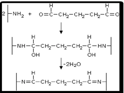
9) Chemical cross-linking of 6PGDH
Many biochemical and biophysical methods can be used to characterize the oligomerization state of proteins. One of the most widely used is glutaraldehyde cross-linking. The simplicity of the procedure, which requires only the mixing of glutaraldehyde with the protein solution and the availability of glutaraldehyde as a common reagent easily found in a biochemical laboratory, and the direct detection of cross-linked products by SDS–PAGE have led to the wide application of this method. The major limitation of the technique arises from the non-specificity of the reagent, which can react with all the nitrogens of a protein and mainly with lysines, tyrosines, histidines, and arginines and . Intra- and intermolecular links are formed that could connect atoms of neighboring but not interacting molecules, yielding artificial protein oligomers that lack biological significance. To eliminate the possibility of artificial interactions, the proper reaction conditions must be established through a detailed and often time-consuming investigation of the influence of
36
many parameters such as protein concentration, reagent concentration, temperature, and time of reaction 61
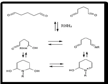
Glutaraldehyde is a linear, 5-carbon dialdehyde. It reacts rapidly with amine groups at around neutral pH 62 and is more efficient than other aldehydes in generating thermally and chemically stable crosslinks. 63. It is a bifunctional reagent because two aldehyde groups can react with two amino groups via Schiff base (Figures 14 and 15). 64
Figure 14: Reaction of the amino groups of proteins with glutaraldehyde.
The simple structure of glutaraldehyde is not indicative of the complexity of its ehaviour in aqueous solution and its reactivity. The structure of glutaraldehyde in aqueous solution has been the subject of more debate than any of the other crosslinking reagents. In fact, glutaraldehyde structure in aqueous solution is not limited to the monomeric form (Figure 16) 63, it can also undergo intramolecular cyclization, for instance producing pyridines (Figure 15). At alkaline pH polymerization of glutaraldehyde makes possible Michael-type additions with the formation of other types of condensation with amino groups (Figures 15 and 16).
37 Figure 15: Formation of pyridine rings after the reaction
of glutaraldehyde with amino groups of proteins.
Glutaraldehyde cross-linking was performed at different concentrations of enzyme and glutaraldehyde. The concentrations range were 0.1-1 mg/ml for the enzyme and 0,05-5 % for glutaraldehyde. Reaction was in 0.04 M sodium pyrophosphate, 25 mM HEPES (pH 7.5), containing 0.5 mM EDTA. Different incubation times (1, 2, 3, 5 minutes) were assayed and the reaction was stopped by adding 40 µl of 1 M TRIS/HCl pH 8.0. Aliquots were then resolved by either 7,5% or 10% SDS-PAGE. The more efficient cross-linking condition was with a final 1 % glutaraldehyde (by mixing 0.5 ml of enzyme with 20 µl of 25 % glutaraldehyde). Cross-linking was made also in presence of either 40 µM NADPH or both 40 µM NADPH and 1mM 4PE.