Farmacia e Scienze della Salute e della Nutrizione
(SSD: MED/04 Patologia Generale)
Dottorato di Ricerca in “Biochimica Cellulare ed
Attività dei Farmaci in Oncologia”
(XXVI ciclo)
The estrogen receptor
is the key regulator of the
bifunctional role of FoxO3a transcription factor in
breast cancer motility and invasiveness
Docente Tutor
Prof. Diego SISCI
Coordinatore
Prof. Diego SISCI
Dottoranda
Dott.ssa Maria Grazia CESARIO
Abstract pag.1
Introduction pag.2
Results pag.12
Cell motility, invasion, and anchorage-independent growth are inhibited in ERα+ breast cancer
cells overexpressing FoxO3a pag.12
The lack of ERα reverses FoxO3a-mediated inhibition of migration, invasion, and colonies
formation pag.15
FoxO3a and E2 synergistically induce caveolin-1 expression in ERα+ cancer cells pag.19 Cav1 is a mediator of FoxO3a-dependent inhibition of migration, invasion, and growth in
suspension in ERα+ breast cancer cells pag.22
FoxO3a binds to and trans-activates the Cav1 promoter in MCF-7 cells pag.24 Nuclear FoxO3a correlates in an opposite way with the tumor grade and the invasive phenotype in
ERα+ and ERα−breast tumors pag.26
Discussion pag.28
Materials and Methods pag.37
Cell culture, conditions, and treatments pag.37
Plasmids and transfections assays pag.37
siRNA-mediated RNA interference pag.38
Migration and invasion assays pag.38
Anchorage-independent growth assay pag.39
RNA extraction, reverse transcription, and real-time (RT)-PCR pag.40
Western blotting (WB) assays pag.40
Chromatin immunoprecipitation (ChIP) pag.41
Tissue collection, immunohistochemistry (IHC), and data analysis pag.41
Abstract
The role of the Forkhead box class O (FoxO)3a transcription factor in breast cancer migration and invasion is controversial. Here we show that FoxO3a overexpression decreases motility, invasiveness, and anchorage-independent growth in estrogen receptor α-positive (ERα+) cancer cells while eliciting opposite effects in ERα-silenced cells and in ERα-negative (ERα−) cell lines, demonstrating that the nuclear receptor represents a crucial switch in FoxO3a control of breast cancer cell aggressiveness. In ERα+ cells, FoxO3a-mediated events were paralleled by a significant induction of Caveolin-1 (Cav1), an essential constituent of caveolae negatively associated to tumor invasion and metastasis. Cav1 induction occurs at the transcriptional level through FoxO3a binding to a Forkhead responsive core sequence located at position −305/−299 of the Cav1 promoter. 17β-estradiol (E2) strongly emphasized FoxO3a effects on cell migration and invasion, while ERα and Cav1 silencing were able to reverse them, demonstrating that both proteins are pivotal mediators of these FoxO3a controlled processes. In vivo, an immunohistochemical analysis on tissue sections from patients with ERα+ or ERα− invasive breast cancers or in situ ductal carcinoma showed that nuclear FoxO3a inversely (ERα+) or directly (ERα−) correlated with the invasive phenotype of breast tumors. In conclusion, FoxO3a role in breast cancer motility and invasion depends on ERα status, disclosing a novel aspect of the well-established FoxO3a/ERα interplay.
Therefore FoxO3a might become a pursuable target to be suitably exploited in combination therapies either in ERα+ or ERα− breast tumors.
Introduction
Breast cancer is one of the most common forms of cancer observed in women. Endogenous estrogens are thought to play a major role in its development and estrogen receptor blockers are important drugs in its treatment (Pike MC et al.,
1983; Mc Guire WL, 1978; Fisher B et al.,2001). It has been shown that longer
exposures to estrogens result in an increased risk for breast cancer (Henderson
IC, 1993). Estrogens have effects on many organ systems, beyond the reproductive system, in both females and males. Breast tissue is particularly dependent on circulating estrogens since there is no breast development in aromatase-deficient women and estrogen therapy of these patients leads to normal pre- and post-pubertal breast development. Estrogen effects are exerted through two types of specific receptor: estrogen receptor alpha (ER) and beta (ER) (Korach KS, 1994; Gustafsson JA, 1999; Dupont S et al, 2000). These nuclear receptors are ligand-dependent transcription factors that mediate the biological effects of estrogens and anti-estrogens. The human estrogen receptors ER and ER contain five functional domains (A–E) as other members of the nuclear receptor superfamily and an additional domain F in their C terminal part
(Kumar V et al, 1987; McKenna NJ et al,1999; Shiau AK et al,1998; Brzozowski AM et al,1997; Moras D et al, 1998; Katzenellenbogen BS et al, 2002; McDonnell DP et al, 2002; MacGregor JI et al; Kato S, 1995). The binding of
estrogen in the hormone-binding domain (HBD) induces a trans-conformational change of the whole molecule allowing unmasking of the activating function 1 (AF1) in domain A/B by removal of chaperone (HSP90), dimerization, activation of activating function 2 (AF2) in the C-terminal part of the E domain and binding to estrogen-responsive element (ERE) on DNA via domain C. Estrogen receptors act mainly by regulating the expression of target genes whose promoters contain specific sequences called estrogen-responsive element (ERE). After ERE-binding of ligand-bound ER dimers, modulation of transcription occurs via interaction with coactivators or corepressors. All
together, these complexes play an important role in the recruitment of transcriptional machinery, the modulation of chromatine structure, and then in the regulation of ER target-gene expression (McKenna NJ et al, 1999). The ER conformation differs with the type of ligand, and there is a marked difference in the topology of the ER surface between agonist and antagonist-bound receptors
(Shiau AK et al, 1998; Brzozowski AM et al, 1997; Moras D et al, 1998).
Moreover, studies conducted with synthetic anti-estrogens, such as tamoxifen, have shown that the agonist/antagonist profile of a ligand varies with the tissue and the target gene considered. This led to the term of selective estrogen receptor modulators (SERMs) to define this class of drug (Katzenellenbogen BS
et al, 2002; McDonnell DP et al, 2002; MacGregor JI et al,1998). ER activity
can also be modulated through indirect activation of the ER by growth factors or cytokines independently of the binding of natural or synthetic hormones (Kato S
et al, 1995; Bunone G et al, 1996). Estrogen and progesterone receptors (ER
and PR) have now been studied in clinical breast cancer for more than 20 years. ERwas found in 50–80% of breast tumors and ERstatus is essential in making decisions about endocrine therapy (Mc Guire WL, 1978; Osborne
CK,1998). Positive receptor status correlates with favorable prognostic features,
including a lower rate of cell proliferation and histologic evidence of tumor differentiation. During the first several years after diagnosis, patients with ER-positive tumors tend to have a lower recurrence rate; however, this is balanced by a higher recurrence rate in subsequent years so that the overall prognostic significance of receptor status is modest. ER and PR have their greatest utility in predicting response to hormonal therapy, both in the adjuvant setting and for advanced disease. Tumors that express both ER and PR have the greatest benefit from hormonal therapy, but those containing only ER or PR still have significant responses (Bardou VJ et al, 2003). Does the ER-negative tumor derive from ER-positive tumor or is it a totally different disease (Zhu K et
appear in tumors as early as carcinoma in situ (Roger P et al, 2000) and the gene expression is substantially different in the two types of invasive carcinoma
(Sheikh MS et al, 1994; Thompson EW et al, 1992). Moreover, ER re-expression in an ER-negative cancer cell is not sufficient to restore the ER -positive phenotype, particularly in terms of mitogenic response and the pattern of gene expression (Garcia M et al, 1992; Jiang SY et al,1992). The great interest on ERis also due to its functional cross-talk with other factors such the forkhead box class protein members.
Forkhead proteins are not among the largest transcription factor families, but display a remarkable functional diversity and are involved in a wide variety of biological processes. The name is derived from the two spiked-head structures in the embryos of the Drosophila fork head mutant, which are defective in anterior
and posterior gut formation (Weigel D et al,1989). With the 1990 discovery of a
110-amino-acid DNA binding domain that was almost perfectly conserved between FORK HEAD and the mammalian HNF-3 transcription factors, it became clear that this motif defined a novel transcription factor family (Weigel
D et al,1990). Among the organisms for which the genome sequences are
completed, or nearly so, there is indeed a correlation between anatomical complexity and forkhead gene number: 4 in Saccharomyces and
Schizosaccharomyces, 15 in Caenorhabditis, 20 in Drosophila, and 39 in Homo.
In 2000, the nomenclature of chordate forkhead transcription factors was revised
(Kaestner KH,2000; Kaestner KH et al,2000). The new nomenclature, which
uses Fox (for “Forkhead box”) as the root symbol, ensures that the same name is used for orthologous genes in different species and reflects phylogenetic relationships by including a letter that indicates subfamily. Within a subfamily, each gene is identified by a number (e.g., FoxO2), the typography follows the conventions used in each species (FOXO3a in Homo, Foxo3a in Mus, and
wingedhelix domain, are characterized by a conserved DNA-binding domain— the forkhead box among invertebrate and mammalian cells (Weigel D et
al,1989; Weigel D,1990; . Kaufmann E et al,1996).
Based on the forkhead box domain, the forkhead genes are grouped into 19 subclasses of Fox genes (Kaestner KH,2000; Kaestner KH et al,2000). FoxO transcription factors, one of largest subgroups of forkhead family members, are characterized by a conserved DNA-binding domain-the forkhead box among invertebrate and mammalian cells (Arden KC,2006; Greer EL et al,2005). The FoxO subfamily contains four members (FoxO1, FoxO3, FoxO4, and FoxO6), which activate or repress multiple genes such as Bim and FasL involved in apoptosis (Finnberg N et al,2004; Tran Het al,2003), p27kip (Dijkers PF et
al,2000) and cyclin D (Schmidt M et al,2002) in cell cycle regulation,
GADD45a in DNA damage repair (Greer EL et al,2005; Finnberg N et al,2004;
Tran Het al,2003; Yang JY et al,2006), manganese superoxide dismutase
(MnSOD) in stress response (Kops GJ et al,2002), and glycogenolytic gene glucose-6-phosphatase (G6pc) in metabolism (Onuma H et al,2006). Recent studies also reveal the importance of FoxOs in preserving the self renewal capacity of hematopoietic stem cells (Miyamoto K et al,2007; Tothova Z et
al,2007), however, the detailed mechanisms are currently a work in progress.
The major consequence of the phosphorylation of FoxO transcription factors by Akt and SGK is a change in the subcellular localization of these transcription factors (Biggs et al., 1999; Brunet et al., 1999; Takaishi et al., 1999). In the absence of growth factors, when Akt and SGK are inactive, FoxO factors are localized within the nucleus. When cells are exposed to growth factors, the PI3K–Akt/SGK cascade is activated and triggers the export of FoxO factors to the cytoplasm. Mutation analyses have revealed that one or two leucine-rich domains in the conserved C-terminal region of FoxO proteins function as a nuclear export sequence (NES) (Biggs et al., 1999; Brunet et al., 2002). In addition, phosphorylated FoxO factors have been shown to specifically interact
with 14-3-3 proteins, which serve as chaperone molecules to escort FoxO proteins out of the nucleus (Brunet et al., 1999, 2002). Several mechanisms have been proposed to explain how 14-3-3 binding to FoxO factors promotes the relocalization of FoxO factors from the nucleus to the cytoplasm. While 14-3-3 proteins are mostly present in the cytoplasm at equilibrium, these chaperone molecules have been found to bind to their substrates in the nucleus (Brunet et
al., 2002). Consistently, 14-3-3 binds to FoxO3 in the nucleus (Brunet et al., 2002). 14-3-3 binding may decrease the ability of FoxO factors to bind DNA,
releasing FoxO proteins from a nuclear DNA anchor (Cahill et al., 2000). 14-3-3 binding to FoxO factors may actively promote the nuclear export of FoxO factors, perhaps by inducing a conformational change in FoxO molecules that would expose the NES and allow interaction with Exportin/Crm1 (Brunet et al.,
2002). 14-3-3 binding to FoxO factors may also prevent the nuclear reimport of
these transcriptional regulators by masking FoxO nuclear localization signal (NLS) (Brownawell et al., 2001; Rena et al., 2001). Finally, the phosphorylation of FoxO factors at Ser322 and Ser325 appears to accelerate FOXO relocalization to the cytoplasm in response to growth factors by increasing the interaction between FoxO and the export machinery (Ran and Exportin/Crm1)
(Rena et al., 2002). These various mechanisms for regulating the translocation
of FoxO transcription factors from the nucleus to the cytoplasm may serve as a fail-safe mechanism to ensure a complete sequestration of FoxO factors away from their target genes. Mutational analysis of the three regulatory Akt/SGK sites have revealed that the phosphorylation of each site contributes to the nuclear exclusion of FoxO factors (Brunet et al., 2001). One attractive possibility is that each site participates in different aspects of the mechanisms that ensure the relocalization of FoxO proteins into the cytoplasm. Thus, phosphorylation of FoxO factors may represent a way of modulating the extent of the relocalization of these transcription factors to the cytoplasm in different cell types or in response to different combinations of signals. The most recently
identified FoxO member, FoxO6, only contains two of the three Akt/SGK regulatory sites (Thr26 and Ser184 in mouse FOXO6) (Jacobs et al., 2003). Unlike the other FoxO isoforms, FoxO6 is mostly nuclear. However, FoxO6 phosphorylation at Thr26 and Ser184 appears to decrease the transcriptional activity of this FoxO isoform (van der Heide et al., 2005). These findings suggest that the regulations and functions of FoxO6 may differ from those of FoxO1, FoxO3, and FoxO4.
The protein phosphatases that dephosphorylate FoxO transcription factors at the sites that are targeted by Akt and SGK remain elusive. These phosphatases would have the capacity to counteract Akt/SGK actions and to rapidly activate FoxO proteins, by allowing these transcription factors to translocate to the nucleus.
As FoxO factors appear to play an important role in cell cycle arrest, identifying ways to activate FoxO factors may be critical to counteract tumor formation. While FoxO transcription factors are mainly regulated via reversible changes in subcellular localization, the degradation of FoxO protein represents an additional and irreversible level of regulation of this family of transcription factors. FoxO protein degradation often accompanies cell transformation (Hu et
al., 2004; Huang et al., 2005), suggesting that this mechanism of regulation may
be a critical initiation step towards tumorigenesis.
The degradation of FoxO transcription factors is mediated by the ubiquitin– proteasome pathway (Matsuzaki et al., 2003; Plasand Thompson, 2003; Aoki et
al., 2004; Hu et al., 2004; Huang et al., 2005).
Akt activity is necessary for ubiquitin-mediated degradation of FoxO3 and FoxO1 (Plasand Thompson, 2003; Huang et al., 2005). In addition, I kappaB kinase (IKK) also causes the proteasome-dependent degradation of FoxO
factors (Hu et al., 2004). IKK is known to activate the transcription factor
NF-kB through the phosphorylation and subsequent degradation of INF-kB, which normally serves as a negative regulator of NF-kB (Karin et al., 2002). IKK
induces the phosphorylation of FoxO3 at Ser644, in the extreme C-terminal portion of the molecule. This phosphorylation results in the ubiquitination and
subsequent degradation of FoxO3 (Hu et al., 2004). Since IKK-induced
tumorigenesis can be suppressed by overexpression of FoxO3 (Hu et al., 2004),
the regulation of FoxO protein degradation by IKK may play an important role
in tumorigenesis. However, Ser644 is not conserved in other FoxO isoforms and is not present in worms and flies. Thus, whether IKK phosphorylates and controls the other FoxO isoforms remains to be determined. It is possible that the degradation of FoxO isoforms is regulated by different protein kinases via independent mechanisms. FoxO1 and FoxO3 protein degradation is regulated by Akt and, at least for FoxO3, by IKK. However, whether FoxO4 and FoxO6 protein degradation is also actively regulated, and if so, whether the mechanisms of regulation are similar, still remains to be established. One major difference between the FoxO family members is that they display overlapping but different patterns of expression. While these differences may be partly due to mRNA expression, it is possible that protein degradation also plays an important role in the distinction between FoxO isoforms in vivo. As tumorigenesis appears to be associated with a loss in FoxO proteins, understanding the regulation of FoxO expression will likely give important insight into mechanisms that govern tumor suppression.
An increasing interest in FoxOs factors has been lately observed in the oncologic research field. In particular, in breast cancer, its role is still controversial, in fact, FoxO3a overexpression has been shown to inhibit tumor growth in vitro and to reduce tumor size in vivo, (Hu MC et al, 2004; Yang JY et
al, 2008; Zou Y et al, 2008) and cytoplasmic location of FoxO3a seems to
correlate with patients poor survival (Hu MC et al, 2004). Moreover, genetic deletion of the FoxOs alleles (FoxO1a, FoxO3a, and FoxO4) generates progressive cancerous phenotypes, such as thymic lymphomas and hemangiomas. These data elucidate FoxOs as bona fide tumor suppressor genes
(Paik JH, 2007). Additionally, FoxO members seem to be important mediators
of the well-established functional cross-talk between estrogens and growth factors, which play a pivotal role in breast cancer development and progression
(Sisci D et al,2007).
In fact, growth factors are known to influence the expression and activity of estrogen receptor α (ERα) and its transcriptional cofactors; conversely, ERα regulates the expression of growth factor receptors and their ligands and signaling intermediates (Lanzino M et al, 2008). In this context, several reports have recently suggested a functional interaction between ERα and FoxO members.
17β-estradiol (E2) has been noted to determine ERα binding to FoxO1a, FoxO3a, and FoxO4, which, in turn, showed either coactivator or corepressor functions on estrogen-responsive element (ERE) sites, depending on the cellular model (Zou Y et al, 2008; Schuur ER et al, 2001; Zhao HH, 2001). Moreover, we introduced the importance of Akt2/FoxO3a axis in the control of ERα-mediated transcription in ERα-positive (ERα+) breast cancer cells. Our results indicate that Akt2 inhibition reduces ERα transcriptional activity through FoxO3a activation, suggesting that FoxO3a, acting as a co-repressor for ERα, could exert a protective role in ERα+ breast tumors (Morelli C, 2010).
In line with this assumption, Belguise et al. showed that ectopic expression of a constitutively active FoxO3a overrode transforming growth factor-B1-mediated invasive phenotype and induced a more epithelial phenotype in ERα+ mouse mammary tumors (Belguise K et al, 2007). However, more recently, FoxO3a has been described to behave in an opposite fashion in several other cancer cell lines, which, interestingly, were all ERα-negative (ERα-); in fact, Storz et al. reported that, in tested cells, nuclear retention of FoxO3a resulted in greatly increased invasion, through the induction of matrix metalloproteinase 9 (MMP-9) and MMP-13(Storz P et al, 200(MMP-9). Due to the inconsistency of the data available from ERα+ and ERα− breast cancer cells, the interplay between ERα
and FoxO3a in tumor metastasis needs further investigations and is the goal of the present study. Since it is well documented that, in breast cancer, ERα signaling strongly correlates with a lower invasiveness and reduced metastatic potential, (Rochefort H et al, 1998) we assume that FoxO3a/ERα interplay could be responsible for the reduction of the migrating and invasive phenotype only in ERα+ cells, while, in ERα− cells, the lack of the α isoform of the receptor might enable FoxO3a to act in an opposite fashion. In ERα+ cells, FoxO3a-mediated events were paralleled by a significant induction of Caveolin-1 (Cav1), an essential constituent of caveolae negatively associated to tumor invasion and metastasis. Caveolae demarcate discrete, highly ordered microdomains of the plasma membrane that serve as dynamic trafficking and signal transduction sub-compartments. Caveolae were originally described morphologically in 1953-1955 as flask-shaped invaginations of the plasma membrane with a diameter of 50-100 nm in endothelial and epithelial cells (Bruns RR et al, 1968). Caveolae occur as both invaginations of the plasma membrane proper and as detached vesicles residing close to the membrane. Caveolae exist in numerous tissues and cell types with varying abundance, the highest levels occurring in fibroblast, adipocytes, endothelial cells, type 1 pneumocytes, epithelial cells and smooth and striated muscle cells (Engelman JA et al,1998; Scherer PE et al,1995;
Scherer PE et al, 1997). By electron microscopy caveolae charatteristically
appear smooth with a distinctive coat appearing as bipolar-oriented, thin striations surrounding the bulb of the caveolae (Rothberg KG et al, 1992; Peters
KR, 1985). The striated coat associated with the cytoplasmatic face of caveolae
is principally formed by homo- and hetero-oligomers of the structural coat proteins the caveolins (caveolin1,2,3) (Scherer PE et al, 1997; Monier S et al,
1995; Sargiacomo M et al, 1995; Tang Z et al, 1996). As a protein family,
caveolins can be defined as cytoplasmatic membrane proper and participate in the sequestration of inactive signaling molecules. As caveolin-1 was the first caveolin member to be identified it has served as the prototype for the study of
caveolins. Based on primary sequence composition and mutation analysis caveolin-1 is predicted to have a central 33 amino acid hydrophobic domain (residues 102-134), thought to form a hairpin structure spanning the membrane, with the hydrophilic amino (1-101) and carboxyl termini (135-178) remaining cytoplasmatic (Monier S et al, 1995; Sargiacomo M et al, 1995; Dupree P et al,
1993).
Thus, the present work was aimed to undertake an accurate study on the molecular mechanisms through which FoxO3a regulates migration and invasion in ERα+ breast cancer cells. Our results offer new interesting insights on FoxO3a activity, elucidating additional mechanisms that could represent novel targets in breast cancer therapy.
Results
Cell motility, invasion, and anchorage-independent growth are inhibited in ERα+ breast cancer cells overexpressing FoxO3a
To assess the role of FoxO3a in the metastatic and invading potential of breast cancer cells, wild-type FoxO3a (F3a) was overexpressed in ERα+ MCF-7. Our results show a significant reduction of migrating and invading MCF-7/F3a cells (Fig. 1A and B), compared with control samples. Ectopic expression of the constitutively active triple mutant of FoxO3a (F3aAAA), where the 3 known PKB phosphorylation sites have been mutated to alanine, so that FoxO3a can no longer be inhibited by PKB-mediated phosphorylation, emphasized the phenomenon (Fig. 1A and B), suggesting that FoxO3a modulation of the migrating and the invading potential could involve the transcriptional induction of Forkhead responsive genes. FoxO3a silencing (siF3a) confirmed these data, since it led to a substantial increase in cell migration and invasion (Fig. 1A and B). Moreover, in agreement with our previous observations, (Sisci D et al, 2010) E2 treatment strongly reduced motility and invasion, and the effect was additive in F3a- and F3aAAAoverexpressing samples, while siF3a only in part was able to counteract E2-mediated effects (Fig. 1A and B).
In addition, anchorage independence, a characteristic of malignancy and tumor progression , was also investigated in F3aoverexpressing and silenced MCF-7 cells through soft agar colony formation assay. We observed a dramatic decrease of the number as well as of the dimensions of the colonies in MCF-7/F3a samples, reaching almost completely the condition of single cells in F3aAAA-expressing cells (Fig. 1 C1 and C2). The same trend was evidenced in E2-treated
samples, showing how FoxO3a, especially in its active form, is able to counteract the well-known positive effect of the nuclear hormone on the colony formation of MCF-7 cells (Manni A et al, 1991). As expected, an increase in the number of colonies was observed following siF3a, and such increase became
more evident in presence of E2 (Fig.1 C1 and C2). Transfections and silencing
efficiency were assessed on total protein lysates (Fig. 1D).
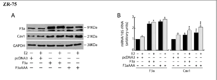
Interestingly, F3a and F3aAAA overexpression in other ERα- positive cell lines, ZR-75 (breast cancer) and Ishikawa (endometrial cancer), led to results that were comparable to those obtained from MCF-7, both in presence or absence of E2 (Fig.2, upper panels)
Figure 1. FoxO3a inhibits migration, invasion and anchorage independent growth in ERα+ MCF-7 breast cancer cells. A double set of MCF-7 cells was transiently transfected with 1 μg/35 mm dish of F3a, F3aAAA, or
pcDNA3 as control. Another double set was silenced for FoxO3a expression (siF3a),using a siScramble as control (60 pmol siRNAs/35 mm dish). After 5 h cells were switched to PRF-SFM, and the next day one of each set of cells was harvested and subjected to migration (A), invasion (B), and soft agar assay (C1 and C2). Migration and invasion assays were conducted as described in “Materials and Methods”, adding 100 nM E2 in the bottom of the wells where indicated. Migrated and invading cells were evaluated after 24 h and 72 h of incubation, respectively. In soft agar assay, colonies >50 μm diameter formed after 14 d from plating were
photographed at 4× magnification (C2) and counted under the microscope (C1). The second set of either transfected or silenced MCF-7 cells was used for total protein extractions and WB analysis to assess transfections efficiency; GAPDH was evaluated as a loading control (D). Results are reported as the mean ± s.d. of at least 3 independent experiments. In all experiments, significance values were as follows: *, P < 0.01 vs. untreated; ●, P < 0.01 vs. corresponding pcDNA3; ♦,P < 0.05 vs. corresponding F3a; □,P < 0.01 vs. corresponding siScramble.
Figure 2 The opposite effects exerted by FoxO3a on migration, invasion and colony formation of ERα+ and ERα cancer cells is not tissue specific.
Double sets of ERα+ (ER+) breast cancer cells (ZR75) and endometrial cancer cells (Ishikawa), and of ERα (ER)breast cancer cells (MDAMB468) and cervical cancer cells (HeLa) were transiently transfected with F3a, F3aAAA or pcDNA3 as described in Materials and Methods. Following 24h of starvation, one of each double set of cells was harvested and subjected to Migration (B), invasion (C1 and C2) or soft agar assays (D1 and
D2), adding 100nM E2 where indicated. Migrated cells were counted after 24h (ZR72 and Ishikawa) or 16h
(MDAMB468 and HeLa) of incubation. Invading cells were counterstained with DAPI after 48h (MDAMB468 and HeLa) or 72h (ZR75, Ishikawa) of incubation (C2), and evaluated by ImageJ software (C1). In soft agar
MTT and counted under the microscope (D2). The second of each set of cells was used to evaluate transfections efficiency by WB analysis on total protein extract; GAPDH was used as a loading control (A). Results are the mean ±s.d. of at least three independent experiments. *, P < 0.01 vs untreated; ,P < 0.01 vs pcDNA3;,P < 0.01 vs F3a.
The lack of ERα reverses FoxO3a-mediated inhibition of migration, invasion, and colonies formation
To assess if the effects of FoxO3a on motility, invasiveness, and colony formation could depend on ERα, silencing experiments were conducted in MCF-7, using specific siRNAs against ERα (siER) (Fig. 3). Interestingly, ERα silencing was able to counteract FoxO3a-mediated inhibition of the above-mentioned pathological features.
In particular, compared with control (siScramble), siER led to an increase in cell migration and invasion, which became even more evident in F3a and, especially, in F3aAAA-expressing cells (Fig. 3A and B), confirming that ERα is a hallmark of a less motile and invading phenotype, (Sisci D et al, 2010; . Platet N et al,
2004) and that FoxO3a’s effect on cell motility and invasiveness can switch
from inhibitory to stimulatory, depending on the presence or absence of ERα, respectively. Moreover, in siER samples, reasonably due to the lack of the receptor, E2 treatment no longer caused the reduction of the invading potential of MCF-7 (Fig. 3B) and even showed the opposite effect on cell motility, which rather increased over the respective controls (Fig. 3A). These evidences suggest that, in absence of a functional ERα, E2 could trigger some other pathway that stimulates cell migration (although not invasion), and that FoxO3a can somehow cooperate with the hormone in this process.
As expected, ERα silencing was able to inhibit both basal and E2 induced MCF-7 growth in soft agar by strongly reducing the number and the dimensions of colonies compared with non-treated and E2-treated siScramble samples, respectively (Fig. 3C). However, as in migration and invasion experiments, the inactivation of the nuclear receptor reversed the effect of ectopic F3a and F3aAAA, which, either in absence or presence of E2 treatment, induced an
increase in the number of colonies, instead of the decrease observed in siScramble samples (Fig. 4C).
The fact that ERα exerts a pivotal role in determining FoxO3a behavior was confirmed by the results obtained in ERα− cells. Indeed, overexpression of FoxO3a in ERα− breast cancer MDA-MB-231 cells was able to induce an evident increase (rather than a decrease, as in ERα+ cells) of the migrating and invading potential (Fig. 4A and B), as well as, when grown in soft agar, F3a-overexpressing cells formed many more and larger colonies compared with
control vector (Fig. 4C1 and C2). Once again, in all experiments, F3aAAA was
more effective than F3a, while an evident reduction of migration, invasion and number and dimensions of colonies was observed in F3a silenced samples (Fig. 4A–C2). Transfections and silencing efficiency were determined concomitantly
(Fig. 4D).
Noteworthy, as in MDA-MB-231, F3a and F3aAAA overexpression led to comparable results in other ERα− breast cancer cell lines (MDA-MB-468 and MDA-MB-435) as well as in ERα− cervical cancer HeLa cells, indicating that FoxO3a functions trough mechanisms that are not tissue-specific (Fig.2, lower panels and data not shown).
Figure 3. FoxO3a mediated inhibition of breast cancer cell migration, invasion and growth in suspension depends on ERα. Two double sets of MCF-7 cells were silenced either for ERα (siER), using siScramble as
control. After 5 h cells were switched to PRF-SFM and transiently transfected with F3a, F3aAAA, or pcDNA3. Next day cells were harvested and one set of each experiment was subjected to migration, invasion, and soft agar assay in the presence or in the absence of E2. Migrated (A) and invading (B) cells were evaluated after 24 h and 72 h of incubation, respectively. In soft agar assay, colonies ≥50 μm diameter formed after 14 d from plating were counted under the microscope (C). The second set of each experiment was used for total protein extraction to evaluate transfections efficiency by WB analysis; GAPDH was used as loading control (D). Results are the mean ±s.d. of at least three independent experiments. *, P < 0.05 vs. untreated; ●, P < 0.01 vs. corresponding pcDNA3; ♦, P < 0.01 vs. corresponding F3a; □ , P < 0.01 vs. corresponding siScramble.
Figure 4. FoxO3a promotes migration, invasion, and anchorage-independent growth in ERα− MDA-MB-231 breast cancer cells. A double set of MDA-MB-MDA-MB-231 cells were transiently transfected with 1 μg/35 mm dish
of F3a, F3aAAA, or pcDNA3 or silenced for FoxO3a expression (siF3a) using a siScramble as control (60 pmol siRNAs/35 mm dish). Both transfection and silencing were made on cells in suspended PRF-GM. After 5 h cells were serum starved and, 24 h later, harvested. One set was subjected to migration (A), invasion (B), or soft agar assay (C1 and C2). Migrated and invading cells were evaluated after 16 h and 48 h of incubation, respectively. In soft agar assay, colonies > 50 μm diameter formed after 14 d from plating were photographed at 4× magnification (C2) and counted under the microscope (C1). The second set of either transfected or silenced MCF-7 cells was used to assess transfections efficiency by WB analysis on total protein extracts; GAPDH was evaluated as a loading control (D). Results are reported as the mean ± s.d. of at least 3 independent experiments. ●, P < 0.01 vs. pcDNA3; ♦, P < 0.01 vs. F3a; □ , P < 0.05 vs. siScramble.
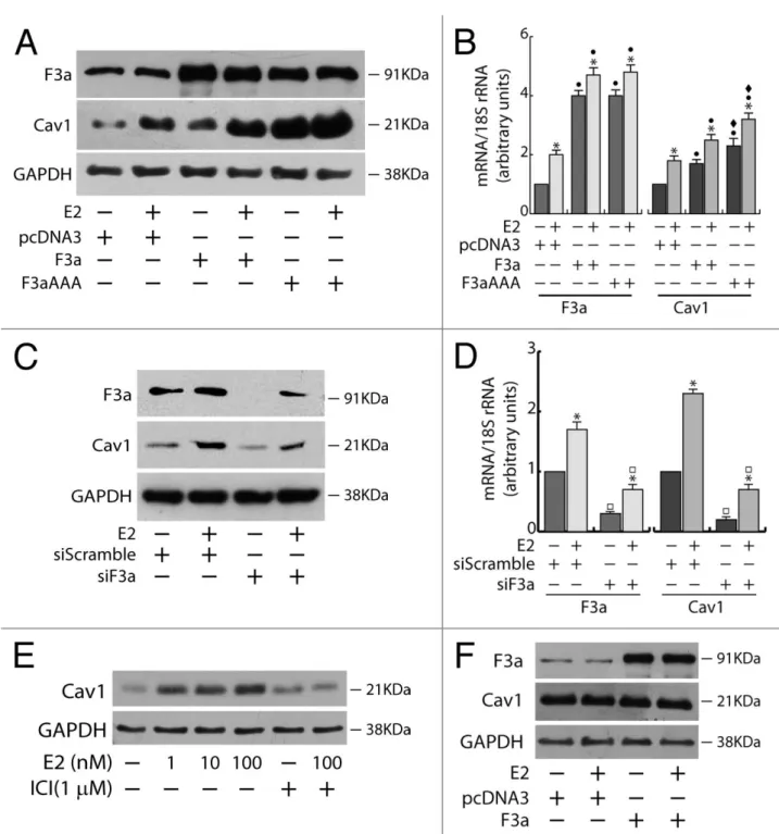
FoxO3a and E2 synergistically induce caveolin-1 expression in ERα+ cancer cells
To the aim of identifying the mechanism through which FoxO3a modulates cell motility and invasiveness, we focused our attention on caveolin-1 (Cav1), a protein that has been reported to be induced by both Forkhead transcription factors (van den Heuvel AP et al, 2005) and E2 (Charpentier AH et al, 2000;
Razandi M, 2002).
Since, in breast cancer, Cav1 has been negatively (Sloan EK et al, 2004) and positively (Joshi B et al, 2008) linked to tumor progression, motility, and invasiveness, we questioned if FoxO3a could control migration and invasion of breast cancer cells through the modulation of Cav1 expression.
In ERα+ MCF-7 cells, the ectopic expression of FoxO3a caused a strong upregulation of Cav1 protein and mRNA, which was even more evident in F3aAAA transfectants, suggesting that FoxO3a induction of Cav1 expression could occur at the transcriptional level. As expected, E2 treatment increased Cav1 levels, and the effect was additive to that exerted by F3a or F3aAAA (Fig.5A and B). Silencing experiments confirmed FoxO3a involvement in Cav1 transcription, leading to a decrease in Cav1 content and attenuating the E2-dependent Cav1 induction (Fig. 5C and D). Notably, Cav1 undergoes similar regulation by E2 and FoxO3a in the other 2 tested ERα+ cell lines, ZR-75 and Ishikawa (Fig. 6). In particular, the induction of Cav1 by E2 is ERα- dependent, since (1) the pure antiestrogen ICI 172.780 was able to abrogate the effect of E2 on Cav1 expression in ERα+ MCF-7 cells (Fig. 5E); and (2) the hormone did not increase Cav1 expression in ERα−, although ERβ+, MDA-MB-231 cells (Fig. 5F).
In light of these evidences we could hypothesize that, in ERα+ cells, FoxO3a might promote a less aggressive phenotype by cooperating with the hormone receptor in CAV1 gene induction.
Figure 5. Cav1 expression depends on E2 and FoxO3a in ERα+ MCF-7 breast cancer cells. A double set of
MCF-7 cells were either transiently transfected with F3a, F3aAAA, or pcDNA3 or silenced for FoxO3a, serum starved after 5 h and treated the next day with 100 nM E2 for 24 h. Cells were then harvested and total proteins and RNA were extracted, and subjected to WB (A and C) and RT-PCR analysis (B and D), respectively, for F3a and Cav1 expression assessment. (E) MCF-7 cells were seeded in growing medium, serum starved the next day for 24 h, pre-treated or not for 1 h with the pure antiestrogen ICI 182.780 and then treated with increasing concentrations of E2 (0, 1, 10, and 100 nM). (F) MDA-MB-231 cells were transiently transfected with F3a or pcDNA3 as control, serum starved for 24 h and then treated or not with 100 nM E2. After 24 h of E2 treatment, total proteins were extracted and subjected to WB analysis. GAPDH was analyzed as loading control in WB assays. For RT-PCR assays, each sample was normalized to its 18S rRNA content.Results are reported as the mean ±s.d. of at least 3 independent experiment. *, P < 0.01 vs. untreated; ●, P < 0.01 vs. pcDNA3; ♦, P < 0.01 vs. F3a; □ , P < 0.05 vs. siScramble.
Figure 6 FoxO3a induces Cav‐1 expression in ERα+cancer cells.
Double sets of ZR75 and Ishikawa cells were transiently transfected with F3a, F3aAAA or pcDNA3, as described in Materials and Methods. Five hours after transfection, cells were starved for 24h and then treated or not with 100nM E2 for additional 24h. Total proteins (A and C) and RNA (B and D) were extracted and subjected to WB and RT‐PCR analysis respectively, to assess FoxO3a and Cav‐1 expression. GAPDH was used as loading control in WB analysis.In RT-PCR assays each sample was normalized to its 18S rRNA content. Results are the mean ±s.d. of at least three independent experiments. *, P < 0.01 vs untreated; , P < 0.01 vs pcDNA3; , P < 0.01 vs corresponding F3a.
Cav1 is a mediator of FoxO3a-dependent inhibition of migration, invasion, and growth in suspension in ERα+ breast cancer cells
Cav1 involvement in FoxO3a-mediated inhibition of motility, invasiveness, and colonies formation was assessed by silencing experiments using specific siRNAs against Cav1 (siCav1) in ERα+ breast cancer cells, (Fig. 7A–D). Cav1 silencing was able to counteract FoxO3a effects, leading to an overall increase of cell migration and invasion in MCF-7 cells, although F3a and F3aAAA overexpression did not contribute to such increase, nor was siCav1 sufficient to completely reverse the inhibitory effect exerted by E2 treatment (Fig. 7A and B). A similar trend was observed in soft agar experiments, where the number of colonies was much greater in siCav1 samples, especially under E2 treatment (note that ERα protein content was not affected by siCav1, Fig. 7D), compared with the respective controls (siScramble) (Fig. 7C). Again, F3a and F3aAAA did not have any additive effect on colony growth (Fig. 7C).
These results show how, in MCF-7, FoxO3a control of cell migration, invasion, and anchorage-independent cell growth depends, in part, on Cav1, while it is strictly linked to ERα expression (Fig. 3). Indeed, in Cav1-negative T47D cells, which, in addition, bear a very low content of ERα, F3a, and F3aAAA overexpression did not lead to any significant decrease in motility, invading potential and colony formation in soft agar, reflecting a sort of compromise between the results observed following either Cav1 or ERα silencing in MCF-7 cells (Figs. 3 and 7E–G), thus indicating that these 2 proteins are mediators of both E2 and FoxO3a activity.
Figure 7. Cav1 is a mediator of FoxO3a dependent inhibition of migration, invasion and growth in suspension of ERα+ breast cancer cells. (A–D) Two double sets of MCF-7 cells were silenced for Caveolin-1
(siCav1), using siScramble as control. After 5 h cells were switched to PRF-SFM and transiently transfected with F3a, F3aAAA, or pcDNA3. Next day cells were harvested and one set of each experiment was subjected to
migration, invasion, and soft agar assay, in the presence or in the absence of E2. Migrated (A) and invading (B) cells were evaluated after 24 h and 72 h of incubation,
respectively. In soft agar assay, colonies ≥50 μm diameter formed after 14 d from plating were counted under the microscope (C). Transfection efficiency was evaluated by WB analysis on total protein extracted by the second set of cells; GAPDH was used as loading control (D). Results are the mean ±s.d. of at least 3 independent experiments. *, P < 0.05 vs. untreated; ●, P < 0.01 vs. corresponding pcDNA3; ♦, P < 0.01 vs. corresponding F3a; □ , P < 0.01 vs. corresponding siScramble. (E–H) A double set of T47D cells were transiently transfected with F3a, F3aAAA or pcDNA3. After 5h cells were switched to PRF-SFM and the next day one set of cells was harvested and subjected to migration (E), invasion (F), or soft agar assay (G), with or without 100 nM E2. Migrated and invading cells were counted after 24 h and 72 h of incubation, respectively. In soft agar assay, colonies formed after 14 d from plating were exposed to MTT and counted under the microscope. The second set of cells was lysed, and total protein was used for WB analysis to assess transfections efficiency; GAPDH was used as loading control (H). Results are the mean ± s.d. of at least 3 independent experiments. *, P < 0.01 vs. untreated.
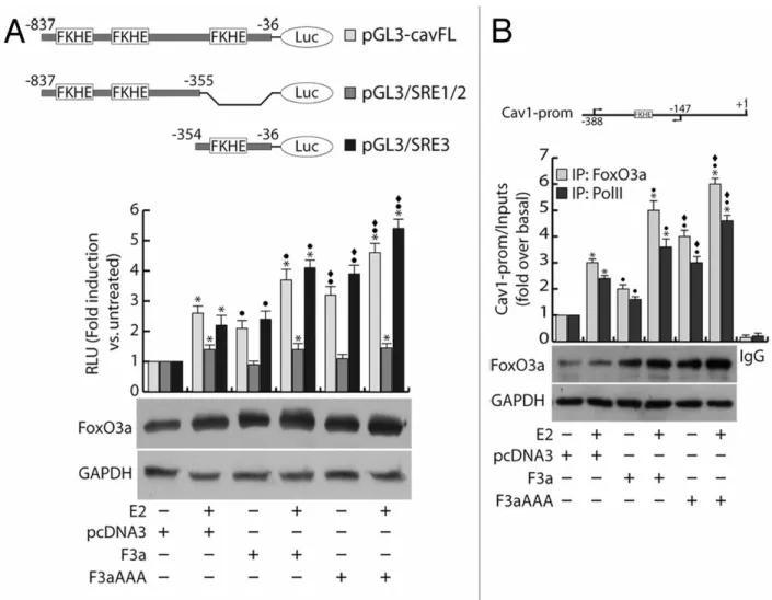
FoxO3a binds to and trans-activates the Cav1 promoter in MCF-7 cells
To deepen the understanding of the mechanism underlying the FoxO3a/ERα interplay in Cav1 induction, through an accurate analysis of the Cav1 promoter (GenBank accession #AF095591.1), we verified the presence of several Forkhead core sequences (FKHE), and we questioned if any of the identified regions may be involved in the FoxO3a/ERα-mediated regulation of Cav1 gene expression in ERα+ breast cancer cells.
To this aim, a vector bearing the luciferase gene under the control of the -837/-36 region of Cav1 promoter (pGL3-cavFL) was co-transfected with F3a or F3aAAA in MCF-7 cells and exposed or not to E2 treatment. In line with the results reported in Figure 5A and B, E2 stimulation significantly induced the Cav1 promoter activity, and such effect was increasingly higher in F3a- and F3aAAA-transfected cells (Fig. 8A). Interestingly, the construct pGL3/SRE1/2 (nt −837/−355), although containing FKHE core sequences, failed to be induced by FoxO3a but still weakly responded to hormone stimulation, most likely for the presence of Sp1 and AP-1 sites; on the contrary, the construct pGL3/SRE3 (nt −354/−36), bearing only one FKHE motif (nt −305/−299) and several Sp1 and AP-1 sites, was induced by both E2 and overexpressed FoxO3a, with a trend comparable to that observed with the pGL3-cavFL construct (Fig. 8A).
The involvement of E2 and FoxO3a in the transcriptional activation of the Cav1 promoter was corroborated by chromatin immunoprecipitation (ChIP)
experiments, which evidenced a significant recruitment of FoxO3a on the region containing the −305/−299 FKHE sequence. Once again, E2 treatment strongly increased FoxO3a occupancy of the promoter, especially in F3a- and F3aAAA-overexpressing samples (Fig. 8B). A similar pattern was observed in Polymerase II (PolII) precipitates, confirming that E2 and FoxO3a, both independently and synergistically, are able to induce Cav1 gene transcription (Fig. 8B).
Figure 8. FoxO3a binds to and transactivates the Cav1 promoter. (A) MCF-7 were seeded in culture
medium on 24-well plates, serum starved for 24 h, co-transfected in PRF-CT with pGL3-cavFL, or pGL3/SRE1/2, or pGL3/SRE3 and pRL-Tk, in presence of either pcDNA3 or F3a or F3aAAA vectors. After 6 h, E2 (100 nM) was added to the medium, where opportune, and the next day cells were harvested, and luciferase activity was evaluated. Cell extracts were also processed by WB analysis to assess F3a and F3aAAA transfection efficiency; GAPDH was used as loading control. (B) ChIP analysis was performed on the nuclear extracts from subconfluent MCF-7 cells seeded in 15 cm dish diameter, switched to PRF-SFM, and transfected with pcDNA3, F3a, or F3aAAA vectors. Twenty-four hours after transfection, the cells were treated with 100 nM E2 for 30 min or left untreated. The FKHE-containing Cav1 promoter region, precipitated with either anti-FoxO3a or anti-PolII pAbs were amplified using a specific pair of primers reported in “Materials and Methods”. E2-treated samples were also precipitated with normal rabbit IgG and used as negative control. FoxO3a expression in transfected samples was analyzed by WB on Cytosolic lysates from the same set of cells. Data represents the mean ±s.d. of 3 independent experiments. *, P < 0.05 vs. untreated; ●, P < 0.05 vs. corresponding pcDNA3; ♦, P < 0.05 vs. corresponding F3a.
Nuclear FoxO3a correlates in an opposite way with the tumor grade and the invasive phenotype in ERα+ and ERα−breast tumors
Tissue specimens from ductal carcinomas in situ (DCIS) and invading ductal carcinomas (IDC) (Fig. 9J) were analyzed to investigate if FoxO3a expression could correlate with the tumor grade and the invasive potential in ERα+ and ERα− breast tumors, as well as with Cav1 expression (in ERα+ tumors only). In all sections, tumor cells were clearly distinguishable from either infiltrating immune cells or stromal cells. In non-invading, well-differentiated ERα+ tumors, FoxO3a was strongly expressed, showing a very high nuclear localization (Fig. 9A).
Strikingly nuclear FoxO3a positivity was gradually lost in invading and less differentiated cells (see insets in Fig. 9B), while cytoplasmic localization was not as indicative. Concomitantly, Cav1 expression tended to decrease from tumors with positive to negative FoxO3a nuclear staining, and was completely lost in highly invading ERα+ tumors (Fig. 9D–F). Statistical analysis of these samples showed that both FoxO3a nuclear expression and Cav1 were inversely correlated with tumor grade and the invasive potential, while cytosolic FoxO3a did not result to be significantly correlated with any clinicopathological feature (Fig. 9K); moreover, Cav1 expression resulted directly correlated with FoxO3a nuclear content (Fig. 9K).
On the contrary, a very weak or even absent FoxO3a nuclear localization was observed in intraductal, well delimited areas of ERα− tumors (Fig. 9G), while a very strong nuclear staining was detected in invading areas of the same samples (Fig. 9H) and in clearly invasive carcinomas (Fig. 9I). This observation was confirmed by statistical analysis that evidenced a direct correlation between FoxO3a expression and both tumor grading and the invasive potential of ERα− breast cancer tissues (Fig. 9L).
Figure 9. Nuclear FoxO3a is highly expressed in non-invasive ERα+, and in invasive ERα− breast tumors.
FoxO3a (A–C) and Cav1 (D–F) expression in ERα+ breast tumors and FoxO3a (G–I) in ERα− breast tumor samples. IHC was conducted on tissue sections deriving from biopsies diagnosed as DCIS (A and D), microinvasive DCIS (B and E), DCIS with contiguous IDC areas (G and H) and highly aggressive IDC (C, F,
and I). Representative fields were photographed at 20× magnification. Insets, showing details of proteins
subcellular localization, were taken at 100× magnification. (J) Samples descriptions and classification; (K) correlation between nuclear FoxO3a or Cav1 content and the tumor grading and invasive potential in ERα+ breast cancer samples; (L) correlation between nuclear FoxO3a content and the tumor grading and invasive potential in ERα− breast cancer samples. The correlation coefficient (r) and the statistical significance (P) are reported.
Discussion
FoxO transcription factors are crucial for regulating a myriad of physiological processes, including proliferation, metabolism, cell differentiation, cell cycle arrest, DNA repair, and apoptosis.
FoxOs also play important roles in tumorigenesis, since they have been shown to be deregulated in many types of human cancers, and restoring their expression/activity has been shown to be effective in tumor suppression (Yang
JY et al,2011).
The involvement of FoxOs in tumor metastasis is controversial, e.g., FoxO3a has been reported to have either a protective or a promoting role on cell motility and invasion (Belguise K et al, 2007; Storz P et al, 2009).Our hypothesis was that such a difference might be ascribed to ERα status, since activated FoxO3a was able to reverse the invasive phenotype of ERα+ breast cancer cells
(Belguise K et al, 2007) while promoting tumor cell invasion in other cancer cell
lines, which, notably, were all ERα− (Storz P et al, 2009). Thus, the present study was aimed to verify if the effect exerted by FoxO3a on the metastatic potential of ERα+ breast cancer could derive from a general mechanism through which FoxO3a cooperates with the nuclear receptor in reducing motility and invasiveness of ERα+ tumors, while in absence of the receptor FoxO3a favors a more migrating and invasive phenotype. Indeed, since Erα signaling is well known to strongly correlate with a lower invasiveness and reduced motility of breast cancer cells (Sisci D et al, 2010) and considering that increasing evidences recognize Forkhead factors as important modulators of ERα transcriptional activity, (Schuur ER et al, 2001; Zhao HH et al, 2001; Morelli C
et al, 2010) it won’t surprise to ascertain that, in ERα+ tumors, FoxO3a could
reduce cell migration and invasion through a functional interaction with ERα. On the other hand, in ERα− tumors, the absence of the receptor could enable FoxO3a to trigger some different pathway that leads to an opposite outcome.
To prove our hypothesis, minimally motile and invasive ERα+ MCF-7 and ZR-75 breast cancer cell lines have been transfected with wild-type F3a and constitutively active F3aAAA mutant, and the effects on cell migration, invasion, and colony formation in soft agar were observed. The results presented here show that FoxO3a overexpression reduces the migratory and invasive potential, as well as anchorage-independent growth (a hallmark of tumor progression), in ERα+ tested cells. It is worth noting that, in all experiments, the constitutively active mutant F3aAAA was always more effective than the wild-type FoxO3a, suggesting that the regulation of the above-mentioned features could occur at the transcriptional level, through the induction of Forkhead responsive genes. Moreover, the expected reduced motility and invasiveness of ERα+ cells upon E2 stimulation (Sisci D et al, 2010) was more evident in F3a and, especially, in F3aAAA-overexpressing cells, providing evidence that E2 and FoxO3a act synergistically on these 2 features (Fig. 1A and B; Fig. 2, upper panels). On the contrary, E2 stimulation does not show an anti-metastatic behavior in presence of growth factors, since it favors the anchorage-independent growth (Manni A et al, 1991), suggesting that other growth factors regulated pathways do prevail on that of ERα in the control of this feature. However, in line with our previous observations (Morelli C et al, 2010), FoxO3a overexpression was able to counteract the proliferative effect of E2, and its silencing led to an increase in basal as well as in E2-dependent cell growth (Fig. 1C1 and C2). Taken together, these results suggest, once again, that FoxO3a
might act as a corepressor (e.g., by quenching E2/ERα dependent proliferative signals (Morelli C et al, 2010)) or a co-activator (e.g., by potentiating E2/ERα mediated inhibition of cell motility and invasion (Sisci D et al, 2010)) for ERα (Zhao HH et al, 2001).
More importantly, ERα is the key regulator of FoxO3a function, as evidenced by the opposite behavior of overexpressed F3a (and F3aAAA) in ERα-silenced cells if compared with the corresponding ERα-expressing samples (Fig.3). Thus,
the lack of the hormone receptor is responsible for the switch of FoxO3a biological function, which shifts from inhibitory (when ERα is present) to stimulatory (when ERα is absent) on cell motility, invasion, and growth in suspension.
This is confirmed by the fact that FoxO3a overexpression exhibits a stimulating (rather than inhibitory as in ERα+ cells) effect on the same features in ERα− MDA-MB-231,MDA-MB-468, and MDA-MB-435S breast cancer cells.
Notably, since the results observed in ERα+ and ERα− breast cancer cells following F3a and F3aaAAA ectopic expression, were similar to those obtained in non-breast cancer Ishikawa (ERα+ human endometrial adenocarcinoma) and HeLa (ERα− human cervical cancer) cell lines, respectively, we could assume that FoxO3a controls cell migration, invasion, and growth in suspension with a general, not tissue-specific, mechanism, which seems to depend on ERα expression (Fig. 4; Fig. 2).
Our results also show how Cav1 represents the ultimate downstream target through which FoxO3a modulates the metastatic potential of ERα+ cells. Cav1 is a multifunctional scaffolding protein that is associated with cell surface caveolae and the regulation of lipid raft domains. Cav1 regulates multiple cancer associated processes, including cellular transformation, tumor growth, cell migration and metastasis, cell death and survival, multidrug resistance, and angiogenesis. In breast cancer, Cav1 seems to function as a tumor suppressor
(Sotgia F et al, 2006). In fact, Cav1 mRNA and protein are downregulated or
absent in primary human cancers as well as in several mouse and human breast cancer cell lines. Forced re-expression of Cav1 in transformed mammary cell lines abrogates numerous of their tumorigenic properties, including anchorage-independent growth and invasiveness (Fiucci G et al, 2002) and suppresses growth of breast cancer cell-derived xenografts in nude mice (Wu P et al, 2008). Moreover, Cav1−/− mice showed an accelerated onset of mammary tumors and lung metastases (Williams TM et al, 2004). In accordance, Cav1 expression has
been inversely related to the grade of the primary breast tumors and its upregulation was found to reduce metastasis to distant organs (Sloan EK et al,
2004).
In light of this evidence, we questioned if FoxO3a could exert a protective role in ERα+ breast cancer cells through the induction of Cav1 expression. Indeed, in all ERα+ cells tested, FoxO3a overexpression increased the RNA and protein amounts of Cav1, and such increase was additive to that observed under E2 treatment, suggesting that ERα is also involved in the transcriptional induction of Cav1 (Fig. 5), which, in turn, seems to be the effector of a less aggressive phenotype, as evidenced by Cav1-silencing experiments (Fig. 7A–D) and by the fact that F3a and F3aAAA overexpression failed to inhibit migration, invasion, and growth in suspension in Cav1-negative T47D cells, despite the presence of a low, but still functional, content of ERα (Fig. 7E–H).
Since the highest induction of Cav1 has always been observed in F3aAAA-transfected cells, Cav1 regulation by FoxO3a and estrogens at the transcriptional level was investigated. In fact, the 5′-flanking region of the CAV1 gene, including the promoter region, bear several perfect and predicted forkhead consensus sequences, one of which (at position −1814, located above the promoter sequence) has been reported to be responsible for forkhead dependent
CAV1 gene regulation (van den Heuvel AP et al, 2005). However, as the same
authors stated, it is possible that other FKHE, also present within the 5′-flanking region, may play a role in Cav1 transcriptional activation by FoxO as well. Indeed, the data presented here clearly show how FoxO3a is able to induce Cav1 transcription by binding to a FKHE motif, mapping nt −305/−299 of its promoter;in addition, the FoxO3a-dependent Pol II recruitment confirms the occurrence of a transcriptional event (Fig. 8). To explain the induction exerted by E2, alone or in combination with FoxO3a, on Cav1 expression, we exclude, at the present stage, the direct involvement of ERα in the transcriptional process, since an integrated analysis of ERα binding sites upstream of the Cav1 gene,
through Myles Brown lab data sets (http://research.dfci.harvard.edu/brownlab/datasets/index.php?dir=ER_whole_h uman_genome/) (Carroll JS et al, 2006) and Cistrome-web application (http://cistrome.dfci.harvard.edu/ap/), evidenced that ERα recruitment to the chromatin occurs at a very large distance from the promoter, on 3 distinct positions around 80–100 Kb upstream of the transcription start site. No ERα binding is reported in the data sets at the promoter level or in its close proximity, as also confirmed by ChIP experiments conducted on several predicted estrogenresponsive motifs identified within the +1/−5000bp region (data not shown). Additionally, neither Sp1 nor AP-1 transcription factors, 2 well-established mediators of the ERα “non-classical” genomic pathway (Safe S et al,
2008) that have been reported to transcriptionally cooperate with FoxO3a, (Lützner N et al, 2012; Luo X et al, 2007) resulted to be involved in Cav1
regulation.
In fact, both Sp1 silencing and c-Jun inhibition achieved through the dominant-negative (DN)/c-fos plasmid (Ahn S et al, 1998) did not lead to any significant decrease in FoxO3a/E2-dependent Cav1 promoter activation, nor to a reduction of Cav1 protein content (data not shown). Despite these observations, the evidence that liganded ERα induces Cav1 expression, and that E2 and FoxO3a, separately or synergistically, lead to a significant increase of Pol II recruitment on the Cav1 promoter region (Fig. 8), suggests that it would be interesting to investigate, by means of the recent and fascinating techniques Chromosome conformation capture (4C) technology and detection of loops in DNA-picked chromatin (DPC), (Simonis M et al, 2007; Abbondanza C et al, 2011) if the combined effect of E2 and FoxO3a on Cav1 expression could be ascribed to the interaction of at least one of the 3 above mentioned ERα binding sites, at 80–100 Kb upstream of the transcription start site, where FoxO3a is recruited to the
CAV1 gene promoter (ongoing experiments). In fact, recent studies using tiled
showed that EREs can function as enhancer elements far away (up to 100 Kb) from gene promoters, and that other cooperating transcription factors (e.g., FoxA1, AP1 and Sp1) can participate with ERα to regulate the expression of E2-induced genes (Carroll JS et al, 2005; Carroll JS et al, 2006).
Taken together, the results obtained in ERα+ cancer cells show that FoxO3a-dependent decrease of migration, invasion, and colony formation is mediated by both ERα and Cav1, as confirmed by knockout experiments of these two factors (Figs. 3, 5, and 7). In particular, ERα cooperates with FoxO3a in the transcriptional induction of Cav1, which, in turn, is responsible of the reduced aggressive phenotype of FoxO3- overexpressing ERα+ cells (Fig. 10).
On the other hand, several reports called into question Cav1 role as a tumor suppressor, since it has been found overexpressed in highly aggressive inflammatory breast cancer (IBC) human specimens and cell lines (Van den
Eynden GG et al, 2006) as well as in invasive human breast cancers samples,
where its expression was significantly associated with basal-like phenotype, high histological grade, shorter disease-free and overall survival, and, more interestingly, lack of steroid hormone receptors positivity(Savage K et al,2007;
Elsheikh SE et al, 2008). Moreover, in ERα− cancer cells, Cav1 has been found
in membrane protrusions, where it promotes tumor cell migration and invasion by regulating either the function of membrane type 1 matrix metalloproteinase (MT1-MMP) (Yamaguchi H et al, 2009), or, when phosphorylated (pY14Cav1), the focal adhesion turnover (Joshi B et al, 2008). Therefore, we investigated if the more aggressive phenotype of FoxO3a overexpressing ERα− cells could depend, also in this case, on Cav1 induction. However, no differences in Cav1 levels or phosphorylation status have been detected in ERα− cells following FoxO3a overexpression, nor E2 treatment, possibly through ERβ, has been able to induce Cav1 expression (Fig. 5, and data not shown).
Although MMP-9 and MMP-13 induction has been proposed as the mechanism through which FoxO3a increases invasion of cells lacking the hormone receptor,
(Storz P et al, 2009) not all the ERα− cell lines tested do express these MMPs,
or do express negligible levels.
Moreover we failed to detect a reproducible increase in MMP-9 transcripts and in MMP-13 mRNA and protein in FoxO3aoverexpressing cells (data not shown), thus other markers are currently being investigated in our laboratory to justify the higher motility and greater invading ability induced by FoxO3a in ERα− cells. However, it is worth to underline that ERα silencing is a sufficient condition to reverse the effect of FoxO3a on migration, invasion and colony formation in ERα+ cells (Fig. 3), thus ERα seems to be a pivotal regulator of FoxO3a function, which switches from protective to malignant depending, respectively, on the presence or absence of the hormone receptor. A schematic representation of our findings is shown in Figure 10.
Finally, an immunohistochemical study from Yoshino’s research group showed that nuclear FoxO3a associates with IDC and lymph node metastasis, and the same authors speculated that, in some cases, aberrant activation of FoxO3a may cause the recruitment of metastasis-related molecules, instead of inducing apoptotic genes (Jin GS et al, 2004). Since no association with ERα status has been considered in this study, it might be possible that nuclear FoxO3a could correlate to a more metastatic phenotype only in the subset of ERα− IDC. In line with this hypothesis, nuclear FoxO3a has been recently proposed as a good prognostic factor in luminal- like breast cancer, which contain principally ERα+ cases, (Bertos NR et al, 2011) where it directly correlates with biomarkers of good prognosis and inversely with mitotic counts and tumor grade. Moreover, with respect to patient outcome, FoxO3a nuclear localization was associated with longer breast cancer specific survival and longer distant metastasis-free interval, independently of the well-established breast cancer prognostic factors
(Habashy HO et al, 2011).
The screening of nuclear FoxO3a on opportunely selected ERα+ and ERα− tissue samples from patients with breast cancer of ductal origin gave results that
perfectly fit with the above-mentioned reports and also confirm the in vitro studies presented in this work. Moreover, the co-expression of Cav1 and FoxO3a in ERα+ tumors, together with the functional link provided by our in vitro data, supports a potentially important role for these 2 proteins in predicting a better tumor prognosis. However, a more systematic evaluation within various subtypes of ERα+ and ERα− non-invasive and invasive breast cancers, in absence or in presence of lymph node and/or long distance metastasis, would help to better clarify the biological and prognostic role of FoxO3a protein expression, also with respect to its subcellular localization.
For instance, since no correlation has been found between FoxO3a and ERα
(Habashy HO et al, 2011), the loss of an active (nuclear) FoxO3a might be
predictive of a worse phenotype in the subset of ERα+ breast cancers that do not respond to therapy. At the same time, a more accurate immunohistochemical analysis on the biological link between FoxO3a and Cav1 in hormone-positive tumors needs to be addressed. In fact, although Cav1 expression has been associated with lack of the steroid hormone receptor (Elsheikh SE et al, 2008), its positivity in luminal-like tumors could represent a good prognostic factor when associated to a FoxO3a nuclear prevalence.
In conclusion, the results presented here give new insights on the functional role of nuclear FoxO3a, whose overexpression seems to be associated to a low motile phenotype in ERα+ breast cancers and to a more metastatic potential in those lacking the hormone receptor, harboring the idea that ERα may represent the molecular switch determining FoxO3a biological behavior.
These evidences clearly suggest that FoxO3a has the potential to become a relevant prognostic factor and a suitable pharmacological target to be exploited in combination therapies for both ERα+ (through FoxO3a activation) and ERα− (through FoxO3a disruption) breast cancer patients.
Figure 10. Proposed model for FoxO3a-mediated control of cell motility and invasiveness in presence or absence of ERα. F3a and ERα synergistically induce the expression of Cav1, which, in turn, reduces cell
motility and invasiveness of ERα+ breast cancer cells. Transcriptionally active F3a binds to a FKHE located on the Cav1 proximal promoter and increases the recruitment of RNA Polymerase II, which is enhanced upon E2 stimulation. The lack of the hormone receptor enables active F3a to behave in an opposite fashion, thus increasing cell motility and invasion. Basal TM, basal transcriptional machinery.