I
INDEX
RATIONALE ...1
BACKGROUND ...6
1. HUMAN ADRENAL GLAND 1.1 The adrenal gland:general structure ...7
1.2 Embriology and development ...8
1.3 Hystology ...10
1.4 Adrenalcortical steroidogenesis ...11
1.5 The steroidogenic regulatory protein ...14
1.5.1 StAR structure and mechanism of action ...15
1.5.2 P450scc ...15
1.5.3 P450c17: 17α-Hydroxylase/17,20-Lyase ...16
1.5.4 Cytochrome b5 and P450c21 (Steroid 21-Hydroxylase ...17
1.5.5 Isozymes of P450c11 and Isozyme of 17β –Hydroxysteroid Dehydrogenase 18 1.5.6 P450arom: Aromatase ...18 1.5.7 Isozyme of 5α-Reductase ...19 1.5.8 3βHSD ...19 2. ADRENOCORTICAL CANCERS 2.1 Introduction ...20 2.2 Epidemiology ...21 2.3 Pathogenesis ...22 2.4 Adrenocortical adenoma ...25 2.5 Adrenocortical carcinoma ...25
2.6 Role of ERα and ERβ activation on ACC development ...29
3. G PROTEIN COUPLED ESTROGEN RECEPTOR: GPER AND ITS LIGAND 3.1 Introduction ...32
II
3.3 Identification and characterization of GPER-selective ligands ...34
3.4 Transcriptional activations mediated by GPER ...37
4. CELL-BASED ASSAYS FOR SCREENING ANDROGEN RECEPTOR LIGANDS 4.1 Introduction ...41
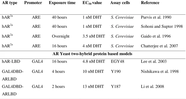
4.2 Yeast-based systems using a β-galactosidase reporter ...43
4.3 β-Lactamase Reporter Model ...47
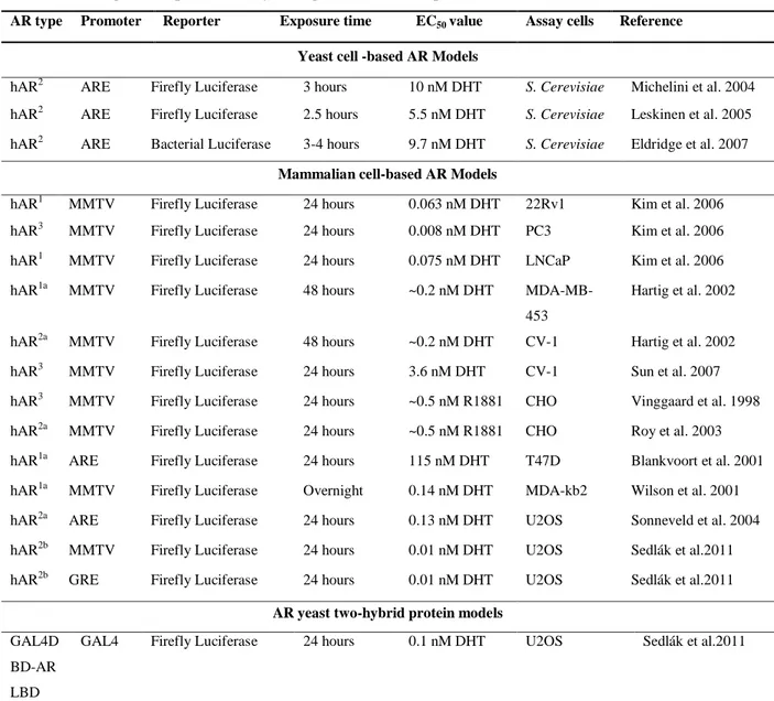
4.4 Lucifeerase Reporte Model ...48
4.4.1 Yeast-AR luciferase models ...50
4.4.2 Mammalian cell AR luciferase models ...50
4.5 Green Fluorescent Reporter Model ...53
4.6 Fluorescent AR trans location bioassay ...55
5. MATERIALS AND METHODS 5.1 GPER agonist G-1 decreases adrenocortical carcinoma (ACC) cell growth in vitro and in vivo ...57
5.1.1 Cell cultire and tissues ...57
5.1.2 RNA extraction, reverse transcription and real time PCR ...58
5.1.3 Western blot analysis ...58
5.1.4 Histopathological and Immunohistochemical analysis ...59
5.1.5 Cytochrome c detection ...59
5.1.6 Cell cycle analysis and evaluation of cell death ...60
5.1.7 Caspase 9 and 3/7 Activity Assay ...60
5.1.8 TUNEL (terminal deoxynucleotidyltransferase-mediated dUTP nick-end labelling) assay ...61
5.1.9 Determination of DNA fragmentation ...61
5.1.10 Assessment of cell proliferation ...61
5.1.11 Gene silencing experiments ...62
5.1.12 Xenograft model ...62
5.1.13 In vivo magnetic resonance analyses ...63
III
5.1.15 Macroarray analysis ...63
5.1.16 Data analysis and statistical methods...64
5.2 Development of a novel cell based androgen screening model ...64
5.2.1 Materials ...64
5.2.2 Cell line ...64
5.2.3 Stable Trasduction ...65
5.2.4 Isolation of RNA and qPCR analysis ...65
5.2.5 Protein extraction and protein assay ...66
5.2.6 Western analysis ...66
5.2.7 AR translocation study...66
5.2.8 Gaussia Luciferase analysis ...66
5.2.9 Sera ...67
5.2.10 Extraction method ...67
6. RESULTS 6.1 GPER agonist G-1 decreases adrenocortical carcinoma (ACC) cell growth in vitro and in vivo ...68
6.1.1 G-1 treatment decreases H295R cell growth in vtiro and in vivo ...68
6.1.2 G-1 induces H295R cell cycle arrest and cell death ...72
6.1.3 G-1 causes cell nuclei morphological changes, DNA damage and apoptosis .73 6.1.4 G-1 treatment causes sustained ERK1/2 phosphorylation ...75
6.1.5 Microarray data ...77
6.2 Development of a novel cell based androgen screening model ...79
6.2.1 AR expression in transduced CV1 cell line ...79
6.2.2 Sensitivity of CV1-ARluc cell line ...81
6.2.3 Selectivity of CV1-ARluc cell line to other steroids ...83
6.2.4 Treatment of CV1-ARluc cell line with different C19 steroids ...83
6.2.5 Effects of a potent anti-androgens on CV1-ARluc ...84
IV
7. DISCUSSION
7.1 GPER agonist G-1 decreases adrenocortical carcinoma (ACC) cell growth in vitro and in vivo ...87 7.2 Development of a novel cell based androgen screening model ...90
1
RATIONALE
During the first part of the PhD program, it was investigated the role of G-1, a new ligand of the novel Estrogen receptor coupled to G-Protein (GPER) in the progression of adrenocortical carcinoma (ACC). The second part of the program was performed in the University of Michigan (Ann Arbor,USA) where it was completed the first project using Microarray tachnology and was performed a second line aimed to develop a novel cell-based androgen screening model.
GPER agonist G-1 decreases adrenocortical carcinoma (ACC) cell
growth in vitro and in vivo
Adrenocortical tumors (ACTs) are common, and most are benign adrenocortical adenomas (ACAs). Malignant adrenocortical carcinoma (ACC) is a rare tumor type and is observed at the rate of one or two cases per million annually. ACTs are classified as either ACAs or ACCs by histopathologic methods that are based on nine Weiss scoring criteria, including the nuclear grade, mitotic rate, presence of necrosis, and others (Erickson et al., 2014). The molecular genetics of adrenocortical tumors remain poorly understood. For decades, molecular studies relied on a small number of samples and were directed to candidate genes. This approach, based on the elucidation of the genetics of rare genetic syndromes in which adrenocortical tumors are a manifestation, has led to the discovery of major dysfunctional molecular pathways in adrenocortical tumors, such as the IGF pathway , the Wnt pathway and TP53(Fassnacht et al., 2013). Interestingly, allelic losses (LOH) at the TP53 locus (17p13) are very frequent and observed in more than 85% of ACC (Bertherat and Bertagna, 2009). Transcriptome analysis suggests also that the Wnt/beta-catenin signalling pathway is activated in ACT. About a third of ACC harbours somatic activating mutations of the betacatenin gene (Bertherat and Bertagna, 2009; Logie et al., 1999). The most consistent and dominant genetic changes in ACC is the perturbation of the insulin-like growth factor II (IGF-II) locus (11p15) that is imprinted. IGF-II is over-expressed in 90% of ACCs determining an autocrine mitogenic effect (Sampaoli et al., 2012). The direct involvement of IGF-II/IGF-IR
2
system in adrenocortical tumor cell proliferation has been also shown in vitro using adrenal cancer cell line NCI H295R (Logie et al., 1999). Moreover, increased levels of the IGF-IR have been found in advanced human ACC, suggesting an important role for the IGF system in adrenocortical carcinogenesis. For this reason inhibitors for IGF-IR are currently in preclinical trials. However, ACC is a disease extremely heterogeneous and this new pharmacological approach could not be enough for the therapy of all forms of ACC, since several molecular mechanisms trigger ACC development. Thus, progress in the understanding of the pathophysiology of ACC is important to improve diagnosis, prognostic evaluation and treatment of different types of ACC. Usually, ACC are more frequent in women than in men, especially in those exposed to estro-progestin (Barzon et al., 2003; Hsing et al., 1996).
Professor Pezzi’s group have demonstrated that ACC are characterized by ERα up-regulation and aromatase (the enzyme involved in the production of estrogens using androgens as substrate) over-expression (Barzon et al., 2008) and that estradiol enhances proliferation of the human adrenocortical carcinoma cell line H295R, whereas antiestrogens upregulate ERβ and inhibit ACC cell growth (Montanaro et al., 2005a). It is well known that tamoxifen and its active metabolite 4-hydroxytamoxifen (OHT), not only exert antiestrogenic activity [9], but also act as full agonist on the G protein-coupled estrogen receptor GPER (from the GPER gene) (Lappano et al., 2013; Vivacqua et al., 2006a). GPER can mediate rapid E2-induced non-genomic signaling events, including stimulation of adenylyl cyclase, mobilization of intracellular calcium (Ca2+) stores and activation of mitogen-activated protein kinase (MAPK) and phosphoinositide 3-kinase (PI3K) signaling pathways (Ariazi et al., 2010; Prossnitz and Barton, 2009). GPER exhibits prognostic utility in endometrial (Smith et al., 2007), ovarian (Smith et al., 2009), and breast cancer (Filardo et al., 2006) and can modulate growth of hormonally responsive cancer cells (Vivacqua et al., 2006b). Expression of GPER has been characterized in the outer zona glomerulosa (ZG) and in the medulla of the human adrenal (Baquedano et al., 2007), however its expression status in ACC is not known. A non-steroidal, high-affinity GPER agonist G-1 (1-[4-(6-bromobenzo [1, 3]dioxol-5yl)-3a, 4, 5, 9b-tetrahydro-3H-cyclopenta-[c]quinolin-8-yl]-ethanone) has been developed to dissect GPER-mediated estrogen responses from those mediated by classic estrogen
3
receptors (Bologa et al., 2006). The biological effects triggered by G-1 appear cell type specific and dependent on the ERs expression pattern (Chimento et al., 2013a; Chimento et al., 2013b; Chimento et al., 2012; Chimento et al., 2010; Chimento et al., 2011). Starting from these observation, by using G-1, we investigated the effects of GPER activation on ACC growth.
Development of a novel cell based androgen screening model
Androgens are hormones that play an essential role in the differentiation and maintenance of primary and secondary male sexual characteristics (Gao et al., 2005). The two main human androgens are testosterone (T), which is involved in the initial virilization phases of the human male embryo, and 5α-dihydrotestosterone (DHT), which is the active hormone in most androgen target tissues (Wiener et al., 1997). T is mainly synthesized by the testicular Leydig cells, in peripheral tissues, as well as to a lesser degree in ovaries and adrenals. T is converted to DHT by 5α-reductases and also can be converted to estradiol by aromatase. DHT is the most active physiologic androgen, inducing ten-fold higher androgen receptor (AR, NR3C4) bioactivity than T (Paris et al., 2002; Raivio et al., 2002). In addition, other endogenously produced steroids exhibit various degrees of androgenic activity (Mitchell, 2012; Rege et al., 2013). Several synthetic androgen-related compounds (AR agonists and antagonists) have also been developed to modulate androgen signaling in therapeutic settings (Fang et al., 2003; Larsson et al., 2011).
Androgens mediate their effects through binding and activation of the AR. AR is a member of the steroid nuclear receptor superfamily (Kato and Fujiki, 2008) and acts as a ligand-dependent transcription factor (Lubahn et al., 1988). Among this family, five steroid receptors are known: estrogen (ESR, NR3A1), progesterone (PR, NR2C3), androgen, mineralocorticoid (MR, NR3C2) and glucocorticoid (GR, NR3C1) receptors. AR activates a wide range of target genes that encode proteins and noncoding RNAs, including regulatory microRNA species (Narayanan et al., 2010).
Similar to the other steroid receptors, unbound AR is located in the cytoplasm. Upon ligand binding, AR goes through a series of conformational changes, dimerization and translocation to the nucleus, which is mediated by a nuclear localization signal.
4
Translocated AR binds to androgen response elements (ARE). These ARE are characterized by a consensus (or near consensus) sequence 5’-TGTTCT-3’, which is located in the promoter or enhancer regions of AR gene targets. The DNA cis-regulatory elements that respond to AR share sequence similarity with cis-regulatory elements for GR, MR and PR. The similarity of the response element for AR and the other steroid receptors, and particularly the wide-spread expression of the GR, has been problematic in the development of selective receptor screening assays.
The determination of androgen levels or the discoveries of new androgenic compounds are key elements for the diagnosis of a number of diseases in children and adults. Assays that detect bioactive serum androgens in a sensitive and selective manner benefit the diagnosis and treatment of several pediatric endocrine disorders, such as precocious puberty and ambiguous genitalia. In addition, androgen bioassays provide a screening tool for androgen abuse and endocrine disruptors (Bagchi Bhattacharjee and Paul Khurana, 2014). Over the past 10 years, several bioassays were developed using different methods (Campana et al., 2015). One of the first assays developed relied on a chloramphenicol acetyltransferase (CAT) reporter model (Xu et al., 2008b). This system was limited by experimental variation due to the transient nature of transgene expression. A luciferase reporter bioassay, using MDA-MB453 cells, was developed by Wilson et al (Wilson et al., 2002b). The major caveat of this assay was that it responds to AR as well as to GR agonists. Other androgen-reporter cell lines were developed but most of them were transiently transfected (Kim et al., 2006a; Sun et al., 2007; Vinggaard et al., 1999). Transient transfection assays (He et al., 2000) can provide similar information with stable assays but may not reflect endogenous levels of receptor. A stable expression of AR in the cells can eliminate the need for repetitious transient transfections, reduce the variability associated with these transient assays and moreover be utilized for high-throughput studies. Until now, a selective androgen-responsive transcriptional activation assay has not been widely available.
The aim of this study was to develop a stable cell-based in vitro bioassay that expresses the human AR (hAR) gene with sensitive and selective reporter readout. For this purpose, a stable cell line was made with CV1 cells stably transduced with hAR and an MMTV promoter-driven luciferase reporter gene. The resulting model is selective for
5
androgens and does not exhibit reporter activation by other steroid receptors. In addition the model appears useful to determine circulating androgenic bioactivity in human serum samples.
6
Background
7
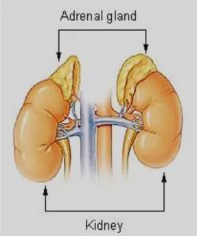
Figure 1.1 Human adrenal gland
1. Human adrenal gland
1.1 The adrenal gland: general structure
In mammals, the adrenal glands (also known as suprarenal glands) are endocrine glands that sit at the top of the kidneys (Figure 1.1).
The adrenal glands are located bilaterally in the retroperitoneum superior and slightly medial to the kidneys. In humans, the right adrenal gland is triangular in shape, whereas the left adrenal gland is semilunar in shape; in non-humans, they are quadrilateral in shape. The combined weight of the adrenal glands in an adult human ranges from 7 to 10 grams. They are surrounded by anadipose capsule and renal fascia.
It is now known that the adrenal gland consists of two ontogenetically, structurally and functionally distinct endocrine tissues, the cortex and the medulla. The cortex is mesodermal in origin and derived from proliferation of the coelomic epithelium. It produces various steroids with specific functions as will be described later. The medulla, on the other hand, is ectodermal in origin and neural crest-derived. It secretes catecholamines, i.e., adrenaline and noradrenaline, that facilitate the acute mammalian stress or “fight-or-flight” response.
8 The adrenal glands affect kidney function through the secretion of aldosterone, and recent data suggest that adrenocortical cells under pathological as well as under physiological conditions show neuroendocrine properties; within normal adrenal glands, this neuroendocrine differentiation seems to be restricted to cells of the zona glomerulosa and might be important for an autocrine regulation of adrenocortical function.
Adrenocortical cells, on the other hand, are of mesodermal origin and synthesise steroid hormones that regulate body homeostasis and mediate chronic stress responses, as part of the endocrine hypothalamic-pituitary-adrenal (HPA) axis and renin-angiotensin system.
1.2 Embriology and development
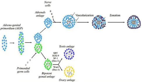
The adrenal gland is two distinct endocrine organs that have separate embryological origins and physiologic functions ; the mesoderm-derived cortex secretes steroid hormones while the neural crest-derived medulla secretes catecholamines (Else and Hammer, 2005). Formation of the adrenal gland occurs in several distinct developmental events (Else and Hammer, 2005; Kim and Hammer, 2007) (Figure 1.2). During the 4th week of gestation in humans (E9.0 in mice), proliferation of mesoderm-derived cells of the coelomic epithelia and underlying mesonephros results in coalescence of the adrenogonadal primordium (AGP), defined by expression of the nuclear receptor NR5a1 (Steroidogenic factor 1, Sf1) (Hatano et al., 1996; Luo et al., 1994). At the 8th week of gestation in humans (E10.5 in mice), the bipotential AGP separates into discrete adrenal primordia (fetal adrenal zone) and gonadal primordial (Hatano et al., 1996; Kim and Hammer, 2007). The segregation of a discrete adrenal primordia from the AGP involves aWilm’s tumor 1 (Wt1) and Cited2-mediated upregulation of Sf1 expression (Val et al., 2007). Once separated from the AGP, the adrenal primordial activates Sf1 expression through an entirely different mechanism – the recruitment of the homeobox protein PKNOX1 (Prep1), homeobox gene 9b (Hox) and pre B-cell leukemia transcription factor 1 (Pbx1) to a fetal adrenal-specific Sf1 enhancer (FAdE) (Zubair et al., 2008). Sf1 itself maintains FAdE-dependent expression of Sf1 in the adrenal primordia over time through autoregulation of Sf1 expression. Proliferation of fetal adrenocortical cells is
9
believed to be under control of fetal pituitary-derived adrenocorticotropic homormone (ACTH) (Mesiano et al., 1997). However, insulin like growth factor 2 (IGF2) is expressed throughout the fetal adrenal cortex and several studies have suggested ACTH mediates some of its effects on proliferation through IGF2 action (Coulter, 2005; Ilvesmaki et al., 1993; Stratta et al., 2003). Concurrent with activation of FAdE-driven Sf1 expression at embryonic day E11.5-12.5 in mice (equivalent to 8–9th week of gestation in humans), neural-crest-derived chromaffin progenitor cells migrate into the central fetal gland. These cells form the adrenal medulla followed by the coalescence of the mesenchymal capsule around the fetal adrenal gland (Else and Hammer, 2005). Before encapsulation is complete, the development of the definitive cortex (definitive zone or adult cortex) is initiated between the capsule and the fetal zone. While the fetal cortex ultimately regresses in all species, the timing of regression is species-specific; in humans the fetal zone regression occurs at birth while in mice the zone persists until puberty in males or the first pregnancy in females (Kim et al., 2009). In humans, functional zonation of the adult cortex into unique concentric steroidogenic regions initiates at birth concurrent with the coalescence of the adrenal medulla (Beuschlein et al., 2002).
10
1.3 Histology
The adrenal cortex is composed of three functionally distinct regions, the zona
glomerulosa (ZG) lying immediately below the capsule and corresponding to
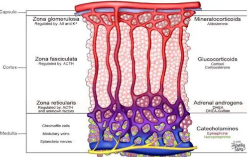
approximately 15% of cortical volume characterized by cells organised in rounded clusters around capillary coils or glomeruli, zona fasciculata (ZF) corresponding to up to 75% of cortical volume, characterized by cells arranged in radial rows separated by trabeculae and by blood vessels and zona reticularis (ZR) that lies next to the medulla, in which cells are located within a uniform reticular net of connective tissue and blood vessels (Miller WL, 2008). The ZG synthesizes mineralocorticoids; the ZF produces cortisol and the ZR secretes the so called adrenal androgens, DHEA and DHEA-sulfate. Each zone is preferentially regulated by different circulating factors that include angiotensin II (Ang II) and potassium (K+) for the ZG, adrenocorticotropic hormone (ACTH) for the ZF, and ACTH plus other yet to be determined factors for the ZR (Wang and Rainey, 2012) (Figure 1.3). It has been established that the reason each zone secretes a unique set of steroids is related to the selective expression of steroid-metabolizing enzymes within each zone (Nguyen and Conley, 2008; Rainey, 1999; Rainey et al., 2002). However, the molecular mechanisms that cause zone-specific expression patterns of enzymes are yet to be resolved. Adrenal steroid production remains an area of active research, which supports the need to develop appropriate cell models that can mimic adrenal physiology or pathology. Primary cultures of adrenocortical cells have proven to be useful for examining the mechanisms controlling many aspects of adrenal physiology (Cardoso et al., 2009; Chen and Hornsby, 2006; Kuulasmaa et al., 2008; Xing et al., 2011; Xing et al., 2010). However, several issues have limited the use of primary adrenal cells as in vitro models. The most common limitations are the constant requirement for fresh tissue and the difficulties associated with the isolation of adequate cortical cells. In addition, cells from different human donors are subject to considerable variability; whereas cells from rodents do not produce cortisol or adrenal androgens due to the lack of steroid 17ahydroxylase (CYP17) expression. To overcome the problems with tissue accessibility and quality, many groups have attempted to establish cell lines from adrenocortical carcinomas. This approach has been somewhat successful leading to adrenal cell lines from several
11 species and we have previously reviewed the overall development of these models (Mountjoy et al., 1994; Rainey et al., 2004).
Fig. 1.3 Adrenal cortex regions
1.4 Adrenalcortical steroidogenesis
Steroid hormones regulate a wide variety of developmental and physiological processes from fetal life to adulthood. Steroid hormones are all synthesized from cholesterol and hence have closely related structures based on the classic cyclopentanophenanthrene 4-ring structure. The human adrenal can synthesize cholesterol de novo from acetate (Mason and Rainey, 1987), but most of its supply of cholesterol comes from plasma low-density lipoproteins (LDLs) derived from dietary cholesterol (Gwynne and Strauss, 1982). By contrast, rodent adrenals derive most of their cholesterol from high-density lipoproteins via a receptor termed scavenger receptor B1, but this pathway appears to play a minor role in human steroidogenesis. The intracellular cholesterol economy is largely regulated by the sterol response element binding protein (SREBPs), a group of transcription factors that regulate genes involved in the biosynthesis of cholesterol and fatty acids (Horton et al., 2002). Adequate concentrations of LDL will suppress 3-hydroxy-3-methylglutaryl co-enzyme A reductase, the rate-limiting enzyme in cholesterol synthesis. ACTH also stimulates the activity of 3-hydroxy-3-methylglutaryl
12
co-enzyme A reductase, LDL receptors, and uptake of LDL cholesterol. LDL cholesterol esters are taken up by receptor-mediated endocytosis, and are then stored directly or converted to free cholesterol and used for steroid hormone synthesis (Brown et al., 1979).
The first step in steroidogenesis takes place within mitochondria. The mechanisms by which cholesterol is transported to and loaded into the outer mitochondrial membrane (OMM) remain an active area of research (Chang et al., 2006; Miller, 2007); the principal action of StAR is to facilitate the movement of cholesterol from the OMM to the inner mitochondrial membrane (IMM). Some cholesterol may be incorporated into vesicular membranes that then fuse with other membranes, thus delivering cholesterol from one intracellular compartment to another, but this appears to be a minor pathway (Soccio and Breslow, 2004). Instead, cholesterol is solubilized by binding to proteins. A steroidogenesis abnormality can often be life threatening. Congenital adrenal hyperplasia (CAH) is one of the most common disorders caused by deficiency of any enzyme involved in steroidogenesis in adrenal glands (Claahsen-van der Grinten et al., 2011; White and Bachega, 2012). Impaired cortisol and aldosterone production increases adrenocorticotropic hormone (ACTH) secretion from the pituitary gland, leading to adrenal hyperplasia and accumulation of adrenal androgens. Female patients are prenatally virilized because of excess androgen and neonates of both genders may suffer from a life-threatening Addisonian crisis. Steroid hormone deficiency also occurs in aging people by hypogonadism.
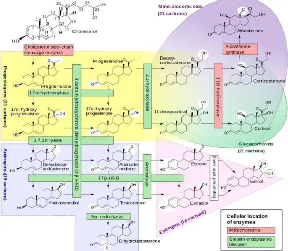
Most enzymes involved in steroid biosynthesis are either cytochrome P450s (CYPs) or HSDs. These steroidogenic enzymes are functionally, if not absolutely, unidirectional, so the accumulation of products does not drive flux back to the precursor. All P450-mediated hydroxylations and carbon-carbon bond cleavage reactions are mechanistically and physiologically irreversible (Hall, 1986) (Figure 1.4).
Cytochrome P450 is a generic term for a group of oxidative enzymes, all of which have about 500 amino acids and contain a single heme group (Gonzalez, 1988). The human genome includes genes for 57 cytochrome P450 enzymes (Lander et al., 2001; Venter et al., 2001). The genes are now formally termed CYP genes. Seven human cytochrome P450 enzymes are targeted to the mitochondria and are termed “type 1”; the other 50
13
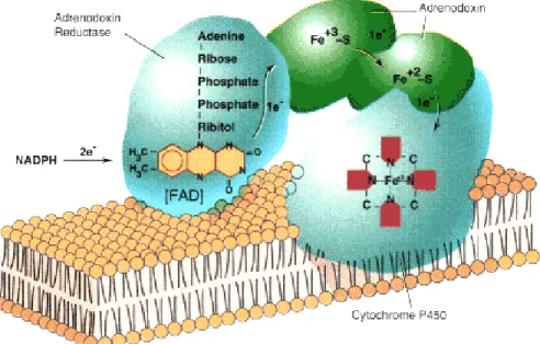
human P450 enzymes are targeted to the endoplasmic reticulum and are termed “type 2.” All P450 enzymes activate molecular oxygen using their heme center and add electrons from the reduced form of nicotinamide adenine dinucleotide phosphate (NADPH). Type 1 enzymes receive electrons from NADPH via a flavoprotein termed ferredoxin reductase and a small iron-sulfur protein termed ferredoxin, whereas type 2 P450 enzymes receive electrons from NADPH via a single 2-flavin protein termed P450 oxidoreductase (POR) (Miller, 2005). Six P450 enzymes are involved in steroidogenesis
Fig. 1.4 Major human steroidogenic pathways
Mitochondrial P450scc is the cholesterol side-chain cleavage enzyme catalyzing the series of reactions formerly termed “20,22 desmolase.” The two isozymes of mitochondrial P450c11, P450c11β (11β-hydroxylase) and P450c11AS (aldosterone synthase), catalyze 11β-hydroxylase, 18-hydroxylase, and 18-methyl oxidase activities. In the endoplasmic reticulum, P450c17 catalyzes both 17α-hydroxylase and 17,20-lyase
14
activities, P450c21 catalyzes 21-hydroxylation in the synthesis of both glucocorticoids and mineralocorticoids, and P450arom catalyzes aromatization of androgens to estrogens.
The HSDs have molecular masses of about 35 to 45 kDa, do not have heme groups, and require nicotinamide adenine dinucleotide (phosphates) (NADH/NAD+ or NADPH/NADP+) as cofactors; Based on their activities, it is physiologically more useful to classify the HSDs as dehydrogenases or reductases. The dehydrogenases use NAD+ as their cofactor to oxidize hydroxysteroids to ketosteroids, and the reductases mainly use NADPH to reduce ketosteroids to hydroxysteroids (Agarwal and Auchus, 2005; Sherbet et al., 2007)
1.5 The steroidogenic regulatory protein
Unlike cells that produce polypeptide hormones, which store large amounts of hormone in secretory vesicles ready for rapid release, steroidogenic cells store very little steroid. Thus, a rapid steroidogenic response (e.g., adrenal secretion of aldosterone and cortisol in response to stress or the “pulsing” of sex steroids in response to an LH surge) requires rapid synthesis of new steroid. ACTH promotes adrenal steroidogenic cell growth. This growth occurs primarily by ACTH stimulating the production of cAMP, which in turn promotes the synthesis of IGF-II (Mesiano et al., 1993; Voutilainen and Miller, 1987), basic fibroblast growth factor (Mesiano et al., 1991), and epidermal growth factor (Coulter et al., 1996). Together, these growth factors stimulate adrenal cellular hypertrophy and hyperplasia, determining the amount of steroidogenic tissue. Second, acting over days, ACTH acts through cAMP, and angiotensin II acts through the calcium/calmodulin pathway to promote the transcription of genes encoding various steroidogenic enzymes and electron-donating cofactor proteins, thus determining the amount of steroidogenic machinery in the cell. Third, ACTH rapidly stimulates StAR gene transcription (Stocco et al., 2005) and phosphorylation of Ser195 in extant StAR (Arakane et al., 1997) to increase the flow of cholesterol from the OMM to the IMM, where it becomes substrate for the first and rate-limiting enzyme, P450scc. This acute response occurs within minutes and is inhibited by inhibitors of protein synthesis (e.g., puromycin or cycloheximide), indicating that a short-lived protein species mediates this
15
process. All microsomal (type 2) cytochrome P450 enzymes, including steroidogenic P450c17, P450c21, and P450aro, receive electrons from POR, a membrane-bound flavoprotein that is a different protein from the mitochondrial flavoprotein, ferredoxin reductase (Miller, 2005). Nuclear magnetic resonance and x-ray scattering data have recently confirmed this view that POR undergoes these dramatic conformational changes while receiving and then transferring electrons (Ellis et al., 2009)
1.5.1 StAR structure and mechanism of action
The short half-life of the 37-kDa cytoplasmic precursor and the longer half-life of the “mature” 30-kDa intramitochondrial form of StAR initially suggested that the 30-kDa form was the biologically active moiety. When expressed in cytoplasm or added to mitochondria in vitro, both the 37- and 30-kDa forms of StAR are equally active (Arakane et al., 1996). When StAR is immobilized on the OMM, it is constitutively active, but StAR is inactive when localized to the mitochondrial intramembranous space or to the matrix (Bose et al., 2002). These data demonstrate that StAR acts exclusively on the OMM (Arakane et al., 1996; Bose et al., 2002), and its activity in promoting steroidogenesis is proportional to its residency time on the OMM (Bose et al., 2002). The interaction of StAR with the OMM involves conformational changes (Baker et al., 2005; Bose et al., 1999) that are necessary for StAR to accept and discharge cholesterol molecules. Although StAR can transfer cholesterol between synthetic membranes in vitro(Tuckey et al., 2008), suggesting that other protein molecules are not needed for its action, this activity can also be seen with the inactive mutant R182L, which is biologically inactive and causes lipoid CAH (Baker et al., 2007). Thus StAR’s action to promote steroidogenesis is distinct from its cholesterol-transfer activity.
1.5.2 P450scc
A cell is said to be steroidogenic if it expresses the cholesterol side-chain cleavage enzyme, P450scc, which catalyzes the first step in steroidogenesis. Conversion of cholesterol to pregnenolone in mitochondria is the first, rate-limiting, and hormonally regulated step in the synthesis of all steroid hormones (Koritz and Kumar, 1970; Macchi and Hechter, 1954). This process involves three distinct chemical reactions, the
22-16
hydroxylation of cholesterol, 20-hydroxylation of 22(R)-hydroxycholesterol, and oxidative scission of the C20–22 bond of 20(R),22(R)-dihydroxycholesterol (the side-chain cleavage event), yielding pregnenolone and isocaproaldehyde. P450scc can use the hydroxysterol intermediates directly as substrate, providing a useful experimental tool because these hydroxysterols are somewhat water-soluble and do not require StAR for access to P450scc. The reactions catalyzed by P450scc are slow, with a net turnover number of about six (Kuwada et al., 1991) to 20 (Tuckey and Cameron, 1993) molecules of cholesterol per molecule of P450scc per second. In human adrenal gene transcription is regulated by ACTH, by gonadotropins in testis and ovary and by unknown factcors in placenta all activated through cAMP as intracellular second messenger (Kimura and Suzuki, 1967). Each catalytic cycle requires a molecule of NADPH and one molecule of oxygen (Figure 1.5).
Fig 1.5 Electron transport to mitochondrial forms of cytochrome P450
1.5.3 P450c17: 17α-Hydroxylase/17,20-Lyase
P450c17 is the microsomal P450 enzyme that catalyzes both 17α-hydroxylase and 17,20-lyase activities, principally in the adrenal and gonads. These two activities were once thought to be catalyzed by separate enzymes that differed in the adrenals and gonads. Clinical observations showed that adrenal 17α-hydroxylase activity (reflected by serum cortisol concentrations) was fairly constant throughout life, This dissociation
17
between adrenal secretion of 17α-hydroxylase products (cortisol) and 17,20-lyase products (DHEA) suggested that distinct enzymes performed the two transformations. the distinction between 17α-hydroxylase and 17,20-lyase is functional and not genetic or structural. P450c17 is encoded by a single gene on chromosome 10q24.3 (Fan et al., 1992; Matteson et al., 1986), which is expressed in the adrenals and gonads (Chung et al., 1987), and not two tissue-specific isozymes as had been thought. This gene, formally called CYP17A1, is structurally related to the gene for P450c21 (21-hydroxylase) (Picado-Leonard and Miller, 1987). These 17-hydroxylated steroids then can be cleaved to give C17/20 DHEA and androstenedione, respectively. When the P450C17 is absent, as in the zona glomerulosa, the products are C-21 17-deoxy steroids such as aldosterone. When the activity of 17--hydroxylase is present products are C-21 17-hydroxysteroids such as cortisol. Instead, when there are 17--hydroxylase and 17, 20 P450C17 liasica activities the products are C-19 precursors of sex steroid hormones.
1.5.4 Cytochrome b5 and P450c21 (Steroid 21-Hydroxylase)
Cytochrome b5 is a small (12–17 kDa) hemoprotein found as a membrane-bound protein in liver and as a soluble protein lacking the C-terminal membrane anchor in erythrocytes. Cytochrome b5 is expressed in both the adrenals and gonads, where it can interact with P450c17; the adrenal expression is specific to the zona reticularis and may contribute to the genesis of adrenarche (Endoh et al., 1996; Nguyen et al., 2008; Suzuki et al., 2000); Although cytochrome b5 can receive electrons from POR, the redox potentials of cytochrome b5 and one electron-reduced P450 are unfavorable for cytochrome b5-to-P450 electron transfer.
The locus containing the CYP21 genes is among the most complex in the human genome and explains why 21-hydroxylase deficiency is one of the most common autosomal-recessive diseases. The human P450c21 protein, found only in the adrenals, is a microsomal P450 that employs the same POR used by P450c17 to transport electrons from NADPH. Much less is known about the enzymology of P450c21 than of P450c17, but the available evidence suggests that, unlike P450c17, P450c21 is not very sensitive to the abundance of POR or cytochrome b5.
18
1.5.5 Isozymes of P450c11 and Isozymes of 17β-Hydroxysteroid Dehydrogenase The final steps in the synthesis of glucocorticoids and mineralocorticoids are catalyzed by two closely related mitochondrial enzymes, P450c11β and P450c11AS (Fardella and Miller, 1996; White and Pascoe, 1992). P450C11 is located in the inner mitochondrial membrane. The human genome has two P450 genes located on chromosome 8 between bands q13 and q22 (Kawainoto et al., 1990).These two human isozymes are encoded by tandemly duplicated genes on chromosome 8q21–22 that have 93% amino acid sequence identity (Mornet et al., 1989). P450C11 is encoded by the gene CYP11B1; it is significantly expressed in the fasciculata zone and is the only with 11--hydroxylase activity. Disorders of P450c11AS cause aldosterone synthase deficiency, formerly termed corticosterone methyl oxidase (CMO) deficiencies, in which aldosterone biosynthesis is impaired whereas the zona fasciculata and reticularis continue to produce corticosterone and DOC.
Multiple reactions are catalyzed by a group of enzymes collectively known as the 17β-hydroxysteroid dehydrogenases (17βHSDs), sometimes also termed 17-oxidoreductases or 17-ketosteroid reductases (Labrie et al., 1997; Moghrabi and Andersson, 1998). These reactions included the interconversions of androstenedione and testosterone, DHEA and androsta-5-ene-3β,17β-diol, estrone and estradiol, androsterone and 5α-androstane-3α,17β-diol, 5α-androstanedione and 5α-DHT, and others.
1.5.6 P450arom: Aromatase
Estrogens are produced by the aromatization of androgens by a complex series of reactions catalyzed by a single microsomal aromatase, P450aro (Grumbach and Auchus, 1999; Simpson et al., 2002; Simpson et al., 1994). This typical cytochrome P450 is encoded by a single gene on chromosome 15q21.1. This gene uses several different promoter sequences, transcriptional start sites, and alternatively chosen first exons to encode aromatase mRNA in different tissues under different hormonal regulation. The CYP19A1gene for P450aro spans over 75 kb (Mahendroo et al., 1991) and contains five different transcriptional start sites (Mahendroo et al., 1993) with individual promoters that permit the tissue-specific regulation of its expression in diverse tissues. P450aro is a glycoprotein, but glycosylation per se does not appear to affect activity
19
(Shimozawa et al., 1993). The p450aro oxidative demethylation action of C19 steroids, mainly androstenedione and testosterone, consumes three equivalents of molecular oxygen and NADPH, yielding formic acid and C18-steroids with an aromatic A-ring (Simpson et al., 1994).
1.5.7 Isozymes of 5α-Reductase
The 5α-reductases are important beyond the context of male genital differentiation and androgen action because both isozymes reduce a variety of steroids in degradative pathways. Progesterone, 17OHP, and related C21steroids are excellent substrates for both 5α-reductases, particularly the type 1; cortisol, cortisone, corticosterone, and related compounds are also good substrates (Frederiksen and Wilson, 1971). Such 5α- (and 5β-) reduced steroids may be metabolized further and conjugated for excretion in the urine. Inhibitors of the type 2 enzyme have been developed for the treatment of prostatic hyperplasia and the prevention of its recurrence after surgery(McConnell et al., 1998): finasteride selectively inhibits human 5α-reductase type 2, whereas dutasteride inhibits both isoenzymes. These drugs are approved for treatment of prostatic hyperplasia in the United States.
1.5.8 3HSD
Once formed, pregnenolone can be converted into 17- idrossipregnenolone by P450C17 or in progesterone by 3--hydroxysteroid dehydrogenase 4-5 isomerase, encoded by the HSD3B gene.
This enzyme presents two activities: 3--hydroxysteroid dehydrogenase and isomerase activities.
In humans there are at least two forms of HSD3B, encoded by different genes: - the gene for HSD3B type I (HSD3B1) is expressed in placenta, skin, mammary gland; - the gene for HSD3B type II (HSD3B2) is expressed in adrenal glands and gonads. Both genes are on band p13 of chromosome 1 (Berube et al., 1989).
20
2. Adrenocortical cancers
2.1 Introduction
Primary carcinoma of the adrenal cortex (Adrenocortical Carcinoma, ACC) is a rare and highly aggressive cancer with a frequently dismal prognosis. It affects worldwide approximately 1–2 new patients per million people a year (Brennan, 1987; Copeland, 1984; Dackiw et al., 2001; Hutter and Kayhoe, 1966; Kebebew et al., 2006; Koschker et al., 2006; Lipsett et al., 1963; Lubitz et al., 1973; Reibetanz et al., 2012), accounting for 0.2% of cancer-related deaths in the United States (Hutter and Kayhoe, 1966; Lipsett et al., 1963). Due to the high and increasing incidence of benign adrenal lesions and incidentalomas, differential diagnosis becomes essential but it's not ever clear preoperatively (Hutter and Kayhoe, 1966) and (Schulick and Brennan, 1999a). With respect to the ability of hormone production, ACCs can be functioning or non-functioning tumors (Bertagna et al., 2008; Schulick and Brennan, 1999b). The rarity of the disease and its dismal prognosis require a multidisciplinary approach to improve results (Bertagna et al., 2008). Indeed diagnosis is often delayed, many patients present at advanced stages and the tumor is quite unresponsive to chemotherapy (Cohn et al., 1986; Crucitti et al., 1996; Wajchenberg et al., 2000). Recurrences, both local and metastatic, are reported in up to 85% of patients after resection (Pommier and Brennan, 1992; Stojadinovic et al., 2003), and overall the prognosis remains poor, with a 5-year survival rate of 16%–47% (Allolio et al., 1989; Dackiw et al., 2001; Fassnacht et al., 2009; Wajchenberg et al., 2000). Radical surgical resection, avoiding tumor rupture, remains the mainstay of therapy and the most important prognostic factor (Kim et al., 2009; Lombardi et al., 2012; Matteson et al., 1986). The very low incidence of the disease has precluded several statistically significant studies that would be needed to improve the management of patients with ACCs. In fact most of recommendations are derived from retrospective series or expert opinions, whereas only few of them are based on prospective clinical trials.
Tumors that originate from the adrenal cortex can be divided into benign adenomas and malignant adenocarcinomas. They differ from other cancers because the cancer may be associated to an endocrine component (Allolio and Fassnacht, 2006).
Secreting forms are responsible for the onset of endocrine syndromes which vary depending on the type of hormone produced in excess:
21
• Cushing's syndrome, caused by hypersecretion of cortisol; • Conn's syndrome, caused by aldosterone hypersecretion;
• hirsutism and virilization, caused by hypersecretion of androgens.
ACC can be asymptomatic or can present with symptoms of hormone excess or complaints referable to the mass (Brennan, 1987; Schulick and Brennan, 1999a). Generally ACC present an immature steroidogenesis and almost all of these tumors exhibit hormonal precursor excess but, approximately, 60% of all ACC patients will present with hormone-related signs and symptoms (so-called “functional tumors”)(Schulick and Brennan, 1999a; Schulick and Brennan, 1999b).
Differential diagnosis between ACA and ACC is of pivotal clinical relevance, as the prognosis and clinical management of benign and malignant ACTs is entirely different. Imaging techniques including computed tomography, magnetic resonance imaging and positron emission tomography with 18F-2-fluoro-2-deoxy-D-glucose (FDG-PET) can be used for assessing malignancy, but none of these techniques are absolutely reliable (Morelli et al., 2013; Terzolo et al., 2011). It is very difficult to establish malignancy in small adrenal tumors and to exclude it in large tumors with the available imaging techniques. Currently used guidelines propose to remove adrenal tumors with a diameter of >6 cm, as they are associated with a risk of malignancy >25% (Aron et al., 2012). Some hormonal features (eg, androgen secretion characteristic for malignant tumors) can also be exploited in diagnosis. Most recent data using urinary steroid hormone metabolomics showed characteristic patterns of steroid secretion and metabolism in ACC samples (Arlt et al., 2011). The histological diagnosis of malignancy is also often difficult (Patalano et al., 2009) and novel markers of malignancy are intensively searched for using bioinformatics approaches to establish an early and specific differential diagnosis between ACC and ACA.
2.2 Epidemiology
ACC is a rare solid tumor (Kebebew et al., 2006; Wajchenberg et al., 2000). The exact incidence is difficult to determine and most authors estimate an incidence of 1–2 per million population (Allolio et al., 1989; Barzon et al., 2003; Dackiw et al., 2001; Wajchenberg et al., 2000). In contrast, adrenal incidentalomas have a prevalence of at least 3% in a population >50 yr of age (ACC constitute <5% of all adrenal incidentalomas) (Barzon et al., 2003; Bovio et al., 2006; Grumbach et al., 2003; Mansmann et al., 2004; Song et al., 2007). However, ACC prevalence depends on the
22
size of the tumor, accounting for 2% of lesions <4 cm, 6% of lesions 4–6 cm, and 25% of lesions >6 cm. ACC affects women more commonly than men with a ratio of 1.5:1 (Bilimoria et al., 2008; Dy, 2013; Koschker et al., 2006; Linnard-Palmer, 2012; Roman, 2006; Wooten and King, 1993). Females with ACC are more likely to have functional tumors. Men with ACC tend to have functional tumors before the age of 20 years and non-functional tumors after the age of 40 years (Brennan, 1987; Cohn et al., 1986; Schulick and Brennan, 1999a). Some reports indicate a bimodal age distribution, with a first peak in childhood (<5 years) and a second higher peak in the fourth and fifth decades (Koschker et al., 2006; Schulick and Brennan, 1999a; Wajchenberg et al., 2000). In adults, the mean age of diagnosis is 45 years. The incidence of ACC is 10–15 times higher in children in southern Brazil, which is related to an inherited germline p53 mutation (Michalkiewicz et al., 2004; Ribeiro et al., 1998). Indeed, while ACC most frequently arises sporadically and without known pathogenesis, it has been also associated with a number of familial tumor syndromes, including multiple endocrine neoplasia type 1 or MEN-1 (mutation of the MEN1 tumor suppressor at 11q13), Li-Fraumeni syndrome (p53 mutation on 17p13), Beckwith–Wiedemann syndrome (alterations of gene clusters on 11p15.5 and 15q11–13), and Carney complex (mutation of PRKAR1A gene at 17q23–24 or mutations at 2p16) (Kjellman et al., 1999b; Libe et al., 2007).
2.3 Pathogenesis
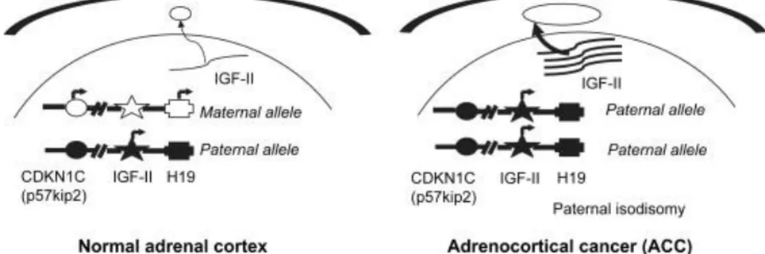
The molecular mechanisms involved in adrenocortical tumorogenesis are still poorly understood. Recent study are focused on alterations of the insuline-like growth factor (IGF) system associated with thase tumors. For instance, there are abnormalities ate the 11p15 region, where the IGF-II gene maps, in more than 90% of malignant adrenocortical tumors (Figure 2.1) (Gicquel et al., 2001).
23
Fig. 2.1 Alterations of 11p15 locus and insulin-like growth factor II (IGF-II) over-expression in adrenocortical cancer (ACC)
The tumors with these abnormalities exhibit strong overexpression of IGF-II gene (Csernus et al., 1999)and large amounts of IGF-II protein (Gicquel et al., 2001; Listrat et al., 1999). The type 1 IGF receptor (Janssen et al., 1997; Wolf et al., 1997) and the IGF-binding protein-2 (IGFB-2) (Boulle et al., 1998) are also specifically overproduced in malignant adrenocortical tumors. These findings strongly implicate the IGF system in adrenocortical tumor progression. The IGF system comprises several elements. The IGFs, IGF-I and IGF-II, are small polypeptides produced in various tissues and cell cultures. They have endocrine and auto/paracrine modes of action (Gockerman et al., 1995). Two structurally different IGF receptors have been described, the type 1 IGF receptor mediating most effects of the IGFs and the IGF-II/mannose-6-phosphate (IGF-II M6P) receptor the function of which should be the internalization and subsequent degradation of II (Clemmons et al., 1995; Gockerman et al., 1995). I and IGF-II can also bind with high affinity to IGFBPs. Six high-affinity IGFBPs have been described to date (Clemmons et al., 1995). These IGFBPs modulate the effects of IGFs either positively or negatively depending on their abundance, their affinity for the growth factors and their cellular localization. IGF-II has been implicates in the growth of various tumors including Wilms’ tumors, hepatomas, colon carcinomas and pheochromocytomas, suggesting the IGF-II plays a central role in tumorogenesis (Karnieli et al., 1996). Similarly, IGF-II may also be involved in adrenocortical tumors. However, direct proof for this role is lacking.
In adrenocortical carcinoma p53 expression is higher than in adenoma (McNicol et al., 1997) and mutations in the p53 gene were found more frequently in malignant tumors (Reinke V, 1997). Additionally, germ line mutations of p53 predispse to childhood adrenocortical cancer (Sameshima et al., 1992) (Wagner et al., 1994).
24
Wnt-signaling has recently been identified as a regulator of a number of endocrine functions in health and disease in addition to its original attribution to developmental biology. Wnts are extracellular ligands on frizzled receptors and on lipoprotein receptor-related protein co-receptors. Ligand binding leads eventually to the activation of intracellular signaling cascades; based on the involvement of the transcriptional co-activator beta-catenin it can be distinguished between canonical (i.e. beta-catenin) and non-canonical Wnt-signaling. Recent studies revealed that canonical Wnt-signaling regulates the function of endocrine organs and contributes to a number of endocrine disorders. Wnt signaling molecules can bind to cell-surface receptors called frizzled and to lipoprotein receptor-related protein (LRP) co-receptors (low density LRP). Frizzled receptors are G-protein-coupled seven-transmembrane receptors. Binding to both receptors activates the canonical Wnt-signaling pathway. By targeting a complex containing adenomatous polyposis coli (APC) and axin the activation of this pathway leads to an inhibition of glycogen synthase kinase-3 (GSK3B). This results eventually in the stabilization of β-catenin. Subsequently, β-catenin, a transcriptional co-activator, translocates to the nucleus to activate T-cell factor (TCF)/lymphoid enhancer factor (LEF) transcription factors on canonical Wnt target-genes. Originally, the Wnt signaling pathway has been identified as a regulator of embryogenesis and has thereafter been associated with tumor development (Logan and Nusse, 2004). In addition, the role of Wnt-signaling agonists and antagonists in adipocyte differentiation has been subjected to a number of studies since its original description in 2000 (Bennett et al., 2002; Christodoulides et al., 2009; Longo et al., 2002; You et al., 2002). This has been the evidence linking Wnt-signaling with metabolic regulation.
A mutation that leads to inactivation of the MEN1 germline is found in approximately 90% of families with multiple endocrine neoplasia type 1 (MEN1). Adrenocortical tumors and / or hyperplasia are observed in 25-40% of patients with MEN1 (Kjellman et al., 1999a; Yano et al., 1989). In most cases these are non-functional adrenocortical adenomas. Hyperplasia was found in a typical way in patients with MEN1 who have hypersecretion of ACTH (Cushing's syndrome), while the ACC has been reported rarely in patients with MEN1. The mutation of the MEN1 gene in somatic cells is very rare in adrenocortical tumors (Wales MM, 1995; Yano et al., 1989).
25
2.4 Adrenocortical adenoma
The adrenocortical adenoma is a benign neoplasm arising from adrenal cortical cells. Dimensions are variable depending on the hormone produced:
adenoma with hyperaldosteronism is usually unilateral and of yellowish color, around 1.5 cm of size and non-enveloped;
adenoma with hypercortisolism is unilateral, has dimensions of about 4 cm, is yellow-brown and is encapsulated;
adenoma with virilization is unilateral, has dimensions of about 5 cm, is red-brown and is encapsulated.
It can present with Cushing’s syndrome (it describes the signs and symptoms associated with prolonged exposure to inappropriately high levels of the hormone cortisol) or primary aldosteronism (it is characterized by the overproduction of the mineralcorticoid hormone aldosterone by the adrenal gland).
Is a well circumscribed, yellow tumour in the adrenal cortex, which is usually 2-5 cm in diameter. The color of the tumour, as with adrenal cortex as a whole, is due to the stored lipid (mainly cholesterol), from which the cortical hormones are synthesized. These tumors are frequent incidental findings at post mortem examination, and appear to have produced no significant metabolic disorder; only a very small percentage lead to Cushing's syndrome. Nevertheless, these apparently non-functioning adenomas are most often encountered in elder obese people. There is some debate that they may really represent nodules in diffuse nodular cortical hyperplasia. Very occasionally, a true adrenal cortical adenoma is associated with the clinical manifestations of Conn's syndrome, and can be shown to be excreting mineralocorticoids.
More frequent with advancing age, adrenocortical adenomas have a peak between 50 and 70 years and the most affected are women (58%) and the right side.
2.5 Adrenocortical carcinoma
ACC is a rare and highly aggressive malignancy with an annual incidence of 0.7–2.0 cases per million population (Kebebew et al., 2006; Kerkhofs et al., 2013). ACC can occur at any age, with a peak incidence between 40 and 50 years, and women are more often affected (55–60%). The incidence in children is particularly high in southern Brazil due to the high prevalence (0.27%) of a specific TP53 germline mutation (R337H) (Custodio et al., 2012). Histologically standpoint are detectable: solid or
26
trabecular areas with fibrous bands interposed between the tumor nodules, necrosis, the presence of large cells with vacuolated cytoplasm, nucleus atypical and hyperchromatic, prominent nucleoli, frequent mitosis, evidence of vascular and capsular invasion.
It is highly aggressive: about 60% of patients have metastases at diagnosis, with a 5-year survival rate of 8% for recurrent and inoperable disease.
Cushing's syndrome is most frequently associated with endocrine cancer. The therapeutic approach of choice for adrenocortical carcinoma is surgery. Surgery should be conducted only after appropriate preoperative diagnostic tests, including biochemical evaluation and imaging. In the setting of adrenal imaging characteristics not clearly excluding malignancy, surgeons are obligated to approach the resection as a cancer operation.
In patients with localized adrenal tumor, suspicious for ACC, surgical resection should be considered. Suspicious features to consider are: tumors size >4 cm, functional tumor, radiologic suspicious characteristics. For tumors invading surrounding tissue or organs, concomitant resection of kidney, liver, spleen, pancreas, stomach, colon and wall of the vena cava should be considered (Schteingart et al., 2005) even if, in primary ACCs, it is quite infrequent that the tumor invades the liver or adjacent kidney. Obviously this is not always predictable during surgery, as in our case in which the tumor was strongly adherent to the kidney, such as to look like a single mass, although histological examination has then denied the spread to the renal parenchyma. The evidence that patients with ACC remain at high risk for tumor recurrence despite complete surgical tumor excision has fueled the search for adjuvant therapies. Even with ostensibly complete resections, rates of local recurrence have typically ranged from at least 19% to 34% in those patients with no residual disease after surgery (Bellantone et al., 1997; Gonzalez et al., 2007). The role of cytotoxic chemotherapy is continuously under investigation. The recommended first-line cytotoxic treatment regimens are etoposide, doxorubicin, cisplatin plus mitotane (Berruti et al., 2005), or streptozotocin plus mitotane (Khan et al., 2000). Mitotane remains the only drug approved by the U.S. Food and Drug Administration and European Medicine Executive Agency for treatment of ACC (Schteingart et al., 2005). The pharmacological mechanism by which mitotane exerts its adrenolytic effect is still not completely understood. Very recently it has been proposed by Sbiera et al, a mechanism where the mitotane causes endoplasmic reticulum stress and profound alteration of lipid-related genes; it was demonstrated that mitotane down-regulates steroidogenesis by inhibition of sterol-O-acyl-transferase 1
27
(SOAT1) and confers adrenal-specific cytotoxicity leading to lipid-induced ER-stress (Sbiera et al., 2015). Mitotane leads with relative specificity to a destruction of the inner zones of the adrenal cortex, the zona fasciculata, and zona reticularis. Several studies have evaluated the efficacy of mitotane as an adjuvant therapy or for advanced ACC as a single treatment or in combination with chemotherapy. Adjuvant treatment is routinely started within 3 months after surgery.
In most patients mitotane abolish steroid secretion but, since uncontrolled hormone secretion might worsen significantly quality of life and may even be life threatening, sometimes additional measures are required to control endocrine symptoms, such as adrenostatic drugs (metyrapone, etomidate). About follow-up, it's repeated every 3 months for the first two years, including abdominal CT or MRI and hormonal markers, and kept on for at least 10 yr. However, the results of the treatment of advanced forms of carcinoma with mitotane are conflicting: some reports attest durable and complete remissions, while others attribute to mitotane a modest antineoplastic activity. Treatments with cisplatin and etoposide in combination with mitotane are placed among the most active for in advanced cancer. Other cytotoxic agents were used in the treatment of this disease such as vincristine, 5-fluorouracil and streptozotocin giving variable results (Berruti A, 1998; Bonacci R, 1998).
A radionucleotide-based approach to therapy of ACC is the use of [131I]IMTO. [123I]IMTO single-photon emission CT imaging showed high tracer uptake in tissue of adrenocortical origin (Hahner et al., 2008), suggesting that [131I]IMTO represents a suitable compound for targeted radionuclide therapy. The clinical utility of this technology, however, needs further evaluation with prospective clinical trials.
Control of the deleterious effects of elevated hormone levels in ACC patients is important. In general, several inhibitors of steroidogenesis as well as direct hormone receptor antagonists can be used to achieve this goal. Several inhibitors of steroidogenesis are in use. During treatment with any of the steroidogenesis inhibitors, patients need to be regularly evaluated for adrenal insufficiency and should be regarded as adrenal-insufficient in times of physical stress (febrile illness or significant injury/surgery). Mitotane inhibits CYP11A1 and CYP11B1 and together with its adrenolytic effects may lead to some control of hormonelevels. Ketoconazole and metyrapone are commonly used to control glucocorticoid excess. Ketoconazole inhibits CYP17A1, CYP11A1, and to some extent CYP11B1 . The usual starting dose is 200 mg twice daily and can be increased to 1200 mg/d. During treatment with ketoconazole,
28
liver enzymes need to be carefully watched. Because it is an inhibitor of several hepatic drugmetabolizing enzymes (eg, CYP3A4, CYP2C9, and CYP1A2), drug interactions need to be carefully reviewed. Another powerful inhibitor of steroidogenesis at the level of CYP11B1 is metyrapone (Hartzband et al., 1988), and 250 mg twice daily is the usual starting dose and can be increased to 2 to 3 g/d in 250-mg intervals. Due to the inhibition of CYP11B1, a relative increase in adrenal androgens may occur, possibly worsening symptoms related to hyperandrogenemia. Other steroidogenesis inhibitors such as aminoglutethimide or etomidate are not in widespread use.
Etomidate is an anesthetic compound often used for rapid induction for intubation or short-term procedures. Even at doses much lower than those used for anesthesia, etomidate is a powerful inhibitor of CYP11B1 and CYP11B2 (Drake et al., 1998). For this effect, it can be used in the inpatient setting. Some centers have experience with a steady low-dose perfusor, which is a last-resort option. Steady infusion can be safe because doses used are only 1/10 of the anesthetic dose (2–3 vs 20–30 mg/h). A direct antagonist used for glucocorticoid excess is mifepristone. However, neither ACTH nor glucocorticoid levels can be used to guide therapy.
Spironolactone can also be used to control androgen effects in women with androgen-secreting tumors and mineralocorticoid effects in patients with mineralocorticoid- secreting tumors. As with other malignancies, local control of ACC is important both for effecting the possibility of a disease cure and for improving symptomatic outcomes. Although traditionally considered ineffective for ACC, radiotherapy has been shown in several recent series to offer a significant improvement in disease control in both the adjuvant and palliative settings (Fassnacht et al., 2006; Hermsen et al., 2010), although such an improvement has not been universally demonstrated (Habra et al., 2013).
In recent years, considerable advances toward understanding the pathogenesis of ACT have been made.
Different strategies have enabled these achievements:
1. Identification of genetic alterations in rare familial syndromes and evaluation of whether the same defects are present in sporadic tumors.
2. Investigation of signaling pathways that were proved important in other tumors types.
3. Employment of high-throughput techniques such as genome wide expression profiling, methylation profiling and microRNA profiling to interrogate novel signaling pathways.
29
4. Studies with animal models with one or more genetic defects in known signaling pathways.
2.6 Role of ERα and ERβ activation on ACC development
A possible involvement of estrogen in tumor development was been suggested by epidemiological evidence and experimental studies: adrenal tumors, especially those secreting, are more frequent in women and the use of estrogen-progestin is a risk factor for tumor development.
Recently, in several tumoral cells the presence of a cross-talk has been reported between the IGF system and estrogens, which is able to activate the same pathway through the action of estrogen receptors (Hamelers and Steenbergh, 2003).
It has been largely demonstrated that the effects of estrogens are mediated by the ERα and ERβ, which act as transcription factors (Nilsson et al., 2001). In the human fetal adrenal gland the mRNA of ERβ was much more expressed than that of ERα and the ERβ protein was detected in the definitive zone of the adrenal cortex (Takeyama et al., 2001). The highly estrogenic environment during pregnancy has been reported to influence steroidogenesis of the primate fetal gland (Albrecht et al., 1999; Hirst et al., 1992) and it has been suggested that the effects of estrogens via ERβ may play an important role in modulating the development of both human and primate fetal adrenal glands (Albrecht et al., 1999; Takeyama et al., 2001).
H295R proliferation seems to be supported by the presence of an autocrine mechanism mediated by E2 through its receptors (Sirianni et al., 2012).
The assessment of response to estrogen receptor antagonists (such as ICI 182 780 and OHT (4-OH tamoxifen)) confirms the E2 involvement in H295R cell proliferation (Montanaro et al., 2005b).
This showed a dose-dependent inhibition of basal and E2-dependent cell proliferation. In particular, OHT induced morphological changes characteristic of apoptosis up-regulating the expression of FasL and inducing autocrine activation of caspases while ICI caused a cytostatic effect that could be explained by the inhibitory effects exerted by ICI on IGF signaling pathway, which is strongly activated in H295R by autocrine IGF-II action through the IGFIR (Sirianni et al., 2012).
ICI mediated inhibition of cell growth is therefore not solely attributable to competition between estrogen and ICI for the estrogen receptor but also to the interruption of the IGF signaling pathway (Montanaro et al., 2005a).
30
ERα and ERβ belong to the steroid/thyroid hormone superfamily of nuclear receptors, members of which share a common structural architecture (Evans, 1988; Katzenellenbogen and Katzenellenbogen, 1996; Tsai and O'Malley, 1994). They are composed of three independent but interacting functional domains: the NH2-terminal or A/B domain, the C or DNA-binding domain, and the D/E/F or ligand-binding domain. Binding of a ligand to ER triggers conformational changes in the receptor and this leads, via a number of events, to changes in the rate of transcription of estrogen-regulated genes. These events, and the order in which they occur in the overall process, are not completely understood, but they include receptor dimerization, receptor-DNA interaction, recruitment of and interaction with coactivators and other transcription factors, and formation of a preinitiation complex (Katzenellenbogen and Katzenellenbogen, 1996; McKenna et al., 1999). Another striking difference between the two receptors is their distinctive responses to the synthetic antiestrogens tamoxifen, raloxifene, and ICI-164,384. On an ERE-based reporter gene, these ligands are partial E2 agonists with ERα but are pure E2 antagonists with ERβ (Batistuzzo de Medeiros et al., 1997; McDonnell et al., 1995; McInerney et al., 1998).
The COOH-terminal, E/F-, or ligand-binding domain (LBD) mediates ligand binding, receptor dimerization, nuclear translocation, and transactivation of target gene expression (Eudy et al., 1998; Giguere et al., 1988; Tsai and O'Malley, 1994).
Crystallographic studies with the LBDs of ERα and ERβ revealed that the AF2 interaction surface is composed of amino acids in helix 3, 4, 5, and 12 and that the position of helix 12 is altered by binding of ligands. When the ERα LBD is complexed with the agonists, E2 or diethylstilbestrol (DES), helix 12 is positioned over the ligand-binding pocket and forms the surface for recruitment and interaction of coactivators (Shiau et al., 1998; Wurtz et al., 1996). In contrast, in the ERα- and ERβ-LBD complexes with raloxifene (Pike et al., 1999) or the ERα-LBD 4-OH-tamoxifen complex (Shiau et al., 1998), helix 12 is displaced from its agonist position over the ligand-binding cavity and instead occupies the hydrophobic groove formed by helix 3, 4, and 5. It is evident that different ligands induce different receptor conformations (McDonnell et al., 1995; Paech et al., 1997) and that the positioning of helix 12 is the key event that permits discrimination between estrogen agonists (E2 and DES) and antagonists (raloxifene and 4-OH-tamoxifen).
Levels of ERβ significantly lower, ERα up-regulation and aromatase over-expression are characteristic of the tumoral condition. In addition, the expression of ER was
31
correlated with the expression of nuclear hormone receptors, suggesting that they may be involved in the modulation of ER. Results of this study suggest that estrogen produced locally by aromatase can induce the proliferation of adrenocortical cells through autocrine and paracrine mechanisms and open new perspectives on the potential use of anti-estrogens and aromatase inhibitors as therapeutic agents against adrenocortical carcinoma.