DOTTORATO DI RICERCA IN
Biologia Cellulare e Molecolare
Ciclo XXXI
Settore Concorsuale: 05/E2
Settore Scientifico Disciplinare: BIO/11
The Helicobacter pylori HrcA repressor: Study of the Global
Transcriptional Response during Heat Shock.
Presentata da: Ojaimi Loibman Stefany
Coordinatore Dottorato Supervisore Prof. Giovanni Capranico Prof. Vincenzo Scarlato
Co-Supervisore Dott. Davide Roncarati
INDICE
1. Introduction 2
1.1 Helicobacter pylori Biology 3
1.2 Pathogenesis 3
1.3 The Helicobacter pylori Genome 7
1.4 Heat-shock Response 10
2. Aim of the Study 15
3. HrcA transcriptional response by RNA-seq 17
3.1 Specific Introduction 18
3.2 Results 18
3.3 Discussion 24
4. In search of new HrcA genomics targets 27
4.1 Specific Introduction 28
4.2 Results 29
4.3 Discussion 35
5. Conclusions 37
6. Materials and Methods 40
7. Bibliography 53
• Supplementary table 1 • Supplementary table 2
1.1 Helicobacter pylori biology
First isolated in 1982, Marshall and Warren (Backert & Blaser, 2016; Warren & Marshall, 1983) described a spiral or curved bacterium from 58 of 100 consecutive histological biopsies of human gastric antral mucosa. This organism demonstrated several similarities to Campylobacter and was then referred to and validated in 1985 as Campylobacter pyloridis. In 1987 the specific epithet was revised for Campylobacter pylori to properly name the latin genus of the pylorus noun.
Only in 1989, given the differences from Campylobacter in many molecular and biochemical aspects as the flagella morphology, the content of fatty acids and the sequence of the 16S rRNA, it was named Helicobacter pylori.
The etymology of the name Helicobacter pylori derives from: helix = spiral; bacterial; pylorus = lower stomach, and currently is described as a Gram-negative bacterium, microaerophilic, spiral or curved shaped and multi-flagellated.
In vitro, Helicobacter species grow quite slowly, normally at 37°C and only under
microaerobic conditions with 10% CO2 and rich media supplemented with 5%
whole blood or serum.
The natural habitats of this bacterium are the deeper extracts of the gastric lining epithelium, just above the carpet of parietal cells and the successful colonization in this hostile and acid environment is achieved through a combination of several virulence factors.
1.2 Pathogenesis
It is estimated that more than half of the world's population is colonized by Helicobacter pylori. The selection of a no-competition habitat with a very acidic pH, which is lethal to most microorganisms, makes H. pylori one of the most successful human pathogens, recognized as the main causative agent of chronic gastritis, gastric and peptic ulcers, and persistent colonization is an important factor in the development of gastric lymphoma and adenocarcinoma.
The mechanisms by which the bacterium causes various diseases with different intensities are not fully established. It is believed that the combination of virulence factors expressed by the bacterium with the environment of the host may justify these various clinical evolutions.
For successful colonization, the first and most important step is to survive in the gastric environment. This is achieved through the expression of a large amount of urease enzyme that converts urea into ammonia and bicarbonate. The ammonia protects H. pylori against the acidic microenvironment and causes damage to the gastric epithelium (Ansari & Yamaoka, 2017). This enzyme is also responsible for the stimulation of mononuclear phagocyte activation, inflammatory cytokine production and causes toxic effects on gastric epithelial cells (Debowsky, 2017). Therefore, the presence of urea allows H. pylori to create a neutral layer around its surface and through adhesion molecules that binds to specific receptors begins the colonization of the gastric mucosa.
Due to its shape and motility caused by the presence of flagella, this microorganism is able to cross the mucus layer of the stomach until it reaches the epithelium, whose degeneration causes the induction of inflammatory infiltration, consisting of leukocytes, neutrophils, lymphocytes and plasma cells. During this process, other enzymes synthesized by the bacterium, such as superoxide dismutase, catalase and arginase confer protection against the lytic activity of macrophages and neutrophils, prevent an efficient response of the host, promoting stimulation of the production and secretion of proinflammatory cytokines by epithelial cells. Helicobacter pylori induces humoral and cellular immune responses, promoting an inflammatory reaction that recruits both polymorphonuclear and mononuclear cells with an increased level of pro-inflammatory cytokines such as IL-1β, tumor necrosis factor alpha (TNF-α), IL-8, and IL-6 (Wilson & Crabtree, 2007).
Adherence factors to the gastric epithelium are important for the pathogenicity of H. pylori and favor its colonization. In this context, a series of proteins called adhesins interact with receptors present in the gastric epithelial cells and allow the pathogen to anchor itself in a stable way. Among some adhesins are: 1) the neutrophil-activating protein, an HP-NAP gene product that induces neutrophil adhesion to endothelial cells and stimulates the production of reactive oxygen and nitrogen species by neutrophils (Satin et al., 2000); 2) babA, a blood group antigen adhesin adhesion factor linked to the Lewis blood group that plays a critical role, facilitating the liberation of other bacterial virulence factors and
damaging the host tissue (Yamaoka, 2008); 3) the H. pylori catalase, a product
survival of this bacterium, mainly in the presence of hydrogen peroxide. The katA knock-out mutant strain is less able to sustain the long-term infection (Harris et al., 2003); and 4) recently, the hopQ gene has been shown to exploit human carcinoembryonic antigen-related adhesion proteins (CEACAM). CEACAM proteins mediate intercellular adhesion and the external cell signaling events: bacterial proteins that interact with CEACAMs have been related to the inhibition of host immune response activation (Moonens et al, 2018).
This bacterium is also associated with the inhibition of the secretory response of mucus cells through the decrease of somatostatin release and consequent increase of gastrin released by the gastric antrum, causing detrimental effect on the primary defense mechanism of the gastric mucosa. In addition, gastric tissue injury is mediated not only by the effects of urease but also by the expression of several genes responsible for the production of cytokines and virulence factors. The ArsS-ArsR two-component system regulates gene expression in response to low-pH and has also been associate to biofilm production (Servetas et al., 2016).
The vacuolating cytotoxin, encoded by the vacA chromosomal gene, damages the epithelium causing vacuolization in epithelial cells.
After binding to the cell surface, the vacuolating cytotoxin is internalized and forms selective channels for anions in the membranes of endocytic compartments. To compensate the increase in intraluminal chloride, the activity of the vacuolar ATPase is intensified, resulting in an increase in proton pumping and a consequent reduction in pH. Membrane-permeated weak bases, such as ammonia, diffuse into late endocytic compartments and become protonated and trapped in these compartments. The osmotic swelling of these compartments results in cell vacuolization. The overall effects of this toxin on gastric epithelial cells include changes in mitochondrial membrane permeability and apoptosis, stimulation of proinflammatory signaling, as well as the increase of plasma membrane permeability and alterations in the endocytic compartments (Cover and Blake, 2005; Necchi et al., 2017).
Another important pathogenic factor of H. pylori is the cytotoxin-associated gene A, encoded by the cagA gene, mapping in a 38 kb multi-operon locus coding for 28 putative ORFs, called cag pathogenicity island (cag-PAI), likely acquired by this bacterium from another organism, as the GC content of this island differs
from that observed in the rest of the genome. The cag-PAI also encodes a type IV secretion system (T4SS) that directly injects CagA into host cells. Once inside the host cell, CagA is phosphorylated by host cell kinases and alters multiple host signaling pathways. To induce the production of interleukin (IL)-8 in the epithelial gastric cells, H. pylori depends on both mechanisms, the T4SS- and CagA expression. Infection with cagA-positive strains increases the risk of gastric cancer by at least one order of magnitude with respect to infection with cagA-negative strains (Boonyanugomol et al., 2018; Jang et al., 2016)
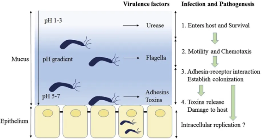
The set of mechanisms that H. pylori uses for colonization and infection of the gastric mucosa are illustrated in Figure 1.
Figure 1. Pathogenesis Model of Helicobacter pylori. The production of urease by H. pylori neutralizes the gastric pH, adjusting the medium for colonization and infection by the bacterial cells. The adhesion of the bacteria to the gastric epithelium is mediated by adhesins, allowing the release of the other virulence factors stimulating strong host immune response and inflammation of the gastric mucosa (Kao et al., Biomedical Journal 39 (2016) 14-23. This is an open access article under the CC BY-NC-ND license (http://creativecommons.org/licenses/by-nc-nd/4.0/).
H. pylori infections can be successfully cured with antibiotic treatment, associated with a proton pump inhibitor (Megraud and Lamouliatte, 2003). Unfortunately, the available antimicrobial therapies are beginning to lose efficacy, principally because of insurgence of antibiotic resistance. Altered expression of gene products sensitive to antibiotic treatment seems to be especially important for resistance to penicillin, nitroimidazoles and clarithromycin. In 2017 WHO (World Health Organization) classified Helicobacter pylori as a high priority pathogen for the production and development of new antibiotics and to explore and identify both bacterial and host markers to diagnose high-risk individuals during severe
infection outcomes, as well as to develop new effective therapeutic strategies. For these reasons, H. pylori is a bacterial pathogen of major medical importance. 1.3 The Helicobacter pylori Genome
The genomic sequencing of multiple H. pylori isolates demonstrated great variation in the sequence and gene content among the analyzed strains, whereas the general genomic organization and the predicted genetic products were quite similar (Alm et al., 1999).
The H. pylori G27 strain, isolated for the first time at Grosseto Hospital (Tuscany, Italy) from an endoscopic patient has been widely used in research.
The G27 genome is a single circular 1.6 kb chromosome AT rich (61,1%) and contains 1,515 open reading frames (ORFs) and strong similarity to other H. pylori genomes of strains 26695, J99 and HPAG with respect to size and composition. G27 contains 58 genes that are not found in 26695, J99 or HPAG and most of these G27-specific genes are predicted to encode hypothetical proteins (Baltrus et. al, 2009).
This bacterium has an interesting peculiarity that is the scarcity of transcriptional factors and predicted regulatory proteins, where genomic analyses have identified only 32 gene products of possible regulatory function in which only 17 are predicted to have a role in transcriptional regulation, that is, approximately half of the number of those reported for H. influenzae, which has a genome of comparable size to H. pylori and less than a quarter of those predicted for E. coli (Tomb et al., 1997).
However, the small number of transcriptional regulators encoded by H. pylori correlates with a small genome size. It has been speculated that the reduction in the number of transcription factors occurred for the absence of selective pressure associated with a restricted and competition-free habitat (Danielli et al.,2010; de Reuse and Bereswill, 2007).
The transcriptional machinery of H. pylori is similar to the one observed in many other organisms, such as E. coli, but with some substantial differences. For
instance, the vegetative sigma factor of H. pylori σ80, RpoD, of the RNA
polymerase enzyme has several differences with respect the σ70 subunit of E.
two alternative sigma factors, named σ54 (RpoN) and σ28 (FliA), and both are
involved in controlling the expression of the flagella components (Kao et al.,
2016). Moreover, mechanisms of post-transcriptional regulation have been
described in H. pylori, through antisense transcripts and the expression of at least 60 small RNAs, as well as through RNA-binding proteins that modulate mRNA stability and translation efficiency (Sharma et al., 2010).
As in other bacteria, coordinated genomic expression is controlled by the transcription regulatory network (TRN), which is commonly organized in hierarchical multilayer structures and composed of regulatory modules (origins) that include the combined activity of transcription factors (TFs) to regulate related physiological functions, so they act as an input signal for the bacteria to respond to changes in the environment (Balazsi and Oltvai, 2005; Danielli et al., 2010). It has been proposed that H. pylori adopts a multilayer TRN of low hierarchy, encompassing its four main origons (motility and chemotaxis, metal and ion homeostasis and heat-shock response), in an unsegregated way of its few transcriptional regulators. Thus, different environmental inputs are interpreted by different combinations of small sets of transcription factors or associated proteins. In this microorganism, the TRN is built unequivocally to maintain homeostasis,
and colonization and survival in the gastric niche depends on the concerted
expression of virulence factors and housekeeping genes.
To initiate the colonization process in the host organism, the H. pylori flagella,
chemotaxis and motor protein are crucial factors in establishing a successful
infection. Their deletion leads to strains with an attenuated or completely
defective ability to establish colonization in animal models (Eaton et al., 1996; Foynes et al., 1999; Josenhans and Suerbaum, 2002).
H. pylori cells usually have a unipolar bundle of two to six flagella with a sheath that allows bacteria to enter and move through the gastric mucosa. Each flagellum exhibits a typical structure as a bulb at its end, being an extension of the outer membrane and has protective function for the flagellar structure at acidic pH of the stomach.
The flagella have three structural elements: a basal body, which is embedded in the cell wall and contains the proteins required for rotation and chemotaxis; an external helically shaped filament that works as a propeller when rotated at its base; and a hook that serves as a joint between the basal body and the flagellar
filament. In H. pylori, more than 40 predicted genes are scattered along the genome whose products are involved in the expression, secretion and assembly of this flagellar complex (Spohn & Scarlato, 2001). The flagellar regulatory
system adopts a regulatory σ short cascade (σ80> σ54> σ28) and a hierarchical
regulation, initiated by the housekeeping σ80 factor, where each σ factor activates
its dedicated target gene class. In addition, each one is also responsible for the
transcription of the next factor in the cascade (Sharma et al., 2010).
As in other enterobacteria, flagellar genes in H. pylori are positively regulated and organized hierarchically into three classes according to their activating sigma factor: 1) class I, which includes the gene targets transcribed by the RNA
polymerase containing the σ80 factor and comprises mainly flagellar regulatory
genes encoding proteins that form flagellar base structures (Kavermann et al.,
2003); 2) class II includes specific targets of the σ54 factor and encodes
components of the basal body and the flagellar hook; finally, class III genes encode late flagellar structures, transcribed by the σ28 factor. Thus, the flagellar
regulatory cascade of H. pylori is appropriate to guarantee the correct sequential expression of early, intermediate and late flagellar components.
Once established in the host, H. pylori cells need to respond rapidly and continuously to a severe acidic environment. Therefore, this microorganism adopts a set of acid acclimation genes, and the transcription of these genes is under the control of the housekeeping factor σ80 and regulated mainly by the essential acid response regulator ArsR. This regulator is part of an ArsS-ArsR two-component system and once acidification of the periplasm is detected by ArsS, the signal is transduced by changes in the protonation of various histidine residues in the extracytosolic sensory domain. This stimulus triggers the phosphorylation of ArsR, thus promoting its DNA binding activity towards a specific set of promoters (Danielli et al., 2010).
The absorption of metal ions is another crucial aspect for the maintenance of H. pylori infection and also for its virulence, since these are important enzymatic cofactors. For example, nickel is a virulence determinant because it is a cofactor of the urease enzyme in the reaction catalyzing the hydrolysis of urea into carbon dioxide and ammonia, which is essential for in vivo colonization (Hu et al., 2017). However, if present in large intracellular amounts the metal ions are toxic, and their homeostasis must be tightly controlled. H. pylori has three systems
dedicated to this fundamental task: the two-component CrdRS system that appears to be involved in copper resistance and apparently is also a sensor for nitrosative stress, the ferric uptake regulator (Fur) involved in iron homeostasis and in detoxification and finally NikR, a homologue of E. coli responsive nickel regulator, typically associated with hydrogenase maturation and acidic survival (Pich et al., 2012; Hung et al., 2015)
1.4 Heat-shock Response
The ability to respond quickly to environmental changes and thus modulate gene expression is a crucial ability exploited by bacteria for their survival. In this context, the widespread human pathogen H. pylori, when in stress conditions, induces the synthesis of a class of highly conserved proteins, called Heat Shock Proteins (HSPs).
These proteins, expressed also under physiological growth conditions, accumulate upon a temperature increase and in response to different types of stress and are employed in assisting the correct folding, assembly, transport and degradation of cellular proteins. The ability to perceive and respond quickly to environmental changes is crucial for their survival during infection in a hostile environment such as the human stomach.
When growth conditions are altered, these proteins play a crucial role, acting against the accumulation of incorrectly folded proteins to avoid the formation of amorphous cytoplasmic aggregates, deleterious to the cell (Narberhaus, 1999). Accordingly, sophisticated regulatory circuits guarantee low expression levels of HSPs under physiological growth conditions and a strong and coordinated induction after the perception of various stimuli of stress. This regulatory circuit, therefore, perceives the signals coming from the environment and translate them into a well-defined gene expression pattern.
In bacteria, the transcriptional regulation of heat-shock genes can be regulated by positive or negative regulatory mechanisms. In systems whose transcriptional regulation is negative, as in H. pylori, transcription is controlled by dedicated repressors whose DNA binding activity changes in response to stress. Under normal growth conditions, these repressors bind to specific operators and repress the transcription of heat shock genes.
In H. pylori the genes coding for the major HSPs are clustered into three multicistronic operons transcribed by the RNA polymerase containing the vegetative sigma factor σ80 and their transcription is rapidly induced by heat
shock and other stress stimuli (Spohn et al., 2002). Under physiological conditions these operons are repressed by two transcriptional regulators: HrcA and HspR, homologues to well-characterized heat-shock regulators found in many other bacterial species with medical relevance such as Streptococcus
pneumoniae and Clostridium difficile (Jain et al., 2017; Chastanet et al., 2001).
The HspR regulator alone represses the transcription of the cbpA-hspR-helicase operon, thereby auto-regulating its own synthesis. On the other hand, both the HspR and HrcA regulators are required to repress transcription of the heat shock hrcA-grpE-dnaK and groES-groEL operons and this model is represented in Figure 2. Specifically, the HspR and HrcA repressors combine to control the transcription of target genes in a way that the HrcA regulon results embedded within the HspR regulon (Roncarati & Scarlato, 2018).
Figure 2. Regulation of H. pylori chaperone expression. Transcriptional repression of the heat shock operons is represented by solid lines that connect HrcA and HspR repressor proteins to their target promoters (Pgro, Phrc, and Pcbp); dashed lines, linking GroES-GroEL to HrcA and CbpA to HspR, represent the posttranscriptional protein-protein feedback control of the regulators.
While HrcA is widely distributed in the prokaryotic kingdom, HspR is found in a restricted number of bacteria. This latter is a homologue of the repressor that controls the dnaK operon of Streptomyces coelicolor and has been shown to bind to inverted repeats in the promoter region, designated HAIR (HspR-associated inverted repeat). In H. pylori, the HAIR sequence is localized in close proximity to the transcription start site of the Pcbp promoter (-59 to +14 on Pcbp), while in the Pgro and Phrc promoters HspR binds far upstream of the core promoter regions,
-120 to -43 on Pgro, from -149 to -78 on Phrc, respectively, while HrcA binds to operators overlapping the core promoter regions (Roncarati & Scarlato, 2017). HrcA is a homologue of the repressor of a set of heat shock genes of Bacillus subtilis that binds to an inverted repeat in the promoter region designated CIRCE (controlling inverted repeat of chaperone expression) with a consensus motif (TTAGC ACTC-N9-GAGTGCTAA) (Narberhaus and Bahl, 1992). In H. pylori, the CIRCE sequence is localized near the transcription start site of the Pgro and Phrc promoters, ranging from -13 to +16 on Pgro and -59 to -34 on Phrc (Roncarati et al., 2017).
The presence of both regulators is, therefore, necessary for maintaining Pgro and Phrc in the repressed state. Although the binding sites of HrcA and HspR are very close to each other, in vitro studies have demonstrated that both bind in an independent manner to their operators, while the HrcA-mediated regulation depends on the presence of HspR. It is not yet known whether this stable complex is due to possible interactions between the repressors with other proteins, so they can form a stable complex capable of repression.
The transcriptional control described, is further assisted by an additional level of post-transcriptional modulation exerted by some HSPs contained in the same operon (GroELS and CbpA), which modulate the binding of HrcA and HspR repressors to DNA (Roncarati et al., 2007; Roncarati et al., 2011; Roncarati et al., 2014).
As in the proposed model for B. subtilis, the chaperonine GroE is able to positively influence the DNA binding affinity of HrcA. As illustrated in Figure 3, during physiological growth conditions, the GroE chaperonin establishes a feedback posttranslational regulatory circuit increasing HrcA DNA binding activity. In contrast, in a stress situation the accumulation of misfolded proteins in the cytoplasm competes with GroE causing a loss of DNA binding affinity for HrcA and consequent derepression of heat-shock promoters (Scarlato & Roncarati, 2018).
Figure 3. Posttranscriptional GroE-mediated regulation of HrcA activity. (a) During physiological growth conditions the GroE chaperonine (represented in grey) is free to interact and fold the HrcA regulator (red oval). Once properly folded this repressor binds and occupies its target promoters, repressing genes transcription. (b) When cells experience stress growth conditions, GroE is titrated by unfolded proteins that accumulate in the cytoplasm and cannot interact with HrcA; The repressor left alone has a conformation with low DNA-binding affinity (red rectangle) and HrcA-dependent heat-shock genes repression is relieved. The RNA polymerase containing the vegetative σ fator is represented by green ovals and Bent arrow indicates the transcription start site. (Roncarati & Scarlato. Int. J. Mol. Sci. 2018, 19, 1702. This is an open access article under the CC BY-NC-ND license (http://creativecommons.org/licenses/by-nc-nd/4.0/).
It has been demonstrated that HrcA is the direct thermosensor of H. pylori. In fact, the DNA binding activity of HrcA is modulated by temperature fluctuations over a very restricted physiological range of temperature (37°C - 42°C).
Under heat stress conditions, due to a major structural alteration, HrcA loses DNA binding activity and the transcription of the chaperone genes is rapidly derepressed. When the ambient temperature drops to physiological levels, GroE chaperonin is able to interact and refold at least a fraction of the heat-inactivated protein and thus, together with the newly synthesized HrcA the repressive state is re-established (Roncarati et al., 2014).
Thus, according to the central function of GroE chaperonin in modulating the HrcA binding activity to the DNA, H. pylori appears to be able to adjust its transcriptional regulation in response to the perception of various stress signals of different intensities. In particular, depending on the extent of a given stress stimulus, the availability of GroE to the functional interaction with HrcA will be differentiated, influencing restoration levels of heat shock gene repression. Despite the detailed characterization of the heat shock regulatory circuit described, not enough is known about the global involvement of the heat-shock HrcA regulator in H. pylori. A global analysis of the heat-shock HrcA regulator would be highly informative, not only for the characterization of all the effectors
involved in protecting the bacterium against environmental stress, but also for the identification of new virulence factors triggered in response to stress signals perceived by the pathogen.
The HrcA protein is a well-known transcriptional regulator of H. pylori involved in
heat shock response. Despite the detailed characterization of the heat shock
regulatory circuit, not enough is known about the role of HrcA at global level in H. pylori. In analogy to other bacterial systems most commonly studied, it is expected that exposure of H. pylori to heat shock conditions causes an altered expression (increased but also decreased expression) of many proteins. The aim of this project is to carry out an integrated study that will answer a series of questions related to heat shock and stress response in H. pylori.
To achieve this goal, we performed a global transcriptome analysis by
RNA-sequencing to identify differentially regulated genes by HrcA and, thus, reveal whether this protein is involved in the regulation of other cellular processes and
interact with additional genes. Then the second part of this study was aimed to
the identification of HrcA binding sites in vivo through a genome-wide approach,
using Chromatin Immunoprecipitation followed by deep sequencing (ChIP-seq),
3. HrcA transcriptional response by
RNA-seq
3.1 Specific Introduction
In H. pylori the heat shock regulon is tightly modulated by the concerted action of two transcriptional repressors, HspR and HrcA. This latter protein negatively regulates transcription from Phrc and Pgro promoters. To study the global heat shock response in H. pylori and to identify potential new genes regulated by HrcA, we performed differential transcriptional analysis of heat shock regulation by RNA-seq, comparing the transcriptome of the G27 (hrcA::km) mutant and the parental G27 wild type strain not exposed to heat shock (t0) and at 30 min (t1) after temperature upshift of the culture to 42°C, using total RNA extracts from mid-exponentially growing cultures (OD600= 0.7-0.8), and results have been validated by qRT-PCR on a selection of targets.
3.2 Results
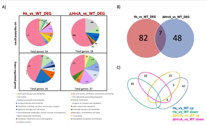
To gain insights into the H. pylori heat-shock response and the global role of the HrcA regulator, we performed a transcript profiling experiment using RNA-seq, comparing the transcriptome of the G27 wild type strain and the G27 (hrcA::km) mutant isolated from exponential growth phase at 37◦C and also from the wild type strain after temperature increase of the culture to 42 °C for 30 min (t1). This analysis showed that a total of 137 genes were differentially expressed (log2 fold change >1 or <-1) by heat shock and hrcA mutation, representing more than 9% of all the H. pylori annotated ORFs (Figure 4 and Supplementary Table 1 and Table 2). After temperature upshift to 42◦C, the heat-shock treatment triggered changes in the transcript levels of 89 of 137 (64,97%) differentially expressed genes (log2FC > 1 padj < 0.01) when compared to the wild type sample not subjected to heat-shock. Of these, 54 genes appeared up-regulated and 35 genes appeared down regulated. As expected, among the up-regulated genes we found the previously characterized heat shock responsive groES, groEL, grpE, dnaK, and cbpA genes.
In addition, each differentially expressed gene was characterized by a specific category of COG (Cluster of Orthologous Group), a database designed to classify proteins from sequenced genomes based on the orthology concept. From the functional annotation and enrichment analysis, we found that 55% of the regulated genes are included in the Unknown Function category. Several
up-regulated genes are involved in “Transcription” and “Post-translational modification, protein turnover, and chaperones and Transcription”. Among the down-regulated genes, also in this case the most of the differentially expressed genes are included in the Unknown Function category but many genes are related with “Cell wall/membrane/envelope biogenesis” and “Replication, recombination and repair” (Figure 4A).
The role of HrcA was established by comparing the transcriptome of the ΔHrcA mutant to that of the H. pylori wild type strain, both grown at 37◦C (ΔHrcA _vs_WT). Intriguingly, we found several HrcA regulatory targets that are not responsive to heat stress.
This analysis showed a total of 55 deregulated genes (log2FC > |1| padj < 0.01) in the hrcA gene deleted condition. Of these, 48 genes were deregulated only in ΔHrcA mutation and not influenced by heat stress response, corresponding to 35% of the differentially expressed genes detected in this study. Among these genes, 33 were down-regulated and 15 were up-regulated (Figure 4 and Supplementary Table 1 and Table 2). Surprisingly, this repressor apparently controls the transcription of several genes, which do not seem to be affected by the heat stress.
The putative HrcA regulon, evidenced by this analysis, is made up of genes coding for proteins involved in several processes. As expected, among the up-regulated genes we found the groES and groEL genes (Roncarati et al., 2007), which is associated to “Post-translational modification, protein turnover, chaperones” functional enrichment (padj = 0.01). As expected, transcription of the grpE and dnaK genes (HPG27_RS00570 – HPG27_RS00575) was unchanged in the HrcA mutant, likely due to a polar effect of the first gene of the operon, hrcA.
Figure 4. Differential transcriptional study between HrcA and heat-shock response. (A) Pie charts showing COGs functional annotation of the differentially expressed genes outlined in the ΔHrcA_vs_WT and HS_vs_WT (Heat-Shock vs. Wilde Type) comparisons, respectively, subdivided into up-regulated (left) and down-regulated (right) groups. The abundance of each category is indicated as a percentage as well as the total number of genes included in each group. (B) Venn diagram illustrating the relation between the deregulated expressed genes in the two set of experiments: heat shock (Hs_vs_WT) depicted by red set, ΔhrcA (ΔHrcA_vs_WT) depicted by blue set. (C) Venn diagram showing the number of genes in common or peculiar in the four previously described gene groups: Hs_vs_WT_ up, Hs_vs_WT_ down, HrcA_vs_WT_up and HrcA_vs_WT_ down, represented in blue, green, yellow and pink respectively.
In addition to these genes, already known to be the target of HrcA regulation, we noticed some functional groups of particular interest: among the up-regulated genes we found some genes (as the rfaJ and HPG27_RS03015 genes) related to “Cell wall/membrane/envelope biogenesis”, Coenzyme transport and metabolism or the dppB gene, which is part of a multicistronic operon (predicted by DOOR, Database of prOkaryotic OpeRons) related to “Inorganic ion transport and metabolism”.
Interestingly, 18% of the down-regulated genes are included in “Cell Motility” category as for example the flaA, flaB, flgL, fliS, flgE, fliK and fliW genes. Additionally, many genes not yet included by COG in cell motility and annotated as "Unknown function” are likely to encode proteins for flagellar structure and biosynthesis, such as the fliD, fliT and flgK genes that are detected as down-regulated genes by the HrcA mutant in this analysis.
Several genes involved in the “Cell wall/membrane/envelope biogenesis” and transposase coding genes, classified in the “Mobilome: prophages, transposons” category enrichment were also down-regulated.
Comparison of the transcriptome of the heat-shock response (89 deregulated genes) to that of the ΔHrcA mutant (55 deregulated genes) led to the identification of 7 genes that were deregulated in both datasets (Figures 4-B). Among these genes, 4 were oppositely regulated (down-regulated in the ΔHrcA mutant and up-regulated in heat-shock response) and 3 were similarly up-up-regulated (Figure 4C). The similarly up-regulated genes included the groES and groEL genes, which are already known to be directly repressed by HrcA and induced by heat- shock (Spohn and Scarlato, 1999). In addition, the HPG27_RS00625 gene coding for a hypothetical protein was up-regulated.
The remaining three genes were induced by heat-shock and repressed in the ΔHrcA strain, thus showing opposite behavior. These include HPG27_RS00565, coding for a hypothetical protein, (ykgB) HPG27_RS02740, coding for a membrane protein, and fliD (HPG27_RS03660) coding for a flagellar filament capping protein FliD. Since HrcA is a transcriptional repressor, the interaction with these genes probably occurs in an indirect way. Moreover, our transcriptome study shows that 15 genes belonging to the HrcA regulon (made up of 55 genes) are not responsive to heat-shock. In addition, our data suggest that HrcA could play a role in the biosynthesis and regulation of the flagellar apparatus of H. pylori. To validate the results obtained by RNA-seq, the relative abundance of selected RNA transcripts of some selected genes was quantified by Real-Time PCR (qRT-PCR), using specific oligonucleotides for each region of interest listed in Table 3. For data analysis, fold-change ratios of ≥ 2.0 or ≤ 0,5 were considered as significantly up-regulated and down-regulated, respectively (Figure 5).
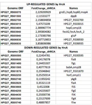
Figure 5. Summary table of the selected up- and down-regulated genes revealed in our transcriptome analysis including the genome ORF name, the fold-change values and the name of the gene.
To validate the up-regulated genes in the ΔHrcA mutant condition, we selected 9 among the 18 genes. Surprisingly, only 2 genes, the HPG27_RS03015 and the known HrcA target groEL (HPG27_RS00070), were in agreement between RNA-seq data and qRT-PCR validation (Fig. 6A). With respect to the down-regulated genes, we selected 13 of the 37 of them and we find a good correlation between the results, in which 9 of the 13 selected genes demonstrated a very similar trend between qRT-PCR and RNA-seq, thus validating our data (Fig. 6B).
Figura 6. (A) Real Time PCR analysis of transcript levels of 9 selected up-regulated genes in the ΔhrcA mutant compared to the wild type not treated using specific primers for the selected genes (table 3). The blue bars indicate the transcript results of the validation and the light grey bars indicates the fold enrichment indicated in the RNA-seq outcomes. The red line indicates the established threshold value of the enrichment at these regions. (B) Real Time PCR analysis of transcript levels of 13 selected down-regulated genes in the ΔhrcA mutant compared to the wild type not treated using specific primers for the selected genes (table 3). The blue bars indicate the transcript results of the validation and the light grey bars indicates the fold enrichment indicated in the RNA-seq outcomes. The red line indicates the established threshold value of the enrichment at these regions.
3.3 Discussion
The Heat-shock response is a universal cellular strategy that allows cells to adapt to their environmental living circumstances and to survive during stress
conditions. In H. pylori, the HrcA protein is a well-known transcriptional reressor
involved in heat shock response, negatively regulating transcription from Phrc
and Pgro promoters.
In this study, through differential transcripts analysis of the heat shock response of wild type H. pylori and a HrcA isogenic (hrcA::km) mutant strain it was possible to verify distinct expression profiles between the heat shock response and the HrcA regulator.
Overall, this analysis highlighted that heat shock treatment of H. pylori determined the majority of differentially expressed genes (89 out of 137), while the HrcA mutant showed 48 deregulated genes (Figure 4). As expected, within the two sets are the genes encoded by three heat-shock multicistronic operons groES-groEL, hrcA-grpE-dnaK and cbpA-hspR-rarA that were up-regulated. Most of the up-regulated genes belong to the functional class “Post-translational modification, protein turnover, and chaperones”. Similar results were recently obtained by a study of the transcriptional response mediated by HspR (Pepe et al., 2018).
One of the most interesting findings that emerged in this study is the interconnection between HrcA and the motility of H. pylori, confirming a previous observation that a ΔhrcA mutant strain is non-motile (Roncarati et al., 2007). Indeed, several flagellar gene transcripts were downregulated in the G27 (hrcA::km) mutant strain and not responsive to heat-shock. Among the down-regulated genes are the flaA, flaB, flgL, fliS, flgE, fliK and fliW genes. Transcription of these motility related genesis under the control of the RNA polymerase-containing one of three sigma factors, σ80, σ54, and σ28 and code for proteins predicted to be involved predominantly to structural components of the flagella as the filament, hook and basal body. Moreover, interconnections between the heat shock repressors and motility have been observed in the closely related bacterium Campylobacter jejuni (Andersen et al., 2005). In H. pylori, motility is a crucial factor to establish a successful colonization and
Recently, some genes known to regulate the flagellar apparatus, such as rpoN
(s54) and fliA (s28), have been related to other important cell functions such as
energy metabolism, oxidative stress and lipopolysaccharide synthesis (Sun et al., 2013; Baidya et al., 2015; De la Cruz et al., 2017). Furthermore, the study carried out by De la Cruz et al., (2017) reports that in the presence of antibiotics, some of these genes may have altered expression at the transcriptional level. In the same study, the rpoN, fliA, flgR and crdR genes were repressed in the presence of kanamycin, chloramphenicol and tetracycline.
Another recent study suggests that the H. pylori flagellar apparatus may play an important structural role during biofilm formation (Hathroubi et al., 2018). In this study, several genes encoding proteins of the flagellar apparatus as FlgB rod protein, the FlgE flagellar hook protein, the FlgK and FlgL hook-filament junction proteins, the FliK hook length control protein, FlaB and the putative flagellin encoded by FlaG appear up-regulated in presence of biofilm. Interestingly, in the same study, transcript levels showed that HrcA appears up-regulated in biofilm cells, showing a close connection between HrcA, motility and biofilm formation (Hathroubi et al., 2018). Similarly, correlation between motility and some flagellar genes were also verified in E. coli and P. aeruginosa (Domka et al., 2007; Sauer et al., 2002).
In the present study, comparison between the heat-shock response of the wild tye strain and the ΔHrcA mutant identified 7 genes that were deregulated in both transcriptomes (Figure 4B). Of these, only 3 were similarly up-regulated (Figure 4C), the groES and groEL genes, which are already known to be directly repressed by HrcA and induced by heat- shock (Spohn and Scarlato, 1999), and the HPG27_RS00625 gene coding for a hypothetical protein.
Among the 4 genes oppositely regulated that were induced by heat-shock and repressed in the ΔHrcA strain we found HPG27_RS00565, coding for a hypothetical protein, (ykgB) HPG27_RS02740, coding for a membrane protein, and fliD (HPG27_RS03660) coding for a flagellar filament capping protein FliD. The scarcity of deregulated genes in both conditions (heat-shock response and mutant HrcA) strongly suggest that HrcA, independently of the heat-shock circuit, acts in several other cellular processes. Interestingly, a study comparing the transcriptome of the heat-shock response to HspR (Pepe et al., 2018) revealed
that 25 genes were deregulated in both datasets, a number approximately 3.5-times greater than our findings for HrcA.
To validate the RNA-seq results the relative abundance of RNA transcripts of selected genes were quantified by Real-Time PCR (qRT-PCR). Surprisingly, only 2 of 9 selected genes, the HPG27_RS03015 and the known HrcA target groEL (HPG27_RS00070) genes were in agreement with RNA-seq data(Fig. 6A). Of note, most of the up-regulated transcripts had a fold-change very close to the established cut-off ratio (Fig. 5), and this may contribute to the observed low rate of validations. In addition, some studies indicate that the validation of results from RNA-seq may depend on the expression levels of the analyzed gene (Wang et al., 2014; Yeri et al. 2018). A high expression of a gene increases the probability of validation.
By contrast, among the down-regulated genes, 9 out of 13 have been validated by qRT-PCR and RNA-seq (Fig. 6B).
Globally, this study shows that the H. pylori HrcA regulon is involved in the regulation of several genes related to crucial cellular processes not responsive to heat-shock and connected to pathogenesis and virulence. In particular, HrcA appears to play a significant role in the biosynthesis and regulation of the flagellar apparatus of H. pylori.
4. In seach of new HrcA genomic
targets
4.1 Specific Introduction
To better understand the interaction of HrcA with its putative targets and define its role in the regulation of the above identified genes, Chromatin immunoprecipitation followed by deep sequencing (ChIP-seq) was set up. The ChIP-seq assay potentially allows the elucidation of transcriptional networks by measuring the binding of transcription factors throughout the genome at great resolution (Park, 2010). Although it is an excellent tool, the identification of genes directly regulated by a target gene is not simple.
This approach involves cross-linking of proteins to specific DNA elements, DNA fragmentation into small segments, followed by immunoprecipitation with specific antibody direct against the protein–DNA complexes and then, the DNA fragments are purified and used to generate libraries which are high-throughput sequenced (Figure 7).
Figure 7. Schematic representation of the ChIP-Seq assay. ChIP-Seq is the most powerful method to investigate in
vivo DNA-protein interactions in a genome-wide approach. In immunoprecipitation experiments bacterial cultures grow to
a mid-exponential phase, protein-DNA complexes are cross-linked with formaldehyde and the genomic DNA is fragmented by sonication. Antibodies direct against the epitope tag are used to select the fusion protein cross-linked to DNA fragments that, upon recovery and purification, are analyzed by Real Time PCR before the sequencing.
Succinctly, the use of a specific antibody is an essential parameter in this technique. Variations in results can be due to differences in affinity and
cross-reactivity of the antibodies, and insufficient enrichment in the expected genomic regions may be masked by protein cross-linking or refolding (Soleimani et al., 2013).
Since the H. pylori HrcA protein appears to be poorly immunogenic and previous attempts to produce antibodies in mice resulted in a production of antiserum with very low affinity in antigen recognition, it is expected that the use of commercial antibodies will allow us to adopt the best strategy to obtain satisfactory results for the HrcA regulator with this technique.
To these aims, H. pylori strains able to express the fusion at the N-terminal of the HrcA protein to an epitope (3XFLAG) were generated and we have set up chromatin immunoprecipitation using α-FLAG antibody (ChIP).
4.2 Results
Transcriptomic analysis performed in this study showed that HrcA seems to be connected to the transcriptional control of other 48 genes involved in diverse cellular processes and not strictly associated to heat-shock. To detect genomic regions, bound in vivo by HrcA and possibly genes directly controlled by this protein, we performed a Chromatin Immunoprecipitation assays. To overcome the apparent scarce immunogenicity of HrcA and to allow the use of commercial antibodies, H. pylori strains expressing a tagged isoform of HrcA were generated. In circumstances where the use of direct specific antibodies is not available, the protein of interest coupled with a tag that can be recognized by commercial antibodies is an excellent alternative for immunoprecipitation experiments. The most commonly used tags include hemagglutinin (HA), Flag, Myc and V5. However, the efficiency of immunoprecipitation using these tags depends on the nature of the protein to which they were linked and the fusion location, if to amino- or carboxy-terminal regions (Kidder et al., 2011).
In the past, different strains of H. pylori expressing HrcA isoforms fused to HA, myc, T7 and V5 tags were tested in our laboratory. Results showed that a tag fused to the C-terminal domain of the regulator yielded a non-complementing protein. To obtain a strain expressing a functional fusion protein, in the present study the epitope tags were fused to the N-terminal domain of the protein, which
harbors the DNA-binding domain (Roncarati et al., 2007). Specifically, strains of H. pylori expressing the HrcA regulator fused to a FLAG, a custom FLAG-FLAG-FLAG, a commercially available 3XFLAG-FLAG-FLAG, and a 6XHis epitope at the N-terminal portion of the protein were generated. Functional analyses showed that all N-terminal tag-fused HrcA were complementing the mutant phenotype (data not shown).
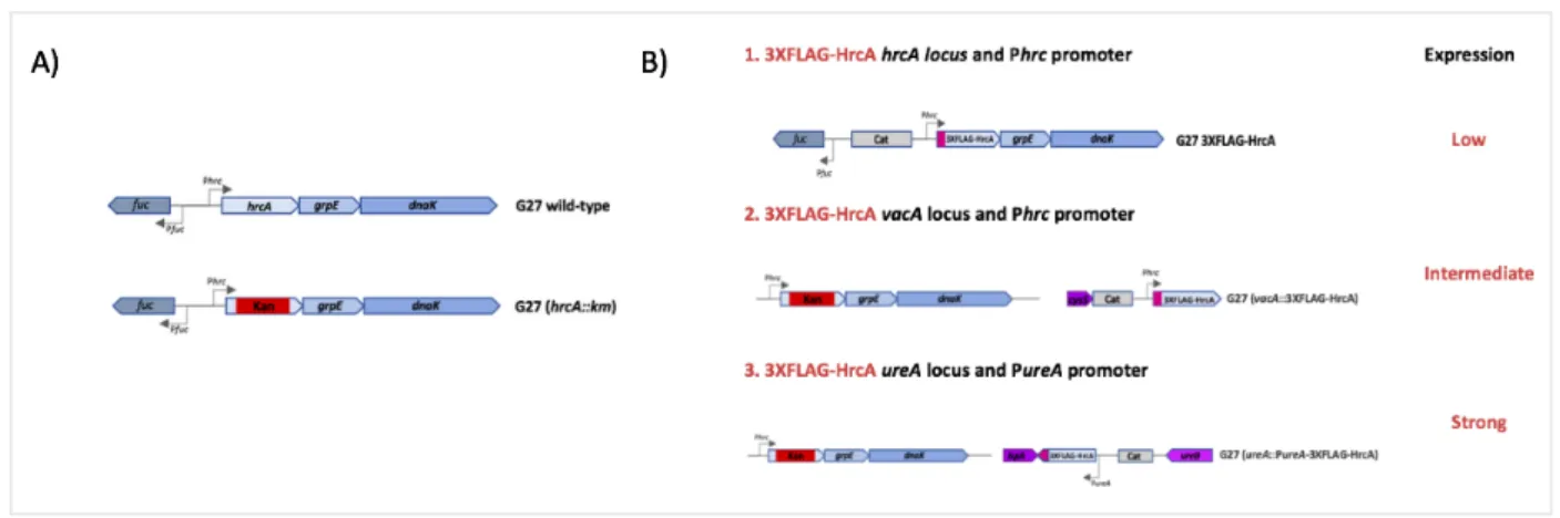
The Tag-HrcA complementing strains were obtained by double homologous recombination by transforming the G27 (hrcA::km) mutant strain (Table 1, Materials and Methods) with the appropriated plasmids (Table 2). The genes coding for the fusion proteins were reintroduced in the genome together with an antibiotic resistance cassette. The schematic representation of the 3XFLAG-HrcA expressing strains, which were selected for further studies, are reported in Figure 8. To find the optimal condition for the immunoprecipitation assays three different strategies aimed to three different expression levels were developed. The first strategy, a strain expressing the 3XFLAG-HrcA fusion protein from its original locus and under the transcriptional control of the hrcA promoter, Phrc, was constructed. Some observations suggested that the HrcA regulator in normal growth conditions is expressed at low levels in the bacterial cells (Martirani et al., 2001; Spohn et al., 2004).
In the second approach we generated a 3XFLAG-HrcA complementing strains expressed from the vacA ectopic and non-essential locus, while maintaining the transcription of hrcA driven by its natural Phrc promoter. Previous works performed in our laboratory have shown that the vacA locus has a positional effect on the inserted gene and usually induces a higher level of proteins compared to the original locus.
Finally, since transcriptional studies highlighted ureA as a gene transcribed by a strong promoter (Akada et al, 2000; Spohn et al, 1997), the ureA promoter, PureA, was selected to increase expression of the 3XFLAG-HrcA fusion protein.
Figure 8. (A) Representation of hrcA-grpE-dnaK operon in the G27 wild type strain and the G27 knockout mutant (hrcA::km). The arrows indicate the ORFs. The kanamycin resistance cassette (km) is reported in the red box. (B) Schematic representation of the three strategies used to construct the complementing 3XFLAG-HrcA strains from the G27 knockout mutant (hrcA::km). In these tagged HrcA strains the pink box is used to depict the position of the 3XFLAG epitope tag; the light grey box underlines a chloramphenicol resistance cassette up- and downstream of the hrcA gene. The two homologous regions flanking the DNA sequence of interest, necessary for the homologous recombination are: 1) the fuc gene (upstream) and the grpE gene (downstream) in the G27 3XFLAG-HrcA strain; 2) a portion of cysS gene (upstream) and a portion of vacA gene in the G27 (vacA::3XFLAG-HrcA) strain; 3) and the ispA gene(upstream) and the
ureB gene (downstream) in the G27 (urea::PureA-3XFLAG-HrcA). The kanamycin resistance cassette (km) is reported in
the red box in the overexpressing strains 2 and 3.
To assess the functionality of the 3XFLAG-HrcA fusion proteins expressed from the above described complementing strains we assayed heat shock response of the groESL operon, a known target of repression for HrcA. That is, bacteria were liquid grown to mid-exponential phase and total RNA was extracted. Then, transcript amounts of the groESL operon was assessed by Real Time PCR and compared to the amount of RNA in the G27 wild type strain). As expected, transcription of the groESL operon was strongly derepressed in the HrcA mutant strain compared to the wild type strain (data not shown). Interestingly, in the complementing strains, groESL repression is restored to almost wild type levels, indicating complementation of the HrcA function by the 3XFLAG-HrcA fusion protein. In contrast, it was observed that in the samples that were submitted to the heat shock, the levels of groESL were induced, being similar levels to the HP G27 hrcA mutant (hrcA::km).
To further confirm expression of the 3XFLAG-HrcA fusion protein in the complementing strains and verify specificity of the antibody, different protein extracts were subjected to Western Blotting analysis with an anti-FLAG tag antibody (Figure 9A).
Subsequently, we immunoprecipitated 3XFLAG-HrcA-bound genomic DNA fragments by chromatin immunoprecipitation assay using α-FLAG antibody. The assays were designed to perform immunoprecipitation of HrcA in the wild-type strain that does not express the tagged protein (negative control) and the strains
that expressed the 3XFLAG-HrcA fusion proteins. Following
immunoprecipitation, enrichment of a known HrcA target was checked by Real Time PCR, with oligonucleotides specific for the groESL binding sites of HrcA (Table 3) with results shown in Figure 9B.
In the first condition tested, that is, in the strain expressing 3XFLAG-HrcA under the Phrc promoter it is possible to appreciate an almost 2,5-fold enrichment of the HrcA binding region (light grey bar) with respect to the wild type strain (dark grey bar). Although promising, this result is not in line with previous observations in our lab that a five-fold enrichment would be the limit required for a significant analysis of the sequencing data.
A similar enrichment level of HrcA binding region (Figure 9B, middle panel) was observed when the immunoprecipitation experiment was carried out on the vac::Phrc-3XFLAG-hrcA strain in which the levels of the HrcA protein was higher (Figure 9A, middle panel). We hypothesized that overexpression of HrcA might alleviate this limitation. Unfortunately, overexpression of 3XFLAG-HrcA showed an even lower amount of immunoprecipitated 3XFLAG-HrcA binding region (Fig. 9B, right panel). Therefore, as also reported by others works, we conclude that overexpression of a protein does not necessarily lead to more effective immunoprecipitation as it may lead to altered genomic binding profiles due to excess protein in the cell and the abundance of protein tends to produce a high signal-to-noise ratio, which significantly increases the background and hinders a good normalization of the experiment (Kidder et al., 2011).
Figure 9. A) About 15 ug of total protein extracts of the H. pylori G27 wild type and complementing 3XFLAG-HrcA strains were fractioned in 12% SDS-PAGE. The band is present only in the strains that express the 3XFLAG-HrcA fusion protein and it completely lacks in the wild type strain (negative control). A Pierce Unstained Protein MW Marker (Thermofisher) was used as molecular weight marker and position of the interest band (32 kDa) is reported on the left side of the autoradiogram. B) Analysis of specific enrichment of known HrcA targets was carried out by Real Time PCR, with oligonucleotides specific for the binding sites of HrcA on groESL promoter in immunoprecipitation assay. Specific primers for the groESL promoter (table 3) were used to specifically test the enrichment of the DNA immunoprecipitated. The light grey bar indicates the fold enrichment of the DNA immunoprecipitated in the complementing 3XFLAG-HrcA strains, compared to the negative control (G27 wild type sample, dark grey bar).
Since the ChIP experiments did not allow the identification of new genomic
targets, to characterize the HrcA interactions on some putative regulated genes,
DNaseI footprinting experiments have been performed on a selection of three validated targets among the list of 18 upregulated genes by HrcA of the
transcriptome analysis: the HPG27_RS01495, HPG27_RS03015 and
HPG27_RS03495 genes.
The probes were radioactively labeled with 32P and incubated in vitro with
increasing amounts of recombinant purified His-HrcA and treated with DNaseI to
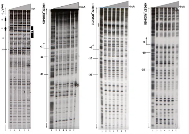
Figure 10 shows DNAseI footprinting experiments performed on the radiolabelled Phrc promoter as positive control and the three putative targets (HPG27_RS01495, HPG27_RS03015 and HPG27_RS03495) identified by transcriptome outcomes. Besides the Phrc promoter that is known to be a direct target of HrcA, no protection was detected in all the other probes submitted to DNaseI footprinting experiments. We conclude that HrcA probably doesn't bind these regions, at least under the experimental conditions tested.
Figure 10. DNaseI footprinting assay with purified recombinant HrcA on Phrc (A), PhbpA (B), PHPG27_RS03015 (C) and
PsabB (D) promoters. A) Schematic representation of the Phrc promoter region. Transcription start site (bent arrow), −10
and −35 boxes are indicated as well as HrcA open reading frame (open arrow). According to our previously published data (Roncarati et al., 2014). HrcA binding site is indicated in black open boxes represent the regions of DNaseI protection, while the black arrowhead indicate bands of hypersensitivity to DNaseI digestion. B, C and D) Radiolabelled DNA probes for putative Phbpa, PHPG27_RS03015 and PsabB promoters were end labeled at their non-coding strands, then were mixed with different amounts of purified HrcA protein and subjected to DNaseI digestion; purified DNA fragments were separated on a polyacrylamide denaturing gel and autoradiographed. Lanes 1 to 7 contain the G+A sequence reaction ladder and 0, 125, 250, 500, 1000 and 2000 ng of His-HrcA, respectively. On the left of each autoradiograph, the numbers refer to the positions with respect to the transcriptional start site (position +1).
4.3 Discussion
In this study, a differential transcriptional analysis allowed us to identify several non-heat shock responsive genes deregulated in the HrcA mutant. Of these, 18 genes appeared up-regulated and 37 genes appeared down regulated by HrcA. These latter are genes are mainly involved in different cellular functions such as cell motility, and transport or cell wall/membrane biogenesis.
To further characterize the HrcA regulon in H. pylori and investigate more in detail its direct or indirect contributions in the regulation of these genes, we attempted setting up a ChIP-seq experiments, the most direct way to the genome-wide profiling of protein-DNA interactions. Several attempts to carry out immunoprecipitation assays in diverse conditions showed a poor enrichment of
DNA fragments bound by HrcA and the overall results of the experiments
performed were not significant for the subsequent step of sequencing, due to a low amount of immunoprecipitated DNA and / or high levels of non-specific signal. It should be pointed out that in bacteria this technique has been used with a limited number of examples and it wasn’t possible to obtain the necessary conditions for the in vivo study of this regulator using this technique, even if we attempted optimization by changing experimental parameters such as conditions of sonication, antibody concentration, number of cells and levels of expression of the HrcA protein. In all tested conditions the immunoprecipitation results were not sufficient to proceed to genome-wide ChIP and the sequencing.
In literature, it has been reported that the HrcA protein from B. subtilis, B. stearothermophilus, S. aureus, C. acetobutylicum and B. thermoglucosidasius shows a common disadvantage characteristic as it is hard to solubilize and has the tendency to form aggregates when expressed in Escherichia coli cells (Watanabe., et al, 2001). Concerning Helicobacter pylori, it has been reported that HrcA is partly toxic and insoluble when expressed in E. coli. In addition, there is no structural information about the conformation of the protein, which seems to be associated to the inner membrane fraction of H. pylori (Roncarati et al., 2007). Possibly this peculiarity contributes to the unsatisfactory results of Chromatin Immunoprecipitation of HrcA-DNA complexes obtained until now.
Since it was not possible to identify new direct targets of HrcA through the
selection of three targets among the list of 18 upregulated genes by HrcA from
the transcriptome analysis with no evidence of DNA-binding (Figure 10).
Possibly, regulation of these genes by HrcA is exerted by an indirect mechanism. In H. pylori, HrcA represses transcription of the hrcA-grpE-dnaK and groES-groEL operons by direct binding to the Pgro and Phrc promoters on positions centered at +9 and -42 of the Pgro and Phrc promoters, respectively (Roncarati et al.,2007). The HrcA binding sites show similarities to a consensus sequence named CIRCE and firstly identified in B. subtilis (TTAGC ACTC-N9-GAGTGCTAA) (Narberhaus and Bahl, 1992).
In H. pylori and in other organisms, such as B. subtillis and S. aureus, it has been demonstrated that the GroE chaperonin is required in vivo to allow HrcA to fold in its active conformation to bind its target promoters and repress transcription of genes of thermal shock. After heat stress, GroE is titrated by misfolded proteins accumulated in the cell, and HrcA becomes inactive and dissociates from its promoter sequence, inducing the HrcA regulon (Roncarati et al., 2014; Chastanet et al., 2003; Mogk et al., 1997). In this model, the interaction between HrcA and GroESL appears very important. A study in C. trachomatis suggested that GroESL is capable not only of increase the binding affinity of HrcA to the CIRCE sequence but also to improve HrcA-mediated transcriptional repression, as HrcA showed efficient bind to the CIRCE in the presence of the GroESL complex (Wilson et al., 2005). Accordingly, this direct correlation between the GroESL complex and the HrcA activity suggests that to properly maintain its function in vivo HrcA requires the presence of chaperonins (Minder et al., 2000). Therefore, the absence of binding on the putative target promoters at the HPG27_RS01495, HPG27_RS03015 and HPG27_RS03495 genes may be explained by the lack of the chaperonin in the invitro technique and the possibility of a direct interaction between HrcA and these genes cannot be a priori disregarded.
Helicobacter pylori is a widespread human pathogen recognized as the main causative agent of chronic gastritis, gastric and peptic ulcers, and persistent colonization is an important factor in the development of gastric lymphoma and adenocarcinoma.
To respond to stress conditions and survive in a hostile environment such as the human stomach, this bacterium induces the synthesis of a class of highly conserved proteins, known as Heat-shock Proteins, which protect the cell from damage, denaturation, aggregation or folding of proteins.
In H. pylori, the genes encoding for the major heat shock proteins are clustered in three multicistronic operons and are repressed by two transcriptional regulators: HrcA and HspR.
In this study, the H. pylori HrcA repressor was demonstrated to be involved in the
regulation of several non heat-shock responsive genes. With a RNA-seq approach it was possible to identify a total of 48 from 137 deregulated genes, induced or repressed, after the almost complete deletion of HrcA in a mutant strain (hrcA::km) and not responsive to heat-shock.
To better characterize the HrcA regulon in H. pylori and investigate more in detail its direct or indirect contributions in the regulation of these genes, we attempted setting up a ChIP-seq experiments. In the present study the epitope tags were fused to the N-terminal domain of the protein, which harbors the DNA-binding domain (Roncarati et al., 2007). However, due to their particular characteristics and the lack of structural information of this protein it wasn’t possible to obtain the necessary conditions for the in vivo study of this regulator and the overall results of the experiments performed were not significant for the subsequent step of sequencing. For future prospects, it would be interesting to obtain more structural information on this protein in order to reallocate the tag used in a more
immunogenic position. Also develop a chromatin tandem affinity purification
(ChTAP) strategy could be an effective alternative to ChIP (Soleimani et al., 2013; Kolodziej et al., 2009).
Furthermore, DNaseI footprinting experiments have been performed on a
selection of three targets among the list of 18 upregulated genes by HrcA from the transcriptome analysis with no evidence of DNA-binding. Several studies suggest that in order to properly maintain its functions in vivo, HrcA requires the presence of chaperonins. Thus, although HrcA probably interacts with most of
these genes in indirect way, the possible direct interaction between HrcA and
these genes can not be a priori disregarded, since in vitro assays may not be
accurate if multiple factors or DNA segments are required for binding (Boyle et al., 2017). Perhaps, developing a protocol that allows the use of in vivo Footprinting may represent an effective alternative.
Globally, this study reveals the involvement of the H. pylori transcriptional
regulator HrcA in crucial cellular processes, most of them interconnected
between then with functions closely associated to the pathogenesis and virulence of this bacteria.
Most of the up-regulated genes in the ΔHrcA mutant are related to Cell wall/membrane/envelope biogenesis, Coenzyme-, Aminoacid- and Inorganic ion-transport and metabolism. Some studies point out genes related to these functions as potential target for the development of new drugs (Sperandeo et al., 2017; Tanaka et al., 2018; Thomas and Tampé, 2018).
Interestingly, this study demonstrates a close connection between the HrcA repressor and the motility of H. pylori. In fact, it was possible to identify several genes that regulate the biosynthesis and function of the flagellar apparatus. This result is in accordance with previous studies in which the mutant strain HrcA is described with non-motile phenotype. Furthermore, a recent study suggested that the H. pylori HrcA regulator and the flagellar apparatus may play an important structural role during biofilm formation (Hathroubi et al., 2018).
An interesting aspect to be verified is whether in a deletion condition of hrcA there are phenotypic changes in the flagellar structure of Helicobacter pylori that agree with its proven loss of motile function.
Although not essential, this study demonstrates that HrcA is interconnected to several cellular functions crucial for the survival, virulence, and maintenance of the H. pylori infection state in the human stomach. As a bacterial pathogen of major medical importance and the available antimicrobial therapies are losing efficacy, principally because of insurgence of antibiotic resistance, the globalized performance of HrcA may be the starting point for a search for new targets and therapeutic strategies.