A
AllmmaaMMaatteerrSSttuuddiioorruumm––UUnniivveerrssiittààddiiBBoollooggnnaa
DOTTORATO DI RICERCA
SCIENZE PNEUMO-CARDIO-TORACICHE
DI INTERESSE MEDICO E CHIRURGICO
Ciclo XXI
Settore scientifico disciplinari di afferenza: MED 10
TITOLO TESI:
IMMUNOGENETICS OF GRANULOMATOUS LUNG DISEASE: A PHENOTYPIC APPROACH TO SUSCEPTIBILITY
Presentata da: Silvia Contini
Coordinatore Dottorato
Relatore
Prof. S. Mattioli Prof. C. Saltini
•
Background……….…...
pag 3•
Immunogenetics of Tuberculosis
Evaluation of a multi-antigen test based on B-cell epitopes peptides for the serodiagnosis of pulmonary tuberculosis……….……pag 4 A model of phenotipic susceptibility to Tuberculosis. Deficient in silico selection of mycobacterium tuberculosi epitopes by HLA alleles………pag 21
•
Immunogenetics of sarcoidosis
M. avium binding to HLA-DR expressed alleles in silico: a model of phenotypic
B
ACKGROUND
Immunogenetics
Immunogenetics is a scientific discipline that uses immunological methods to study the inheritance of traits. Traditionally, immunogenetics has been concerned with moieties that elicit immune response, that is, with antigens (antigenic determinants). It has now broadened its scope to study also the genetic control of the individual's ability to respond to an antigen. The immunological methods used in immunogenetics are of two principal kinds, serological and histogenetical. In serological methods, antibodies are used to detect antigens, either in solution or on a cell surface. In histogenetical methods, immune cells (lymphocytes) are used to detect antigens on the surface of other cells. In modern immunogenetics research, the serological and histogenetical methods are combined with molecular methods in which the researcher isolates and works with the genes that code for the traits. This approach of going back and forth from classical to molecular methods has proved to be very successful and has led to the elucidation of several complex genetic systems.
Granulomatous Lung Disease
Granulomatous lung diseases, such as sarcoidosis, hypersensitivity pneumonitis, Wegener's granulomatosis, and chronic beryllium disease, along with granulomatous diseases of known infectious etiologies, such as tuberculosis, are major causes of morbidity and mortality throughout the world. Clinical manifestations of these diseases are highly heterogeneous, and the determinants of disease susceptibility and clinical course (e.g., resolution vs. chronic, progressive fibrosis) are largely unknown. The underlying pathogenic mechanisms of these diseases also remain poorly understood. Within this context, these diseases have been approached using genomic and proteomic technologies to allow us to identify patterns of gene/protein expression that track with clinical disease or to identify new pathways involved in disease pathogenesis. The results from these initial studies highlight the potential for these "-omics" approaches to reveal novel insights into the pathogenesis of granulomatous lung disease and provide new tools to improve diagnosis, clinical classification, course prediction, and response to therapy. Realizing this potential will require collaboration among multidisciplinary groups with expertise in the respective technologies, bioinformatics, and clinical medicine for these complex diseases.
IMMUNOGENETICS OF TUBERCULOSIS
E
VALUATION OF A MULTI-
ANTIGEN TEST BASED ONB-
CELL EPITOPES PEPTIDES FOR THE SERODIAGNOSIS OF PULMONARY TUBERCULOSISAbstract
SETTING: Two sample panels: (i) twenty pulmonary tuberculosis (TBp) patients and ten healthy subjects (HS) from a country with low incidence of TB (Italy) and (ii) forty-seven TBp patients and 26 HS from a country with high incidence of TB (Morocco).
OBJECTIVE: To identify a combination of Mycobacterium tuberculosis (Mtb) peptides useful for the serodiagnosis of active TBp.
METHODS: Fifty-seven B-cell epitopes peptides of Mtb were evaluated by immunoenzymatic assay and the data were analyzed using logistic regression analysis and Random Forest method. RESULTS: The best discriminating peptide between TBp patients and HS from the sample of the country with low incidence of TB has been the 23 amino acid peptide of the Rv3878 protein. Thus, the sensitivity and specificity was 65 % and 100 %, respectively. In contrast, the same peptide showed 47% and 100% as sensitivity and specificity respectively in the country with high incidence of TB. In addition, the best peptides combination was a pool of nine peptides which showed a sensitivity of 70.2% and a specificity of 100% in the country with high incidence of TB.
CONCLUSIONS: The 9-peptides pool can be useful in identifying patients with active TBp
Introduction
Tuberculosis (TB) remains a major public health problem in most developing countries. The global annual incidence of tuberculosis has been estimated at 8 to 10 million cases, with approximately 1.7 million deaths (1). The control of the disease depends largely on early detection and the treatment of active cases.
At present, efforts at developing immunological tests are directed towards the identification of novel antigens that are associated with active disease. Thus the major objective of current immunological studies in TB is the identification of species-specific antigens and determination of the significance of corresponding immune responses. In this context, it is reasonable to hypothesize that proteins preferentially expressed by Mtb in models of intracellular growth and infection might be ideal targets for the design of new, highly sensitive diagnostic tests of Mtb infection and, eventually, for vaccine design (2, 3).
Serodiagnosis tests like enzyme linked immuno-sorbent assay (ELISA) are promising in view of their ease of performance and cost-effectiveness, many studies have been carried out by using quite complex antigens, such as whole bacteria, culture filtrates, bacterial extracts, and tuberculin or their purified protein derivatives (PPD) (4-6). However, a large variability in diagnostic accuracy has been reported, depending on the antigen employed and on the heterogeneity of the antibody response in TB patients(7).
The use of biochemical purification methods and recombinant DNA techniques of natural and recombinant proteins to obtain mycobacterial antigens is a challenging task with high cost and low specificities of the assays based on these antigens (8, 9). Therefore, we used synthetic peptides to assess their antibody responses in tuberculosis patients. Furthermore, the in silico approach based on screening of Mtb coding sequences from the proteins databases by using the prediction programs algorithms to identify B-cell epitopes is promising, since it reduce the cost of synthesizing peptides by reducing the number of peptides required for experimental evaluation for antibody reactivity (10, 11).
In this study, we assessed the potential use of the in-silico selection approach of M. tuberculosis-specific synthetic peptides in the serodiagnosis of active tuberculosis.
Materials and methods
Study populationTo identify the immunological relevant peptides between the in-silico selected peptides, a first panel of sera from a country with low incidence of tuberculosis (Italy) were analysed. Specifically, 10 sera from skin test negative (TST-negative) healthy subjects without any history of TB exposure and 20 sera from active TB patients enrolled at Division of Respiratory Medicine of the University of Rome “Tor Vergata”, at the “L. Spallanzani” Institute (Roma, Italy). The diagnosis of active TB was microbiologically confirmed by culture isolation of Mtb in all TB cases. Patient’s details are shown in Table 1.
A second panel of sera from a country with high level of TB incidence has been used to evaluate the relevant peptide and the pool of peptides selected with the first study population. To this end, 47 patients with pulmonary active tuberculosis from Morocco were evaluated (Table 1). All patients underwent to smear and culture examination for Mtb according to standard procedures. Specifically, 31 Smear and culture positive (S+C+) patients and 16 smear-negative, culture-positive (S-C+) TB patients were evaluated in the study (table 1). Further, sera from twenty-six skin test positive healthy voluntary donors were collected from the Blood transfusion Regional Center (CRTS) of Rabat, Morocco.
All stud y s ubj ects were foun d to be H IV negativ e. Sera from all patients were collected before anti-tuberculous treatment and were stored at -20°C until use.
The B-cell epitopes selection
Fifty-seven synthetic peptides containing potential B-cell epitopes were selected by in-silico strategy from 25 proteins of Mtb. Briefly, the sequence of Mtb proteins belonging to RD1 genomic region (12) and of proteins expressed during Mtb growth in human macrophage (2, 3), was obtained by the Swiss-Prot database (Table 2). Each single protein was screened for potential B-cell epitopes by using three computer algorithms of antigenicity: the Jameson-Wolf index, the Hopp-Woods method and the Parker index. These algorithms, available with Genrunner software [Hastings Software, Inc., Hastings, N.Y.] and Antheprot software (13-15), predict antigenic regions based on probability of surface exposure, local hydrophobicity, beta turns amino acid sequence, atomic flexibility and experimental HPLC retention times of synthetic peptides. Only protein regions identified as containing B-cell epitopes by at least two algoritms were selected.
The sequence of all the selected peptides is reported on Table 2.
Peptides
The peptides were synthesized by Fmoc chemistry as free amino acid termini by ABI (http://www.abi.it/). All peptides were purified by reverse-phase chromatography (RPC). The purity of the purified peptides was higher than 90%. Sequence and purity were confirmed by mass spectrometry and analytical RPC. Lyophilized peptides were diluted in DMSO at stock concentrations of 10 mg/ml for each peptide and stored at – 80°C until use.
Peptide-based ELISA
Ninety-six wells high-binding capacity flat-bottom microplate (Greiner) was coated with 1 µg per well of each peptide in a Phosphate Buffered saline (PBS) buffer (pH 7.4) and kept at 4°C overnight. Then the plate was washed with 0.05% (v/v) Tween-20 in PBS and blocked with 3 % of bovine serum albumin (BSA, Sigma) for 1 hour. Each sample was assayed at a 1:50 dilution (2 hours, at room temperature). A horseradish peroxidase (HRP) conjugated anti-human IgG was used as the second antibody. The reaction was revealed using Ortho-Phenyl-Diamine (OPD)/H2O2
(Sigma) and quantified using an ELISA reader at 450 nm after stopping the reaction with 0.5M H2S04. Blank wells were included as negative control in this assay.
Statistical analysis
Test results are presented as means ± standard deviation (SD) of the mean. ELISA tests were scored positive when individual readings were greater than two standards deviations above the optical density average of healthy subjects. Comparisons between different groups were made by Mann-Whitney test.
The best peptides discriminating between TB patients and controls sera were identified by using univariate logistic regression model using Statistical Package for Social Science™ (SPSS version 15), the Random Forests Method [http://genesrf.bioinfo.cnio.es/] (16) and stepwise multivariate logistic regression model using SPSS program.
The Receiver Operating Characteristic (ROC) curve was used to evaluate the performance of the ELISA tests with the two categories (TB patients and controls) ; the Area Under the Receiver Operating Characteristic curve (AUC) was used as a measure of diagnostic quality. The cut-off value used here is the one that leads to a ≥ 50% probability of being a case.
Results
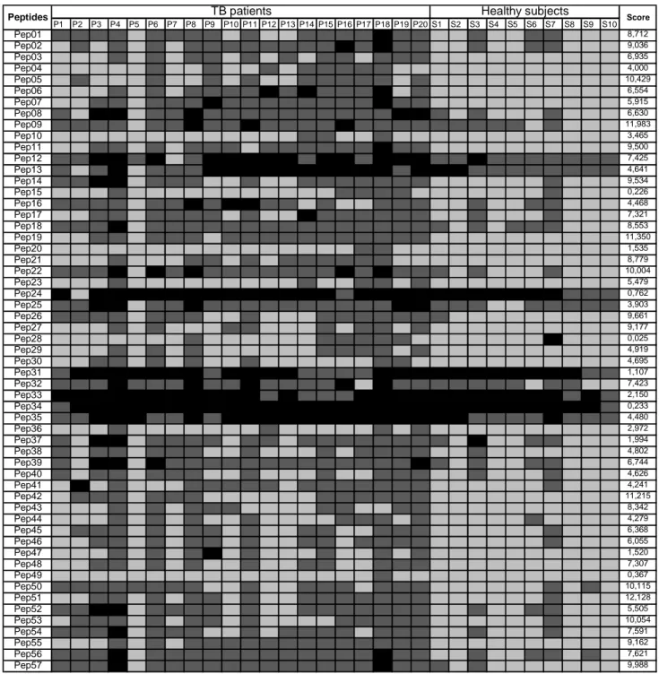
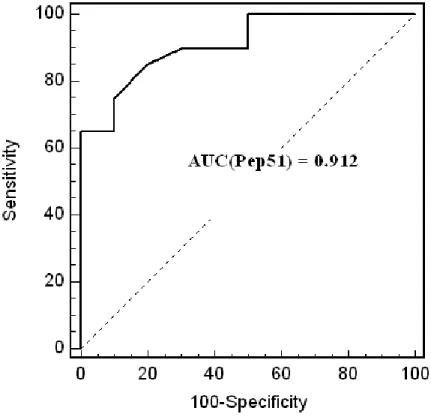
In order to identify peptides capable of being recognized by TB patients, a first panel of TB and control sera from a low incidence country for TB was tested. Figure 1 shows the IgG response against each one of the fifty-seven selected peptides containing potential B–cell epitope (Figure 1). The antibody responses against the peptides were variable (figure.1). Univariate logistic regression method showed that the best discrimination score value between TB and healthy controls was 12.128 obtained with the peptide Pep51 of the Rv3878 protein (see for sequence table 2). With a 2SD cut-off value, the sensitivity and specificity was 65 % and 100% respectively and the area-under-the-ROC curve was 0.912 (Figure 2).
When multivariate logistic regression method and random Forest method were used, the best peptides combination identified was: Pep5 (Rv0747), Pep11 (Rv1114), Pep14 (Rv1434), Pep26 (Rv1979c), Pep42 (Rv3736), Pep48 (Rv3874 also known as CFP10), Pep51 (Rv3878), Pep55 (Rv3883) and Pep57 (Rv3883).
The peptides identified were then used in a second population from a country with high incidence of TB in order to evaluate their performances in the diagnosis of active TB.
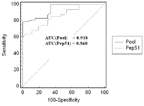
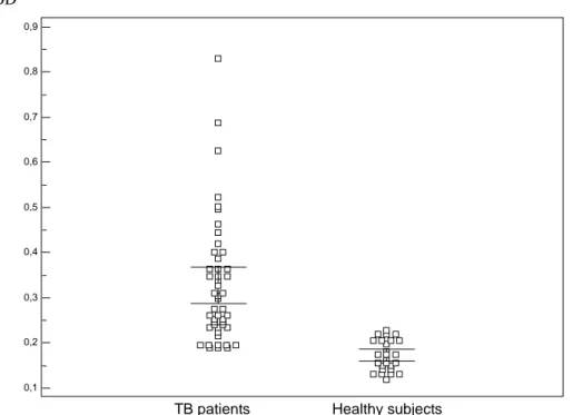
The results of the ELISA based on the peptide Pep51 alone among the forty-seven TB patients and the twenty-six healthy controls showed a sensitivity and specificity of 47% and 100% respectively with a cut-off value at 2SD of the controls and the area-under-the-ROC curve was 0.842 (Figure 3). However, the results among the same population but with the ELISA based on the selected 9-peptides showed a higher value (0.938) of the area-under-the-ROC curve (P value=0.04) (Figure 4). Further, figure 5 shows the box plots of the antibody titer obtained with the second panel sera of TB and healthy controls. With a cut-off value at 2SD of the controls, the sensitivity and specificity was 70.2 % and 100% respectively (Figure4).
Discussion
The availability of mycobacterial genome sequences, the development of chemical synthesis techniques of peptides and the development of algorithms for the identification of immunodominant B-cell epitopes, might determine the rapid identification and testing of epitopes with diagnostic potential for active TB with the advantage of reducing the number of experimental evaluations for antibody reactivity. In fact, the use of ELISA test based on synthetic peptides would in principle circumvent the technical complexity and potential antigen variability associated with antigen purification. Further, synthetic peptides used as antigens have the added advantage of avoiding contamination with low-level impurities derived from cloning vectors required to produce recombinant proteins. Such impurities could decrease the specificity of the assay if they were cross-reactive with M. tuberculosis antigens. Finally, using synthetic peptides as the diagnostic antigen permit the uniformity and standardization of antigen preparations (17).
However, the main problem in setting immunodiagnostic systems is the identification of immunodominant antigen(s) specific of the active disease phase. In this context, previous studies (2, 3), have shown that Mtb is capable of expressing differential transcription programs in response to different environments. In particular, Mtb can express different gene sets in synthetic media compared to macrophage cultures. For instance, RD1 proteins are expressed both in early and late phase cultures, are variably expressed in in vivo in murine models, but are expressed at very low level in macrophage cultures (2, 3, 18-20).
With this background, we hypothesized that the Mtb proteins overexpressed in a model of Mtb infection in macrophage cultures could be the specific target of a strong immune response in active TB subjects. Thus, we combined the use of relevant Mtb antigen in a model of Mtb infection with in
silico approach to identify B-cell epitopes to evaluate their potential use in TB serodiagnosis.
We were able to identify a peptide belonging to the Mtb protein Rv3878, as the best discriminating peptide between TB patients and healthy controls among a population of low exposure level of TB. Rv3878 is one the proteins encoded by RD1, a region present in Mtb and virulent M. bovis genomes but missing from the DNA of all substrains of M. bovis Bacillus Calmette-Guerin (BCG) (12). Although our findings are not in agreement with previous observation by Brusasca et al (21) who reported that only 3–7% of human TB patients responded to RD1 region proteins, including Rv3878, it is well known that all RD1 antigens elicited a high antibody response in guinea-pigs infected with Mtb (21). Further, in a recent study (22), it has been shown that the Rv3878 protein elicited strong humoral immune response in TB patients in Indian population, supporting the notion
that the antibody response to a specific antigen often depends on the geographical location and ethnic background of the population being studied (23).
Several studies indicate that combinations of mycobacterial antigens are likely to have better diagnostic value (24, 25). Using both multivariate logistic regression analysis and random Forest method, we were able to identify a pool of 9-peptides beloning to the proteins Rv0747, Rv1114, Rv1434, Rv1979c, Rv3874, Rv3736, Rv3878 and Rv3883 as the best discriminating peptides combination between TB patients and healthy controls.
Recent reports indicate that the antigen Rv3874 is frequently recognized by antibodies in patients with TB, from regions where TB is not endemic (21) and from regions where TB is endemic (26). Further, Rv3883c or Mycosin-1 is an extra-cellular protein that is membrane- and cell wall-associated, and is shed into the culture supernatant. The protein is expressed after infection of Macrophages (27), but Rv3883C is not expressed in the attenuated M. bovis strain BCG, although the gene for mycosin-1 is present in the genome of this organism. This gene was found to be situated 3700 bp from the RD1 deletion region (28).
Moreover, recombinant Rv1979c has been identifies as an antigen capable to induce IFNγ in correlation with extent of lesions at necropsy (29). Finally, no information is available in the literature about the immunoreactivity of the other proteins identified as relevant in discriminating TB patient sera.
Both genetic and environmental factors also influence the antibody response. For example, the varied antibody response to Mtb is managed by HLA types. That fact, therefore, that our pool of peptides identified in an population has strong diagnostic value also for another population with a different genetic background, suggest that these peptides may form the basis of a test that can be used independent of location.
Our results, in accordance with those obtained by other investigators, confirmed higher sensitivity for the smear-positive patients than for the smear-negative patients (P<0.001), due probably to a greater exposure to antigens in patients with high bacillary loads (30, 31). However, in developing countries such as Morocco where TB disease is prevalent, serological tests might play a major role in the setting of the diagnosis of patients with suspect TB where acid-fast bacilli examination is negative. Our results showed that with the ELISA based on the peptide pool, TB can be diagnosed with 44% sensitivity in acid fast bacilli smear-negative TB patients. This results is interesting given the facts that the are no simple fast alternatives (e.g. culture takes 6 weeks) in these clinical settings, and even thought that x-ray is a standard tool of TB diagnosis it could be quit questionable in some
Finally, it is worth to consider that in the study population with high TB prevalence we evaluated, all controls were TST-positive. Thus, the peptide based ELISA was able to distinguish between active and inactive tuberculosis infection, while other immunological based tests such as TST and gamma–interferon based assays are not capable (33, 34). Nevertheless it would be important to explore how the ELISA based on the peptide pool performs in recently exposed individuals as well as in subjects with cured tuberculosis or under chemotherapy treatment that are known to present high anti-Mtb IgG levels (35).
In conclusion, with the limitation of the small study population evaluated, this study indicates the applicability of the combination of nine well-defined synthetic peptides to the serodiagnosis of active TB. Their use in an ELISA would be important because of their stability, reproducibility and capability to identify strong humoral immune responses in patients with active pulmonary TB. The
Mtb-complex specificity of these peptides strongly encourages further evaluation of this pool of
peptides as a reagent for TB-specific immunodiagnostic assays on larger study populations including other disease groups such as patients with pulmonary disease other than TB, or seropositive patients for HIV.
REFERENCES
1. OMS. 2006. Global tuberculosis control: Surveillance, planning, financing. Genève.
2. Cappelli, G., E. Volpe, M. Grassi, B. Liseo, V. Colizzi, and F. Mariani. 2006. Profiling of Mycobacterium tuberculosis gene expression during human macrophage infection: upregulation of the alternative sigma factor G, a group of transcriptional regulators, and proteins with unknown function. Research in microbiology 157:445-455.
3. Mariani, F., G. Cappelli, G. Riccardi, and V. Colizzi. 2000. Mycobacterium tuberculosis H37Rv comparative gene-expression analysis in synthetic medium and human macrophage.
Gene 253:281-291.
4. Chiang, I. H., J. Suo, K. J. Bai, T. P. Lin, K. T. Luh, C. J. Yu, and P. C. Yang. 1997. Serodiagnosis of tuberculosis. A study comparing three specific mycobacterial antigens. Am
J Respir Crit Care Med 156:906-911.
5. Harboe, M., H. G. Wiker, and S. Nagai. 1992. Protein antigens of mycobacteria studied by quantitative immunologic techniques. Clin Infect Dis 14:313-319.
6. Raja, A., U. D. Ranganathan, R. Bethunaickan, and V. Dharmalingam. 2001. Serologic response to a secreted and a cytosolic antigen of Mycobacterium tuberculosis in childhood tuberculosis. Pediatr Infect Dis J 20:1161-1164.
7. Lyashchenko, K., R. Colangeli, M. Houde, H. Al Jahdali, D. Menzies, and M. L. Gennaro. 1998. Heterogeneous antibody responses in tuberculosis. Infection and immunity 66:3936-3940.
8. Mustafa, A. 2005. Progress towards the development of new antituberculosisvaccines. In
Focus on Tuberculosis Research. e. Smithe LT, ed. Nova Publishers, New York. p. 47-76.
9. Mustafa, A. S. 2002. Development of new vaccines and diagnostic reagents against tuberculosis. Mol Immunol 39:113-119.
10. Hagmann, M. 2000. Computers aid vaccine design. Science (New York, N.Y 290:80-82. 11. Zerpa, N. C., A. Wide, J. Noda, H. Bermudez, R. Pabon, and O. O. Noya. 2006.
Immunogenicity of synthetic peptides derived from Plasmodium falciparum proteins.
Experimental parasitology 113:227-234.
12. Mahairas, G. G., P. J. Sabo, M. J. Hickey, D. C. Singh, and C. K. Stover. 1996. Molecular analysis of genetic differences between Mycobacterium bovis BCG and virulent M. bovis.
Journal of bacteriology 178:1274-1282.
13. Deleage, G., F. F. Clerc, and B. Roux. 1989. ANTHEPROT: IBM PC and Apple Macintosh versions. Comput Appl Biosci 5:159-160.
14. Deleage, G., F. F. Clerc, B. Roux, and D. C. Gautheron. 1988. ANTHEPROT: a package for protein sequence analysis using a microcomputer. Comput Appl Biosci 4:351-356.
15. Deleage, G., C. Combet, C. Blanchet, and C. Geourjon. 2001. ANTHEPROT: an integrated protein sequence analysis software with client/server capabilities. Comput Biol Med 31:259-267.
16. Heidema, A. G., J. M. Boer, N. Nagelkerke, E. C. Mariman, A. D. van der, and E. J. Feskens. 2006. The challenge for genetic epidemiologists: how to analyze large numbers of SNPs in relation to complex diseases. BMC Genet 7:23.
17. Liang, F. T., A. C. Steere, A. R. Marques, B. J. Johnson, J. N. Miller, and M. T. Philipp. 1999. Sensitive and specific serodiagnosis of Lyme disease by enzyme-linked immunosorbent assay with a peptide based on an immunodominant conserved region of Borrelia burgdorferi vlsE. Journal of clinical microbiology 37:3990-3996.
19. Rogerson, B. J., Y. J. Jung, R. LaCourse, L. Ryan, N. Enright, and R. J. North. 2006. Expression levels of Mycobacterium tuberculosis antigen-encoding genes versus production levels of antigen-specific T cells during stationary level lung infection in mice. Immunology 118:195-201.
20. Shi, L., R. North, and M. L. Gennaro. 2004. Effect of growth state on transcription levels of genes encoding major secreted antigens of Mycobacterium tuberculosis in the mouse lung.
Infection and immunity 72:2420-2424.
21. Brusasca, P. N., R. Colangeli, K. P. Lyashchenko, X. Zhao, M. Vogelstein, J. S. Spencer, D. N. McMurray, and M. L. Gennaro. 2001. Immunological characterization of antigens encoded by the RD1 region of the Mycobacterium tuberculosis genome. Scand J Immunol 54:448-452.
22. Mukherjee, P., M. Dutta, P. Datta, A. Dasgupta, R. Pradhan, M. Pradhan, M. Kundu, J. Basu, and P. Chakrabarti. 2007. The RD1-encoded antigen Rv3872 of Mycobacterium tuberculosis as a potential candidate for serodiagnosis of tuberculosis. Clin Microbiol Infect 13:146-152.
23. Weldingh, K., I. Rosenkrands, L. M. Okkels, T. M. Doherty, and P. Andersen. 2005. Assessing the serodiagnostic potential of 35 Mycobacterium tuberculosis proteins and identification of four novel serological antigens. Journal of clinical microbiology 43:57-65. 24. Gennaro, M. L. 2000. Immunologic diagnosis of tuberculosis. Clin Infect Dis 30 Suppl
3:S243-246.
25. Houghton, R. L., M. J. Lodes, D. C. Dillon, L. D. Reynolds, C. H. Day, P. D. McNeill, R. C. Hendrickson, Y. A. Skeiky, D. P. Sampaio, R. Badaro, K. P. Lyashchenko, and S. G. Reed. 2002. Use of multiepitope polyproteins in serodiagnosis of active tuberculosis. Clinical and
diagnostic laboratory immunology 9:883-891.
26. Greenaway, C., C. Lienhardt, R. Adegbola, P. Brusasca, K. McAdam, and D. Menzies. 2005. Humoral response to Mycobacterium tuberculosis antigens in patients with tuberculosis in the Gambia. Int J Tuberc Lung Dis 9:1112-1119.
27. Dave, J. A., N. C. Gey van Pittius, A. D. Beyers, M. R. Ehlers, and G. D. Brown. 2002. Mycosin-1, a subtilisin-like serine protease of Mycobacterium tuberculosis, is cell wall-associated and expressed during infection of macrophages. BMC microbiology 2:30.
28. Brown, G. D., J. A. Dave, N. C. Gey van Pittius, L. Stevens, M. R. Ehlers, and A. D. Beyers. 2000. The mycosins of Mycobacterium tuberculosis H37Rv: a family of subtilisin-like serine proteases. Gene 254:147-155.
29. Cockle, P. J., S. V. Gordon, A. Lalvani, B. M. Buddle, R. G. Hewinson, and H. M. Vordermeier. 2002. Identification of novel Mycobacterium tuberculosis antigens with potential as diagnostic reagents or subunit vaccine candidates by comparative genomics.
Infection and immunity 70:6996-7003.
30. Bartoloni, A., M. Strohmeyer, F. Bartalesi, D. Messeri, E. Tortoli, A. Farese, F. Leoncini, S. Nutini, R. Righi, A. Gabbuti, F. Mazzotta, and F. Paradisi. 2003. Evaluation of a rapid immunochromatographic test for the serologic diagnosis of tuberculosis in Italy. Clin
Microbiol Infect 9:632-639.
31. Pottumarthy, S., V. C. Wells, and A. J. Morris. 2000. A comparison of seven tests for serological diagnosis of tuberculosis. Journal of clinical microbiology 38:2227-2231.
32. Linh, N. N., G. B. Marks, and A. B. Crawford. 2007. Radiographic predictors of subsequent reactivation of tuberculosis. Int J Tuberc Lung Dis 11:1136-1142.
33. Miguel, G., Madariaga, J. Ziba, and S. Susan. 2007. Clinical Utility of Interferon Gamma Assay in the Diagnosis of Tuberculosis. J Am Board Fam Med 20:540-547.
34. Ravn, P., M. E. Munk, A. B. Andersen, B. Lundgren, J. D. Lundgren, L. N. Nielsen, A. Kok-Jensen, P. Andersen, and K. Weldingh. 2005. Prospective evaluation of a whole-blood
test using Mycobacterium tuberculosis-specific antigens ESAT-6 and CFP-10 for diagnosis of active tuberculosis. Clinical and diagnostic laboratory immunology 12:491-496.
35. Amicosante, M., S. Barnini, V. Corsini, G. Paone, C. A. Read, Jr., P. L. Tartoni, M. Singh, C. Albera, A. Bisetti, S. Senesi, and et al. 1995. Evaluation of a novel tuberculosis complex-specific 34 kDa protein in the serological diagnosis of tuberculosis. Eur Respir J 8:2008-2014.
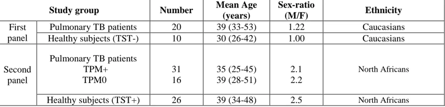
Table 1: Demographic characteristics of the study subjects
Study group Number Mean Age (years)
Sex-ratio
(M/F) Ethnicity
First panel
Pulmonary TB patients 20 39 (33-53) 1.22 Caucasians Healthy subjects (TST-) 10 30 (26-42) 1.00 Caucasians
Second panel Pulmonary TB patients TPM+ TPM0 31 16 35 (25-45) 39 (28-51) 2.1 2.2 North Africans
Healthy subjects (TST+) 26 39 (34-48) 2.5 North Africans TP = Pulmonary tuberculosis, TPM+ = Smear positive pulmonary tuberculosis, TPM0 = Smear positive pulmonary tuberculosis, M/F = male /female, TST+ = Tuberculin Skin Test positive, TST- = Tuberculin Skin Test negative.
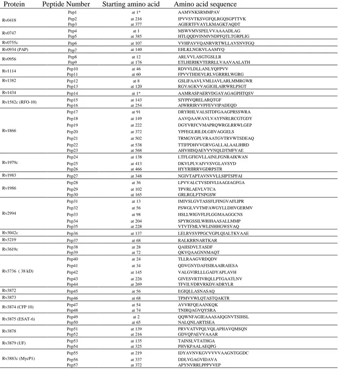
Table 2: List of the peptides used in this study.
*Peptide epitopes starting from amino acid position 1 in the protein sequence where two Alanine were added to the N-terminus of the peptide in order to increase binding affinity to the epitope.
Protein Peptide Number Starting amino acid Amino acid sequence
Rv0418 Pep1 at 1* AAMVNKSRMMPAV Pep2 at 216 IPVVSVTKSVGFQLRGQSGPTTVK Pep3 at 377 AGIERTFVAYLKMAGKTAQDT Rv0747 Pep4 at 1 MSWVMVSPELVVAAAADLAG Pep5 at 385 HTLQQDVINMVNDPFQTLTGRPLIG Rv0755c Pep6 at 107 VVHPAVVQANRVRTWLLAVSNVFGQ
Rv0934 (PAP) Pep7 at 140 EHLKLNGKVLAAMYQ
Rv0956 Pep8 at 12 ARLVVLASGTGSLLR Pep9 at 176 ETLHERIKVTERRLLVAAVAALATH Rv1114 Pep10 at 46 RDVVLDLLANLYQFPVV Pep11 at 60 FPVVTHDEVLRLVGRRRLWGRG Rv1382 Pep12 at 8 GSLIFAAVLVMLIAVLARLMMRGWR Pep13 at 120 RGVAGKVVAGIGILAIRWRLPSGT Rv1434 Pep14 at 1* AAMRASPAERVDGAYAGAGPHTQSV
Rv1582c (RFO-10) Pep15 at 143 SIYPIVQRELARQTGF
Pep16 at 254 AIWRRIRVVPFEVVIPADEQD Rv1866 Pep17 at 91 DRYRHLVALSITDFGAAGPRSSWRA Pep18 at 149 AAVQAAWAVLVAYFNRLRCGTGDY Pep19 at 222 DGYVRFCVMAPRQWRGLRRWLGEP Pep20 at 372 YPFEGLRILDLGIIVAGGELS Pep21 at 502 TRMGYGPLVRAATGVTRVWTSDEAQ Pep22 at 538 TTIFPDHVVGRVGALLALAALIHRD Pep23 at 568 AHVHISQAEVVVNQLDTMFVAE Rv1979c Pep24 at 138 LTFLGFIGVLLAINLFGNRAIKWAN Pep25 at 413 DKVLPLVAIVVSVGLAVSYD Pep26 at 466 HYYRIIRRVGDRPSTR Rv1983 Pep27 at 348 NGIVTAPTAVNVVLLSIPTSPFAI Rv1986 Pep28 at 36 LPVVALCTVSDIVLIAAGIAGFGA Pep29 at 102 TPVRLAEVLVTCA Pep30 at 165 GRLRGLFTNPGSW Rv2994 Pep31 at 13 IMIVSLGVTASSFLFINGVAFLIPR Pep32 at 56 PSWGLVVTMFAWGYLLDHVGERMV Pep33 at 98 HSLLWIGVFLFLGGMAAGGCNS Pep34 at 204 SPYRGSSILWRIHAASALLMMP Pep35 at 228 VTVTFMLVWLINHHGWSVAQ Rv3042c Pep36 at 137 LELRVSVPPGCVGPLQIALTKVAAE Rv3219 Pep37 at 68 RALKRRNARTKAR Rv3619c Pep38 at 28 QAIISDVLTASDF Pep39 at 72 QKVQAAGNNMAQT Rv3736 ( 38 kD) Pep40 at 24 TLLRAAGVRDQDV Pep41 at 34 QDVGNYDAFISIRAAIRAIESA Pep42 at 145 VALGVIRLLLGADYAPLAVH Pep43 at 226 GIVESVRTIVRQLLPTGAATLNV Pep44 at 269 TFVILVDRVRKDVADRYLR Rv3872 Pep45 at 56 EGIQLLASNASAQ Rv3873 Pep46 at 68 TPMVVWLQTASTQAKTR Rv3874 (CFP 10) Pep47 at 54 AVVRFQEAANKQK Pep48 at 74 TNIRQAGVQYSRA
Rv3875 (ESAT-6) Pep49 at 2 QQWNFAGIEAAASAIQGNVTSIHSL
Pep50 at 65 NALQNLARTISEA
Rv3878 Pep51 at 139 PRVVATVPQLVQLAPHAVQMSQN
Pep52 at 216 GDVQPAEVVAAAR
Rv3879 (UF) Pep53 at 135 TAINSLVTATHGA
Pep54 at 325 PHVKPAALAEQPG
Rv3883c (MycP1)
Pep55 at 219 IDYAVNVKGVVVVVAAGNTGGDC
Pep56 at 337 DDLVGAGVIDAVA
Figure 1 : Graphical value representation of the optical density (OD)
obtained for each peptide and sera from the first panel by ELISA showing the heterogeneity of the antibody responses in TB patients and healthy subjects against Mycobacterium tuberculosis peptides.
Antibody level (OD): O OD<0.100 ; 0.100≤OD<0.200 and 0. 200≤OD.
The right column shows the score value for each peptide obtained by unvariate regression analysis.
P1 P2 P3 P4 P5 P6 P7 P8 P9 P10 P11 P12 P13 P14 P15 P16 P17 P18 P19 P20 S1 S2 S3 S4 S5 S6 S7 S8 S9 S10 Pep01 8,712 Pep02 9,036 Pep03 6,935 Pep04 4,000 Pep05 10,429 Pep06 6,554 Pep07 5,915 Pep08 6,630 Pep09 11,983 Pep10 3,465 Pep11 9,500 Pep12 7,425 Pep13 4,641 Pep14 9,534 Pep15 0,226 Pep16 4,468 Pep17 7,321 Pep18 8,553 Pep19 11,350 Pep20 1,535 Pep21 8,779 Pep22 10,004 Pep23 5,479 Pep24 0,762 Pep25 3,903 Pep26 9,661 Pep27 9,177 Pep28 0,025 Pep29 4,919 Pep30 4,695 Pep31 1,107 Pep32 7,423 Pep33 2,150 Pep34 0,233 Pep35 4,480 Pep36 2,972 Pep37 1,994 Pep38 4,802 Pep39 6,744 Pep40 4,626 Pep41 4,241 Pep42 11,215 Pep43 8,342 Pep44 4,279 Pep45 6,368 Pep46 6,055 Pep47 1,520 Pep48 7,307 Pep49 0,367 Pep50 10,115 Pep51 12,128 Pep52 5,505 Pep53 10,054 Pep54 7,591 Pep55 9,162 Pep56 7,621 Pep57 9,988
Figure 2: The ROC curve of the peptide Pep51 evaluated with the panel of TB
and control sera from a low incidence country for TB. (AUC = Area under the
Figure 3: The ROC curves of the peptide Pep51 and the 9-peptides pool
evaluated with the panel of TB and control sera from a high incidence country
for TB. (AUC = Area under the curve)
Figure 4: Box plot representation of the distribution of levels of antibodies (IgG)
to the 9-peptides pool of Mycobacterium tuberculosis in
TB patients and
healthy subjects originated from a high incidence country for TB.
(Error
bars : 95% confidence interval for mean).
0,9 0,8 0,7 0,6 0,5 0,4 0,3 0,2 0,1
TB patients Healthy subjects OD
A
MODEL OF PHENOTYPIC SUSCEPTIBILITY TOT
UBERCULOSIS:
D
EFICIENT IN SILICO SELECTION OFM
YCOBACTERIUM TUBERCULOSIS EPITOPES BYHLA
ALLELESAbstract
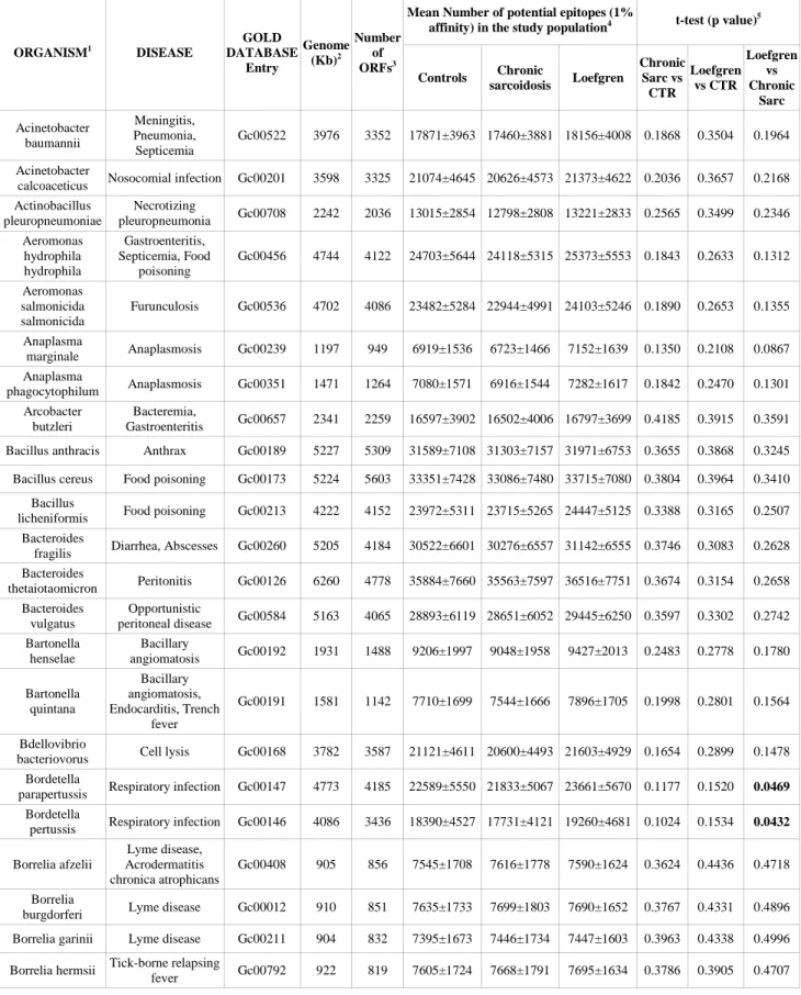
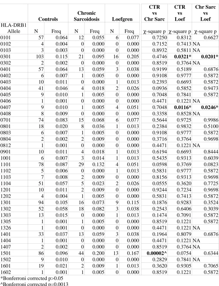
.HLA-DR allelic variants have been associated with tuberculosis (TB) susceptibility in different populations with risk ratios of 3.7 to 7.2.We hypothesized that the genetic susceptibility to TB depends upon the reduced capability of HLA-class II alleles of TB patients to bind and select peptide antigen from the Mycobacterium tuberculosis (MTB) expressed genome. To test this hypothesis, we developed a software that can predict HLA-DR restricted epitopes within the whole MTB genome based on quantitative peptide binding matrices.We analyzed the number of MTB epitopes recognized in two previously described populations of TB patients and matched controls and in a control population comprised of individuals affected by a sarcoid-like granuloma induced by beryllium and by healthy exposed controls. The number of putative epitopes within the whole MTB genome which could be bound by any HLA-DR allele (HLA-DR immunome of MTB) was 405,422 out of 1,304,277 possible 9-mers i.e., 31.08% of the global capability, instead of the expected 35%. When tested at an affinity level equivalent of the 1% of the best binder peptides, the HLA-DR alleles (HLA-DRB1*0801, *0802, *1401, *1501 and *1502) associated with TB susceptibility recognized a significantly lower mean number of MTB-epitopes (7,862±4,258) than the MTB-epitopes recognized by HLA-DR alleles (HLA-DRB1*0301, *0701, *1101, *1102, *1301 and *1302) negatively associated
with TB (11,376±1,984, p<0.032). The number of epitopes bound at high affinity out of the whole MTB genome by the combination of the two HLA-DR alleles carried by each individual was lower in TB patients [TB-population 1: 11,341±908 (mean+SEM); TB-population 2: 15,303±657] than in matched healthy controls (population 1: 13,587±605, p<0.03 vs TB-population 1; CTR-population 2: 1,6841±555, p<0.04 vs TB-CTR-population 2). No difference was seen in individuals with the sarcoid-like granuloma induced by beryllium compared to the exposed healthy (beryllium-hypersensitivity: 17,593+447; controls 18,014±421; p=0.57). The data suggest that HLA-DR alleles associated
with susceptibility to tuberculosis may be endowed with a reduced capability to bind at high affinity T-cell epitopes and select them for antigen presentation. The same alleles may contribute to determine the reaction to mycobacteria in non tuberculous granulomatous disorders. (Sarcoidosis Vasc Diffuse Lung Dis 2008; 25: 21-28).
Key words
: tuberculosis, susceptibility, HLA, epitope prediction, T-cell response, sarcoid-likeIntroduction
Classic tuberculosis (TB) epidemiologic studies indicate that the genetic background may play an important role in susceptibility to TB (1).With the expansion of molecular genetics studies, a large number of genes have been associated with TB, leading to think that susceptibility might be the result of an imbalance between the effects of susceptibility genes and of protective genes such as the natural resistance- associated macrophage protein Nramp1, the IFN-α receptor 1, the IL-12 receptor β1 genes and the HLA genes. In this regard, a number of allelic variants of the HLA locus which have been associated with TB risk in population studies indicating DQB1*05, *06 and DRB1*08, *14, *15 and *16 as susceptibility genes with risk ratios ranging from 3.7 to 7.2, and HLA-DRB1*03, *07, *11 and *13 as “protective” genes (2-10). HLA genes code for surface receptors that are known to play a pivotal role in the generation of antimicrobial immunity (11). HLA class II proteins (HLA-DP, -DQ and -DR) bind peptides derived from the digestion of microbes in the phago-lysosome of antigen presenting cells, carry them to the cell surface and present them to cytokine-producing CD4 T-cells. Structure-function studies of HLA class II molecules have indicated that the selection of antigenic peptides by the HLA receptors is dictated by the chemico-physical interaction between the amino acid side chains lining receptor-like pockets on the floor of the HLA antigen binding groove and the the agretopes i.e., the aminoacid side chains of the antigenic peptides (12, 13). As most of the polymorphisms generating allelic variability of the HLA molecules code for aminoacid changes in the peptide binding groove’s pockets, each HLA allele will bind a unique set of aminoacid side chains hence selecting a discrete set of antigenic peptides for antigen presentation (14, 15). As a consequence, the ability of HLA alleles to select a peptide antigen repertoire from a given microbe for antigen presentation, hence to induce a protective immune response, may vary widely, leading to greater susceptibility to infection of the subjects carrying the HLA alleles less efficiently binding and presenting antigens (16). In this context though, since each individual coexpresses at least 2 HLA-DR molecules on the cell surface of the APC, it is reasonable to think that individual susceptibility to infection shall be determined by the capability of each subject’s two HLA-DR molecules combined to recognize microbial antigen epitopes, rather than the carriage of a single susceptibility allele. Moreover, among the HLA-DR alleles, there is a predominant expression of HLA-DRB1 alleles, being expressed at a level five times higher than its paralogues DRB3, DRB4 and DRB5 (17-19). The assessment of HLA-associated susceptibility to TB, as model for susceptibility to granulomatous disorder mediated by mycobacteria, might thus require an analysis of disease associated phenotypes instead of disease associated alleles. To assess this hypothesis, we took advantage of bioinformatics tools allowing the
identification of antigenic peptides in whole microbial genomes by quantitative peptide binding motifs analysis for the HLA alleles (20). We developed software that can predict HLA-DR restricted epitopes in the whole MTB genome based on quantitative implemented peptide binding matrices and used this tool to determine the number of epitopes potentially recognized in the MTB genome in two already described populations of TB patients and matched healthy controls (21, 22). In addition, a population of patients affected by the sarcoid-like granulomatous reaction induced by beryllium and matched beryllium-exposed subjects (23).
Methods
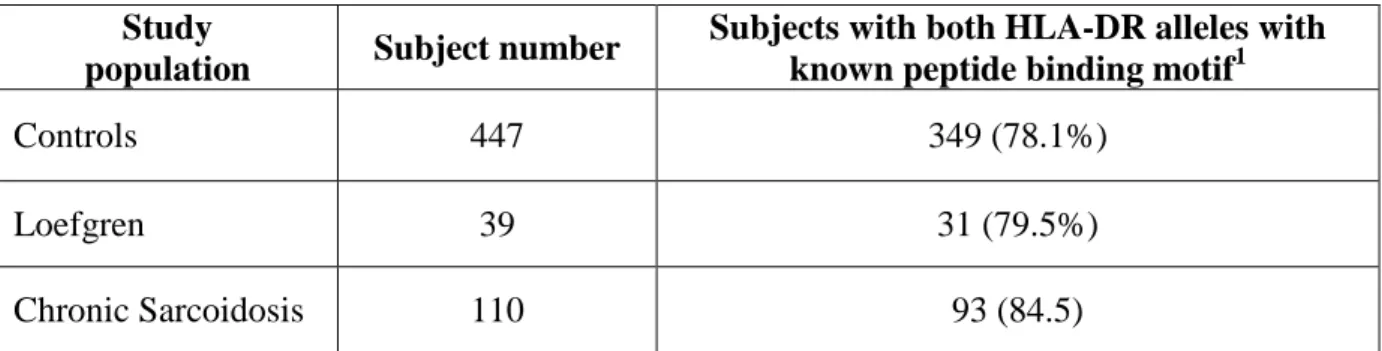
Patients’ characteristics: The study populations was composed by the TB patients and matched controls of two already described separate reports on the genetic susceptibility to TB in which HLA-DR high resolution typing was available for all study subjects (21,22). A population of patients with beryllium hypersensitivity and matched beryllium-exposed unaffected subjects were used as disease control population (23). They were 160 patients with tuberculosis (TBpopulation 1), and 200 controls (CTR-population 1) (see table 1) included in the Kim HS et al. study (22) and the 127 patients with tuberculosis (TB-population 2) and 120 matched controls (CTR-population 2) included in the Vejbaesya S et al. study (21) (see 22 A model of phenotypic susceptibility to Tuberculosis (table 1). The disease control population included 74 subjects with beryllium hypersensitivity (BeH) and 86 beryllium exposed matched controls (Be-CTR) from the Amicosante et al. study (23). The quantitative implemented peptide binding motifs are available only for 52 over more than 300 HLA-DRB1 alleles (24, 25). All together they may cover, with at least one allele, about 90% of the HLA-DR variability of different human populations (26). For the purpose of this study, only the subjects carrying both HLA-DR alleles with an available HLA-DR binding motif were used (see table 1). Specifically, 106 out of 320 (33%) control subjects and 85 out of 287 (30%) TB patients could be subjected to the analysis, as 168 (52%) controls and 151 (53%) TB patients had only one HLA-DR allele with an available binding motif, while 48 (15%) controls and 51 (18%) TB patients had both HLADR alleles without an available binding motif. Consequently, the alleles which were analyzed in the study populations were: HLA-DRB1* 0101, 0102, 0301, 0401, 0402, 0404, 0405, 0408, 0410, 0701, 0802, 0806, 1101, 1102, 1104, 1106, 1301, 1302, 1307, 1401, 1501, 1502. The selected subgroups did not differ for demographical characteristics from the subgroups of subjects excluded from the study for having one or both HLA-DR alleles without a known binding motif (data not shown). Genomes The genome of Mycobacterium tuberculosis H37Rv strain (NC 000962.2) composed of 4048 genes transcribed into 3,989 proteins and the genome of Escherichia coli K12 (NC 000913.2) were used in this study for the immuno-informatic analysis. Software for the identification and enumeration of epitopes in whole genomes To enumerate the T-cell epitopes present in data sets of proteins as large as a microbial genome, we eveloped a software for the identification and enumeration of peptide binding epitopes to HLA-DR molecules. This software was developed on LabView platform (National Instruments, US) using a graphic language. Basically, it is an open system in which two different databases are uploaded and crossed. Briefly, the first input database is represented by the protein sequences that can be uploaded from a file in FASTA format. The second database is represented by matrice(s) describing
the peptide binding capabilities of the HLA-DR alleles under analysis. The present version of the software is equipped with a set of 52 additional matrices for HLA-DR peptide binding profiles (15, 27).
The matrices database includes also the threshold values for different affinity levels as reported in the
original packages. The distribution of the matrix results of all the 209 possible peptides that can theoretically bound HLA-DR molecule is automatically generated as well as theoretical affinity thresholds. The analysis can be customized by selecting single/multiple proteins and HLA alleles among the set of data loaded, threshold and other parameters. The software generates all the possible nonamer peptides in a protein sequence and analyse them on the HLA-peptide binding matrices in analysis. For each protein and each HLA allele, all the peptides presenting a permissive 23 aminoacid (AA) in relative position P1 are stored in memory together with its matrix score and relative position in the protein. For the purpose of this work, two analysis were implemented in the software. A first analysis is represented by the identification of the epitopes recognised, in each single protein of the data set at the affinity threshold applied for each HLA-DR allele in analysis. This allows the enumeration of the epitopes in the whole data set and the identification and enumeration of proteins that present a defined number of epitopes, such as the proteins that are putatively not recognised by the HLA-DR allele at the affinity threshold used, as they present zero epitopes. A second specific analysis for the identification of the epitopes recognised in a set of proteins by 2 HLA-allelestogether has been developed to mime the situation of the HLA-DR recognition in a single subject. In this analysis, the software identify and enumerate the common and the different peptide epitopes recognised by the two HLA-DR alleles of the subject under evaluation by the position in the test set of proteins at the threshold of affinity applied. The whole MTB and E. coli genomes were analysed for the HLA-DR alleles negatively and positively associated to TB and for the enumeration of epitopes recognized by single subjects in analysis at the different thresholds of affinities equivalent to the 1%, 2%, 3%, 4% and 5% of the best binding natural peptides for HLA-DR alleles (27). The Threshold of affinity is a preselected numerical value used to differentiate between binders and non binders, any peptide frame scoring higher than this value is predicted as binder or vice versa; it correlates with the peptide score (15) and therefore with HLA-ligand interaction, therefore it is an indicator for the likelihood that predicted peptide is capable of binding to a given HLA-molecule. To express the results, we have chosen, the percentage of 1% in order to lower the false positive rate. The number of epitopes recognized by
mean + standard deviation of the mean (SD). Comparisons between groups are made by Student’s t test. Results 1. The immunome of MTB H37Rv HLA-DR molecules bind a core of nine
aminoacids long protein fragments, when they carry in the relative position 1 (hereafter named P1) non-polar residues (I, L, M, F, W, Y, V) i.e., 7 out of 20 aminoacids or 35% of all the aminoacids. Analysis of the genome of M. tuberculosis H37Rv strain (NC_000962), which comprises 4,048 genes that can be transcribed into 3,989 proteins, allowed to estimate that, independently of the relative affinity of the epitopes for the HLA-DR molecules, the number of putative epitopes capable of binding any HLA-DR allele in the whole MTB genome was 405,422 out of 1,304,277 possible 9-mers, that we define as the HLA-DR immunome. This indicates that the HLA-DR MTB-immunome encompasses 31.08% of the whole MTB nonamer population. This number is equivalent to the 88% of the expected theoretical recognition that is equivalent to 35% of the all the peptides, thus suggesting that MTB encompasses a lower number of epitopes than expected if its genome presented a normal aminoacid distribution. Differently from MTB, the number of putative eepitopes capable of binding any HLA-DR allele in the E.coli genome was 537,294 out of 1,566,080 nonamers (34.30%). This is equivalent to 98% of the expected theoretical recognition, a fraction that is significantly higher than that of the MTB HLA-DR immunome (p<0.0001). 2. Impaired MTB proteins recognition by DR alleles associated with TB susceptibility. HLA-DR alleles HLA-HLA-DRB1*0801, *0802, *1401, *1501, *1502, which have been associated with TB susceptibility in previous studies, recognized significantly lower number of MTB-epitopes (7,862+4,258) than the HLA-DR alleles HLADRB1* 0301, *0701, *1101, *1102, *1301 and *1302, associated with TB resistance [11,376+1,984 (P<0.032)], at the affinity level of 1%. Consequently, there was a significantly higher number of MTB proteins (1,268+686) which could not be recognized by the HLA-DR alleles associated with TB susceptibility compared to HLA-DR alleles negatively associated with TB (776+232; p<0.001). 24 A model of phenotypic susceptibility to Tuberculosis 25 3. Phenotypic analysis of the ability of HLA-DR alleles of TB patients to bind whole MTB genome peptides. When this analysis was applied to those individual subjects carrying a pair of HLA-DR alleles with known peptide-binding motifs, the number of MTB epitopes recognized by TB patients in both populations was significantly lower than that recognized by controls [TB-population 1: 15,303±657, CTR-population 1: 16,841±555; p=0.038 compared to TB population 1; TB-population 2: 11,341±908, CTR-population 2: 13,587±605, p=0.035 compared to TB-population 2 (Figure 1 panel A)]. Interestingly, when a population of individuals affected by the sarcoid-like granulomatous reaction to beryllium was analyzed as a control, no differences where observed between granuloma-affected and unaffected subjects in their recognition ability of MTB genome peptides (beryllium-hypersensitivity: 17,593±447; beryllium-exposed controls 18,014±421;
p=0.608). Finally, when the the three population groups were tested for their ability to recognize E. coli genome peptides, no differences were seen between TB and their matched controls nor between Be-hypersensitive subjects and their matched controls (figure 1 panel B), suggesting that the epitope binding defect seen in TB patients was restricted to, or more pronounced for, the MTB genome.
Discussion
The binding of antigenic peptides by the host HLA proteins expressed by antigen presenting cells is thought to represents a limiting step in the development of an antimicrobial immune response. In the
context of the importance of HLA-DR genes in the immune response to MTB (28, 29) and of the observations positively or negatively linking different alleles of the HLA-DR, HLA-DQ, HLA-DP, and
HLA class I genes to TB susceptibility in HLA association studies (10), it is reasonable to think that altered peptide binding by susceptible HLA molecules may be the cause of susceptibility to disease (11).
With this as a background and in the context that TB susceptibility is increased in homozygous, compared to heterozygous twins (30), it is reasonable to hypothesize that a deficient antigen recognition capability of the immune system might be at the basis of the inefficacious response to MTB in susceptible individuals, and that the determinant of susceptibility to MTB infection and disease may be the combination of the recognition abilities of both HLA-DR alleles expressed by each subject, i.e., the HLA-DR phenotype. The finding of this in silico model that the number of MTB epitopes ecognized by the combination of the two HLA-DR alleles by TB subject was significantly lower than the matched Fig. 1. Total number of epitopes recognized by M. tuberculosis and E. coli. The number of M. tuberculosis (panel A) and E. coli (panel B) epitopes recognised in silico by each study subject with the combination of the two carried HLA-DR alleles at the threshold affinity of 1% in the two genomes. The three populations evaluated (TB-population 1, TB-population 2, and Be-exposed control population) are presented separately. Open circles, control subjects; closed circles, TB patients; grey circles Be-exposed control population. p value has been determined by Student’s t-test. 26 S. Contini, M. Pallante, S. Vejbaesya, M. Hee Park, N. Chierakul, et al controls in both study populations suggest that TB patients present may be affected by a deficient capability of recognition of the MTB proteome compared to the non affected subjects in the population. Interestingly, Delgado et al. have recently reported a highly significant association between progressive pulmonary TB and homozygosity for HLADQ beta57-Asp alleles where a single polymorphism in the HLA-DQ beta chain played a critical role in the binding of ESAT-6, a highly immunogenic MTB protein and in the ensuing CD4+ T-cells immune response. Although they do not explain the mechanism of susceptibility to the development active TB, these data provided a functional link between an HLA polymorphism and susceptibility to progressive tuberculosis infection (31). In contrast to infection, current concepts are that in hypersensitivity and
autoimmune diseases susceptibility is associated with excessive HLA binding or the binding of specific (neo)-antigens by the HLA allele associated to the diseases (32).
In this context, it has been shown that the immune reaction to non-tuberculous mycobacteria can be characterized by an exaggerated reaction leading to the hypersensitivity pneumonias of the hot tub
lung (33), to the metalworking fluid-associated hypersensitivity pneumonitis (34), or to the formation of granulomas within the bronchial walls leading to the formation of bronchiectasis (35), a condition that’s been associated with HLA-DR 6, i.e., the alleles 13 and 14 (36). Mycobacteria have also been implicated in sarcoidosis, where acid fast rods have been seen in affected tissues (37), wall-deficient form (l-form) of mycobacteria have been isolated (38), and mycobacterial DNA has been detected (39). Interestingly, a reaction to mycobacterial antigens has been described in sarcoidosis as T-cell and antibody responses to MTB ESAT-6 and KatG protein (40), HSP70 (41), and superoxide dismutase (SodA) (42) have been observed in patients with sarcoidosis and some antigens such as the Heat Shock Protein (HSP) and Catalase-Peroxidase (KatG) have been detected by immunohistochemistry in sarcoid tissues (43, 44). It is worth noticing in this regard, that the same HLA allelic variants which have been associated with susceptibility or resistance to tuberculosis have also been implicated in susceptibility to sarcoidosis. The HLA-DRB1*03 alleles, which have been negatively associated with TB, being therefore dubbed as “resistance” genes (45), have been associated with acute, self limiting, sarcoidosis (46). In contrast, the HLA-DRB1*15 alleles, which have been associated with the susceptibility to develop active TB (45), have been associated with stage III, or chronic sarcoidosis (47). Thus, one might hypothesize that the lack of HLA binding and presentation of mycobacterial antigens could determine disease progression in tuberculosis as well as in sarcoidosis, the type of reaction –extensively necrotizing versus non-necrotizing, being possibly driven by the expression of allelic variants of the host’s genes of the innate immune response (48) or by variants expressed in the infected organism of the parasite virulence genes (49). The virtual approximation to truth made by in silico models, together with the limitation imposed by the reduced number of HLA-DR phenotypes that can be analyzed in the different populations, require that in silico results be confirmed by studies using conventional biological techniques. With this caveats, is counceivable that the use of immuno-informatic tools for the prediction of T-cells epitopes also on other HLA class I and II alleles on data sets as large as an the entire MTB genome, will help with generating mechanistic hypothesis on the determinants of HLA-associated genetic susceptibility to TB, as well as to other granulomatous disorders caused, or
References
1. Grigg ER: The Arcana of Tuberculosis. Am. Rev. Tuberc. 1958, 78:426-53.
2. Vidal SM, Malo D, Vogan K, Skamene E, Gros P: Natural resistance to infection with intracellular parasites: isolation of a candidate for Bcg. Cell 1993, 73: 469-85.
3. Bellamy R, Ruwende C, Corrah T, McAdam KP, Whittle HC, Hill AV: Variations in the NRAMP1 gene and susceptibility to tuberculosis in West Africans. N Engl J Med 1998, 338: 640-4.
4. Greenwood CM, Fujiwara TM, Boothroyd LJ, Miller MA, Frappier D, Fanning EA, Schurr E, Morgan K: Linkage of tuberculosis to chromosome 2q35 loci, including NRAMP1, in a large aboriginal Canadian family. Am J Hum Genet 2000, 67: 405-16.
5. NewportMJ, Huxley CM, Huston S, Hawrylowicz CM, Oostra BA, Williamson R, Levin M: A mutation in the interferon-gamma-receptor gene and susceptibility to mycobacterial infection. N Engl J Med 1996, 335: 1941-9.
6. Jouanguy E, Altare F, Lamhamedi S, Revy P, Emile JF, Newport M, Levin M, Blanche S, Seboun E, Fischer A, Casanova JL: Interferongamma-receptor deficiency in an infant with fatal bacille Calmette-Guerin infection. N Engl J Med 1996, 335: 1956-61.
7. Altare F, Durandy A, Lammas D, Emile JF, Lamhamedi S, Le Deist A model of phenotypic susceptibility to Tuberculosis 27 F, Drysdale P, Jouanguy E, Doffinger R, Bernaudin F, Jeppsson O, Gollob JA,Meinl E, Segal AW, Fischer A, Kumararatne D, CasanovaJL: Impairment of mycobacterial immunity in human interleukin-12 receptor deficiency. Science 1998, 280: 1432-5.
8. de Jong R, Altare F, Haagen IA, Elferink DG, Boer T, van Breda Vriesman PJ, Kabel PJ, Draaisma JM, van Dissel JT, Kroon FP, Casanova JL, Ottenhoff TH: Severe mycobacterial and Salmonellainfections in interleukin-12 receptor-deficient patients. Science 1998, 280: 1435-8.
9. Baghdadi JE, Orlova M, Alter A, Ranque B, Chentoufi M, Lazrak F, Archane MI, Casanova JL, Benslimane A, Schurr E, Abel L: An autosomaldominant major gene confers predisposition to pulmonary tuberculosis in adults. J Exp Med 2006, 203: 1679 - 1684. 10.Kettaneh A, Seng L,Tiev KP,Toledano C, Fabre B, Cabane J: Human leukocyte antigens
and susceptibility to tuberculosis: a meta-analysis of case-control studies. Int J Tuberc Lung Dis 2006, 10: 717-25.
11.Ghodke Y, Joshi K, Chopra A, Patwardhan B: HLA and disease. Eur J Epidemiol. 2005, 20: 475-88.
12.Davenport MP, Quinn CL, Valsasnini P, Sinigaglia F, Hill AV, Bell JI: Analysis of peptide-binding motifs for two disease associated HLA-DR13 alleles using an M13 phage display library. Immunology 1996, 88: 482-486.
13.Mallios RR: Predicting class II MHC/peptide multi-level binding with an iterative stepwise discriminant analysis meta-algorithm. Bioinformatics 2001,17: 942-8.
14.Germain RN: MHC-dependent antigen processing and peptide presentation: providing ligands for T lymphocyte activation. Cell 1994, 76: 287-99.
15. Sturniolo T, Bono E, Ding J, Raddrizzani L, Tuereci O, Sahin U, Braxenthaler M, Gallazzi F, Protti MP, Sinigaglia F, Hammer J: Generation of tissue-specific and promiscuous HLA ligand databases using DNA microarrays and virtual HLA class II matrices. Nat Biotechnol 1999, 17: 555-61.
16.Scherer A, Frater J, Oxenius A, Agudelo J, Price DA, Gunthard HF, Barnardo M, Perrin L, Hirschel B, Phillips RE, McLean AR; Swiss HIV: Quantifiable cytotoxic T lymphocyte responses and HLA-related risk of progression to AIDS. Proc Natl Acad Sci U S A 2004,101: 12266-70.
17.Stunz L, Karr RW, Anderson R: HLA-DRB1 and -DRB4 genes are differentially regulated at the transcriptional level. J Immunol 1989, 143: 3081– 86.
18. Berdoz J, Gorski J,Termijtelen A, Dayer J, Irle C, Schendel D, Mach B: Constitutive and induced expression of the individual HLA-DR h and a chain loci in different cell types. J Immunol 1987, 139:1336–41.
19.Cotner T, Charbonneau H, Mellins E, Pious D: mRNA abundance, rather than differences in subunit assembly, determine expression of HLA-DRh1 and DRh3 molecules. J Biol Chem 1996, 264: 11107 –11.
20.De Groot AS, Bosma A, Chinai N, Frost J, Jesdale BM, Gonzalez MA, Martin W, Saint-Aubin C: From genome to vaccine: in silico predictions, ex vivo verification. Vaccine 2001, 19: 4385-95.
21.Vejbaesya S, Chierakul N, Luangtrakool K, Srinak D, Stephens HA: Associations of HLA class II alleles with pulmonary tuberculosis inThais. Eur J Immunogenet 2002, 29: 431-4. 22.Kim HS, Park MH, Song EY, Park H, Kwon SY, Han SK, Shim YS:Association of
HLA-DR and HLA-DQ genes with susceptibility topulmonary tuberculosis in Koreans: preliminary evidence of associations with drug resistance, disease severity, and disease recurrence.Hum Immunol 2005, 66: 1074-81.
23.Amicosante M, Berretta F, Rossman M, Butler RH, Rogliani P, van den Berg-Loonen E, Saltini C: Identification of HLA-DRPhebeta47 as the susceptibility marker of hypersensitivity to beryllium in individuals lacking the berylliosis-associated supratypic marker HLADPGlubeta69. Respir Res 2005, 6: 94.
24.Marsh SGE: Nomenclature for factors of the HLA system, update January 1998. Tissue Antigens 1998, 51: 582-3.
25.Marsh SG:WHO Nomenclature Committee for Factors of the HLA System. Nomenclature for factors of the HLA system, update September 2006. Tissue Antigens 2006, 68: 540-2. 26.Wilson CC, Palmer B, Southwood S, Sidney J, Higashimoto Y, Appella E, Chesnut R, Sette
A, Livingston BD: Identification and antigenicity of broadly cross-reactive and conserved human immunodeficiency virus type 1-derived helper T-lymphocyte epitopes. J Virol 2001, 75: 4195-207.
27.Singh H, Raghava GP: ProPred: prediction of HLA-DR binding sites. Bioinformatics 2001, 17: 1236-7.
28.Uma H, Selvaraj P, Reetha AM, Xavier T, Prabhakar R, Narayanan PR: Influence of HLA-DR antigens on lymphocyte response to Mycobacterium tuberculosis culture filtrate antigens and mitogens in pulmonary tuberculosis. Tuber Lung Dis 1999, 79: 199-206. 29.Sriram U, Selvaraj P, Kurian SM, Reetha AM,Narayanan PR: HLADR2 subtypes &
immune responses in pulmonary tuberculosis. Indian J Med Res 2001, 113: 117-24.
30.Comstock GW: Tuberculosis in twins: a re-analysis of the Prophit survey. Am Rev Respir Dis 1978, 117: 621-4.
31.Delgado JC, Baena A, Thim S, Goldfeld AE: Aspartic acid homozygosity at codon 57 of HLA-DQ beta is associated with susceptibility to pulmonary tuberculosis in Cambodia. J Immunol 2006, 176: 1090-7.
32.Czaja AJ,Donaldson PT. Genetic susceptibilities for immune expression and liver cell injury in autoimmune hepatitis. Immunol Rev. 2000; 174: 250-9.
33.Embil J,Warren P, Yakrus M, Stark R, Corne S, Forrest D, Hershfield E: Pulmonary Illness Associated With Exposure to Mycobacterium-avium Complex in Hot Tub Water: Hypersensitivity Pneumonitis or Infection? Chest 1997; 111: 813-6.
35.Fujita J, Ohtsuki Y, Suemitsu I, Shigeto E, Yamadori I, Obayashi Y,Miyawaki H, Dobashi N, Matsushima T,Takahara J: Pathological and adiological changes in resected lung specimens in Mycobacterium Avium intracellulare complex disease. Eur Respir J 1999; 13: 535-40.
36. Kubo K, Yamazaki Y, Hanaoka M, Nomura H, Fujimoto K, Honda T, Ota M, Kamijou Y. Analysis of HLA antigens in Mycobacterium avium-intracellulare pulmonary infection. Am J Respir Crit CareMed 2000, 161: 1368-71.
37.Van?k J, Schwarz J: Demonstration of acid-fast rods in sarcoidosis. Am Rev Respir Dis 1970, 101: 395-400.
38. Almenoff PL, Johnson A, Lesser M, Mattman LH: Growth of acid fast L forms from the blood of patients with sarcoidosis. Thorax 1996, 51: 530-3.
39. Mangiapan G, Hance AJ.Mycobacteria and sarcoidosis: an overview and summary of recent molecular biological data. Sarcoidosis 1995, 12: 20-37.
40.Drake WP, Dhason MS, Nadaf M, Shepherd BE, Vadivelu S, Hajizadeh R, Newman LS, Kalams SA: Cellular recognition of Mycobacterium tuberculosis ESAT-6 and KatG peptides in systemic sarcoidosis. Infect Immun 2007, 75: 527-30.
41.Dubaniewicz A, Trzonkowski P, Dubaniewicz-Wybieralska M, Dubaniewicz A, Singh M, MyÊliwski A: Mycobacterial heat shock protein-induced blood T lymphocytes subsets and cytokine pattern: comparison of sarcoidosis with tuberculosis and healthy controls. Respirology 2007, 12: 346-54.
42.Allen SS, Evans W, Carlisle J, Hajizadeh R, Nadaf M, Shepherd BE, Pride DT, Johnson JE, Drake WP: Superoxide dismutase A antigens derived from molecular analysis of sarcoidosis granulomas elicit systemic Th-1 immune responses. Respir Res 2008; 9: 36.
43.S. Contini, M. Pallante, S. Vejbaesya, M. Hee Park, N. Chierakul, et al 43. Dubaniewicz A, Dubaniewicz-Wybieralska M, Sternau A, Zwolska Z, Izycka-Swieszewska E, Augustynowicz-Kopec E, Skokowski J, Singh M, Zimnoch L: Mycobacterium tuberculosis complex and mycobacterialheat shock proteins in lymphnode tissue from patients with pulmonary sarcoidosis. J Clin Microbiol 2006, 44: 3448-51.
44.Song Z, Marzilli L, Greenlee BM, Chen ES, Silver RF, Askin FB, Teirstein AS, Zhang Y, Cotter RJ, Moller DR: Mycobacterial catalase- peroxidase is a tissue antigen and target of the adaptive immune response in systemic sarcoidosis. J Exp Med 2005, 201: 755-67.
45.Kettaneh A, Seng L,Tiev KP,Toledano C, Fabre B, Cabane J: Human leukocyte antigens and susceptibility to tuberculosis: a meta-analysis of case-control studies. Int J Tuberc Lung Dis 2006, 10: 717-25.
46.Seitzer U, Gerdes J, Müller-Quernheim J: Evidence for disease phenotype associated haplotypes (DR.TNF) in sarcoidosis. Sarcoidosis. Vasc Diffuse Lung Dis 2001, 18: 279-83. 47.Rutherford RM, Brutsche MH, Kearns M, Bourke M, Stevens F, Gilmartin JJ: HLA-DR2
predicts susceptibility and disease chronicity in Irish sarcoidosis patients. Sarcoidosis Vasc Diffuse Lung Dis2004, 21: 191-8.
48. Burgner D, Jamieson SE, Blackwell JM: Genetic susceptibility to infectious diseases: big is beautiful, but will bigger be even better? Lancet Infect Dis 2006, 6: 653-63.
49.Rollenhagen C, Sörensen M, Rizos K, Hurvitz R, Bumann D: Antigen selection based on expression levels during infection facilitates vaccine development for an intracellular pathogen. Proc Natl Acad Sci U S A 2004, 101: 8739-44.
DESIGN OF IMMUNOGENIC PEPTIDES FROM M. TUBERCULOSIS
GENES EXPRESSED DURING MACROPHAGE INFECTION
Abstract
In vitro diagnosis of MTB-infection uses MTB proteins coded for by genes of the region of differentiation 1 (RD1) of the MTB genome. This study wants to test if proteins preferentially expressed by during MTB intracellular growth might provide new targets for the diagnosis of MTB infection.
To this end Seventy-five multiepitopic HLA-promiscuous MTB-peptides were designed by quantitative implemented peptide binding motif analysis from 3 MTB-protein genes expressed in activated human macrophages (MA), 4 genes expressed during growth in non-activated human macrophages (MN-A), 12 housekeeping genes (HKG) and 6 genes of the RD1 region (RD1) as control. ELISpot for IFN- was performed to measure the responses of PBMCs deriving from 45 patients affected by active Tuberculosis and 34 controls. In active TB patients, the mean response to RD1 derived peptides was higher than that to either MA (p<0.01), MN-A (p<0.008) or HKG (p<0.01) derived peptides. In TSpositive subjects all selected peptides elicited significant IFN- T-cell responses (p<0.02 compared to TST-negatives), but without differences between the subgroups. Further, T-cell responses to RD1 peptides were lower in the 23 active-TB treated patients than in the untreated ones (p<0.01). The response to MA peptides in treated active-TB was higher than when untreated (p<0.01). These results demonstrate that the use of in vitro models of MTB-intracellular infection to select MTB gene products for further in silico and in vitro assessment of their immunogenicity has the potential to identify novel antigens amenable to the design of new tools for diagnosis and monitoring of tuberculosis.
KEYWORDS: