UNIVERSITÁ POLITECNICA DELLE MARCHE
DOCTOR OF PHILOSOPHY IN
“BIOMEDICAL SCIENCES”
Coordinator: Prof. Andrea Giacometti
Chemopreventive effects of Manuka and Strawberry tree honey on human
colon cancer HCT-116 and LoVo cells: a focus on molecular targets
Ph. D. dissertation by
Sadia Afrin, B.Sc., M.Sc.
Supervisor: Prof. Maurizio Battino
Co-supervisor: Dr. Francesca Giampieri
TABLE OF CONTENTS
ABSTRACT………... 1
ITALIAN SUMMARY………. 4
LIST OF ABBREVIATIONS……….. 7
CHAPTER 1. INTRODUCTION……… 11
1.1. General overview on colorectal cancer………... 12
1.1.1. Epidemiology of colorectal cancer……… 12
1.1.2. Risk factor and etiology of CRC……… 13
1.1.2.1. Inflammation and CRC………. 15
1.1.2.2. Oxidative damage and CRC………. 17
1.1.2.3. Alteration of oncogenes and tumor suppressor genes……….. 18
1.1.2.3.1. Wnt signaling……… 18 1.1.2.3.2. MAPK signaling……… 19 1.1.2.3.3. PI3K/Akt signaling……… 22 1.1.2.3.4. EGFR signaling………. 23 1.1.2.3.5. TGF- β signaling……… 23 1.1.2.3.6. NFκB signaling………. 24 1.1.2.3.7. Nrf2 signaling……… 25 1.1.2.3.8. Notch signaling……….. 25 1.1.2.3.9. JAK/STAT3 signaling………... 26
1.1.2.4. The role of endoplasmic reticulum stress in CRC………. 27
1.1.2.5. The role of AMPK signaling in CRC……… 28
1.1.2.6. The role of matrix metalloproteinases in CRC……….. 30
1.1.2.7. The role of epithelial-mesenchymal transition in CRC……… 30
1.1.2.8. Genetic instability of CRC……… 31
1.1.2.9. Epigenetic alterations in CRC……….. 33
1.1.2.9.1. DNA methylation in CRC……… 33
1.1.2.9.2. Histone modifications in CRC………. 34
1.1.2.9.3. The role of microRNAs in CRC……… 34
1.1.3. Current treatment and management of CRC……… 34
1.2. Honey as a food……….. 36
1.3. Bioactive profile of honey……….. 37
1.3.1.1. Sugar in honey……….. 38
1.3.1.2. Proteins, amino acids and enzymes in honey……… 39
1.3.1.3. Organic acids in honey……….. 40
1.3.1.4. Vitamins in honey………. 40
1.3.1.5. Minerals in honey………. 41
1.3.1.6. Aroma and volatile compounds……… 41
1.3.2. Phenolic profile of honey………... 42
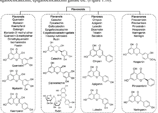
1.3.2.1. Flavonoids in honey………. 42
1.3.2.2. Phenolic acid in honey……….. 43
1.3.2.3. Antioxidant properties of honey……… 45
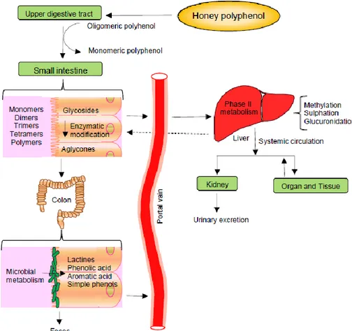
1.4. Bioavailability and metabolites of honey……… 47
1.5. The potential impact of honey on human health……… 48
1.5.1. Honey and cancer………... 49
OBJECTIVE……….. 52
CHAPTER 2. MATERIALS AND METHODS………. 53
2.1. Honey samples……… 54
2.2. Reagents and antibodies……….. 54
2.3. Determination of total polyphenol and flavonoid content……….. 55
2.4. Extraction, identification and quantification procedure for phenolic compounds……….. 55 2.4.1. Solid-phase extraction and HPLC condition……….. 55
2.4.2. Determination of flavonols and phenolic acids……….. 56
2.5. Determination of total protein and free amino acid content……… 56
2.6. Determination of total antioxidant capacity……… 57
2.6.1. Ferric reducing antioxidant power assay……… 57
2.6.2. Trolox equivalent antioxidant capacity assay……… 58
2.6.3. Diphenyl-1 picrylhydrazyl assays……….. 58
2.7. Preparation of artificial honey………. 59
2.8. Cell culture……….. 59
2.9. Determination of cell viability by MTT assay……… 59
2.10. Cell cycle analysis by Tali® Image-Based Cytometer………. 60
2.11. Determination of apoptotic cells by Tali® Image- Based Cytometer………….. 61 2.12. Determination of intracellular ROS levels by Tali® Image-Based Cytometer… 62
2.13. Cellular lysates preparation………. 63
2.14. Determination of oxidative stress markers……….. 63
2.14.1. Determination of lipid peroxidation by TBARS assay……….. 63
2.14.2. Determination of protein carbonyl content……… 64
2.15. Determination of antioxidant enzyme activity………. 64
2.15.1. Determination of glutathione peroxidase ……….. 64
2.15.2. Determination of glutathione transferase………... 65
2.15.3. Determination of glutathione reductase………. 65
2.15.4. Determination of superoxide dismutase ……… 65
2.15.5. Determination of catalase………... 66
2.16. Bioenergetic assay……… 66
2.16.1. Determination of the mitochondrial functionality………. 66
2.16.2. Determination of the glycolysis function………... 68
2.17. Wounding assay……… 68
2.18. Colony formation assay……… 69
2.19. Protein extraction and western blotting……… 69
2.20. Statistical analysis……… 70
CHAPTER 3. RESULTS AND DISCUSSION……….. 71
RESULTS: PART I……….. 72
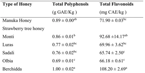
3.1. Nutritional characterization of Manuka honey (MH) and Strawberry tree honey (STH)……… 72 3.1.1. Phytochemical content of MH and STH……… 72
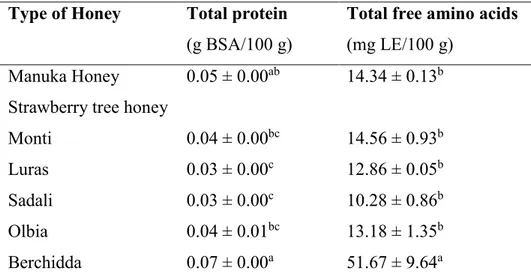
3.1.2. Total protein and free amino acid content of MH and STH………... 76
3.1.3. Total antioxidant capacity of MH and STH………... 76
3.1.4. Correlations between biochemical parameters and antioxidant potentials of MH and STH………. 77 DISCUSSION: PART I……… 79
RESULTS: PART II………. 83
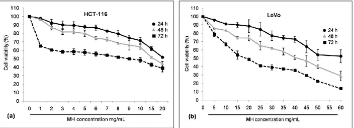
3.2. Chemopreventive effect of MH and STH on human colon cancer HCT-116 and LoVo cells……… 83 3.2.1. Effect of MH and STH on HCT-116 and LoVo cells proliferation……... 83
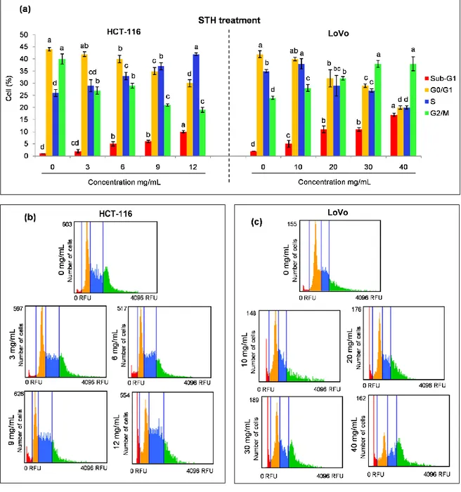
3.2.2. Effect of MH and STH on cell cycle arrest on HCT-116 and LoVo cells. 86 3.2.3. Effect of MH and STH on apoptosis on HCT 116 and LoVo cells……… 90
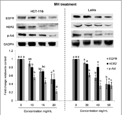
3.2.4. Anti-inflammatory effect of MH and STH on HCT-116 cells…………... 94 3.2.5. Effect of MH and STH on MAPK and EGFR signaling pathways on
HCT-116 and LoVo cells………... 95
3.2.6. Effect of MH and STH on oxidative stress induction on HCT-116 and LoVo cells……….
99
3.2.6.1. Effect of MH and STH induces intracellular ROS production on HCT-116 and LoVo cells………
100
3.2.6.2. Effect of MH and STH on the biomarkers of oxidative stress and the antioxidant system on HCT-116 and LoVo cells……….
103
3.2.7. Effect of MH and STH on ER stress induction on HCT-116 and LoVo cells……….
109
3.2.8. Effect of MH and STH on HCT-116 and LoVo cells metabolism………. 111 3.2.8.1. Effect of MH and STH on mitochondrial resperiation on HCT-116
and LoVo cells……… 111
3.2.8.2. Effect of MH and STH on glycolysis on HCT-116 and LoVo cells……….
115
3.2.8.3. Effect of MH and STH on AMPK signaling pathway on HCT-116 and LoVo cells………
117
3.2.9. Effect of MH and STH on metastasis activity on HCT-116 and Lovo cells……….
118
3.2.9.1. Effect of MH and STH on migration and invasion ability of HCT-116 and Lovo cells……….
118
3.2.9.2. Effect of MH and STH on the colony formation ability of HCT-116 cells……….
122
3.2.9.3. Effect of MH and STH on the expressions of EMT-related genes in HCT-116 and LoVo cells………
124
3.2.9.4. Effect of MH and STH on the metastasis promoting factor expression CXCR4 and NF-κB in Lovo cells………
125
DISCUSSION: PART II………... 127 RESULTS: PART III……… 139
3.3. MH and STH potentiates anticancer effects of 5-FU chemotherapy on human colon cancer HCT-116 and LoVo cells………..
139
cells proliferation……… 3.2.2. Effect of 5-FU in a combination of MH and STH on cell cycle arrest on
HCT-116 and LoVo cells……… 142
3.3.3. Effect of 5-FU in a combination of MH and STH on apoptosis induction on HCT 116 and LoVo cells………
145
3.3.4. Anti-inflammatory effect of 5-FU in a combination of MH and STH on HCT-116 cells……….
148
3.3.5. Effect of 5-FU in a combination of MH and STH on MAPK and EGFR signalling pathways in HCT-116 and LoVo cells………
149
3.3.6. Effect of 5-FU in a combination of MH and STH on oxidative stress induction on HCT-116 and LoVo cells………
152
3.3.6.1. Effect of 5-FU in a combination of MH and STH on intracellular ROS production on HCT-116 and LoVo cells………
152
3.3.6.2. Effect of 5-FU in a combination of MH and STH on the biomarkers of oxidative stress and the antioxidant enzyme on HCT-116 and LoVo cells………..
154
3.3.7. Effect of 5-FU in a combination of MH and STH on the ER stress induction on HCT-116 and LoVo cells………
160
3.3.8. Effect of 5-FU in a combination of MH and STH on HCT-116 and LoVo cells metabolism ……….
162
3.3.8.1. Effect of 5-FU in a combination of MH and STH on mitochondrial respiration on HCT-116 and LoVo cells………
162
3.3.8.2. Effect of 5-FU in a combination of MH and STH on glycolysis on HCT-116 and LoVo cells………
165
3.3.8.3. Effect of 5-FU in a combination of MH and STH on AMPK signaling pathway on HCT-116 and LoVo cells………
168
3.2.9. Effect of MH and STH on metastasis activity on HCT-116 and LoVo cells……….
170
3.2.9.1. Effect of MH and STH on the migration and invasion ability of HCT-116 and LoVo cells………
170
3.2.9.2. Effect of 5-FU in a combination of MH and STH on the colony formation of HCT-116 and LoVo cells………
172
HCT-116 and LoVo cells……… 3.2.9.4. Effect of MH and STH on the metastasis promoting factor expression CXCR4 and NFκB in Lovo cells……….
176
DISCUSSION: PART III……….. 177
CONCLUSIONS……… 185
REFERENCES………. 187
1
ABSTRACT
Despite the major advances in colon cancer treatment, including postoperative care, recurrence and mortality rates remain high; hence the urgent need to complement the current therapies. Numerous investigations have been made on plant bioactive compounds and colon cancer prevention in the recent decades. Honey is a natural food product known to modulate several biological activities including cancer. The main objective of the present work was to evaluate the anticancer potential of Manuka and Strawberry tree honey on in vitro colon cancer models targeting different molecular aspects.
Manuka and Strawberry tree were used for phytochemical characterization and total antioxidant capacity. The phenolic compounds were indentified and quantified by HPLC assay. The growth inhibitory effects of Manuka and Strawberry tree honey on human colon adenocarcinoma cells (HCT-116) and Dukes’ type C, grade IV, colon metastasis cells (LoVo) were observed by cell viability, cell cycle, apoptosis and intracellular reactive oxygen species (ROS) production assay. The oxidative stress were assessed through the determination of lipid peroxidation, protein carbonyl content and DNA damage, as well as the antioxidant enzyme activities on cells lysates. The expression of proteins related to inflammation, apoptotic, endoplasmic reticulum (ER) stress and EGFR signalling was determined by western blotting. Bioenergetic characterization was evaluated by observing several parameters of mitochondrial respiration (oxygen consumption rate) and glycolysis (extracellular acidification rate) in both cell lines. Anti-metastasis effects were observed through migration and colony formation assay, and the expression of invasion, epithelial mesenchymal transition (EMT) and metastasis markers were observed by western blotting. Finally, the synergistic or addictive effects of Manuka and Strawberry tree honey on 5-fluorouracil (5-FU) chemotherapy were evaluated in both colon cancer cell lines.
Manuka and Strawberry tree honey represent a good source of phenolic compounds, flavonoids and antioxidant capacity. Both types of honey exhibited profound inhibitory effects on the growth of human colon cancer cells, without showing any toxic effect on normal non-cancer cells. At different concentrations, a strong induction of cell cycle arrest, apoptosis and oxidative stress was observed after the treatments since they increased the accumulation of ROS and elevated the damage of protein, lipid and DNA of the HCT-116 and LoVo cells. Anti-inflammatory effects were observed through the suppression of NFκB, p-IκB-α and other pro-inflammatory cytokines expression, while the suppression of Akt indicated anti-proliferative effects; at the same time the increased levels of
p-2
p38MAPK and p-Erk1/2, after honey treatments, indicated the up-regulation of apoptosis rate. Furthermore, suppression of Nrf2 dependent antioxidant enzyme expression (superoxide dismutase (SOD), catalase and hemeoxygenase 1) and the activity of catalase, SOD, glutathione peroxidase, reductase and transferase enzymes were observed in both cell lines after honey treatments. The ER stress was also inducted, as suggested by the elevation of ATF6 and XBP1 protein expression. In HCT-116 and LoVo cells, both honeys decreased mitochondrial function that is correlated with cell survival potential and concurrently, decreased the extracellular acidification rate (glycolysis). Moreover, the expressions of p-AMPK/AMPK, PGC1α and SIRT1 were suppressed after the treatments which were involved in the survival of HCT-116 and LoVo cells under metabolic stress condition. In addition, the migration and colony formation abilities were reduced, as well as the invasion abilities were suppressed by both types of honey through the expression of matrix metallo-proteinases (MMPs). Expressions of EMT markers, such as E-, N-cadherin and β-catenin and tumor metastasis promoting factor CXCR4 and NF-κB, were also down-regulated by Manuka and Strawberry tree honey treatments.
Both honey varieties potentiate the anticancer effects of 5-FU chemotherapy on human colon cancer cells at less concentration compared to single dose of 5-FU. In the presence of Manuka or Strawberry tree honey, 5-FU induced synergistic effects by suppressing the expression of inflammation markers, EGFR pathways and antioxidant enzyme activities, while it increased MAPK pathway and ROS production at less concentration of 5-FU alone compared to IC50 values. Moreover, additive effects were observed by arresting cell cycle and increasing lipid peroxidation, protein carbonyl content and ER stress. All the parameters of mitochondrial respiration and glycolysis function were decreased in a similar way after all the treatments, while the expression of AMPK pathway was suppressed more after the combination treatments compared to single doses of 5-FU. In addition, anti-metastasis effects of 5-FU also increased when it was combined with Manuka and Strawberry tree honey by decreasing the migration ability, as well as suppressing the expression of MMP-2, MMP-9, E-cadherin, NFκB and CXCR4 proteins.
The above findings indicate that Manuka and Strawberry tree honey induce growth inhibitory effects of HCT-116 and LoVo cells, while the anticancer potential of 5-FU chemotherapy is enhance in the presence of both honey varieties at less doses. These preliminary results are interesting and suggest a potential chemopreventive action which
3
could be useful for further studies in order to develop chemopreventive agents for colon cancer treatment.
4
ITALIAN SUMMARY
Il cancro al colon rappresenta una delle principali cause di morbosità e mortalità per neoplasia in tutti i Paesi occidentali. Nonostante i numerosi progressi ottenuti nei trattamenti pre- e postoperatori, i tassi di recidiva rimangono ancora elevati; da qui l'urgente necessità di integrare le attuali terapie convenzionali. Negli ultimi decenni numerose ricerche hanno valutato l’effetto dei composti bioattivi naturali nella prevenzione del cancro del colon. In questo contesto, il miele rappresenta un prodotto alimentare naturale noto per modulare diverse attività biologiche e prevenire alcune patologie, tra cui il cancro. L'obiettivo principale del presente lavoro è stato valutare in vitro il potenziale antitumorale del miele di Manuka e del miele di corbezzolo su due diversi modelli cellulari di cancro al colon, che differiscono tra loro per l’espressione di alcuni geni di metastatizzazione.
I due mieli sono stati analizzati per determinare la composizione fitochimica, mediante analisi di HPLC, e la capacità totale antiossidante. Gli effetti antitumorali esercitati dal miele di Manuka e di corbezzolo sono stati valutati indue linee di adenocarcinoma del colon (HCT-116 e LoVo), attraverso la determinazione della vitalità cellulare, del tasso di apoptosi e dei livelli intracellulari di specie reattive dell’ ossigeno (ROS). Lo stress ossidativo è stato misurato mediante la determinazione della perossidazione lipidica, del contenuto di gruppi carbonili e del danno del DNA, nonché mediante la valutazione delle attività degli enzimi antiossidanti nei lisati cellulari. I livelli di espressione delle proteine correlate all'infiammazione, all'apoptosi, allo stress del reticolo endoplasmatico (RE) e alla via molecolare dell’ EGFR sono stati determinati attraverso saggi di Western blot. La caratterizzazione bioenergetica è stata analizzata valutando diversi parametri di respirazione mitocondriale (tasso di consumo di ossigeno) e glicolisi (velocità di acidificazione extracellulare) in entrambe le linee cellulari. Gli effetti anti-metastatici sono stati osservati attraverso il saggio di migrazione cellulare e di formazione delle colonie; l'espressione di marcatori di invasione, transizione mesenchimale epiteliale (EMT) e metastatizzazione è stata osservata mediante Western blot. Infine, sono stati analizzati gli effetti sinergici del miele di Manuka e di corbezzolo con il chemioterapico 5-fluorouracile (5-FU) in entrambe le linee cellulari.
I risultati hanno dimostrato che il miele di Manuka e di corbezzolo rappresentano una buona fonte di composti fenolici, flavonoidi e possiedono una notevole capacità antiossidante. Entrambi i tipi di miele hanno dimostrato profondi effetti inibitori sulla
5
crescita delle cellule tumorali, senza mostrare, al tempo stesso, alcun effetto tossico in cellule sane non cancerose. Nello specifico, dopo il trattamento con il miele, è stata evidenziata una forte induzione dell'arresto del ciclo cellulare, dell'apoptosi e dello stress ossidativo con un significativo aumento di ROS e del danno a carico di proteine, lipidi e DNA in entrambe le linee cellulari. Gli effetti antinfiammatori sono stati osservati attraverso la riduzione di NFκB, p-IκB-α e di altre citochine pro-infiammatorie, mentre la soppressione della proteina p-Akt ha evidenziato effetti antiproliferativi; allo stesso tempo, l’incremento dei livelli di p-p38MAPK e p-Erk1/2, dopo il trattamento con il miele, ha indicato l’induzione dell’ apoptosi. Inoltre, è stata osservata la riduzione dell'espressione degli enzimi antiossidanti (superossido dismutasi (SOD), catalasi ed eme-ossigenasi 1) dipendenti da Nrf2, così come l'attività della catalasi, SOD, glutatione perossidasi, reduttasi e transferasi in entrambe le linee cellulari. Il trattamento con il miele ha indotto anche lo stress del RE, come suggerito dall'incremento dell’ espressione di ATF6 e XBP1. E’ stata evidenziata anche una riduzione della funzionalità mitocondriale, correlata con il potenziale di sopravvivenza cellulare, e, contemporaneamente, una diminuzione del tasso di glicolisi. Inoltre, il trattamento con il miele ha ridottoi livelli di espressione di p-AMPK/AMPK, PGC1α e SIRT1 coinvolte nella sopravvivenza delle cellule HCT-116 e LoVo in condizioni di stress metabolico. Inoltre, è stata evidenziata una diminuzione delle abilità di migrazione cellulare e di formazione delle colonie, cosi come delle capacità di invasione e metastatizzazione, come dimostrato dalla riduzione dell’espressione delle metallo-proteinasi, delle E-, N-caderine, delle β-catenine e di CXCR4 e NF-κB.
Infine, è stato riscontrato che entrambe le varietà di miele sono in grado di potenziare gli effetti antitumorali del chemioterapico 5-FU. In presenza dei mieli, infatti, il 5-FU ha soppresso l'espressione dei marcatori di infiammazione, dell’ EGFR e le attività degli enzimi antiossidanti, mentre ha stimolato le MAPK e la produzione di ROS, ad una concentrazione minore rispetto a quando utilizzato da solo. Inoltre, sono stati osservati effetti additivi nell’arresto del ciclo cellulare e nell’induzione della perossidazione dei lipidi, nel contenuto di carbonili e nello stress del RE. Il trattamento combinato ha inoltre ridotto tutti i parametri di funzionalità mitocondriale e glicolitica e l'espressione delle proteine coinvolte nella via dell’ AMPK, delle MMP-2, MMP-9, E-cadherina, NFκB e CXCR4 .
Questi risultati indicano che il miele di Manuka e di corbezzolo sono in grado di esercitare effetti inibitori sulla crescita delle cellule HCT-116 e LoVo ed effetti sinergici con il
6
chemioterapico 5-FU; queste interessanti scoperte, sebbene preliminari, suggeriscono una potenziale azione chemiopreventiva che potrebbe risultare utile per sviluppare agenti chemiopreventivi per il trattamento del cancro al colon.
7
LIST OF ABREVIATIONS A
Annexin binding buffer, ABB; 2,2׳-azino-bis(3-ethylbenzothiazoline-6-sulfonic acid), ABTS; acetyl-CoA carboxylases, ACC; ACF, aberrant crypt foci; activate metalloproteases in the disintegrin and metalloprotease, ADAM; artificial honey, AH; Akt, protein kinase B; adenosine monophosphate, AMP; AMP-activated protein kinase, AMPK; adenomatous
polyposis coli, APC; activator protein-1, AP-1; antioxidant response element, ARE;
apoptosis signal-regulating kinase , ASK; activating transcription factor, ATF; American type culture collection, ATCC.
B
B-cell lymphoma 2 associated death promoter, Bad; B-cell lymphoma 2 associated X protein, Bax; B-cell lymphoma, Bcl; B rapidly accelerated fibrosarcoma, BRAF; bovine serum albumin, BSA.
C
(+)-catechin equivalents, CAE; complementary and alternative medicine, CAM; cyclin-dependent kinase inhibitor 2A, CDKN2A; 1-chloro-2,4-dinitro benzene, CDNB; caudal type homeobox transcription factor 2, CDX2; CCAAT-enhancer-binding protein homologous protein, CHOP; combination index, CI; CpG island methylator phenotype, CIMP; chromosomal instability, CIN; cyclooxygenase 2, COX-2; colorectal cancer, CRC; C-X-C chemokine receptor type 4, CXCR4.
D
Diode array detector, DAD; 2-Deoxy-D-glucose, 2-DG; DNA methyltransferase 1, DNMT1; dinitrophenylhydrazine, DNPH; 2,4-dinitrophenol, 2,4-DNP; 2,2-diPhenyl-1-picrylhydrazyl, DPPH.
E
Eukaryotic translation initiation factor 4E-binding protein 1, 4E-BP1; extracellular acidification rate, ECAR; epidermal growth factor, EGF; epidermal growth factor receptor, EGFR; eukaryotic initiation factor 2 alpha, eIF2α; epithelial–mesenchymal transition, EMT; endoplasmic reticulum, ER; extracellular-signal-regulated kinase 1/2, Erk 1/2.
8
F
Familial adenomatous polyposis, FAP; first apoptosis signal, FAS; cellular Fos proto-oncogene, c-Fos; ferric reducing antioxidant power, FRAP; 5-fluorouracil, 5-FU.
G
Glyceraldehyde-3-phosphate dehydrogenase, GADPH; gallic acid equivalents, GAE; guanosine diphosphate, GDP; G-protein-coupled receptors, GPCRs; glutathione peroxidase, GPx; glutathione reductase, GR; glucose-regulated protein-78, GRP-78; glycogen synthase kinase 3-β, GSK-3β; glutathione transferase, GST; guanosine triphosphate, GTP.
H
Human dermal fibroblast, HDF; human epidermal growth factor receptor, HER; hairy-enhancer-of-split, Hes; helicase like transcription factor, HLTF; hereditary non-polyposis colorectal cancer, HNPCC; hemeoxygenase 1, HO-1; high-performance liquid chromatography, HPLC; harvey rat sarcoma viral oncogene homolog, HRAS.
I
Inflammatory bowel disease, IBD; 50% inhibitory concentration, IC50; insulin-like growth factor-binding protein 7, IGFBP7; inhibitor of kappa B, IкB; interleukin, IL; inducible nitric oxide synthase, iNOS; inositol requiring kinase-1, IRE-1; integrin alpha-4 precursor, ITGA4.
J
Jun N-terminal kinase, JNK; Jun, Jun proto-oncogene.
K
Kirsten rat sarcoma virus oncogene homolog, KRAS.
L
9
M
Mitogen-activated protein kinase, MAPK; mitogen-activated protein kinase kinase, MAPKK; mitogen-activated protein kinase-activated protein kinase, MAPKAPK; malondialdehyde, MDA; myocyte enhancer factor-2, MEF2; O6-methylguanine DNA methyltransferase, MGMT; Manuka honey, MH; MutL Homolog, MLH; mismatch repair, MMR; matrix metalloproteinases, MMP; mitogen-activated protein kinase interacting protein kinase, MNK; maximal respiration rate, MRC; MutS homologs, MSH; mitogen- and stress-activated protein kinase, MSK; microsatellite instability, MSI; 3-(4,5-Dimethyl-2-thiazolyl)-2,5-diphenyl-2H-tetrazolium bromide, MTT; mammalian target of rapamycin complex 1, mTORC1; Myc proto-oncogen, c-Myc.
N
Nicotinamide adenine dinucleotide phosphate, reduce form, NADPH; blue tetrazolium, NBT; nuclear factor kappa B, NFκB; Notch intracellular domain, NICD; nuclear related factor 2, Nrf2.
O
Oxygen consumption rate, OCR; 8-Oxoguanine DNA glycosylase, OGG1; oxidative phosphorylation, OXPHOS.
P
Poly (ADP-ribose) polymerase, PARP; plating efficiency, PE; protein kinase-like ER kinase, PERK; peroxisome proliferator activated receptor gamma coactivator 1-alpha, PGC1α; prostaglandin E2, PGE2; propidium iodide, PI; phosphatidylinositol 3-kinase, PI3K; phosphatidylinositol-4,5-bisphosphate, PIP2; phos-phatidylinositol-3,4,5-triphosphate, PIP3; ribosomal protein S6 kinase beta-1, P70S6K; picomoles per minute, pmole/min; p38-regulated/ activated protein kinase, PRAK; protein patched homolog 1, PTCH1.
R
Rapidly accelerated fibrosarcoma, RAF; RBPJ-associated module, RAM; reactive nitrogen species, RNS; rat sarcoma virus oncogene, RAS; reactive oxygen species, ROS; retention time, RT; receptor tyrosine kinases, RTKs.
10
S
Standard deviation, SD; secreted frizzled related protein, SFRP; sirtuin 1, SIRT1; superoxide dismutase, SOD; solid-phase extraction, SPE; spare respiratory capacity, SRC; signal transducer and activator of transcription, STAT; strawberry tree honey, STH.
T
Total antioxidant capacity, TAC; thiobarbituric acid, TBA; thiobarbituric acid-reactive substances, TBARS; Tris HCl buffered saline with Tween 20, TBST; trichloroacetic acid, TCA; T-cell factors, TCF; Trolox equivalents, TE; trolox equivalent antioxidant capacity, TEAC; total flavonoid content, TFC; transforming growth factor beta, TGF-β; tumor necrosis factor alpha, TNF-α; total polyphenol content, TPC; 2,4,6-tripyridyl-S-triazine, TPTZ; tuberous sclerosis, TSC.
V
Vascular endothelial growth factor, VEGF.
X
11
CHAPTER 1. INTRODUCTION
12
1.1. General overview on colorectal cancer 1.1.1. Epidemiology of colorectal cancer
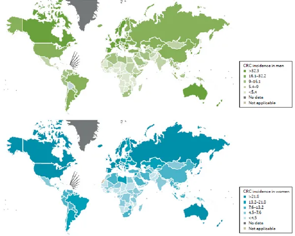
Globally, colorectal cancer (CRC) is the third most widespread cancer in both men and women, while over 1 million new cases are diagnosed each year accounting for 9.7% of all cancers apart and consequently more than 693,933 deaths per annum corresponding to 8.5% of the total number of cancer deaths (Ferlay et al., 2015). In the United States, colon cancer is the second most prevalent cause of death from cancer in men and women after lung cancer; in 2017, an estimated 71,420 men and 64,010 women new cases of colon cancer and 27,150 men and 23,110 women deaths were reported (Siegel et al., 2017a). In Europe, CRC is the second most common cancer, with almost 500,000 new cases diagnosed accounting for 13.0% of all cancers apart and 214,866 death which correspond to 12.2% of the total number of cancer deaths in 2012 (Ferlay et al., 2015).
The incidence rate of CRC gets higher with increasing age. At the time of diagnosis greater number of patients affected with sporadic cancer are more than 50 years of age, with 75% of patients with rectal cancer and 80% of patients with colon cancer being more than 60 years of age. The age standardized incidence rate of CRC is higher in men (20.6 per 100,000 individuals) than in women (14.3 per 100,000 individuals) (Kuipers et al., 2015). The incidence of CRC mostly varies on geographic distinctions worldwide, with almost 55 % of the cases occurring in more developed countries (Figure.1.1). These geographic variations may be attributable to diverse dietary and environmental revelations that are imposed upon a background of genetically determined susceptibility. The age standardized mortality rate of CRC is higher in men (10.0 per 100,000) than in women (6.9 per 100,000) which reflects disease incidence (Kuipers et al., 2015). In 2012, the cumulative risk of CRC in persons aged under 75 was 1.95% worldwide (2.36% in men, 1.57% in women) and 3.51% in Europe (4.48% in men, 2.73% in women) (Ferlay et al., 2013). In developed countries, such as Australia, Canada, United States, New Zealand and European countries etc. higher mortality rates exits compared to developing countries, such as Africa, Central America, Japan, China, Singapore and Korea etc. in the recent years (Siegel et al., 2016). Mortality rate also depends on the stage of diagnosis, which is motivated by the availability of a population screening program and by the level of care in each country (Kuipers et al., 2015).
Like the incidence and mortality, survival rates vary by biological and sociological factors (Jackson-Thompson et al., 2006). Most of the marked global and regional disparity in
13
survival is likely due to differences in access to diagnostic and treatment services (Boyle and Langman, 2000). The prevalence rate of CRC after 5 years was estimated at 3,543,582 worldwide (68.2 CRC survivors per 100,000 population) and 1,203,943 in Europe (192.3 CRC survivors per 100,000 population) in 2012 (Ferlay et al., 2013). On the global economy, CRC has a great impact on the medical care. By 2020, CRC is estimated to exceed $17 billion in healthcare system only in the USA (Mariotto et al., 2011).
1.1.2. Risk factor and etiology of CRC
Multiple factors are associated with the development of CRC (Figure 1.2). The incidence of CRC is rising in the age group. An individual’s risk of CRC increases with advancing age, with the livelihood of cancer diagnosis increasing after 40 years of age and rising progressively after 50 years of age. According to the WHO and the center for disease control, obesity and cancer are two main epidemics in current century, and prevalence has significantly increased in the last few decades. High consumption of processed foods,
Figure 1.1. Geographical variations in colorectal cancer (CRC) rates. Data from the WHO demonstrating
14
animal fat, and a high calorie diet are risk factors for CRC development (Vucenik and Stains, 2012).
In the last 20 years, a benefit of physical activity on cancer etiology has been growing interest. Several evidences support that the prevention strategies with convincing data that physical activity decreases CRC risk (Winzer et al., 2011). Diet has an essential function in the development of CRC. Calcium, fiber, milk, and whole grains have been related with a lower risk of CRC, while high content in fat and/or red meat and processed meat have been connected with an increased risk (Song et al., 2015).
Other environmental factors including cigarette smoking are associated with 20% while alcohol
abuse is associated with 60% of greater risk to the CRC development (Derry et al., 2013). Furthermore, chronic inflammatory bowel disease (IBD) increased the incidence of ulcerative colitis and Crohn’s disease, which contributed the CRC progression (Terzić et al., 2010). Insulin resistance, metabolic consequences of obesity (Meyerhardt et al., 2003), intestinal dysbiosis (Gao et al., 2015) and circadian clock alter digestion (Derry et al., 2013) could also influence the CRC.
CRC can be developed through the mutation of specific gene related to the tumor suppressor, oncogene and DNA repair mechanism. About 70% of CRC cases occur sporadically due to transformation of specific morphological changes starting from adenoma to carcinoma state, 5% are related to inherited (familial adenomatous polyposis (FAP), hereditary non-polyposis colorectal cancer (HNPCC) or Lynch syndrome and MUTYH-associated polyposis) and the rest of the 25% of CRC are associated with familial, only a few are known with high microsatellite instability, with DNA mismatch repair (MMR) deficiency (Stoffel and Kastrinos, 2014).
In the colonic epithelial cells, the environmental and genetic factors cause CRC progression by promoting the acquisition of characteristic behaviors of CRC (Figure 1.3).
Figure 1.2. Several risk factors associated
15
Several genetic and epigenetic alterations may increase the accretion of mutation and epigenetic changes of oncogenes and tumor suppressor genes, and alteration several signaling pathways which drive the malignant transformation from early neoplastic lesions (aberrant crypt foci (ACF), adenomas and serrated polyps) to cancer progression sequence (Vogelstein et al., 1988).
1.1.2.1. Inflammation and CRC
Even through the physiologic levels of inflammation are preventive and encourage tissue repair, extreme inflammation is harmful and lies at the origin of IBD that is eventually connected to the expansion of CRC (Clevers, 2004). Genetic alteration could also be promoted by inflammation in the intestinal tract by generating pro-inflammatory cytokine, chemokine, reactive oxygen species (ROS) and reactive nitrogen species (RNS) (Munkholm, 2003). Continuous production of cytokines and other mediators are associated with the progression of colitis-associated tumors by generating a microenvironment favoring colonic epithelial proliferation, survival and invasiveness (Munkholm, 2003) (Figure 1.4).
16
Figure 1.4. Inflammation and oxidative stress induces CRC.
Tumor necrosis factor alpha (TNF-α) activated from this course form macrophage and T-cells and induced DNA damage, angiogenesis and nuclear factor kappa B (NFκB) activation. Activated NFκB also observed by the intestinal bacteria breaches the colonic epithelial barrier by the Toll like receptors (TLRs) (Abreu, 2010). It helps to increase other pro-inflammatory cytokines (interleukin (IL)-1β and IL-6) and mediators (inducible nitric oxide synthase (iNOS), cyclooxygenase 2 (COX-2) and prostaglandin E2 (PGE2)) (Klampfer, 2011) to progression of CRC development by increasing chromosomal instability, mutations of adenomatous polyposis coli (APC) gene, elevating the levels of DNA methyltransferase 1 (DNMT1). DNMT1 enzyme is overexpressed in IBD and stimulates tumor promotion, angiogenesis and inhibits apoptosis (Foran et al., 2010). Additionally, the stimulation of a number of oncogenic signaling pathways, Wnt/β-catenin, kirsten rat sarcoma virus oncogene homolog (KRAS), phosphatidylinositol 3-kinase (PI3K)/ protein kinase B (Akt) and signal transducer and activator of transcription (STAT3) has been observed for development of inflammation associated CRC (Ullman and Itzkowitz, 2011).
17
1.1.2.2. Oxidative damage and CRC
In oxidative stress, there is an imbalance between production of ROS or other oxidants and their abolition by defensive mechanisms of antioxidant enzymes. In the carcinogenic pathway oxidative stress has the potential impact to enhance the malignant transformation and proliferation of initiated CRC cells (Figure 1.4) (Li and Martin, 2016). Disorder in the redox equilibrium, such as increased production of ROS and nitrogen species, hampers immune system and may lead to damage of lipids, proteins and DNA (Nogueira and Hay, 2013).
ROS play an important role to modulate multiple signal transduction pathways, such as NFκB, PI3K/Akt, heat shock proteins and mitogen-activated protein kinase (MAPK), which is associated with inflammation, proliferation, growth, differentiation and apoptosis. In cancer cells, there are an unbalanced ROS level due to the defective cell metabolism and it acts as a pro-tumorigenic (Nogueira and Hay, 2013). The helpful or destructive role of ROS depends on their concentrations on the cells. The high ROS content in cancer cells renders them more susceptible to oxidative stress-induced cell death, and can be exploited for selective cancer therapy.
ROS initiates lipid peroxidation by a chain of reaction that produces free radicals and other substances (malondialdehyde (MDA), hydroperoxides, lipoperoxides, and toxic aldehydes), which facilities membrane permeability and increases inflammation (Mena et al., 2009). Additionally, lipid peroxidation may act as a signal transducer for cells proliferation and modulates gene expression in the DNA bases which may contribute to CRC (Marnett, 1999). ROS restrain proteolytic system by oxidizing structural proteins and accretion of damaged proteins in cancer cell, which acts as an inhibitor of the proteasome (Grune et al., 2003). This process induces accumulating misfolded and damaged proteins and affects the lysosomal system, which hampers protein turnover and gradually leads to further structural and functional alteration for promoting cancer initiation (Brunk and Terman, 2002). Reproducing cells are highly sensitive against DNA damage by arresting cell cycle or induction of transcription, replication error, initiation of signaling pathways and genomic alteration, all of which associated with CRC progression (Valko et al., 2006).
18
1.1.2.3. Alteration of oncogene and tumor suppressor gene
The development of the CRC from initiation to progression depends on a serious of well-characterized histopathological changes from mutation of several oncogenes and tumor suppressor genes. At least four chronological genetic changes require ensuring CRC development, such as oncogene, KRAS, as well as the tumor suppressor genes APC, Smad4 and TP53, are the main targets of these genetic changes. Mutations of APC gene can promote the destruction of β-catenin and therefore activate of the wingless-type (Wnt) pathway, a common mechanism for initiating polyp to cancer progression (Vogelstein et al., 1988). p53 (transcriptional factor) that controls cell cycle, apoptosis and DNA repair mechanisms. Mutation of this gene is one of the familiar genetic changes in the development of CRC (Rodrigues et al., 1990). In addition, mutations of oncogenes, KRAS or B rapidly accelerated fibrosarcoma (BRAF) proto-oncogene, occur in approximately 55% to 60% of CRCs, aberrantly activating the MAPK signaling pathway (Samowitz et al., 2005). Multiple additional genetic alterations contribute to carcinoma formation and metastases by altering cellular growth, metabolism, migration and invasive capabilities, and angiogenesis. Major signaling pathways have been described in the below following part which is modulated at the alteration of oncogene and tumor suppressor gene.
1.1.2.3.1. Wnt signaling
Activation of the Wnt signaling pathway through the mutation of the APC gene is a crucial incident in the development of colon cancer. Inactivation of the APC gene is the single gene defect responsible for FAP, and about 90% of sporadic colon cancers harbor activating mutations within the Wnt pathway; most of these mutations destroy APC function (Miyaki et al., 1994). On the basis of the biological activity of Wnt over expression, it has been classified into two types: canonical (β-catenin-dependent) and non-canonical (β-catenin-independent) (Figure 1.5).
19
APC gene encodes the APC protein that combines and degrades the downstream signaling component cytoplasmic β-catenin. Cytoplasmic β-catenin is arrested by a destruction complex composed of APC, glycogen synthase kinase 3-β (GSK-3β), Axin, and other components. In the absence of functional APC, the distraction of the destruction complex discharges β-catenin and allows it to accumulate in the nucleus along with lymphoid enhancer factor/ T-cell factors (LEF/TCFs). As nuclear concentrations increase, LEF/TCFs
recruit β-catenin to target genes transcription, such as Myc proto-oncogen (c-Myc), Axin2 and LEF1 which leading to constitutive activation of the Wnt signaling pathway. A similar effect can arise via mutations in β-catenin and Axin2, however, these are significantly less recurrent mutations than in APC (Najdi et al., 2011). At the same time, Wnt pathway activation evidently helps to the development of adenomas by activation of other additional signaling pathways, such as those mediated by the proto-oncogene KRAS, which is necessary for optimal nuclear localization of β-catenin to the nucleus and promotion of the carcinoma program (Najdi et al., 2011). For the development of the precancerous adenomas by the alteration of APC/ β-catenin gene, canonical dependent Wnt signaling is the main focus. Furthermore, recently non-mutational Wnt signal activation has been observed for the development of cancer by the non-canonical pathway.
1.1.2.3.2. MAPK signaling
The MAPK are serine/ threonine (proXser/ThrPro) kinases and they can convert various extracellular signals into intracellular responses through serial phosphorylation cascades. When mitogen binds to the kinase it activates guanosine triphosphatase (GTPase) by converting its guanosine diphosphate (GDP) to GTP and then phosphorylates the downstream of MAPK. The major MAPK families containing of extracellular-signal-regulated kinase 1/2 (Erk1/2), Jun N-terminal kinase (JNK), and p38 MAPK, are known to
20
relay, amplify and integrate signals from a diverse range of stimuli in controlling cellular proliferation, differentiation, cell cycle, inflammatory responses, apoptosis, invasion and angiogenesis (Zhang and Liu, 2002). In CRC, dysregulated Erk1/2, p38MAPK and JNK pathway have been found which are associated with the destruction of the normal mechanism of colonic epithelial cells (Figure 1.6).
Figure 1.6. MAPK signaling pathway in CRC.
The Erk1 and Erk2 module are activated upon phosphorylation by MEK1 and MEK2 which are activated when phosphorylated by BRAF. Erk1/2 signaling is a particular consequence to CRC and activated by numerous extracellular stimuli and growth factors. In this pathway, ligand-mediated activation of receptor tyrosine kinases triggers GTP loading of the rat sarcoma virus oncogene (Ras) GTPase which can then recruit rapidly accelerated fibrosarcoma (RAF) kinases to the plasma membrane for activation. Mutation of the KRAS and BRAF genes is observed in the early step of CRC, which leads to constitutive activation of Erk1/2 signaling cascade and increases the cell proliferation (Dhillon et al., 2007). Additionally, this signaling pathway is also involved in the
21
metastasis process through increasing epithelial to mesenchymal transition, migration and invasion (Urosevic et al., 2014). Conversely, the Erk1/2 pathway is connected to the activation of apoptosis because they are often stimulated in response to different cellular stress and growth factors (Jeon et al., 2005).
In mammals, four genes have been identified that encode p38αMAPK, p38βMAPK, p38γMAPK and p38δMAPK. Several extracellular stimuli can activate p38MAPK pathways and allow cells to respond properly by generating a plethora of diverse biological effects such as inflammation, cell proliferation differentiation, and apoptosis in a tissue-specific and signal-dependent manner. A variety of combination of upstream kinases control the activation of p38 isoforms by activating mitogen-activated protein kinase kinase (MAPKK)3 and MAPKK6 which are activated by their upstream kinases MEKK1/2/3/4 and the apoptosis signal-regulating kinase 1 (ASK1) and another way that is MAPKK independent. Non-phosphorylated p38MAPK is inactivated and become activated by phosphorylation of two Thr-Gly- Tyr motifs. Phosphorylated p38 proteins can activate several transcription factors, such as activating transcription factor 1/2/6 (ATF 1/2/6), CCAAT-enhancer-binding protein homologous protein (CHOP)-1, myocyte enhancer factor-2 (MEF-2), p53, and Elk-1 and a variety of kinases, including mitogen-activated protein kinase interacting protein kinase (MNK)1, MNK2, mitogen- and stress-activated protein kinase (MSK)1, MSK2, p38-regulated/ activated protein kinase (PRAK), mitogen-activated protein kinase-mitogen-activated protein kinase (MAPKAPK)2 and MAPKAPK3 are involved in regulating cytoplasmic or nuclear signaling networks and response to growth factors, cytokines, toxins and pharmacological drugs (Grossi et al., 2014). In colon cancer, p38 MAPKs can play a dual role: they can either mediate cell survival or promote cell death through different mechanisms. According to the previous evidence, it was suggested that the p38α can act as a tumor suppressor by suppressing the inflammation associated colon tumor, negatively regulate the malignant transformation by mutation of harvey rat sarcoma viral oncogene homolog (HRAS) and increased apoptosis through upregulation of first apoptosis signal (FAS), B-cell lymphoma 2 associated X (Bax) and downregulation of Akt survival pathway. Conversely, aberrant activation of p38MAPK has been observed in high grade of CRC and IBD (Grossi et al., 2014; Gupta et al., 2014).
The JNK proteins are encoded by three genes, JNK1, JNK2 and JNK3, each of which has 2 to 4 isoforms and can be activated by the upstream MKK4 and MKK7 kinases kinases via dual phosphorylation of the Thr-Pro-Tyr motif. The activated JNK can not only regulate a
22
variety of transcription factors such as c-Jun (cellular Jun proto-oncogene), c-Fos (cellular Fos proto oncogene), ATF-2, activator protein-1 (AP-1), p53 and Elk but also phosphorylate many cytoplasmic substrates such as cytoskeletal proteins, mitochondrial proteins like B-cell lymphoma (Bcl)-2 and Bcl-xl. In colon cancer, the oncogenic role of JNKs is based on their aptitude to phosphorylate Jun and to activate AP-1 that controls cell survival, cell cycle, apoptosis and metalloproteinases, while tumour suppressor functions of JNK are correlated to their pro-apoptotic activity (Ogunwobi and Beales, 2007; Wagner and Nebreda, 2009).
1.1.2.3.3. PI3K/Akt signaling
The PI3K/Akt signaling pathway can be activated by several growth factors and by G-protein-coupled receptors (GPCRs) upon stimulation with phospholipids and chemokines which can activate the signals for cell survival and proliferation, cell cycle and anti-apoptotic effects leading to CRC carcinogenesis (Huang and Chen, 2009). The growth induced activation of PI3Ks that are recruited to receptor tyrosine kinases (RTKs) consists of two subunits: catalytic subunit p110 and regulatory subunit p85. In the case of GPCRs induced activation, p110 PI3K subunits directly bind to the αβγ-trimeric G protein. Binding and activation of PI3K to membrane-bound RTKs stimulate the conversion of phosphatidylinositol- 4,5-bisphosphate (PIP2) to phos- phatidylinositol-3,4,5-triphosphate (PIP3) that in turn phosphorylates Akt which thereby becomes activated (Figure 1.7).
Activated Akt inhibits apoptosis through preventing cytochrome c release, phosphorylating B-cell lymphoma 2 associated death promoters (Bad) and increasing NFκB. Additionally, Akt stimulates cell proliferation through blocking kinase activity by reducing GSK3β which decreases Cyclin d1 degradation and elevates β-catenin accumulation. Furthermore, AKT-mediated phosphorylation of tuberous sclerosis (TSC) 2 releases TSC-inhibition of
Figure 1.7. PI3K/Akt signaling pathway
23
the GTPase, which activates mammalian target of rapamycin complex 1 (mTORC1) kinase. Activated mTORC1 phosphorylates eukaryotic translation initiation factor 4E-binding protein 1(4E-BP1) and ribosomal protein S6 kinase beta-1 (P70S6K) which allows protein translation initiation and elongation (Zhang et al., 2013a).
1.1.2.3.4. EGFR signaling
Gene amplification of the epidermal growth factor receptor (EGFR) gene encodes for a transmembrane glycoprotein involved in signaling pathways that affect cell growth, differentiation, proliferation, and angiogenesis. EGFR is over-expressed in up to 60 to 80% of CRC and high tumor EGFR overexpression is related with worse prognosis (Goldstein and Armin, 2001).
EGFR belongs to the ErbB family of receptor which includes ErbB1 (EGFR or human epidermal growth factor receptor (HER) 1), ErbB2 (HER2), ErbB3 (HER3), and ErbB4 (HER4)). The EGFR signaling can be activated by numerous ligands, such as epidermal growth factor (EGF), transforming growth factor, heparin-binding EGF, epiregulin (EREG), and betacellulin binding to the receptor dimerization with another EGFR monomer (homodimerization) or with a monomer of another ErbB family member. Several tyrosine residues in the intracellular domain are phosphorylated creating a series of high-affinity binding sites for various transducing molecules. The two major signaling pathways RAS–RAF–MAP and PI3K-Akt pathway are activated by EGFR signaling (Saletti et al., 2015). These mechanisms are relevant because of the availability of pharmacologic agents that can influence these signaling pathways and affect cell growth.
1.1.2.3.5. TGF- β signaling
Transforming growth factor beta (TGF-β) signaling pathway has the potential to control a range of biological processes such as cell growth, differentiation, apoptosis, extracellular matrix modeling, and immune response. TGF-β signaling pathway acts as a tumor suppressor and alterations in this pathway promote CRC cell growth, migration, invasion, angiogenesis and metastasis (Ramamoorthi and Sivalingam, 2014).
Once activated, the TGF-β ligands regulate cellular processes by binding to three high-affinity cell surface receptors- the type 1 TGF-β receptor (TβRI), the type 2 TGF-β receptor (TβRII), and the type 3 TGF-β receptor (TβRIII) serine/ threonine kinases- on the cell surface and induce heterotetrameric receptor complexes. In the early stage of CRC,
24
TGF-β signaling acts as a tumor suppressor and it encourages colon cancer cell growth, migration, invasion, angiogenesis and metastasis in later stages (Ramamoorthi and Sivalingam, 2014). There is a lack of function of TβRI and TβRII receptors which are accountable to alter the TGF-β induced growth inhibition in colon cancer. Over expressed TGF- βI is observed in the primary stage of CRC, while TGF-βII expression is higher in advanced stage of CRC which promotes colon cancer progression and metastasis. Similarly, the expression of TβRI, TβRII, Smad4 and Smad7 is not so much increased in early stage but in advanced stage, Smad7 is overexpressed while Smad4, TGF-β1 and TβRII expression is reduced. The overexpression of TβRIII is increased TGF-β signaling pathway through phosphorylation of Smad 2/5/8 and p38 and downregulation of p21 and p27 levels and thus enhances proliferation of colon cancer cells (Zhang et al., 2013a).
1.1.2.3.6. NFκB signaling
Aberrant NFκB activation has been detected in more than 50% of colorectal and colitis-associated tumors (Kojima et al., 2004). The transcription factor, nuclear NFκB pathway, plays a leading to inflammation to carcinogenesis by modulating several genes responsible for the generation of pro-inflammatory mediators and
cytokines (Figure 1.8).
In presence of multiple pro-inflammatory stimuli, inhibitor of kappa B (IκB) is rapidly degraded by the ubiquitin-proteasome pathway. The degradation of IκB induces the translocation of NFκB subunits into the nucleus, and the NFκB subunits bind to the promoter regions of target genes, including iNOS, COX-2, TNF-α and IL-1β, and stimulate transcription genes which encode cytokines, growth factors, chemokines, and anti-apoptotic factors. Furthermore, activated NFкB pathway also increases cell proliferation, anti-apoptosis, genomic instability, glycolysis and drug resistance in colon cancer cells (Sakamoto et al., 2009).
Figure 1.8. NFҡB signaling
25
1.1.2.3.7. Nrf2 signaling
Nuclear related factor 2 (Nrf2) transcription factors play a vital role in cellular protection by regulating many antioxidant and detoxification enzyme genes via the antioxidant response element (ARE). In the presence of oxidative stress, Nrf2 translocates to the nucleus and binds to ARE sequences to regulate the transcription of several types of genes. Nrf2 downstream genes are involved in intracellular redox-balancing, metabolism, autophagy and apoptosis. Depending on the function of the genes, activated Nfr2 protects the stressed cells form toxic exposure (Menegon et al., 2016).
Previously, Nrf2 has been considered as a tumor suppressor for its cytoprotective effects which are believed to the main cellular protection mechanism against exogenous and endogenous insults, including xenobiotics and oxidative stress. Conversely, several recent studies revealed that hyperactivation of the Nrf2 signaling creates a favorable environment for survival of malignant cells, protecting them against oxidative stress, chemotherapeutic agents and radiotherapy (Menegon et al., 2016). In CRC, the cytoprotective effects are associated by inhibiting Nrf2 repressor Keap1, which maintains Nrf2 expression at the basal levels and decreases the risk of CRC. Conversely, after inhibiting Nfr2 signal, protection of other external signal disappears and increases the risk of CRC. However, overexpressed Nfr2 also increases the risk of CRC by arising Nrf2 mediated inflammation and chemotherapy resistance (Gonzalez-Donquiles et al., 2017).
1.1.2.3.8. Notch signaling
Notch signaling is important for maintaining the cellular proliferation, differentiation, apoptosis, angiogenesis and migration of cancer cells (Schroeter et al., 1998). The activation of Notch signaling is concerned in colon carcinogenesis due to the initiation of pro-survival signaling in colonic epithelial cells. Overexpression of Notch elements, such as receptors, ligands and downstream target genes, is connected with promoting progression, metastatic potential, recurrence and poor prognosis and clinical outcome in various cancers including CRC (Suman et al., 2014).
The activation of Notch signaling depends on cell to cell communication in which Notch ligands bind to Notch receptors, which is present on a neighboring cell and activate metalloproteases in the disintegrin and metalloprotease (ADAM) family, resulting in the cleavage of the extracellular domain (Figure 1.9). Consequently, activated γ-secretase
26
breaks within the transmembrane domain of Notch receptors, resulting in the release of the Notch intracellular domain (NICD), translocates to the nucleus and binds via its RBPJ-associated module (RAM) and ankyrin domains to the DNA-binding transcription factor CSL (CBF1, Suppressor of Hairless, Lag-1). Once created, this complex displaces the co-repressors and binds to the transcription factors, recruits transcriptional co-activators and induces the expression of target genes, such as hairy-enhancer-of-split (Hes-1) and Hes-related protein gene families, that subsequently implement pro-survival functions (Suman et al., 2014).
1.1.2.3.9. JAK/STAT3 signaling
The Janus kinase/ signal transducers and activators of transcription (JAK/STAT) signaling pathway is important in cytokine mediated immune responses, proliferation, apoptosis and migration. In the CRC, JAK/SATA3 signaling is one of the important pathways for colonic tumorigenesis and has a critical function in different aspects together with initiation, promotion and progression (Slattery et al., 2013). The JAK family consists of four non-receptor protein tyrosine kinases, JAK1, JAK2, JAK3 and TYK2, ubiquitously expressed in the mammals cells (Figure 1.10).
In the presence of growth factors and cytokines, RTK promotes activation of JAKs. Activated JAKs auto-phosphorylate and recruit signal transducers and activators of transcription factor STATs. Five additional STATs have been identified: STAT3, STAT4, STAT5A, STAT5B, and STAT6, and in CRC STAT3 is
more activated. Activated STAT3 complex then translocates from the cytoplasm to the nucleus where they initiate transcription of STAT3 targets genes by combining with DNA consequence elements, such as BcI-xL, c-Myc, cyclin D1, vascular endothelial growth
Figure 1.9. Notch signaling pathway
in CRC.
Figure 1.10. JAK/STAT3
27
factor (VEGF) and survivin, which are involved in CRC cells inflammation, proliferation, differentiation, invasion and immune function (Wang and Sun, 2014).
1.1.2.4. The role of endoplasmic reticulum stress in CRC
The endoplasmic reticulum (ER) is the main intracellular organelle responsible for protein folding, translocation and post-translation modification of secreted and membrane proteins, maintenance of calcium homeostasis and lipid biosynthesis. Accretion of unfolded proteins and disorder of calcium homeostasis within ER causes ER stress (Kim et al., 2008b). Targeting ER stress has gained a great deal of attention as the molecular pathway that can lead to the cell death through apoptosis intrinsic or extrinsic-mediated pathways to eliminate the damaged cells as well as enhance the sensitivity of tumor towards chemotherapeutic agents (Kim et al., 2008b).
In various stressful conditions, ER stress may restore homeostasis and make favorable environment for colon cancer cells survival and proliferation by activating unfolded protein response. Three ER transmembrane proteins: protein kinase-like ER kinase (PERK), ATF6, and inositol requiring kinase-1 (IRE-1) primary regulate the ER stress response and play key role for colon tumorigenesis (Figure 1.11). These three proteins remain inactive and bound to the ER-resident chaperone glucose-regulated protein-78 (GRP-78) in normal conditions. In the period of ER stress, GRP-78 separates from these proteins complex and autophosphorylation of detached PERK and IRE-1 and transcolation of ATF6 to the Golgi apparatus where is sequentially cleaved by protease for making active nuclear ATF6. These variations cause activation of their downstream signaling pathways. Activated PERK then phosphorylates
eukaryotic initiation factor 2 alpha (eIF2α) to attenuate protein translation and decrease ER protein overload, while expanded ER stress induces apoptosis by increasing CHOP expression. This is one the therapeutic targets for the ER stress inducing cancer cell death. Furthermore, activated IRE-1 acts as an endonuclease to enhance the X-box binding
Figure 1.11. Unfolded protein response
28
protein 1 (XBP1) splicing, thereby leading to upregulation of genes for cell survival during ER stress (Yadav et al., 2014). Interestingly, elevated levels of the unspliced form of XBP1 increase the apoptosis of cancer cells (Davies et al., 2008).
1.1.2.5. The role of AMPK signaling in CRC
Adenosine monophosphate (AMP)-activated protein kinase (AMPK) is an evolutionarily conserved serine/threonine kinase that is a master regulator of metabolic and energy homeostasis in tumor cells in energy stress environments by promoting both glycolytic and mitochondrial metabolism (Chaube et al., 2015). AMPK consists of a catalytic α subunit and regulatory β and γ subunits, each of which has at least two isoforms; the activation of this pathway happens by binding of AMP to the γ subunit, and phosphorylation of Thr172 in the activation loop of the α catalytic subunit by upstream kinases, such as liver kinase B1 (LKB1). The activated AMPK acts as a tumor suppressor because this signaling network contains a number of tumor suppressor genes including LKB1, p53 and TSC2, while LKB1 is an upstream activator of AMPK, and p53 and TCS2 are direct AMPK substrate (Hardie, 2011; Mihaylova and Shaw, 2011). Under nutrient depletion, mTOR regulates the cancer cells growth and proliferation, while AMPK directly inhibits mTOR through phosphorylating raptor, one of the molecules of TOR complex (Laplante and Sabatini, 2012).
However, recent evidence recommended that AMPK activation is necessary to regulate oxidative stress and enhance cancer cell proliferation in metabolic stress microenvironment (Chaube et al., 2015; Jeon, 2016; Li et al., 2015). In cancer cells under energy diminution condition, AMPK induces metabolic adaptation by initiating signaling cascade resulting in the reduction of ATP consuming pathways with concomitant stimulation of biochemical reactions that generate ATP (Figure 1.12).
29
Under stress condition, nicotinamide adenine dinucleotide phosphate in its reduced form (NADPH) generate by pentose phosphate pathway is damaged but AMPK initiates alternative pathway to inhibit cancer cell death by maintains NADPH. The suppression of the acetyl-CoA carboxylases (ACC) 1 and ACC2 by AMPK sustains NADPH levels by reducing NADPH consumption in fatty-acid synthesis and elevating NADPH production in fatty-acid oxidation. Therefore AMPK, in addition, maintaining in ATP homeostasis has an another role in NADPH maintenance, which is vital for cancer cell survival under energy stress conditions (Chaube et al., 2015; Jeon, 2016; Jeon et al., 2012; Li et al., 2015). Additionally, in gastric cancer cells, AMPK activation protects the anticancer drugs induced cell death by diminishing apoptosis (Kim et al., 2008a).
Activation of AMPK increases the expression of downstream targets peroxisome proliferator activated receptor gamma coactivator 1-alpha (PGC1α), and its associated genes for cancer cells survive under energetic stress conditions (Chaube et al., 2015). PGC-1α is a major transcription co-activator and it encourages oxidative, mitochondrial and fatty acid metabolism to enhance cancer cell proliferation (Jones et al., 2012) and metastasis (LeBleu et al., 2014). Sirtuin 1 (SIRT1) is another downstream target of AMPK, depending on nature and phase of the tumor; it acts as a tumor suppressor and an oncoprotein (Chalkiadaki and Guarente, 2015). In CRC patients, elevated levels of SIRT1 expression promote carcinogenesis and are associated with a poor prognosis (Chen et al., 2014).
Figure 1.12. Role of AMPK signaling pathway in cancer. Figure slightly
30
1.1.2.6. The role of matrix metalloproteinases in CRC
Matrix metalloproteinases (MMPs) play an important role for maintaining extracellular homeostasis by sharing proteolytic activity against other extracellular matrix molecules. Several studies investigated that the expression and activation of MMP-2 and MMP-9 in CRC is associated with cancer progression, angiogenesis, invasion and metastasis (Mook et al., 2004). MMP-2 and MMP-9 contain the gelatinase sub-family of MMPs, and though the principal substrates for these enzymes are type IV collagen and gelatin. Integrins are necessary for the cell adhesion, in colon cancer cells MMP-2 degrades the β1 integrins activity for increasing motility and decreasing cell adhesion capability (Kryczka et al., 2012). Gelatinase can activate by multiple signaling pathways. Smad proteins are involved in TGF-β signaling and modulating of colon cancer cell migration by regulating MMP-9 activity. However, in colon cancer cells, overexpression of p38γMAPK has been observed to lead to enhanced c-Jun synthesis, resulting in increased 9 transcription and MMP-9-dependent invasion. Furthermore, MMP-9 is highly expressed in colitis associated colon cancer through modulating Notch and Wnt signaling (Said et al., 2014).
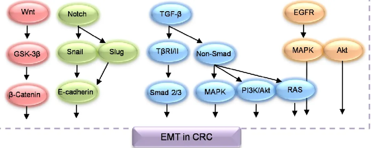
1.1.2.7. The role of epithelial-mesenchymal transition in CRC
The transformation of epithelial cells to mesenchymal cells in a specific pathophysiological condition is known as epithelial-mesenchymal transition (EMT). By modulating several transcriptional regulator, epithelial cancer cells maintain a basal-apical polarity by connecting with several types of junction to undergo various biochemical’s changes; these modifications allow them to disrupt cell to cell adherence by losing E-cadherin levels, remodel the cytoskeleton of cells by increasing vimentin levels and obtain mesenchymal characteristics such as increased migration capability, invasiveness, anti-apoptosis activity by enhancing the production of N-cadherin, osteopontin, Snail, slug and other EMT machineries (Boyer and Thiery, 1993). In CRC, EMT plays a vital role in in situ infiltration and distant metastasis by modulating a complex transcriptional regulator (Figure 1.13).
31
Figure 1.13. Signaling pathways involved in the EMT of CRC. Figure slightly modified form
Jin et al., (2015).
A decrease in the expression of E-cadherin and an increases in the expression of vimentin in CRC patients indicate the presence of lymph node associated metastasis, poor tumor differentiation and prognosis (Cao et al., 2015). In the cancer cells, E-cadherin expression is lossed by methylation of the promoter, degradation and phosphorylation of targets protein and enhances the expression of transcriptional repressors such as Snail, Slug, Sip1 and Zed1. Loss of E-cadherin expression can increase β-catenin in the cytoplasm and in the nuclease, bind to transcription factors TCF/LEF and induce the transcription of target genes of Wnt signaling pathways. Therefore, activation of E-cadherin/β-catenin complex and Wnt signaling potentially encourage colon cancer development and metastasis (Jin et al., 2015). Another important EMT mechanism is overexpression of N-cadherin, while E-cadherin is decreased. In this circumstance, epithelial cancer cells are chopping from the primary focal, invade nearby tissues and migrate via lymphatic and blood circulation (Rosivatz et al., 2004).
Furthermore, the progression of CRC are through to undergo in a dynamic process of EMT by developing several signaling routes (Cao et al., 2015), including TGF-β induced activation of Smad dependent and non-Smad dependent signaling, Wnt signaling, Notch signaling and EGFR induced signaling which are already described in the above section.
1.1.2.8. Genetic instability of CRC
One of the basic processes driving the initiation to progression of CRC is the accretion of a diversity of genetic changes in colonic epithelial cells. Chromosomal instability (CIN),