Alma Mater Studiorum – Università di Bologna
DOTTORATO DI RICERCA IN
BIOLOGIA CELLULARE E MOLECOLARE
Ciclo
XXXI
Settore Concorsuale: SSC 05/E2
Settore Scientifico Disciplinare: SSD BIO/11
TITOLO TESI
Atypical LONELY GUY protein in Bordetella pertussis
synthetizes a cytokinin-like compound negatively
related to oxidative stress
Presentata da:
Filippo Moramarco
Coordinatore Dottorato
Supervisore
Prof. Giovanni Capranico
Prof. Vincenzo Scarlato
Co-Supervisore
Prof. Enrico Balducci
2
Table of Contents
ABBREVIATIONS AND ACRONYMS 4
1. INTRODUCTION 6
1.1. Pertussis Disease 7
1.1.1. Infection and transmission 7
1.1.2. Clinical course 8
1.2. Bordetella Pertussis 9
1.2.1. The two-component regulatory system BvgAS 10
1.2.2. Virulence factors 11
1.3. LONELY GUY and cytokinins 13
2. AIM OF WORK 17
3. MATERIALS AND METHODS 20
3.1. Bioinformatics analysis 21
3.2. Bacterial strains and growth conditions 21
3.3. Generation of E. coli expressing recombinant BP1253 protein and
purification procedure 21
3.4. Size-exclusion chromatographic analysis 22
3.5. Multi-angle light scattering 23
3.6. Generation of the bp1253 knockout strain 23
3.7. High-throughput purification of bp1253 mutants 24
3.8. SDS-page and western blot 24
3.9. Phosphoribohydrolase activity assay 25
3.10. Generation of mouse immune sera 25
3.11. Surface plasmon resonance experiments (Biacore) 26
3.12. Identification and characterization of cytokinins by LC-MS/MS 26
3.13. Phenotypic characterization 27
3.14. Statistical analysis 27
3
4. RESULTS 29
4.1. Production and purification of recombinant BP1253 30
4.2. In silico-based identification of BP1253 as a member of the LONELY
GUY family 32
4.3. BP1253 binds nucleotides 34
4.4. BP1253 has phosphoribohydrolase activity 38
4.5. BpLOG synthesizes 6-O-Methylguanine 45
4.6. BpLOG is prevalently expressed in clinical isolates 48
4.7. BpLOG is negatively related to oxidative stress 49
5. DISCUSSION 52
4
ABBREVIATIONS AND ACRONYMS
5-FU 5-fluorouracil
ACT Adenylate cyclase toxin
AMP Adenosine 5'-monophosphate
aP Acellular pertussis
B. Bordetella
Bp Bordetella pertussis
cAMP cyclic adenosine 5'-monophosphate
CHASE Cyclase/histidine kinases-associated sensing extracellular
DNT Dermonecrotic toxin
FHA Filamentous haemagglutinin
FhaC Filamentous haemagglutinin transporter protein
FIM Fimbriae HAP 6-N-hydroxylaminopurine HK Histidine kinase Hpt Histidine phosphotransferase Kd Dissociation constant LDC Lysine decarboxylase
LOG LONELY GUY
LOS Lipooligosaccharide
M. Mycobacterium
MALS Multi-angle light scattering
MCD Mature C terminus distal
NO Nitric oxide
NOS Reactive nitrogen species
P. Pseudomonas pHBA para-hydroxybenzaldehyde PLP Pyridoxal 5’-phosphate PRN Pertactin PT Pertussis toxin Rec Receiver RTX Repeats in toxin
5
RMS Root Mean Squared
ROS Reactive oxygen species
SEC Size-exclusion chromatographic
SPR Surface Plasmon resonance
StIR Stimulated Innate Resistance
TCT Tracheal cytotoxin
VFT Venus flytrap
6
7
1.1 PERTUSSIS DISEASE
Pertussis is a highly contagious infection of the human respiratory tract. The disease is commonly known as “whooping cough”, because of the characteristic inspiratory sound that infected people emit after cough fits. In fact, the violent and uncontrollable cough that distinguishes this disease often makes hard to breathe, leading ill people to deep breaths, which ring out as a “whooping” sound. Pertussis disease is a significant cause of morbidity and mortality in infants worldwide and despite high vaccination coverage, it continues to be a public health concern (World Health Organization 2015).
The causative agent of whooping cough is the human pathogen Bordetella (B.) pertussis (Bp), a Gram-negative bacterium isolated and described for the first time by Jules Bordet and Octave Gengou in 1906 (Lynfield and Schaffner 2014, Kuchar, Karlikowska-Skwarnik et al. 2016). Pertussis disease is endemic in all countries and affects people of all ages, but it could be particularly intense with a fatal clinical course for young infants, especially of prevaccination age (Paddock, Sanden et al. 2008).
1.1.1. INFECTION AND TRANSMISSION
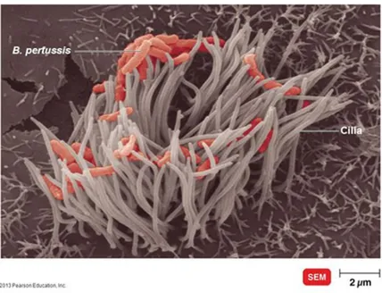
The site of the infection is the respiratory tract (Mallory and Hornor 1912). Indeed Pertussis colonization begins with the adherence of B. pertussis solely to the tufts of ciliated epithelial cells of the upper respiratory tract: nasopharynx and trachea (Tuomanen and Hendley 1983) (Fig. 1). Following this attachment B. pertussis starts to exert its virulence producing several factors such as adhesins, immune-modulators and toxins which damage the cilia and cause airways to swell. The combined action of all these virulence factors prevents rapid clearance of the bacteria and allows them to replicate and to disseminate to the lower areas of the respiratory tract (de Gouw, Diavatopoulos et al. 2011, Zlamy 2016).
Bordetella pertussis is transmitted from infected to susceptible individuals via
aerosolized respiratory droplets released through coughing and sneezing (Warfel, Beren et al. 2012, Trainor, Nicholson et al. 2015). Whooping cough is highly contagious at the first stages of the disease and could be transmitted for three weeks or more without therapeutic intervention. In particular, vaccinated adults and adolescent, who do not manifest infection symptoms but host the pathogen, represent the major source of
8
transmission of bacteria to unvaccinated young infants (World Health Organization 2015). It has been demonstrated that acellular pertussis (aP) vaccines protect against the developments of pertussis symptoms but not against colonization and transmission (Warfel, Zimmerman et al. 2014).
Figure 1. Bordetella pertussis colonization. Image at SEM of Bordetella Pertussis while adheres exclusively to cilia of epithelial cells in the upper respiratory tract during infection. From https://slideplayer.com/slide/4651572/
1.1.2 CLINICAL COURSE
Pertussis clinical course may vary depending on various factors: age, presence of transplacentally acquired antibodies and previous infection or vaccination (McGregor, Ogle et al. 1986, Elliott, McIntyre et al. 2004, Kowalzik, Barbosa et al. 2007). Classical pertussis, seen mainly in unimmunized children, has an average of incubation period of 7 to 10 days and it could last from 6 to 12 weeks. The illness is characterized by three stages: catarrhal, paroxysmal and convalescent phase. The catarrhal phase lasts from 1 to 2 weeks and presents symptoms similar to those of many upper respiratory infections, like nasal congestion, lacrimation, rhinorrhea, tolerant cough and in few cases fever. In fact, it is not immediate to diagnose whooping cough at this stage. When the cough gets worse; pertussis progresses to the paroxysmal phase. Characteristic of this stage is the paroxysm episodes that increase in incidence and severity. They are
9
repetitive series of forceful coughs during a single expiration, followed by forced inspiration that produces the distinctive whoop. The post-tussive vomiting is usually a consequence of the paroxysms (Melvin, Scheller et al. 2014). The paroxysmal stage may last from 2 to 6 weeks and represents a very critical phase for infected infants less than 6 months. Unfortunately they do not have the strength to have a whoop and therefore, fall into a state of apnea resulting in bradycardia, cyanosis and unresponsiveness (Christie and Baltimore 1989). Only in the last phase, the
convalescent stage, the adaptive immunity begins to execute the bacterial clearance.
However the cessation of symptoms, paroxysms, whooping and vomiting, occurs gradually in a period of time ranging from 1 to 12 weeks (Melvin, Scheller et al. 2014, Nieves and Heininger 2016).
In immunized or partially immune children, adolescents and adults, the clinical manifestations are much milder and could include coryza and cough, but without whoop (Nieves and Heininger 2016). In infants, less than a year old, the clinical course could evolve in a much more severe scenario. The bacteria can disseminate in lungs and induce bronchiolitis necrosis, intra-alveolar hemorrhage and fibrinous edema (Paddock, Sanden et al. 2008). In severe cases an excessive lymphocytosis could trigger a pulmonary hypertension, respiratory failure and death (Paddock, Sanden et al. 2008).
1.2 BORDETELLA PERTUSSIS
B. pertussis, belonging to the genus Bordetella, is an encapsulated Gram-negative,
human-specific aerobic coccobacillus equipped with fimbriae. It is motile and non-sporulating with a size approximately of 0.5 x 1 µm.
The survival of the B. pertussis in the host depends on its ability to produce virulence factors able to reshape the immune system to its advantage (Higgs, Higgins et al. 2012). The main virulence factors are: pertussis toxin (PT), adenylate cyclase toxin (ACT), tracheal cytotoxin (TCT), lipooligosaccharide (LOS), filamentous haemagglutinin (FHA), pertactin (PRN) and type 2 and 3 fimbriae (FIM). Pertussis pathogenesis is not yet entirely understood, but it is demonstrated that FHA, PRN and FIM promote the attachment to the epithelial cells, whilst, PT, TCT and ACT contribute to elude host immunity and to harm the epithelium (Higgs, Higgins et al. 2012). The expression of all these and other factors is under the control of BvgAS locus, which encodes a
two-10
component regulatory system (Weiss, Hewlett et al. 1983). This system, named BvgAS, perceives environmental changes and activates the expression of hundreds of genes (Cummings, Bootsma et al. 2006, Nicholson 2007).
1.2.1 THE TWO-COMPONENT REGULATORY SYSTEM BvgAS
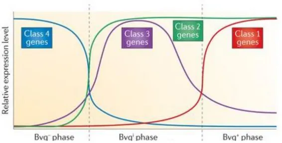
BvgS is a 135-kDa sensor kinase composed of several domains. At N-terminal there are two periplasmic venus flytrap (VFT) domains followed by a transmembrane region, a cytoplasmic PAS domain, a histidine kinase (HK) domain, a receiver domain (Rec) and a last histidine phosphotransferase (Hpt) domain. BvgA is a 23-kDa response regulator protein containing a Rec domain at amino terminus and a DNA-binding helix-turn-helix domain at carboxyl terminus (Decker, James et al. 2012). Specific chemical stimuli, not yet identified, activate BvgS which uses ATP to phosphorylate a histidine residue in the HK domain, triggering the enzymatic cascade (Uhl and Miller 1994). The phosphoryl group is relocated from domain to domain until BvgA Rec domain. Following this phosphorylation BvgA dimerizes and binds to specific DNA sequences to positively or negatively regulate transcription (Boucher, Murakami et al. 1997, Boucher, Maris et al. 2003). The genes regulated by BvgAS belong to four classes and their transcriptional regulation corresponds to three phenotypic phases: Bvg+, Bvgi and Bvg- (Fig.1). Bvg+ occurs when BvgAS is fully active. Class 1 genes (like toxins PT and ACT) and class 2 genes (like FHA and FIM), known also as virulence-activated genes (vag-genes), are maximally expressed in this phase. Phosphorylated BvgA activates further the repressor protein BvgR that represses the expression of virulence-repressed genes (vrg-genes) or class 4 genes. Bvg+ phase is necessary and sufficient to cause infection in vivo (Cotter and Miller 1994). Bvgi, intermediate phase, occurs when BvgAS is partly active and the class 2 and class 3 genes are maximally expressed. Until now only bipA that encodes for an outer membrane protein, has been characterized as class3 genes. It is supposed that Bvgi phenotype may be involved in the transmission of the bacteria. Bvg- phase, instead, occurs when BvgAS is inactive and it is constituted by the highest expression of class 4 genes. The inactivation of BvgAS results under nutrient-limiting condition, therefore Bvg- phase may be essential for bacteria survival in the external environment (Porter, Parton et al. 1991).
11
Figure 2. Schematic representation of the effects of BvgAS activation on the various classes of genes in the three phenotypic phases. The Bvg- phase is characterized by maximal expression of class 4 genes
and occurs when BvgAS is inactive. When instead the BvgAS is partially active, the Bvgi phase begins
with the highest expression of class 2 and class 3 genes. With the fully activation of the two-component regulatory system, Bvg+ phase starts and BvgAS induces the maxima expression of class 1 and class 2 genes. Adapted from (Melvin, Scheller et al. 2014).
1.2.2 VIRULENCE FACTORS
B. pertussis pathogenesis is the result of the action of several virulence factors that interact with host cells and coordinately alter their function. The most known B.
pertussis virulence factors expressed during the virulent Bvg+ phase are proteins that
can be classified as adhesins and toxins. The first category encloses surface proteins mainly involved in the adhesion of the bacteria starting the colonization of upper respiratory tract. The second one includes released proteins primarily implicated in impairing the epithelium and in evading host immunity. Filamentous haemagglutinin, pertactin and the fimbrie are the leading adhesins. FHA represents the dominant adhesin and is synthesized as pre-pro-protein of ~370 kDa (FhaB) to be then processed by peptidases to the mature form of ~250 kDa. During this process it is translocated through cytoplasmic membrane by Sec translocation system and across the outer membrane by filamentous haemagglutinin transporter protein (FhaC) (Mazar and Cotter 2007). Mature FHA has a rod shape with its functionally essential C terminus (MCD) distal to the surface (Mazar and Cotter 2006). It was reported that FHA could act as an immune suppressor in the first steps of colonization (Inatsuka, Julio et al. 2005,
12
Villarino Romero, Osicka et al. 2014). PRN is a protein of 69 kDa and belongs to the classical autotransporter family of outer membrane proteins (Henderson, Navarro-Garcia et al. 2004). In vivo PRN contributes to the adherence of the B. pertussis to epithe1ium (Edwards, Groathouse et al. 2005). It was also demonstrated its involvement in the resistance to neutrophil-mediated clearance (Inatsuka, Xu et al. 2010). The Fimbriae are filamentous appendages on the cell surface also known as type I pili. They are at the basis of Bordetella serotyping and the major serological types are FIM2 and FIM3. As for FHA, FIM favors the bacterial adherence to ciliated respiratory epithelium (Guevara, Zhang et al. 2016) and acts as suppressor of airways inflammation (Vandebriel, Hellwig et al. 2003). Regarding the second category, the most relevant members are pertussis toxin, adenylate cyclase toxin and dermonecrotic toxin (DNT). PT is a 105 kDa ADP-ribosylating AB5-type toxin (Stein, Boodhoo et al. 1994)
composed of A catalytic subunit and five B membrane-binding or transport subunits, which compose the B pentamer. The complex is assembled in the periplasm and is released in the extracellular space by type IV secretion system (Kotob, Hausman et al. 1995). PT enters in the host cells through a receptor-mediated endocytosis following a backward transport route up to endoplasmic reticulum from which the subunit A exits (el Baya, Bruckener et al. 1999). Once in the cytoplasm, the A subunit catalyzes the ADP-ribosylation of a cysteine residue of heterotrimeric G proteins α-subunits blocking the activities of enzymes regulated by G proteins. In particular it prevents G proteins to inhibit the adenylate cyclase activity with the consequent increase of cyclic AMP (cAMP). Overall the ADP- ribosylation induces a primary suppression of inflammatory cytokine production and an inhibition of recruiting immune cells (Graf, Codina et al. 1992, Locht, Coutte et al. 2011). ACT belongs to RTX (repeats in toxin) toxin family and is secreted by type I secretion system (Glaser, Danchin et al. 1988, Carbonetti 2010). It consists of C-terminal domain, which contains the RTX repeats and of N-terminal domain, which possesses calmodulin-dependent adenylate cyclase activity. ACT through its C-terminal binds with high affinity to complement receptor 3 situated on neutrophils, macrophages and dendritic cells surfaces. Upon binding the RTX forms a cation-selective pore in plasma membrane inducing variation in ion permeability and the calmodulin-dependent adenylate cyclase domain moves inside the host cells catalyzing the conversion of ATP in cyclic AMP (Glaser, Elmaoglou-Lazaridou et al. 1989, Ladant, Michelson et al. 1989, Sakamoto, Bellalou et al. 1992, Guermonprez, Khelef et al. 2001, El-Azami-El-Idrissi, Bauche et al. 2003). Increased level of cAMP
13
inhibits phagocytosis and oxidative burst in neutrophils (Confer and Eaton 1982) and prevents the complement-dependent phagocytosis by macrophages (Kamanova, Kofronova et al. 2008). DNT is a cytoplasmic toxin with a size of 160 kDa able to induces necrosis (Cowell, Hewlett et al. 1979). It possesses glutaminase activity and can activate Rho GTPases (Schmidt, Goehring et al. 1999). In the toxin category is also included the tracheal toxin, although not regulated by BvgAS system. TCT is a disaccharide-tetrapeptide monomer of peptidoglycan (Cookson, Tyler et al. 1989) that impairs the respiratory epithelium inducing the production of Interleukin 1 and nitric oxide (Heiss, Moser et al. 1993).
1.3 LONELY GUY AND CYTOKININS
LONELY GUY (LOG) proteins are key enzymes in plants for the production of phytohormones named cytokinins. The gene encoding LOG was identified in a rice mutant in an attempt to screen for defects in shoot meristem activities (Kurakawa, Ueda et al. 2007). Since the flowers of these mutants often contained only one stamen but no pistil, for analogy the protein was named LONELY GUY. It is involved directly in the synthesis of cytokinins converting inactive cytokinins nucleotides to the biological active form by its phosphoribohydrolase activity (Kurakawa, Ueda et al. 2007). The active form of cytokinins is predominantly N6-substituted adenine derivatives. When the modified adenine is conjugated with sugar, such as in nucleosides and nucleotides, shows a minor or completely absent biological function in activating cytokinins receptors (Bajguz and Piotrowska 2009). LOGs hydrolyze the N-glycosidic linkage between the base and the ribose, leading to the formation of free active cytokinins. In plants these phytohormones are involved in many important physiological processes, such as growth and branching (Werner, Motyka et al. 2001), chloroplast development (Cortleven and Schmulling 2015), delay of leaf senescence (Gan and Amasino 1995) and activation of plant defense responses (Bari and Jones 2009).
Over time the study of LOG proteins was limited, because they have been erroneously annotated as lysine decarboxylase enzymes (LDC) due to the presence of a highly conserved PGGxGTxxE motif (Kukimoto-Niino, Murayama et al. 2004, Jeon, Allard et al. 2006). Recently, some LOG-like proteins from non-plant organism, annotated as LDCs, have been identified to have phosphoribohydrolase activity and to produce
14
cytokinins (Dzurova, Forneris et al. 2015, Samanovic, Tu et al. 2015, Seo, Kim et al. 2016). Indeed many plant-interacting microbes and fungi produce mis-annotated LOG proteins able to synthetize cytokinins (Hinsch, Vrabka et al. 2015, Radhika, Ueda et al. 2015). These plant-like hormones are recognized by the plants and lead to a variation of resource allocation and to a morphological reconfiguration of the plant tissue that result in benefits for the pathogen. In fact the uncontrolled cytokinin signaling with the consequent delay of leaf senescence and creation of new organs such as galls and nodules, provides pathogens a continuous nutriment and protection against biotic and abiotic agents (Giron, Frago et al. 2013). Nevertheless a possible role of LOGs and their product cytokinins in a microbial cell-interaction has yet to be elucidated. Surprisingly cytokinin-activating homologous of plant LOG have been recently also identified in human pathogens Mycobacterium (M.) tuberculosis (Samanovic, Tu et al. 2015) and
Pseudomonas aeruginosa PAO1 (Seo and Kim 2018). Interestingly, RV1205, the gene
encoding M. tuberculosis LOG, was detected in an attempt to understand why the mycobacterial resistance to nitric oxide (NO) is linked to proteasome. Through a genetic screen in a proteasome-deficient strain, Rv1205 product was identified as proteasome regulated substrate and its accumulation in proteolysis mutants was responsible for NO sensitivity. In the absence of proteasome, which regulates and keeps LOG protein production to low levels, the amount of the cytokinins as well as of their breakdown products increase. In particular the cytokinin p-topolin degradation product para-hydroxybenzaldehyde (pHBA) resulted to be lethal in proteolysis mutant of M.
tuberculosis in presence of NO (Samanovic, Tu et al. 2015). Despite this, the
physiological role of LOGs in mammalian pathogens and why they synthetize cytokinins are completely unknown so far. Few studies suggest that cytokinins may have suppressive effects on both innate and humoral immune responses (Lappas 2015) or may induce cellular differentiation or apoptosis in immune cells (Casati, Ottria et al. 2011). Mammalian-pathogens, therefore, are likely able to reshape surrounding environment releasing plant-like cytokinins as modulator of the immune system. The specific effects that these metabolites may exert on immune cells depend on concentrations (Ciaglia, Pisanti et al. 2014). Another hypothesis is that LOGs may be involved in a signaling pathway at the inter-bacterial interface. However, a putative role of LOG protein during an infection is suggested by identification of a LOG homolog through a specific screen of genes expressed in a frog model of infection with M.
15
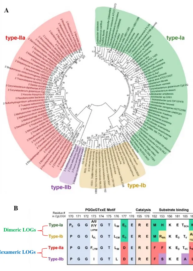
The LOG domain is highly conserved among all kingdoms and this leads scientists to investigate new interesting research areas. Structural and molecular studies have proposed that the two highly conserved amino acids Glu and Arg, are the residues involved in the catalysis (Kukimoto-Niino, Murayama et al. 2004), the PGGxGTxxE motif holds the AMP moiety of substrate nucleotides (Seo, Kim et al. 2016) and that the active site needs two LOG domains for its formation (Seo, Kim et al. 2016). Notwithstanding these overall aspects, a phylogenetic analysis of LOG proteins from various organisms showed differences in the oligomeric state, in the amino acids of the binding site and variation at the PGGxGTxxE motif (Fig.2 A, B) (Seo and Kim 2017). According to this analysis it is possible to classify LOGs into at least two clusters based on the oligomeric state, the dimeric LOGs as type-I LOGs and the hexameric LOGs as type-II LOGs. Type-I and type-II LOGs may be in turn subdivided into two sub-groups, type-Ia and type-Ib, type-IIa and type-IIb respectively (Seo and Kim 2017). Type-II LOGs implement a hexameric form through an additional α-helix (Kukimoto-Niino, Murayama et al. 2004, Seo and Kim 2017). The existence of two structural clusters further divided and variations in amino acids of the binding site suggest the presence of different LOG-like proteins able to produce various cytokinin classes.
16
Figure 3. Phylogenetic analysis and sequences alignment from various subtypes of LOG proteins in comparison. (A) Unrooted maximum likelihood phylogenetic tree of LOGs represented in a circular
model. The four subgroups are schematized and marked with different colors: green for type-Ia and yellow for type-Ib, red for type-IIa and violet for type-IIb. Bootstrap values are shown at each node. (B) Amino acids sequence alignment of key residues of the four subgroup of LOG proteins involved in the PGGxGTxxE motif, catalysis and substrate binding. The oligomeric state of each LOG type is shown. Adapted from (Seo and Kim 2017).
Dimeric LOGs
Hexameric LOGs
17
18
In order to defeat and prevent Pertussis disease, in the late 1940s was introduced the whole-cell pertussis (wP) vaccine, consisting of suspensions of killed B. pertussis organisms (Kuchar, Karlikowska-Skwarnik et al. 2016). The introduction of the vaccine led to a rapid reduction in both the incidence and the number of deaths caused by the infection. Because of its reactogenicity for the high number of endotoxins and comprehensive concerns about its safety, in the late of 1990s it was replaced with acellular pertussis (aP) vaccine in many developed countries (Kuchar, Karlikowska-Skwarnik et al. 2016). aP is based by one to five purified antigens: PT, FHA, PRN and FIM 2/3. Unfortunately, notwithstanding a high vaccine coverage all over the world, an increase in pertussis cases has been recorded in adolescent and adults, concerning the public health about whooping cough resurgence. Although difficult to contextualize, several causes have been hypothesized, like waning immunity due to the lasting immunity induced by aP shorter than wP (Mooi, Van Der Maas et al. 2014), epidemiological changes in the circulating strains (Bart, Zeddeman et al. 2014, Bart, van der Heide et al. 2015) and the different both humoral and cell-mediated immunological response triggered by aP compared to the immunity generated by wP and natural infection (Higgs, Higgins et al. 2012). Furthermore, the difficulty of diagnosis in the first phase of the disease, leads to a loss of effectiveness of antibiotic treatment and, in severe cases, the patients may also not respond to the therapies due to breathing difficulties (Paddock, Sanden et al. 2008). The continuous increase of pertussis cases in all age groups is seriously worrying the public health, above all for the associated risk of transmission to incompletely or unimmunized infants. To solve this concern, the scientific community is operating on several fronts. On one hand it is trying to improve the current vaccine in order to have an acceptable safety, prevent transmission and provide a longer lasting immunity; on the other hand is trying to develop new therapeutic agents and targeted treatment strategies to reverse the disease at the time of diagnosis. However, to obtain all of these, it is necessary and indispensable to discover new features related to Bordetella pathogenesis and microbiology. On the basis of the observations above described, this PhD project aims to improve the knowledge of the physiology and pathology of Bordetella, which can help to improve the quality of vaccine and to develop alternative therapeutic interventions. Specific topics studied in this work were the following:
19
Identification and characterization of the biochemical function of new selected proteins, potentially involved in Bordetella pathogenesis;
Identification of factors playing a key role in the complex communication circuit between the host and the pathogen.
Concerning the aims, we focus our attention on protein pertussis BP1253, a putative exported protein with an unknown function, classified as possible lysine decarboxylase.
20
21
3.1 BIOINFORMATICS ANALYSIS
The BLAST algorithm was used to investigate the sequence homologies between BP1253 and LOGs, while Cluster omega to realize amino acid sequence alignment with specific type-I and type-II LOGs. The structural analysis were performed through PDB Viewer, either to develop the modeling of BP1253 that to determinate the coordinates of atoms in the overlapping. To realize the figures, ESPript 3.0 software was used to represent the sequence alignment, instead, the graphic software PyMOL to visualize the overlaps. Regarding the phylogenetic tree, it was generated with MAFFT and figured through the software tool Archaeopteryx.js.
3.2 BACTERIAL STRAINS AND GROWTH CONDITIONS
The following B. pertussis strains were used in this study: Tohama I-derivative BP536 (Weiss and Falkow 1983 153, 304-9), the clinical isolates B3629, B3621. Bacteria were stored at -80 °C and recovered by plating on Bordet-Gengou (BG) agar plates, supplemented with 15% (v/v) sheep blood, for 3 days at 37°C. Bacteria were then inoculated at initial 600 nm optical density (OD600) of 0.05–0.1 in Stainer-Scholte
medium supplemented with 0.4% (w/v) L-cysteine monohydrochloride, 0.1% (w/v) FeSO4, 0.2% (w/v) ascorbic acid, 0.04% (w/v) nicotinic acid, 1% (w/v) reduced
glutathione. Cultures were grown in rotary shakers at 37 °C.
3.3 GENERATION OF E. COLI EXPRESSING RECOMBINANT BP1253 PROTEIN AND PURIFICATION PROCEDURE
The synthetic gene1253 from Bordetella pertussis was synthesized and assembled by GeneArt (Thermofisher) starting from synthetic oligonucleotides and/or PCR products. The fragment was inserted into pET24b(+)_D455 as a fusion construct with a carboxyl-terminal 6x histidine tag separated from the last amino-acid of the protein by a linker of 2 amino-acid residues. The plasmid DNA was purified from transformed bacteria and concentration determined by UV spectroscopy. The sequence congruence verified by sequencing of the final construct within the insertion sites was 100%. Escherichia coli
22
strain BL21 (DE3) cells (New England Biolabs) were transformed with the above construct and used for protein expression. Cells were grown using BioSilta medium (Enpresso B Animal-free growth systems), at 30°C for 8 h with gentle shaking (160 rpm). The expression of Bp1253 gene was induced to an A600 of 0.5–0.6 by the addition
of 1mM isopropyl β-D-1-thiogalactopyranoside (IPTG) 24 h at 17°C. Cells were harvested by centrifugation and re-suspended in lysis buffer containing 50 mM NaH2PO4, 300 mM NaCl, pH 7.4 and protease inhibitor EDTA free (Boheringer
Mannheim) at a ratio of 10 ml of lysis buffer per 1 g of bacterial pellet. After lysis, performed via sonication (Qsonica Q700), cell lysates were clarified by centrifugation at 15000 x g for 50 min at 4°C, and the supernatant, containing the expressed protein, filtered using a 0.22 μm membrane filters (EMD Millipore filters) before starting the first chromatography step. BP1253 was purified by a Co2+-affinity chromatography (5mL HiTrap TALON crude, GE Healthcare) at room temperature (RT, 25°C) using an AKTA purifier 100 system (GE Healthcare). The column was equilibrated with buffer A (50 mM Tris pH 8, 300 mM NaCl). After loading the crude extract, the column was washed with 10 bed volumes of buffer A. Bound proteins were eluted with buffer A containing 500 mM imidazole. The content of lipopolysaccharide (LPS) on the purified protein was checked using the Endosafe nexgen-PTS system (Charles River). When the content of LPS was out of the range it was removed using the EndoTrap Red columns (Hyglos). The purity of the protein was checked using 4–12% SDS–PAGE gradient gels in MES buffer, after the identification of the fractions containing BP1253, samples were pooled and stored at -20°C for subsequent analysis.
3.4 SIZE-EXCLUSION CHROMATOGRAPHIC ANALYSIS
The investigation of BP1253 oligomerization was performed using an analytical size-exclusion chromatography. The chromatographic step was performed using a BEH200 column 4.6x300mm (Waters) at a Flow of 0.4 ml/min with a buffer containing 10 mM NaH2PO4 and 400 mM (NH4)2SO4 pH 6.0. Protein samples of 15 µl 0.62 mg/ml and 3
mg/ml were analyzed. The molecular weights of the different forms of BP1253 were calculated based on the calibration curve standard proteins.
23
3.5 MULTI-ANGLE LIGHT SCATTERING
Multi-Angle Light Scattering (MALS) analysis was performed in an HELEOS (WYATT Technology) in combination with a SEC separation. SEC was carried out with Sepax Zenix SEC-300 3 mm 7.8 x 300 mm at a flow of 0.5 ml/min with PBS as running buffer. The BP1253 for the analysis was diluted in PBS buffer at a final concentration of 0.6 mg/ml. Results were analyzed using the ASTRA software 3.1.
3.6 GENERATION OF THE BP1253 KNOCKOUT STRAIN
The deletion strain for the genes BP1251-1252-1253 of the B. pertussis BP536 Tohama I-derivative was constructed as follows. The 5′ and 3′ extremities of the locus were amplified by PCR using Bp chromosome as template and the oligonucleotides
FlankingUP-Fw ccgGAATTCCGAAAACCGTAGCGGTCGAA and FlankingUP-rev ggaGGATCCGGACCGATGTCGGCCAATTT, FlankingDOWN-Fw ggaGGATCCCGCGTCTATGTCGACCACG and
FlankingDOWN-rev cccAAGCTTCGAACTGCACCTGACCATCC as primers,
respectively.
The amplicons were then successively introduced as EcoRI-BamHI and BamHI-HindIII fragments into pUC19, together with a BamHI-BamHI fragment encoding the kanamycin resistance cassette for selection. The resulting EcoRI-HindIII fragment was then purified and introduced into the EcoRI-HindIII sites of pSORTP1, a mobilizable suicide plasmid used for conjugation between Bp and E. coli. Conjugation was performed on BG-blood agar plates containing 10 mg/ml MgCl2 for 5 hours, and
co-integrates were selected on BG-blood agar plates containing 10 µg/ml gentamicin and 20 µg/ml nalidixic acid to prevent growth of the E. coli donor. Single crossing overs determine the insertion of the pSORTP1 vector in the chromosome and confer gentamicin resistance and streptomycin sensitivity. Double crossing overs were selected by a successive step on BH-blood plates containing 25 µg/ml of kanamycin and 400 µg/ml of streptomycin. After 3 to 4 days growth on selective media, isolated kanamycin and streptomycin-resistant colonies were analyzed by PCR to confirm the deletion.
24
3.7 HIGH-THROUGHPUT PURIFICATION OF BP1253 MUTANTS
Single point conservative and not conservative mutations were inserted into the Bp1253 gene to generate six different mutants. Mutated genes were synthesized and assembled by GeneArt (Thermofisher) starting from synthetic oligonucleotides and/or PCR products using the same plasmid and procedure as above described. The mutants generated were R120A, R120K, K121A, K121R, E143A and E143D. Purification of mutants was performed under vacuum conditions in a 96-well Vacuum plate. Cells were lysed using B-PER (Sigma) and applied on a His Multitrap HP 50ml NiSepharose High Performance 96 wells previously washed with water and equilibration buffer (300 mM NaCl, 50 mM NaH2PO4, pH 8). After loading the samples, the plate was washed with
80 vol. of washing buffer (300 mM NaCl, 50 mM NaH2PO4, 20 mM imidazole, pH 8) at
room temperature. The His-fusion proteins were eluted in two steps by addition of 6 vol. of elution buffer (300 mM NaCl, 50 mM NaH2PO4, 500 mM imidazole, pH 8)
twice. All the elutions related to the same protein were subsequently pooled. All purification steps were executed applying a vacuum not exceeding the maximum pressure of 5 mmHg.
3.8 SDS-PAGE AND WESTERN BLOT
SDS-PAGE and western blot (WB) analyses were performed to monitor expression, purity and identity of BP1253 during purification. Samples mixed with 1X NuPage LDS loading buffer and 1X NuPage Sample reducing agent (Life Technologies), were heated at 70°C for 10 minutes before loading 20 μl of the mixture onto a 4-12% gradient NuPAGE Bis-Tris gel (Life Technologies). SeeBlue Plus Prestained Standard markers were run onto each gel, which was stained with Comassie Blue to visualize the proteins. Separated proteins were electro-transferred onto nitrocellulose membranes with iBlot 2 Dry Blotting System (Life Technologies). The membranes were blocked for 60 min with 0.1% Tween 20 and 10 % milk in phosphate buffer solution (PBS), incubated for 1 hour with specific mouse polyclonal α-BP1253 antibodies (1:500 dilution) in 0.1% Tween-20 and 3% milk buffer in PBS. After three washes with 0.1% Tween-20 in PBS (T-PBS) the membrane was incubated with a secondary rabbit α-mouse horseradish peroxidase conjugated antibody (Jakson Immune Research Laboratory, 1:1000
25
dilution), for 30 min at room temperature. Bound antibodies were visualized, after washings membranes thrice with T-PBS, with ECL immunoblotting detection system (Amersham) according to the manufacturer’s instructions. The supernatants of the clinical isolates were previously TCA precipitated before western blot analysis. Briefly, the supernatants were incubated 10 min at 4°C with 20% TCA and then centrifuged at 14000 x g for 5 min. The pellets were washed twice with 200 μl of cold acetone to remove TCA. After washings, pellets were dried in heat block for 10 min to dry off acetone.
3.9 PHOSPHORIBOHYDROLASE ACTIVITY ASSAY
Phosphoribohydrolase activity was assessed by detecting the nitrogenous base ring compounds after separation with a thin layer chromatography (TLC) as described (Seo, Kim et al. 2016 6, 31390). Briefly, enzyme reaction was carried out in a mixture containing, in a final volume of 20 µl, 40 mM Tris-HCl (pH 8), 20 mM Nucleotide Mono Phosphate (NMP) as substrate and the amount of purified recombinant protein as specified in figure legends. After incubation at 30°C, samples (5 µl) were denatured at 95°C for 2 min and then 1 µl dotted on PEI-Cellulose-F-plastic TLC sheet (Merck Millipore). Since the solubility of guanine is poor in water and to enhance its visibility on TLC, the inactivation was performed with 1 M NaOH (v/v). The nitrogenous base ring was separated from the phosphoribosyl moiety using a TLC method with a mobile phase containing 1 M sodium chloride except for pyrimidine rings where the mobile phase was acetone/water 30/70 (v/v) with the addition of 0.2 mol/l NaCl. After the run, the sheet was completely dried and the separated dots visualized by UV lamp (264 nm).
3.10 GENERATION OF MOUSE IMMUNE SERA
BALB/c mice (10 females/group, 6-week old) (Charles River Laboratories International Inc., Wilmington, MA) received three intraperitoneal immunizations, with a 4-week interval, with aluminum hydroxide (2 mg/ml) adjuvanted recombinant BP1253-His (10 µg per dose) at one fifth of a human dose. Sera were collected before immunization and 2 weeks after the third immunization. Control mice immunized with adjuvant only were included in the experiments. All the experiments involving animal were performed in
26
compliance with the Italian law with the approval of the local Animal Welfare Body (AWB 2014/06) followed by authorization of Italian Ministry of Health.
3.11 SURFACE PLASMON RESONANCE EXPERIMENTS (BIACORE)
SPR experiments were performed using a Biacore T200 instrument (GE Healthcare). The recombinant BP1253 protein was amine-coupled at 20 µg/mL in 50 mM NaOAc at pH 4.5 to the surface of a CM5 sensor chip (GE-Healthcare), activated with 50 mM 1-ethyl-3-(3-dimethylaminopropyl)-carbodiimide (EDC) / 50 mM N-hydroxysuccinimide (NHS). Typically, response levels of 8000-9000 RU were achieved. After priming the system with HBS-EP+ buffer (10 mM HEPES; 150 mM NaCl; 3 mM EDTA; 0.05% Surfactant P20; pH 7.4), affinity data were generated under steady state conditions, injecting ligand nucleotides dissolved in HBS-EP+ at 30 µl/s and 25°C for 60 s, then allowing to dissociate for another 60 s. Blank-subtracted sensorgrams were analyzed using the Biacore T200 Evaluation Software v. 3.0. Response values were evaluated 4 seconds before the end of each injection and averaged over a window of 5 sec.
3.12 IDENTIFICATION AND CHARACTERIZATION OF CYTOKININS BY LC-MS/MS
For the analysis of the cytokinins, the supernatant was obtained spinning down 40 ml bacterial growth broth at 9000 x g for 20 min. filtered and treated as described (Dobrev, Hoyerova et al. 2017 1569, 31-39). Briefly, the supernatant was cleared by molecules with MW higher than 3kDa using a protein concentrator 3K (Amicon) following vendor’s instructions. Subsequently the samples were concentrated up to 1 ml volume through a Speed Vac and loaded on a solid phase extraction (SPE) column (MCX matrix, Oasis). Before loading, samples were previously acidified to pH 3 with formic acid. The column was then washed with 0.5 mL SPE load solvent (1 M formic acid), followed by 1 mL water. The samples of interest were eluted with 0.5 mL of freshly prepared solvent 2 (0.35 M ammonium hydroxide in 70 % methanol, to 70 mL methanol were added 2.5 mL of 25% ammonium hydroxide filled to 100 mL with distilled water) and the flow-through collected into new 2 mL microcentrifuge tube was
27
dried in SpeedVac at 10 mBar and 40 °C. The dried fractions were stored at −20 °C until LC-MS analysis. At the time of starting the analysis in LC-MS, the dried samples were dissolved into 100 μL 5 % methanol in water and centrifuged at 20000 × g, 4 °C for 20 min. The supernatant was transferred into an autosampler vial and used for the analysis without further modification. LC-MS/MS analysis was performed using an LTQ-Orbitrap XL coupled with an Ultimate 3000 HPLC system equipped with a reverse phase column Luna C18 (2) 100mm x 2mm, 3 µm, 100 Å. The mobile phase A and B used for the analysis of the sample were 0.1% formic acid in water and 0.1% formic acid in acetonitrile respectively. The gradient started with 100% of A, maintained for 3 minutes and the phase B increased up to 20% in 12 minutes. Then after two minutes the phase B reached the 100% and it was hold constant for two minutes. The flow rate was set to 200 µl/min., UV at 268 nm, while a positive ion mode with electrospray was used as mass spectrometric detection. The ESI ion source spray voltage was set to 5 kV, the capillary temperature was 300 °C, tube lens and capillary voltage were 110 and 35 V respectively. The sheat and auxiliary gas (N2) were set to 20
and 5 a.u., respectively. The mass spectra and the MS/MS spectra were recorded using the data dependent scan mode at resolution of 30000, while the fragmentation mass spectra were recorded in low resolution with collision energy 25 a.u.
3.13 PHENOTYPIC CHARACTERIZATION
Bacteria stored with glycerol were inoculated at initial 600 nm optical density (OD600)
of 0.05–0.1 in Stainer-Scholte medium supplemented with 0.4% (w/v) L-cysteine monohydrochloride, 0.1% (w/v) FeSO4, 0.2% (w/v) ascorbic acid, 0.04% (w/v)
nicotinic acid, 1% (w/v) and reduced glutathione. Cultures were grown in rotary shakers at 37 °C for 24 h. After that we tested the bacterial susceptibility to oxidative stress by treating cultures with 0.1 and 0.3 M H2O2 (Sigma) for 30 minutes as specified in Figure
legend. The viability of the Bp strains was measured after treatment.
3.14 STATISTICAL ANALYSIS
Data are presented as the means ± SD and the Kruskal-Wallis test was used to analyze significance of data. Values of P ≤ 0.05 were considered and referred to as significant.
28
3.15 PROTEIN ASSAY
Protein concentration was measured with the bicinchoninic acid (BCA) protein assay (Smith, Krohn et al. 1985 150, 76-85) (Thermo Scientific-Pierce, Rockford, IL, USA), using BSA (2 mg/ml) as a standard, equal amounts of proteins were resolved by SDS-PAGE.
29
30
4.1 PRODUCTION AND PURIFICATION OF
RECOMBINANT BP1253
In order to obtain the Bp protein BP1253, we transformed BL21 (DE3) competent E.
coli cells with known concentrations of pET24b(+)_D455 plasmid containing the
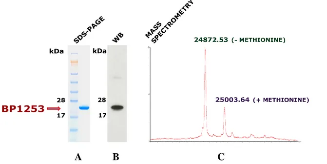
corresponding synthetic gene. BP1253 was expressed as His-tagged recombinant protein after IPTG induction and purified by a single chromatographic step on an immobilized metal (Co2+) affinity chromatography. SDS-PAGE analysis gave a single pure band (Fig. 4A) and western immunoblot (Fig. 4B) confirmed the identity of the purified protein, instead the MS analysis of BP1253-His, resulted in an experimental mass of 24872.53 Da (Fig 4C). Due to overexpression in the bacterial system, not all the methionine residues of the recombinant protein have been removed from the proteins after translation, leading to a second peak corresponding to 25003.64 Da in intact mass measurement.
Figure 4. Expression, purification and BP1253 identification. BP1253 was cloned in E. Coli BL21 (DE3) and purified after IPTG induction trough HiTrap TALON crude. (A) The eluate, 2 µg, was evaluated by SDS-PAGE in 4-12% gradient NuPAGE Bis-Tris gel stained with Coomassie Blue. Molecular mass markers (kDa) are indicated on the left (B) The identity of the purified protein was further confirmed by immunoblot. The experiment was performed with 25 ng of BP1253 and with mouse polyclonal α-BP1253 (1:500 dilution) as described in Material and methods. Molecular weights of proteins markers are shown on the left. (C) Intact mass measurement revealed a BP1253-His molecular weight of 24872.53 Da. The weight of 25003.64 Da refers to BP1253-His proteins with unremoved methionine.
B
A
C
kDa 28 1731
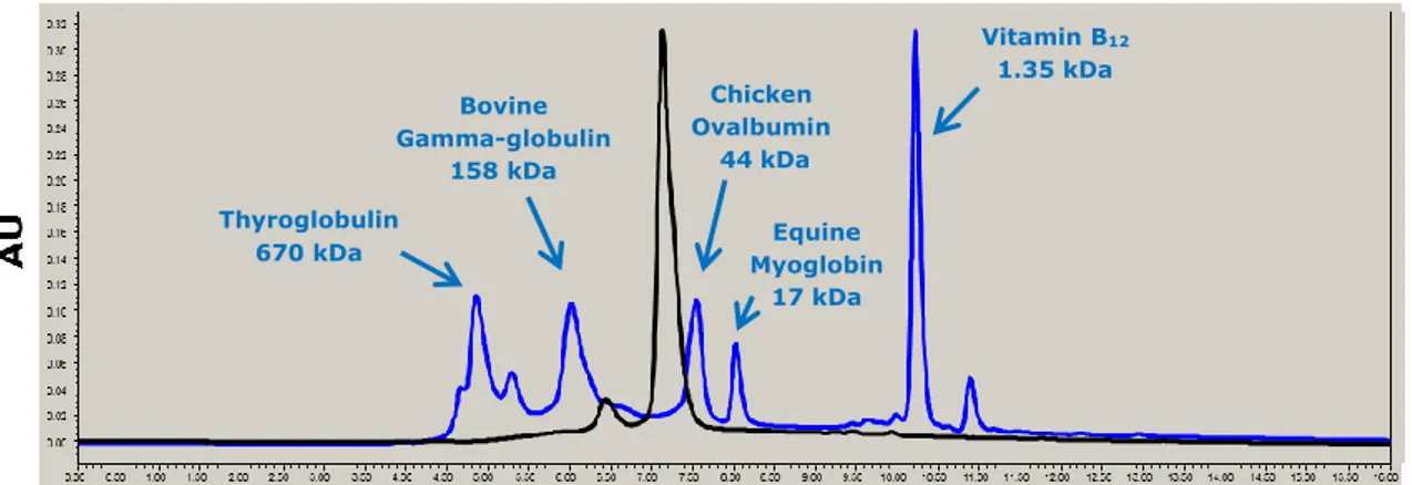
To characterize the functional unit and the oligomeric state of BP1253, we performed a size exclusion chromatography (SEC) (Fig. 5). The SEC gave a main peak with a retention time of 7.20 minutes. Using the molecular weights and the retention times of the protein standards, through a graphic interpolation we calculated that this peak refers to a molecule of about 50 kDa. The analysis revealed also a second, but very small peak approximately of about 100 kDa. To exclude the hypothesis that the oligomeric state of the protein depended on the concentration, we performed SEC analysis with two different concentrations of BP1253, 0.6 mg/ml and 3 mg/ml respectively. The two chromatograms acquired did not display any differences to each other suggesting that the dimeric form is the functional unit of BP1253 (data not shown).
Figure 5. BP1253 is a dimer in size-exclusion chromatography. BP1253 was eluted as dimeric form with a molecular weight of 50 kDa. The analysis was carried out with concentrations of 0.6 mg/ml and 3 mg/ml of protein. The peak corresponding to BP1253 is marked in black, while the proteins standards with their own molecular weights are highlighted in blue. The figure refers to 3 mg/ml protein concentration.
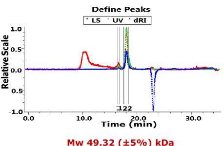
To consolidate the results obtained in SEC, we performed a multi-angle light scattering (MALS) study (Fig. 6). This analysis showed further the presence of a protein with a molecular weight of 49.32 (±5%) kDa with a polydispersity of 1.010 (±7.7%). MALS definitively confirmed that the oligomeric state of BP1253 is a dimer.
Thyroglobulin 670 kDa Bovine Gamma-globulin 158 kDa Chicken Ovalbumin 44kDa Vitamin B12 1.35 kDa Equine Myoglobin 17 kDa
32
Figure 6. Multi-Angle Light Scattering confirms the dimeric state of BP1253. MALS analysis confirmed the dimer as the oligomeric state of BP1253. The study was performed in combination with SEC using a final protein concentration of 0.6 mg/ml. The data were analyzed with ASTRA software 3.1.
4.2 IN SILICO-BASED IDENTIFICATION OF BP1253 AS A
MEMBER OF THE LONELY GUY FAMILY
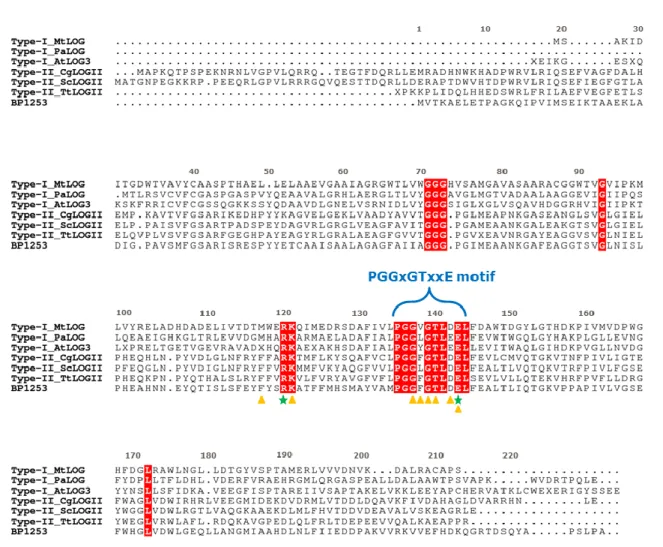
Recently, in the literature it has emerged that many proteins from different organisms of various kingdoms, like Plantae, Animalia, Bacteria and Fungi, are mis-annotated as lysine decarboxylase because the presence of PGGxGTxxE motif in their sequence (Naseem, Bencurova et al. 2018). A more accurate analysis classifies them as members of the LOG protein family. This mis-annotated classification included also BP1253 considered a possible LDC so far. Through bioinformatics analysis and following sequence alignments we observed that BP1253 shares with LOG proteins not only the highly conserved motif PGGxGTxxE, but also the catalytic core complying with the peculiar distance among the catalytic Arg and Glu residues (Fig. 7).
33
Figure 7. Sequence alignment between LOG proteins from various subtypes and BP1253. Primary
structures alignment among LOG proteins type-I and type-II and BP1253 was generated by Cluster
omega and drawn with ESPript 3.0 software. The residues involved in the catalysis are indicated with
green-colored stars while, the amino acids that accommodate AMP in the catalytic site are indicated by orange-colored triangles. The PGGxGTxxE motif is specified by a blue bracket. The amino acids shared by all the LOG proteins are highlighted in red. MtLOG, PaLOG and AtLOG, are abbreviations of type-I LOG proteins from M. tuberculosis, P. aeruginosa and A. Thaliana. CgLOG, ScLOG and TtLOG are abbreviated name of type-II LOGs from C. glutamicum, S. coelicor and T. thermophiles.
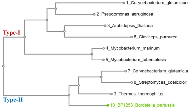
We also noticed from sequence alignment, that BP1253 presents a higher sequence homology towards the LOGs type-II than the LOGs type-I. As described, the cluster type-II of LOG proteins assembles phosphoribohydrolase enzymes with a hexameric form, but we demonstrated (Fig. 5, 6) that the oligomeric state of BP1253 is dimeric. A phylogenetic tree between BP1253 and proteins described in literature to possess phosphoribohydrolase activity and classified as type-I or type-II, indicates how our protein could represent a further sub-group of LOG type-II with a dimeric form (Fig. 8).
34
Figure 8. Likelihood phylogenetic tree between described LOGs and Bordetella pertussis BP1253. The phylogenetic tree was realized with MAFFT and visualized through the software tool Archaeopteryx.js. BP1253 is highlighted in green. The proteins are divided in two branches; one represents type-I LOGs while the other one the type-II LOGs. The dimeric BP1253 is grouped with Type-II LOGs characterized by having a hexameric form.
4.3 BP1253 BINDS NUCLEOTIDES
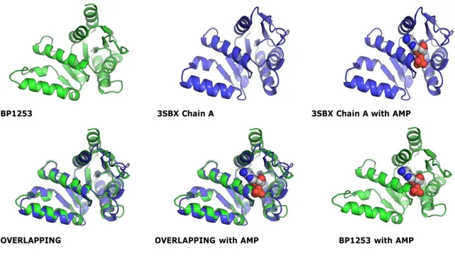
LOG proteins bind and cleave preferentially adenosine 5'-monophosphate (AMP). To verify whether BP1253 is able to bind AMP like the known LONELY GUY proteins, we at first resorted to a theoretical approach overlapping a modeling of BP1253 with a monomer of a Mycobacterium marinum protein classified in the maximum likelihood phylogenetic tree (Seo and Kim 2017) (Fig. 9) as type-I LOG protein with dimeric form (MmLOG). Characteristic of this M. marinum protein with PDB ID 3SBX is that it was crystallized in complex with AMP (Baugh, Phan et al. 2015). The modeling was based on a monomer of Thermus Thermophilus protein TT1465 (TtLOG), PDB ID 1WEK, with which BP1253 shares the 48% of homology. TT1465 is annotated in the phylogenetic tree as LOG type-II. Usually two related structures have a Root Mean
Type-I
35
Squared (RMS) value between 0 – 2Å. In this case the overlap produced a RMS value of 1.21 Å and revealed a great similarity between the 3SBX and BP1253 binding pockets.
Figure 9. Overlapping between monomer structures of MmLOG and a modeling of BP1253. The prediction data obtained from the overlapping gave a RMS value of 1.21 Å and showed the analogy of the two binding sites. The modeling was created through PDB Viewer on the base of 48% homology among BP1253 and TtLOG sequences. The overlapping was realized with PDB Viewer and the figures of single monomers and of overlaps were created via the graphic software PyMOL.
To validate definitively the capability of BP1253 to bind AMP, we performed surface plasmon resonance (SPR) studies. Recombinant BP1253-His was immobilized on the surface of a golden chip CM5 and a ranging concentration from 0.78 to 400 μM of AMP was applied to measure the dissociation constant (kd). Analysis by Biacore indicated that BP1253 binds AMP with a kd of 5.7 µM (Fig. 10) suggesting a high affinity for this compound.
36
Figure 10. Surface plasmon resonance study with AMP. Biacore analysis displays that BP1253 has the ability to bind AMP with a dissociation constant of 5.7 µM. BP1253-His was immobilized via amine coupling procedure on Sensor chip CM5. Concentrations from 0.78 to 400 μM were used to measure the kd. The results shown are representative of three independent experiments giving identical results.
To investigate if this protein is able to bind other monophosphate nucleotides, both purines and pyrimidines such as GMP, IMP, CMP, TMP and UMP were analyzed. Results shown in Fig. 11 indicate that both GMP and IMP are able to bind BP1253 with kd of 38 µM and 71.8 µM respectively; a lower affinity was obtained with TMP and UMP (Table I).
Figure 11. Biacore analysis of GMP and IMP. SPR studies between BP1253 and various monophosphate nucleotides revealed the BP1253 capability to bind GMP and IMP withgood affinity. For GMP and IMP was used a ranging concentration from 0.78 to 400 μM. Identical results were obtained by independent experiments.
37
Table I. Purines and pyrimidines analyzed in surface plasmon resonance studies. List of monophosphate nucleotides evaluated by Biacore on BP1253 and the related kd.
The binding experiment was then extended to other nucleotide metabolites (Tab. II). Among them NAD, NADH and NADP revealed a kd of 0.3 µM for NAD, 0.4 µM for NADH and 4.4 µM for NADP (Fig. 12). For the other substrates a very high kd or no binding was recorded. This suggests a binding capacity also for these metabolites.
Figure 12. Biacore analysis of nucleotide metabolites. SPR investigations performed on nucleotide substrates NAD, NADH and NADP using Sensor chip CM5 and concentration of substrates from 0.097 to 50 μM. Nucleotides Dissociation constant (kd) AMP 5.7 µM GMP 38 µM IMP 71.8 µM TMP 2.4 mM UMP 6.1 mM CMP ND
38
Table II. List of nucleotide metabolites investigated in SPR studies.
4.4 BP1253 HAS PHOSPHORIBOHYDROLASE ACTIVITY
LONELY GUY catalytic activity consists in the hydrolysis of the N-glycosidic linkage between the N6-modified adenine and ribose 5’-monophosphate, leading to the
formation of free and active cytokinins. Shown in Fig. 13 is the reaction catalyzed by phosphoribohydrolases.
Figure 13. Schematic representation of phosphoribohydrolase catalyzed reaction.
Nucleotide metabolites
NAD Nicotinamide adenine dinucleotide
NADH Nicotinamide adenine dinucleotide reduced
NADP Nicotinamide adenine dinucleotide phosphate
α-NAD α-Nicotinamide adenine dinucleotide
ε-AMP ε-Adenosine monophosphate
ADPR Adenosine diphosphoribose
ATP Adenosine triphosphate
NGD Nicotinamide guanine dinucleotide
39
Sequence alignments with LOG proteins showed that BP1253 has a high amino acid sequence identity with type-II LOGs and that shares with all of them the PGGxGTxxE motif and the catalytic residues. Additionally, the SPR analysis revealed the capability of the pertussis protein to bind AMP. These results led us to hypothesize that BP1253 could be a LOG protein. To examine the biochemical activity of BP1253, we performed phosphoribohydrolase activity assay. As substrate we used AMP, described to be the preferred substrate of LOG (Samanovic, Tu et al. 2015, Seo, Kim et al. 2016, Seo and Kim 2017, Seo and Kim 2018, Seo and Kim 2018). AMP and the product of the reaction adenine run at different height for their different hydrophilicity on TLC and are visible as spots under UV light at 264 nm. In dose-response experiment AMP was incubated at 30°C with different concentration of the protein and the reaction resulted BP1253 dose-dependent as shown in Fig 14 A. In the time course experiment, the substrate was incubated with 24 µM of enzyme and the reaction stopped at different time points. It resulted that the catalytic activity increased over the time (Fig.14 B). As negative control for the experiments was used the heat inactivated protein (Fig.14 C).
Figure 14. Dose-response and time-course experiments reveal phosphoribohydrolase activity in BP1253. The experiments were performed at 30°C in the presence of 20 mM AMP as substrate. After heat inactivation of the protein at 95°C for 2 min, the products were separated by TLC in 1 M NaCl using Cellulose F plastic sheets. The dots were evidenced under UV light at 264 nm. (A) Dose-response experiments carried out after 6 hours of incubation. The BP1253 concentrations used were 6, 12, 24 and 48 µM. (B) Time course assay carried out with 24 µM of enzyme at different incubation time. (C) TLC with the heat-inactivated protein as negative control performed after 8 hours of incubation.
40
Since the SPR study for GMP resulted in a dissociation constant of 38 µM comparable to that of AMP, in order to investigate if BP1253 is able to cleave the N-glyosidic linkage in GMP, we decided to carry out the enzymatic assay using GMP as substrate (Fig. 15).
Figure 15. GMP is cleaved by BP1253. The assays were performed at 30°C in the presence of 20 mM GMP. To improve the solubility of GMP, the inactivation was performed with 1 M NaOH (v/v) and then the products of reaction separated by TLC in 1 M NaCl using Cellulose F plastic sheets. The dots were visible under UV light at 264 nm. (A) Dose-response experiments carried out in 20 minutes. 1.2, 2.4 and 4.8 µM were the enzymatic concentrations used for the assay (B) Time course experiment performed with 2.4 µM of BP1253 at different incubation time.
As clearly shown in Fig. 15, BP1253 recognizes GMP as substrate showing phosphoribohydrolase activity. Of note in the assays with GMP we used a 10-fold lower concentration of enzyme compared to that one used for the AMP. The substrate was almost completely hydrolyzed after 30 minutes of incubation indicating GMP a substrate more efficient compared to AMP for BP1253. For a more complete characterization of BP1253 we examined the protein activity with several ribonucleotides and deoxyribonucleotides (Tab. III). Among them a high phosphoribohydrolase activity was recorded also for CMP (Fig. 16). This partially explains the high kd measured by biacore for CMP, the substrate is so fast cleaved that appears as no binding was present.
41 Nucleotides 20 minutes 24 hours
AMP - + GMP ++ ++ IMP + ++ CMP ++ ++ TMP - + UMP - - dAMP - - dGMP - + dCMP + ++ dTMP - + dUMP + ++
Figure 16. BP1253 cleaves CMP. The assay was carried out at 30°C in the presence of 20 mM CMP for 20 minutes. The inactivation was performed at 95°C for 2 min. To separate the products by TLC on Cellulose F plastic sheets the mobile phase was acetone/water 30/70 (v/v) with the addition of 0.2 mol/l NaCl. The dots were visible under UV light at 264 nm.
Table III. Ribonucleotides and deoxyribonucleotides tested with BP1253 in phosphoribohydrolase assay. The different nucleotide substrates were incubated with 2.4 µM of BP1253 for 20 minutes and 24 hours at 30°C. The reaction was blocked with 1 M NaOH for those compounds having guanine as nitrogen base, whilst by heat at 95°C for 2 minutes for the others. The products were separated by TLC on Cellulose F plastic sheets. We used as mobile phase 1 M NaCl for purine separation while, the mobile phase for pyrimidines was acetone/water 30/70 (v/v) with the addition of 0.2 mol/l NaCl. The dots were visualized under UV light at 264 nm. We registered different activities from BP1253 depending on the incubated substrates. We indicate with - those reactions for which no phosphoribohydrolase activity was recorded, as + we specify reactions with a minimal activity, as ++ we identify high catalytic activity.
42
As previously described BP1253 contains the typical amino acids of LOG catalytic core. Therefore, to probe if the R120, E143 residues and K121 probably involved in substrate binding, may have a role in enzymatic activity, we generated by site-specific mutagenesis a panel of recombinant BP1253 mutant proteins. In these single point mutants R120, E143 and K121 were changed first with Ala and then with the conservative amino acid: Lys, Asp and Arg respectively. Among the amino acids implicated in the binding, we chose the K121 residue for its closeness to the catalytic amino acid R120. The mutants were expressed as His-tagged recombinant proteins in BL21 competent E. coli and purified by two step of high-throughput immobilized metal (Ni2+) affinity chromatography. SDS-PAGE analysis and western immunoblotting confirmed the identity of the purified single point mutant proteins (Fig 17).
Figure 17. Purification profile of BP1253 mutants. BP1253 variants with specific site mutations with non-conservative and conservative amino acids were cloned in E. Coli BL21 (DE3) and purified in high-throughput through a His Multitrap HP 96-well Vacuum plate after IPTG induction. (A) SDS-PAGE (2 µg of proteins) and Immunoblotting (20 ng of proteins) performed on R120A, K121A and E143A. Mouse polyclonal α-BP1253 (1:500) was used as the primary antibody. Molecular weights of proteins markers are shown on the left. (B) SDS-PAGE (2 µg of proteins) and Immunoblotting (20 ng of proteins) carried out on R120K, K121R and E143D. Molecular mass markers (kDa) are indicated on the left and the mouse polyclonal α-BP1253 (1:500) was used for the WB analysis.
After purification, we tested the R120A, K121A and E143A mutant proteins with the substrates GMP (Fig. 18 A, B panel) and CMP (Fig. 18 E, F panel) in phosphoribohydrolase assays at 20 and 40 minutes. The non-conservative amino acid substitutions resulted in a complete loss of the enzymatic activity. When we tested conservative, R120K and E143D mutants resulted in a total absence of activity, while
28 17 kDa 28 17 kDa A B
43
the replacement of K with R at the residue 121 produced a minimal hydrolysis with GMP and CMP as substrates though not comparable to wild type (Fig. 18 C, D, G, H). These results confirm that Arg 120 and Glu 143 are the catalytic residues implicated in the phosphoribohydrolase activity and that the positive charge of the K at 121 residue is essential for the catalysis. AMP was assayed with conservative mutants for a time of 6 and 18 hours (Fig. 18 I, J). Also in these cases, R120K and E143D replacements caused the loss of the enzymatic activity and, K121R a hydrolysis almost undetectable on TLC. The results from the mutagenesis are in agreement with the recently described LOG proteins and this confirms that BP1253 is a member of the LOG family and, henceforth, we will refer to it as BpLOG.
A
C
B
44
Figure 18. Phosphoribohydrolase activity assays with recombinant BP1253 mutant proteins. The experiments were all performed at 30°C in the presence of 20 mM of substrate. The incubation time was 20 and 40 minutes for GMP and CMP, and 6 and 18 hours for AMP. The reaction was blocked by heat inactivation for enzymes incubated with CMP and AMP and, with 1 M NaOH (v/v) for the proteins incubated with GMP. The products were separated by TLC on Cellulose F plastic sheets and the mobile phase was NaCl for purine and acetone/water 30/70 (v/v) with the addition of 0.2 mol/l NaCl for CMP. The dots were evidenced under UV light at 264 nm. As positive control we used wild type BP1253. (A) Non-conservative mutations incubated with GMP for 20 minutes and (B) for 40 minutes. (C) Conservative substitutions assayed with GMP for 20 minutes and (D) 40 minutes. (E) Alanine substitutions tested with CMP for 20 minutes and (F) for 40 minutes. (G) Conservative mutants assessed with CMP for 20 minutes and (H) 40 minutes. (I) Conservative replacements incubated with AMP for 6 hours and (J) 18 hours. For positive control in AMP experiments, refer to the figure number 14.
G H
I J
45
4.5 BpLOG SYNTHESIZES 6-O-METHYLGUANINE
BpLOG being a member of the LOG family and in analogy with its homologous proteins should be able to produce cytokinins. Therefore, we decided to try to identify the physiological product of BpLOG. For this purpose, we generated from Tohama I-derivative BP536 a Bordetella strain deleted of the operon in which is located Bp1253 gene in order to compare the production of cytokinins between the wild type strain and the knockout. (Bp1253 gene is located inside an operon along with other two genes,
Bp1251 and Bp1252 that encode for a putative toxin and putative exported protein
respectively. However, currently, the real function of both proteins is unknown). The attempt of this investigation is to identify the produced cytokinins by the two strains, to separate them in reverse phase through HPLC and to visualize them as peaks via UV detector exploiting the intrinsic capability of these compounds to adsorb UV light. In plants the cytokinins are hormones, therefore, they are produced in small quantities and released in the extracellular space. For this reason, we focused our attention first on supernatant. The knockout and the wild type strains were grown in Stainer-Scholte medium at 37°C until 48 hours, after that the supernatants were filtered, cleared by proteins and molecules with molecular weight higher than cytokinins as described in Methods (Dobrev, Hoyerova et al. 2017). At the end of the purification procedure, the dried samples, re-suspended in 100 μL 5 % methanol were run in HPLC and analyzed in MS. The two chromatographic spectra related to Tohama I strain supernatant and to Δ1253 supernatant arising from UV detector were identical, but we observed that at retention time of 5 minutes in wild type spectrum there was a small peak completely absent in Δ1253 (Fig. 19). This peak was isolated and analyzed in MS. It turned out that this peak corresponded to a compound with molecular weight of 165 Da, 166 Da in the mass chromatogram because of electrospray with a positive ion mode used as mass spectrometric detection, and with an apparent molecular formula of C6H7N5O. In data
banks the compound with this molecular formula that fitted better with our study was the 6-O-Methylguanine. We tested it on HPLC at the same conditions used for supernatants samples and noted that the retention time of 6-O-Methylguanine was very close to retention time of BpLOG physiological product (Fig. 19)