PhD School of Neuropharmacology
XXVI Cycle
ROLE OF ACETYL-L-CARNITINE IN HEPATIC
ENCEPHALOPATHY
Doctorate Thesis
Michele Malaguarnera
Coordinator: Prof. Salvatore Salomone
Tutor: Prof. Fabio Galvano
Co-Tutor: Prof. Filippo Caraci
3
Acknowledgements
I wish to express my sincere gratitude to:
Professor Fabio Galvano, my tutor, for sharing his knowledge and for introducing me to the field of nutrition.
Professor Filippo Caraci, my co-tutor, for his essential contribution for this thesis a for continous support.
Professor Filippo Drago, for introducing me to the field of neuroscience and for his precious suggestions.
Professor Giovanni Li Volti, Professor Claudia Di Giacomo and Dottor Ezio Barbagallo for the knowledge and the availability to teach during my labs work of these years
Doctor Vicente Felipo and Doctor Marta Llansola for following me during this last year in the Neurobiology Lab, “Centro de Investigación ‘Principe Felipe’”, Valencia, Spain.
The colleagues and friends at the Neurobiology Lab for the kindness and an environment that I wish to find in the following years.
4
Professor Giovanni Pennisi, Professor Rita Bella, Professor Liborio Rampello, Professor Giuseppe Nunnari, Doctor
Marco Vacante, Professor Massimo Motta and
doctor Maria Giordano for the precious collaboration
And last but not least, My family and Andrea Cabrera-Pastor, my girlfriend, for their love and the continuous support during my PhD program.
List of abbreviations
HE = Hepatic Encephalopathy
MHE = Minimal Hepatic Encephalopathy ALF = Acute liver failure
PCS = Portacaval Shunt CBF = Cerebral Blood Flow BBB =Bloood Brain Barrier EEG = Electro Encephalogram GS = glutamine synthetase
mGluR = metabotropic glutamate receptor NAc = the nucleus accumbens
MDT = mediodorsal thalamus VMT = ventromedial thalamus ATP = Adenosine Triphosphate
5
ICP = intracranial pressure CSF = cerebrospinal fluid
BCAA = branched chain amino acids
Preface
Hepatic encephalopathy (HE) is a debilitating complication of cirrhosis which presents as a spectrum of neurological and
neuropsychiatric dysfunction, affecting the patients
consciousness, intellect, personality and neuromuscular activity. Hepatic encephalopathy as a complication of cirrhosis leads to physical complication that affect function and performance of daily life such as fatigue, muscle cramps and asterixis, to neuropsychological complication such as shortened attention, disorientation in time or space, changes in personality and inappropriate behavior.
Understanding the various factor that affect the patient’s quality of life depends in our realization of the multifaceted issues of cirrhosis and their effects on health status
6
DESIGN OF PRESENT RESEARCH
Design of the present researchThe present thesis has focused on:
1) Elucidate pathophysiological mechanism of HE to date 2) The role of Acetyl-L-Carnitine
3) Examine the role of Acetyl-L-Carnitine in neurocognitive symptoms of HE
4) Examine the role of Acetyl-L-Carnitine in fatigue symptoms of HE
7
9
INTRODUCTION
Epidemiology
When the liver fails, owing to acute liver failure (ALF) or chronic liver disease such as chronic hepatitis or cirrhosis, the normal detoxification of endogenous and exogen compound is compromised and those compounds may reach the brain and affect cerebral function (Felipo 2013). (Felipo 2013) Although the incidence of ALF is low (around 2,000 people per year in the United States or Europe), mortality rates remain high. By contrast, chronic liver diseases affect 5.5 million individuals in the United States alone.
(Rose 2012) MHE (Covert Hepatic Encephalopathy) is characterized by decreased attention, poor concentration, impaired memory, sleep disturbances, reduced speed of information processing, and altered motor abilities. In addition, the subclinical cognitive impairment that characterizes MHE increases the risk of having a car accident. As such, MHE has a significant impact on patients’ health-related quality of life and their ability to carry out day-to-day functions.(Stewart el al. 2007) As many as 80% (20–80%, depending on the severity of the disease) of patients with chronic liver disease may have MHE. Its presence also identifies patients with a fourfold higher risk of developing OHE. (Poordad, et al. 2007)
(Rosso Stepanova 2010) Overt HE occurs in approximately 30%– 45% of patients with cirrhosis, while minimal HE may
10
affect up to 60% of patients with chronic liver disease and up to 80% with cirrhosis. (Stepanova, et al. 2010)
The definitive data on the true incidence and prevalence of HE is lacking, mainly because of large differences in the etiology and severity of HE and relevant issues in diagnosing minimal HE (Lewis M and Howdle PD 2003; Poordad, et al. 2007; Randolph et al. 2009). Development of HE is associated with a poor prognosis. Specifically, in the presence of chronic liver disease, HE typically heralds hepatic decompensation, and its development is usually associated with high mortality, indicating the need for liver transplantation. (Benhaddouch Z, et al. 2007 ; Udayakumar N, et al. 2007 ; Findlay JY, et al. 2011; Bustamante J, et al. 1999).
Classification
Patients with clinical HE are classified into four grades. Patients in grade 1 show a trivial lack of awareness; experience euphoria or anxiety; have a shortened attention span; and have impaired performance in basic arithmetic, such as addition or subtraction. In grade 2, symptoms include lethargy or apathy; minimal disorientation in time or space; subtle changes in personality; and inappropriate behaviour. Patients in grade 3 experience somnolence to semistupor but remain responsive to verbal stimuli. They also experience confusion, gross disorientation and
11
exhibit bizarre behaviour. Patients in grade 4 are comatose. (Conn HO, et al. 1977)
In the last years, HE has been classified in covert and overt, the first one does not present clinical signs of the overt HE. Covert HE shows mild cognitive impairment, attention deficits, psychomotor slowing and impaired visuomotor and bimanual coordination. (Felipo V, 2013)
Pathophysiology
HE is characterized by neurocognitive and neuromotor impairment, this pathology measibly affect the quality of life of HE patients.
There is a general consensus that indicate ammonia and inflammation as a major characters in HE pathophisiology. The two main underlying factors are hyperammonaemia and inflammation. The only mechanism for ammonia detoxification in the brain involves its incorporation into glutamine by glutamine synthetase, an enzyme that is present in the brain only in astrocytes. It has been proposed that glutamine accumulation in astrocytes owing to ammonia detoxification results in water entry and increased osmotic forces, which leads to astrocyte swelling and cytotoxic oedema. Glutamine-induced astrocyte swelling was considered to be a main mediator in all types of HE (Felipo V 2013).
12
Ammonia
Ammonia by-product of nitrogen metabolism, is produced mainly within the gut through the deamination of glutamine by glutaminase in the enterocytes of the small intestine and colon, as well as through the hydrolysis of urea, catalyzed by urease-producing bacteria that exist abundantly in the human gut. Gut-derived ammonia is transported and absorbed across the mucosal epithelium into the hepatic portal circulation, from which, in the case of a healthy liver, it is removed primarily through the urea cycle. This low-affinity, high-capacity ammonia detoxication system is present in the periportal hepatocytes located around the portal vein. Glutamine synthetase, another important ammonia-removing pathway located in the liver, catalyzes the conversion of glutamate into glutamine, thereby removing an ammonia molecule. This high-affinity, low-capacity reaction takes place in the perivenous hepatocytes located around the hepatic vein and acts as a scavenger for the ammonia that escapes periportal urea synthesis. The production of ammonia within the gut and its detoxication by the liver are the main pathways through which ammonia homeostasis is maintained in the body. However, other organs also contribute to ammonia metabolism. In addition to the liver and intestines, glutamine synthetase is found in the muscles and the brain (particularly in the astrocytes) and in phosphate-activated glutaminase in the kidneys and the brain (primarily in the neurons). In the presence of a healthy liver, blood ammonia levels are maintained in the low range of 35–60 µmol/l (Figure 2). However, during liver disease, given the reduced hepatic capacity for ammonia removal, the extrahepatic interorgan
13
ammonia metabolism is altered (including glutamine metabolism) (Wright G , et al. 2011), thus upsetting the balance between ammonia-producing/removing organs and ammonia homeostasis (Figure 2) ( Rose 2012) However, other organs also contribute to ammonia metabolism. In addition to the liver and intestines, glutamine synthetase is found in the muscles and the brain (particularly in the astrocytes) and in phosphate-activated glutaminase in the kidneys and the brain (primarily in the neurons).. This results in a two to fivefold increase in blood ammonia, leading to an increase in ammonia levels in the brain, with deleterious consequences (Bosoi CR and Rose CF 2009 ; Felipo V and Butterworth RF 2002)
Inflammation
Neuroinflammation also contributes to encephalopathy and brain oedema in rats with ALF (Acute Liver Failure) (Jiang W et al., 2009) . The mechanisms and time course of these changes are different in various brain regions: ALF in rats increases cerebral blood flow (CBF) in the cortex but reduces it in the cerebellum. In patients with ALF, inflammation, cerebral oedema and increased CBF and lactate contribute to ICP and death (Jalan R, et al. 2004)
Hyperammonaemia per se induces neuroinflammation. Rats with chronic hyperammonaemia but without liver failure show microglial activation and neuroinflammation, especially in the cerebellum. Treatment of these rats with anti-inflammatory drugs restores cognitive function (Cauli O, et al. 2009) , which
14
suggests that hyperammonaemia-induced neuroinflammation has a major role in neurological impairment observed in MHE. In cultured microglia, ammonia upregulates the microglial activation marker allograft inflammatory factor 1 (also known as IBA1), which is also increased in the brain of patients with HE (Zemtsova I, et al. 2009) . Therefore, ammonia may act directly on microglia to induce their activation but could also induce neuroinflammation through peripheral effects that are then transduced to the brain. This may occur by transfer of pro-inflammatory cytokines from blood to the brain in the circumventricular organs in which the BBB is more permeable, or by direct infiltration of blood immune cells. Blood cytokines may also stimulate receptors on endothelial cells and trigger the release of inflammatory factors into the brain. Rats with MHE show neuroinflammation and cognitive and motor alterations that are reversed with anti-inflammatory drugs (Cauli O, et al. 2009; Cauli O, et al. 2007; Rodrigo R, et al. 2010). Inflammation exacerbates the cognitive deficits induced by hyperammonaemia (Marini JC and Broussard SR 2006), and hyperammonaemia has been shown to reduce motor coordination in rats with inflammation (Jover R, et al. 2006). The combination of hyperammonaemia and inflammation over a certain threshold induces mild cognitive impairment in humans even in the absence of liver disease (Felipo V, 2012). Together, the results from these studies suggest that targeting neuroinflammation may restore cognitive and motor function in patients with MHE (Felipo V, 2013).
15
Urea Cycle
(Felipo-Butterworth 2002) Hyperammonemia is associated with profound effects on Cerebral Brain Flood (CBF). These effects are dependent upon the severity and duration of hyperammonemia and show region selectivity. For example, in chronic mild hyperammonemia associated with liver cirrhosis, CBF is decreased in proportion to the deterioration of neuropsychiatric status (Posner JB and Plum F, 1960; James IM and Garassini M, 1971). Some studies demonstrated that there is a regional selectivity of the cerebral metabolic changes in hyperammonemia like a reduction in cortical structures and a concomitant increase in some sub-cortical areas (O’Carroll RE et al., 1991). Intracarotid infusions of ammonia sufficient to cause EEG slowing were found to result in increased cerebral metabolic rate for the glucose, which was confined to deep grey matter structures (Lockwood AH et al., 1982).
Since brain lacks carbamoyl-phosphate synthase I and ornithine transcarbamylase, it is unable to remove ammonia in the form of urea. Consequently, brain ammonia is metabolized almost exclusively to glutamine via the GS reaction. Glutamine synthesis remains the predominant route for ammonia removal in brain under both normal and hyperammonemic conditions (Cooper AJ and Plum P, 1987). In the brain, Glutamine synthetase is located only in astrocytes (Norenberg MD and Martinez-Hernandez A, 1979). Thus, it is the astrocyte rather
16
than the neuron that is uniquely responsible for ammonia detoxification in brain. Surprisingly, in contrast to peripheral tissue such as skeletal muscle, there is no significant induction of GS expression in brain in hyperammonemic states (Cooper AJ et al., 1985; Lavoie J et al., 1987). Moreover, , it is important to underline that since the enzyme functions at near maximal capacities under normal physiological conditions (Cooper AJ and Plum P, 1987).
Brain Energetic metabolism
Ammonia causes significant alterations of mitochondrial function and, consequently, changes in cerebral Energy metabolism. Ammonia stimulates glycolysis in brain extracts by activation of phosphofructokinase (Sugden PH and Newsholme EA, 1975) and acute ammonia toxicity in normal rats leads to brain ammonia concentrations in the 1.4–1.5 mM range resulting in increased brain glucose utilization (Hawkins RA et al., 1973). Increased brain glucose concentration in acute hyperammonemia may be the consequence of an increased in expression of the endothelial cell/astrocytic glucose transporter GLUT-1 as was recently reported in experimental ALF (Desjardins P et al., 2001). Increased brain glucose uptake was accompanied by increased brain lactate concentrations, which occurred without any loss of high Energy phosphates. The increased brain glucose uptake and lactate accumulation due to acute ammonia exposure appears to be predominantly an astrocytic phenomenon since expression of the neuronal glucose
17
transporter GLUT-3 is not affected by ammonia (Desjardins P et al., 2001).
During hyperammonaemia, it was also reported a reduction in ATP (McCandless DW and Schenker S, 1981; Kosenko E et al., 1994), but several groups have found but most of these articles show this reduction in animal models exposed to an hyperamonemia of 3 mM, similar or sometimes higher than an hepatic coma. Infact, there is little convincing evidence to suggest that hyperammonemia resulting from acute or chronic liver failure results in a loss of ATP in brain at least until stages of encephalopathy characterized by prolonged coma (Mans AM et al., 1994; Hindfelt B et al., 1977). Likewise, studies in cirrhotic patients with end-stage liver failure using spectroscopic techniques have so far not provided convincing evidence for a primary cerebral energy deficit (Taylor-Robinson SD et al., 1994; Lockwood A et al., 1997).
Two distinct mechanisms have been proposed to explain ammonia-induced reductions in brain ATP:
(1) inhibition of the tricarboxylic acid cycle (TCA);
(2) a mechanism involving N-methyl-d-aspartate (NMDA) receptors.
In favour of the first mechanism, McKhann and Tower (1961) reported ammonia-induced inhibition of the TCA cycle in brain with accumulation of alpha-ketoglutarate and pyruvate. Subsequently, Lai and Cooper (1986) described a significant inhibition of the rate-limiting TCA cycle enzyme alpha-ketoglutarate dehydrogenase (alpha-KGDH) in brain mitochondrial preparations exposed to ammonia with an EC50 of
18
2 mM. Consistent with alpha-KGDH inhibition and a consequent reduction in entry of pyruvate into the tricarboxylic acid cycle are the findings of increased brain lactate concentrations in various hyperammonemic disorders (Hawkins RA et al., 1973; Hindfelt B et al., 1977; McCandless DW and Schenker S, 1981; Therrien G et al., 1991; Mans AM et al., 1994; Chatauret N et al., 2001). Furthermore, hypoxia significantly exacerbates the effects of lethal injections of ammonium salts in mice (Warren and Schenker, 1960). Cerebrospinal fluid lactate concentrations are increased in both acute (Chatauret N et al., 2001) and chronic (Therrien G et al., 1991) liver failure and are positively correlated with the severity of HE in these disorders. Increased CSF lactate has also been reported in human HE (Yao H et al., 1987). According to this hypothesis involving inhibition of the TCA cycle by ammonia is the report that increased ammonia levels in animals injected with U 14C glucose resulted in a reduction in the amount of label incorporated into the amino acids glutamate and GABA (Prior RL and Visek VJ, 1972). In support of a pathogenetic role of NMDA receptors, it has been shown that ammonia-induced depletion of brain ATP in vivo is prevented by administration of a wide range of glutamate (NMDA) receptor antagonists (Kosenko E et al., 1994). Based upon these observations, it was suggested that ammonia-induced activation of NMDA receptors results in ATP depletion via the activation of Na+, K+, ATPase as well as by decreased synthesis of ATP due to impairment of Ca2 + homeostasis (Kosenko E et al., 2000).
19
There are several possible explanations for the absence of cerebral energy deficit in chronic liver failure.
1) Proliferation of astrocytic mitochondria has been reported in conditions of chronic hyperammonemia (Gregorios JB et al., 1985; Norenberg MD and Lapham LW, 1974), a phenomenon that has been attributed to increased energy requirements.
2) It has been reported that chronic hyperammonemia similar in magnitude to that observed in end-stage chronic liver failure leads to down-regulation of functional NMDA receptors (Peterson C et al., 1990; Marcaida G et al., 1995).
3) The impairment of signal transduction pathways associated to NMDA receptors could determinate the lack of energy depletion in chronic hyperammonemia (Hermenegildo C et al., 1998).
Neurotrasmission
Alterations in glutamatergic and GABAergic neurotransmission in different brain regions contribute to altered motor function in MHE. The neuronal circuits between the basal ganglia, thalamus and cortex that modulate motor activity are altered in patients with MHE as well as in rats with hyperammonaemia and MHE, and lead to motor impairment, including hypokinesia (Cauli O, et al. 2006; Agusti A, et al. 2011). In vivo microdialysis studies show that in control rats, metabotropic glutamate receptor (mGluR) activation in the nucleus accumbens (NAc) increases
20
the level of extracellular dopamine, which activates dopamine receptors, inducing GABA release in the ventral pallidum and a reduction of GABA levels in the mediodorsal thalamus (MDT). In turn, this reduced inhibition by GABA leads to increased glutamate release in the cortex, resulting in increased motor activity. However, hyperammonaemia or MHE lead to the activation of an ‘alternative’ circuit involving the NAc, the SNr and the ventromedial thalamus (VMT). In rats with MHE, mGluR activation in the NAc does not increase extracellular dopamine levels but does increase glutamate levels, which activates AMPA receptors, inducing GABA release in the SNr and a reduction of GABA levels in the VMT. This reduced inhibition by GABA results in an increased level of extracellular glutamate in the cortex and enhances motor activity in rats with MHE but not in control rats (Cauli O, et al. 2006; 2007b).
Under normal conditions (without the exogenous activation of mGluRs in the NAc described above), rats with MHE show less spontaneous motor activity than normal rats and similar motor impairments to those observed in patients with HE (hypokinesia). This hypokinesia is due to increased extracellular glutamate levels in the substantia nigra pars reticulata (SNr) of rats with MHE, which results in excessive mGluR1 activation, which then increases extracellular GABA levels in the VMT and reduces extracellular glutamate levels in the cortex. Blocking mGluR1 by stereotaxic injection of the selective antagonist CPCCOEt in the SNr normalizes extracellular GABA levels in the VMT and glutamate levels in the cortex, and eliminates hypokinesia , supporting the idea that excessive levels of
21
extracellu-lar glutamate and activation of mGluR1 in the SNr are responsible for hypokinesia. A main contributor to increased extracellular glutamate levels in the SNr of MHE rats is the reduced amount and function of the glutamate transporters EAAC1 (also known as excitatory amino acid transporter 3) and GLT1 (also known as excitatory amino acid transporter 2) in the SNr. The amount of glutamate transporter is normalized and extracellular glutamate levels are reduced in rats with MHE by administration of an anti-inflammatory drug (ibuprofen) that reduces neuroinflammation and also eliminates hypokinesia. These findings demonstrate that, as mentioned above, both hyperammonaemia and neuroinflammation contribute to motor alterations in MHE. Although non-steroidal anti-inflammatory drugs such as ibuprofen are not recommended in cirrhotic patients, owing to risk of secondary effects on the kidneys, inhibitors of p38 also reduce neuroinflammation and eliminate hypokinesia in rats with MHE and may improve motor function in patients with MHE or clinical HE.
Activation of the glutamate ionotropic receptor N-methyl-D-aspartate (NMDA) has been demonstrated to play an important role in the pathophysiology of hepatic encephalopathy (Llansola M, et al. 2007). The opening of the channel is controlled by a powerful voltage-dependent block by external magnesium ions (Mayer ML, et al. 1984; Nowak L, et al. 1984). It is believed that ammonia, by raising the membrane potential,removes the magnesium block rendering NMDA receptors susceptible to activation. However the mechanism and the degree in which is
22
involved ammonia in the removing of the magnesium ion block in these receptors has not been clarified. Pathophysiological concentrations of ammonia directly raise the membrane potential in both astrocytes and neurons, however, not sufficiently to activate voltage-gated channels or generate an action potential in neurons (Bosoi CR and Rose CF 2009). These mechanisms could effectively minimize the impact of mechanism involving N-methyl-d-aspartate (NMDA) receptors and prevent loss of ATP due to NMDA receptor activation. Consistent with these possibilities, it has been shown that chronic moderate hyperammonemia in rats completely prevents the depletion of ATP induced by subsequent acute lethal injections of ammonia (Kosenko E et al., 1993).
Studies in rats support the idea that cerebral damage in ALF involves an initial disruption of BBB permeability leading to vasogenic oedema in certain areas such as the cerebellum but not in the frontal cortex. At early stages of ALF, oedema is mainly vasogenic and is associated with increased intracranial pressure (ICP) (Cauli O, et al. 2011) , indicating that astrocyte swelling is not the initial trigger of oedema and ICP. As brain ammonia and glutamine progressively increase, cytotoxic oedema (which is probably due to astrocyte swelling) ensues in many areas, further increasing ICP.
At least 16 enzymatic pathways in brain result in the formation of ammonia. One of the most important is glutamate dehydrogenase, which catalyses the reversible oxidative deamination of glutamate. It has been proposed that both in
23
dehydrogenase is ammonia producing, particularly in astrocytes and, in this way, may provide a mechanism for the removal of excess nitrogen from certain catabolyzed amino acids (Cooper AJ and Plum P, 1987). A catabolic role for glutamate dehydrogenase is also consistent with the finding of decreased brain glutamate concentrations in a wide range of hyperammonemic syndromes (Lavoie J, et al., 1987; Swain M, et al. 1992; Ratnakumari L, et al. 1994). L-Glutaminase is widespread in brain and is particularly abundant in nerve endings of glutamatergic neurons where it forms an integral part of the glutamate-glutamine cycle in which a molecule of ammonia is transferred from the astrocyte to the neighbouring neuron. There is evidence to suggest that at least part of the increased glutamine encountered in brain in hyperammonemia results from inhibition of glutaminase (Tyce GM, et al. 1981). Enzymes of the purine nucleotide cycle may also be responsible for generating a significant fraction of brain ammonia (Schultz V and Lowenstein JM, 1978).
NMDA receptors modulate learning and memory, and the glutamate–nitric oxide–cGMP pathway has an important role in these processes. In rats with MHE, reduced functioning of this pathway in the hippocampus leads to impaired long-term potentiation (LTP) and spatial learning in the Morris water maze. Furthermore, reduced signalling through this pathway in the cerebellum has been reported in rats with hyperammonaemia or MHE in vivo and correlates with impaired learning of a conditional discrimination task in a Y maze. In patients that died for HE, lower cGMP formation in the cerebellum is thought to be
24
involved in their reduced learning ability. Restoration of extracellular cGMP levels in the cerebellum restores learning in rats, and this can be achieved by inhibiting phosphodiesterase 5. Studies on the mechanisms by which HE impairs signalling through the glutamate–nitric oxide–cGMP pathway have identified other modulators of the cGMP pathway that could be pharmacologically targeted to restore learning.Chronic hyperammonaemia has also been shown to increase tonic activation of NMDA receptors in the cerebellum, leading to the activation of calcium/calmodulin-dependent protein kinase II (CaMKII) and increased phosphorylation of neuronal nitric oxide synthase (NOS) on Ser847. This reduces the enzyme’s activity and, thus, the formation of nitric oxide and cGMP. Chronic hyperammonaemia also leads to a subcellular redistribution of NOS, as reduced amounts reach synaptic membranes. As a result, activation in response to NMDA receptor activation is reduced, and nitric oxide and cGMP formation is further decreased. Increases in tonic activation of GABA A receptors in the cerebellum have also been reported, leading to reduced functioning of the pathway and cGMP formation. Blocking GABA A receptors with bicuculline restores signalling and learning in rats. Neuroinflammation has also been shown to mediate some of the effects of hyperammonaemia on the glutamate–nitric oxide–cGMP pathway. Treatment of MHE rats with anti-inflammatory drugs (ibuprofen) reduces microglial activation and neuroinflammation in the cerebellum and restores learning . Microglial activation and neuroinflammation could also be reduced with MAPK p38
25
inhibitors, which have been shown to restore learning ability in rats with HE.
Cerebral Oedema
A large amount of studies demonstrated that cerebral oedema is implicated in all of the forms of HE, but in most of these studies was used an high ammonia concentration that does not reflect the concentration found in chronic HE patients (Felipo 2013). In vivo data in patients with chronic liver disease do not support a role for astrocyte swelling in their HE. The findings from functional MRI (fMRI) studies in patients with chronic liver disease and HE suggest that they develop vasogenic oedema, as an increase in the apparent diffusion coefficient was detected rather than a decrease (which would be expected in cytotoxic oedema). Thus, cytotoxic oedema is unlikely to be the main cause of neurological alterations in MHE. Rats with MHE also show cognitive and motor alterations in the absence of oedema.
Other causes
in addition to ammonia, chronic liver failure results in the accumulation of other toxins including manganese (Pomier Layrargues G, et al. 1995), mercaptans and short-chain fatty acids (Zieve L, et al. 1974), all of which may have deleterious effects on brain function.
The role of Acetyl-L-Carnitine in the treatment of HE
L-Carnitine (LC), acylcarnitines, and various carnitine enzymes constitute the carnitine system that play a pivotal role in cellular energy production. The system is ubiquitous
26
and the mitochondrial carnitine system has an obligatory role in beta oxidation of long-chain fatty acids by their transport into the mitochondrial matrix. LC and its esters are present in different concentrations in human serum (L-carnitine/acetyl-L-carnitine / propionil-L-(L-carnitine/acetyl-L-carnitine = 5:1:0,1). Carnitines are involved in the removal of accumulated toxic fatty acyl-CoA metabolites and helping in the balance between free and acyl-CoA. The toxic effects of poorly metabolized acetyl groups can be lowered with transesterification from CoA and excretion of ALC esters by carnitine acetyltransferase (CAT) and carnitine palmitoyltransferases (CPT-1 and CPT-2).
L-carnitine was first used in Reye Syndrome, a syndrome characterized by a deficiency of Carnitine. Reye syndrome is biochemically distinct from the clinically similar syndromes of systemic carnitine deficiency. In this syndrome patients show an impaired liver function and some signs similar to hepatic encephalopathy such as increased ammonia levels and a damaged brain function.
One of the first pathophysiological explanation proposed for hepatic encephalopathy was the altered brain metabolism caused by a decrease in ATP concentration due to a decreased ATP production because of non-optimal operation of the Krebs cycle and inhibition of the mechanism for introducing reducing equivalents into mitochondria (O’Connor JE, et al. 1984). On the basis of this evidence L-carnitine was tested in mice with an acute ammonia intoxication. L-carnitine showed decreased ammonia concentration in these mice finally increasing the
27
survival rate (O’Connor JE, et al. 1984). The possible explanation of the lowering ammonia concentration effect of L-Carnitine was also explained with the blocking of malate aspartate shuttle leading to increase ATP production via oxidative phosphorylation and also for the increase of Acetyl CoA due the avaibility of acetylic moieties for the increase of the beta-oxidation reaction in the mitochondria. Acetyl CoA should enhance the synthesis of N-acetylglutamate, the physiological activator of carbamyl phosphate synthetase I, and thus urea synthesis with a concomitant increase in ammonia utilization (O’Connor JE, et al. 1984).
28
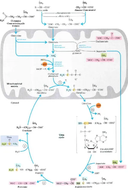
Figure 1. Urea cycle and reactions that feed amino groups into the cycle. The enzymes catalyzing these reactions (named in the text) are distributed between the mitochondrial matrix and the cytosol. One amino group enters the urea cycle as carbamoyl
29
phosphate, formed in the matrix; the other enters as aspartate, formed in the matrix by transamination of oxaloacetate and glutamate, catalyzed by aspartate amino-transferase. The urea cycle consists of four steps 1) Formation of citrulline from ornithine and carbamoyl phosphate (entry of the first amino group); the citrulline passes into the cytosol. 2) Formation of argininosuccinate through a citrullyl-AMP intermediate (entry of the second amino group). 3) Formation of arginine from argininosuccinate; this reaction releases fumarate, which enters the citric acid cycle. 4) Formation of urea; this reaction also regenerates ornithine (Lehninger)
30
Figure 2. Links between the urea cycle and citric acid cycle. The interconnected cycles have been called the "Krebs bicycle" The pathways linking the citric acid and urea cycles are known as the aspartate-argininosuccinate shunt; these effectively link the fates of the amino groups and the carbon skeletons of amino acids. The interconnections are even more elaborate than the arrows suggest. (Lehninger)
L-Carnitine is acylated by L-carnitine acyltransferases (e.g. palmitoyltransferase) and it is transported into the mitochondrial matrix by L-carnitine translocases, which exchange L-carnitine with carnitine. In the mitochondrial matrix, acyl-L-carnitine is used to form acyl-CoA by acyltransferases (Fritz IB, 1959; Haeckel R et al., 1990).
Figure 3. Fatty acid entry into mitochondria via the acyl-carnitine/carnitine transporter. After fatty acyl-carnrtine is formed at the outer membrane or in the intermembrane space, it moves into the matrix by facilitated diffusion through the transporter in the inner membrane. In the matrix, the acyl
31
group is transferred to mitochondriat coenzvme A, freeing carnitine to return to the intermembrane space through the same transport.
Primary genetic disorders of L-carnitine metabolism are due to inherited enzyme deficiencies, for example, carnitine palmitoyltransferase (CPT I or CPT II) deficiencies. Secondary deficiencies (reduction in plasma concentration) may be due to a number of conditions affecting intermediary metabolism: organic acidemias, inherited fatty acid oxidation disorders due to deficiencies in enzymes or proteins involved in mitochondrial beta-oxidation or respiration or in the urea cycle. Some nongenetic disorders also result in reduced plasma L-carnitine, for example, AIDS, chronic haemodialysis, or treatment with sodium valproate or with antibiotics that contain pivalic acid. In most cases carnitine deficiency is associated with hyperammonemia (Breningstall GN, 1990; Haeckel R, et al., 1990).
Reye’s-like syndrome is also induced by valproate, an anti-epileptic drug that may cause hepatotoxicity, hyperammonemia, hypoketonemia, and a decrease of L-carnitine levels. L-Carnitine treatment of patients with valproate-induced hepatotoxicity restores plasma ammonia levels and improves hepatic function (Bohan TP, et al. 2001).
Many studies report a recovering of energy metabolism and urea cycle enzymes, in different animal models of hyperammonemia (Ratnakumari L, et al. 1993 ; Horiuchi M, et al. 1992 ; Hearn TJ, et al. 1989).
32
In PCS (Portocaval-shunt) rats, that is one of the main animal models used to study first grade of HE showed that L-Carnitine prevents increase of ammonia levels in cerebrospinal fluid (CFS) and normalizes levels of alanine and lactate in CFS (Therrien G, et al. 1997). Alanine and lactate used to increase during hepatic encephalopathy, as I previously showed, for an impairment in the aminoacid metabolism and for the increase of glycolitic pathway. Different studies have been carried out trying to unveil the mechanism of this protective effect of L-carnitine. Other quaternary amines (betaine, choline, or trimethylamine N-oxide) like L-carnitine also have a protective effect against ammonia toxicity and the authors suggested that osmoregulation is involved in the mechanism of this protective effect (Kloiber O, et al. 1988). The protection due the osmoregulation property of quarternary amines were confirmed using same compounds and others with similar chemistry property (trimethylamine N-oxide, choline, acetylcholine, carbachol, and acetyl-L-carnitine). At low concentration these quaternary amines showed a protection at low concentration during ammonia toxicity in mice (Miñana 1996)
Dr. Felipo’s group from the center of investigation “Principe Felipe” of Valencia, have studied the neurobiology and possible treatment of HE. In the 1992 they demonstrated that acute ammonia toxicity in the brain is mediated by excessive activation of NMDA glutamate receptors (Marcaida et al., 1992). Ten different antagonists of NMDA receptors acting on three different sites of the receptor prevent ammonia toxicity in rats and mice injected with lethal doses of ammonia acetate
33
(Hermenegildo et al., 1996). They studied whether prevention of ammonia toxicity by L-carnitine is due to prevention of glutamate neurotoxicity, using primary cultures of cerebellar neurons to study the effect of L-carnitine on glutamate neurotoxicity. Treatment of these neurons in culture with 1 mM glutamate causes death of 80% of neurons. In this system glutamate neurotoxicity is mainly mediated by activation of NMDA receptors. Addition of 1 mM L-carnitine 15 min before glutamate prevented neuronal death caused by glutamate. However the high concentration of carnitine required is in agreement with the large doses of L-carnitine necessary to completely prevent ammonia toxicity in animals. In the same study, they tested whether L-carnitine affects the binding of glutamate to its receptors in hippocampal rat membranes. L-Carnitine increased the affinity of [ 3 H]-glutamate binding to the receptors. This increase was due to an increase of binding affinity to “quisqualate” receptors, whereas the affinity for the binding to NMDA and kainate receptors was slightly decreased (Felipo et al., 1994). In 1994, a specific agonist for “quisqualate” receptors was not available. For this reason they used Quisqualate, an unspecificic agonist that activates AMPA and metabotropic glutamate receptors. This suggests that in the presence of L-carnitine, glutamate binding to metabotropic receptors is increased. They also assessed whether the increase in the binding affinity of metabotropic glutamate receptors induced by L-carnitine is involved in its protective effect against glutamate neurotoxicity. AP-3, an antagonist of metabotropic glutamate receptors prevented the protective effect of L-carnitine
34
against glutamate neurotoxicity. Moreover, pre-incubation with t-ACPD, an agonist of metabotropic glutamate receptors also prevented glutamate neurotoxicity (Felipo et al. 1994).
L-carnitine and trimethylamine-containing compounds do not prevent neurotoxicity induced by NMDA, this fact supports the idea that the protective effect of these compounds is mediated by an increase of glutamate binding to metabotropic glutamate receptors. The affinity of NMDA for metabotropic receptors is not significant and NMDA would not activate these receptors in the presence of either carnitine or the other protective compounds. Some of the trimethylamine-containing compounds (acetylcholine, carbachol) are agonists of acetylcholine receptors. Atropine, an antagonist of acetylcholine receptors, also prevents the protective effect of most of these compounds, including that of t-ACPD (agonist of metabotropic glutamate receptors), against glutamate neurotoxicity.
Injection of atropine also prevents the protective effect of some of the trimethylamine-containing compounds against ammonia toxicity in mice. The protective effect of L-carnitine and betaine is not prevented by atropine (Miñana et al., 1996). These results show that antagonists of both acetylcholine and metabotropic glutamate receptors prevent the protective effect of
trimethylamine-containing compounds against glutamate
neurotoxicity, suggesting that there is an interplay between both types of receptors in the protective effects of L-carnitine against glutamate neurotoxicity (Llansola M, et al. 2002)
Subsequently, the same group published a study demonstrating that the activation of the mGluR5 subtype is responsible for the
35
protective effect of metabotropic glutamate receptor agonists (Montoliu et al., 1997). Recently, it was assessed that LAC treatment in mice could increase mGluR2 expression but not mGluR3 levels in hippocampus (Cuccurazzo 2013), but no studies have been conducted to assess a particular role of these receptors during ammonia intoxication. According to the literature, activation of mGluR5 is associated with activation of phospholipase C. Phospholipase C hydrolyzes inositol phospholipids releasing inositol triphosphate (IP 3) and diacylglicerol (DAG). Inositol triphosphate induces release of calcium from internal organelles and DAG activates protein kinase C.
In hippocampal slices, addition of the metabotropic glutamate receptor agonist t-ACPD induces an increase in phospholipid hydrolysis. They expected that L-carnitine would induce an increase in t-ACPD-induced phospholipid hydrolysis, i.e., a greater activation of metabotropic glutamate receptors. However, pre-incubation with L-carnitine inhibited t-ACPD-induced hydrolysis of phospholipids in a dose-dependent manner. Moreover, L-carnitine also inhibited phosphoinositide hydrolysis induced by arterenol, an agonist of noradrenergic receptors, and partially inhibited the effect of carbachol, agonist of acetylcholine muscarinic receptors (Llansola and Felipo, 1998). These results suggest that L-carnitine affects phosphoinositide hydrolysis induced by different types of receptors. . The same group also examines whether L-carnitine affects G-protein affects G-protein function by assessing the effect of L-carnitine on phosphoinositide hydrolysis induced by AlF 4 ¡, that directly
36
activates G-proteins. L-Carnitine inhibited partially (45%) phosphoinositide hydrolysis induced by AlF 4-. This suggests that L-carnitine affects some types of heterotrimeric G-proteins (Llansola and Felipo, 1998).
37
38
Oral acetyl-L-carnitine therapy reduces
fatigue in overt hepatic encephalopathy:
a randomized, double-blind,
placebo-controlled study*
Michele Malaguarnera1, Marco Vacante2, Maria Giordano2, Giovanni Pennisi3, Rita Bella3, Liborio Rampello3, Mariano Malaguarnera2, Giovanni Li Volti1, and Fabio Galvano1 1Department of Biological Chemistry, Medical Chemistry, and
Molecular Biology, University of Catania; 2Departmnent of
Senescence, Urological and Neurological Science, University of Catania; 3Department of Neurosciences, University of Catania,
Catania,
Abstract
Background: Fatigue is frequently reported in hepatic encephalopathy (HE) and may be related to hyperammonemia. Acetyl-L-carnitine (ALC) offers neuroprotective benefits and improves mitochondrial energetics and function.
Objective: This study evaluated the effect of exogenous ALC on physical and mental fatigue, fatigue severity, and physical
activity in patients with mild and moderate
39
Design: A total of 121 patients with overt HE were recruited to the study and were subdivided into 2 groups according to their initial HE grade [HE1 (n = 61) or HE2 (n = 60)]. Thirty-one patients with HE1 and 30 with HE2 received 2 g ALC, and 30 patients with HE1 and 30 patients with HE2 received placebo twice a day for 90 d. All patients underwent clinical and laboratory assessments and automated electroencephalogram analysis.
Results: At the end of the study period, the ALC-treated patients in the HE1 group showed significantly better improvement than did the placebo group in mental fatigue score (−1.7 compared with −0.3; P < 0.05), the fatigue severity scale (−6.4 compared with 2.3; P < 0.001), 7-d Physical Activity Recall questionnaire score (17.1 compared with −2.5; P < 0.001), and Short Physical Performance Battery (2.1 compared with 0.2; P < 0.001); the HE2 group showed significantly better improvement in the fatigue severity scale (−8.1 compared with −5.1; P < 0.001) and 6-min walk test (19.9 compared with 2.3; P < 0.05). Significant decreases in NH4+ were observed in both groups (P < 0.001). Conclusion: Patients with HE treated with ALC showed a decrease in the severity of both mental and physical fatigue and an increase in physical activity. This trial was registered at clinicaltrials.gov as NCT01223742.
* published in Am J Clin Nutr 2011;93:799–808. Printed in
40
1. Introduction
Hepatic encephalopathy (HE) is a neuropsychiatric complication of cirrhosis. Overt HE can be diagnosed clinically, and a mild-to-moderate grade of disease might be present in a considerable proportion of ambulatory patients with cirrhosis. Overt HE is a syndrome of neurologic and neuropsychiatric abnormalities. Affected patients exhibit alterations in psychomotor, intellectual, cognitive, emotional, behavioral, and fine motor function. Fatigue is frequently reported in HE and can also be related to hyperammonemia (1). Ammonia is recognized as a crucial component in the pathogenesis of HE, but other factors, such as oxygen free radicals, circulating opioid peptides, nitric oxide,
inflammatory cytokines, reduction in serotoninergic
neurotransmitters, depletion of endogenous antioxidants, neurosteroids, and manganese, are also implicated in the development of the disease (2). In recent years, fatigue has been researched as the main symptom of elevated ammonia in HE (1, 3). The treatments that remove ammonia from the body or that decrease ammonia production and absorption through the gastrointestinal tract improve mental status and cognitive function, but no effects have yet been shown in fatigue treatment. Our previous study showed a protective effect of L-carnitine against ammonia-evoked encephalopathy in cirrhotic patients, and another study showed that acetyl-L-carnitine (ALC) administration improved neurologic symptoms and plasma variables in selected cirrhotic patients with hepatic coma. Finally, other studies showed that ALC treatment reduces fatigue
41
in the elderly and in centenarians (4–7). ALC is an endogenous molecule synthesized in mitochondria by the enzyme ALC transferase and is the predominant acylcarnitine in normal tissues. Acylcarnitine is the fatty acid–bound form of L-carnitine, which has an important role in the transport of long-chain fatty acids into mitochondria and in their β-oxidation (8–10). Serum acylcarnitine is mainly composed of short-chain fatty acid carnitine, especially ALC. Although 99% of the amount of L-carnitine is intracellular, the relation between serum acylL-carnitine and free L-carnitine is highly sensitive to intramitochondrial metabolic alterations (11). ALC treatment restores the altered neurochemical abnormalities, cerebral energy metabolites in ischemia and aging and, in particular, ammonia-induced cerebral energy depletion (12). It also facilitates the removal from the mitochondria of excess short- and medium-chain fatty acids that accumulate during metabolism (13). Some of ALC's proposed neuroprotective benefits involve improved mitochondrial energetics and function, antioxidant activity, stabilization of membranes, protein and gene expression modulation, and enhancement of cholinergic neurotransmission (14). Patients with fatigue show reduced exercise tolerance and postexercise fatigue induced by minimal physical activity, which suggests decreased muscle function. During physical activity, the rate of free radical formation may overcome the various protective defense mechanisms and induce systemic oxidative stress through plasma accumulation of secondary products of lipid peroxidation (15). The aim of this study was to evaluate the effect of exogenous ALC on physical and mental fatigue, fatigue
42
severity, and physical activity in patients with mild and moderate encephalopathy.
2. Subjects and Methods
Subjects
A total of 121 cirrhotic patients (22 with hepatitis B virus infection, 65 with hepatitis C virus infection, 9 with alcoholism, and 25 with cryptogenetic cirrhosis) meeting the following inclusion criteria were enrolled in the study:
1) Chronic hepatitis with spontaneous manifest HE
(mental state grade 1 or 2 according to the West Haven criteria) and a Number Collection Test-A performance time >30 s
2) Hyperammonemia (venous ammonia concentration
>50 mmol/L)
3) Cooperative, hospitalized adult patients with liver
cirrhosis diagnosed by clinical, histologic, and ultrasonographic findings (reduced dimensions of the liver as well as splenomegaly) and esophageal varices at stages 2 and 3 observed by endoscopy Exclusion criteria
43
1) Major complications of portal hypertension, such
as gastrointestinal blood loss, hepatorenal syndrome, or bacterial peritonitis
2) Acute superimposed liver injury
3) Patient with other neurologic disease and
metabolic disorders, diabetes mellitus, unbalanced heart failure, and/or respiratory failure or end-stage renal disease
4) Alcoholic-toxic cirrhosis because toxic brain
damage may interfere with the assessment of HE
5) Severe HE
6) Administration of anti-HE medications, such as
neomycin and branched-chain amino acids
7) Any additional precipitating factors, such as high
protein intake (additional high-protein meals), constipation, or intake of psychostimulants,
sedatives, antidepressants, benzodiazepines,
benzodiazepines-antagonists (flumazenil),
neuromuscular blocking agents, and certain antibiotics
8) Patients with fever, sepsis, or shock were also
excluded to avoid variations caused by body temperature
9) Illiteracy
The study protocol was received and approved by the Institutional Review Board of the Hospital following the guidelines of the 1975 Declaration of Helsinki (16). All patients
44
gave written informed consent before any study procedures were initiated.
Study design
This was a randomized, double-blind, placebo-controlled study. The study was performed between June 2002 and December 2007. Patients meeting the inclusion criteria were randomly assigned to either a 90-d treatment with ALC (group A) or placebo (group B). Randomization was based on a computer-generated list. All study subjects were subdivided into 2 groups on the basis of the initial grade of HE: mild (grade 1; HE1) or moderate (grade 2; HE2) according to the West-Haven criteria (17). Group A consisted of patients with initial HE1 (ALC group: n = 31; placebo group: n = 30 placebo); group B consisted of patients with initial HE2 (ALC group: n =30 patients; placebo group: n =30 patients). The effectiveness of therapy was compared and evaluated separately in the different subgroups. Methods
Clinical and laboratory assessment and automated
electroencephalogram (EEG) analysis were performed for all patients. The diagnosis of HE grade was based on the evaluation of consciousness, intellectual functions, behavior, and neuromuscular functions and was made when appropriate laboratory and diagnostic testing excluded other causes of mental status changes. The investigators were blinded to the patients’ ammonia concentrations. Patients whose clinical course was not consistent with HE were excluded. Mental status was assessed
45
and graded on admission according to the West Haven criteria introduced by Conn (18).
Prerandomization phase
The subjects were required to document all caloric intake with the use of a diary, which was completed every 2 d. This prerandomization period was designed to nullify the effects of dietary changes on metabolic markers. During the initial 2-wk phase, subjects were instructed by a dietitian to follow an ad libitum diet as follows: 25–30% total fat, <7% saturated fat, ≤10% polyunsaturated fat, ≤20% monounsaturated fat, 50–60% of total energy as carbohydrate, ≈15% of total energy as protein, and <200 mg cholesterol/d (19). Patients were seen by a dietitian every month; at each visit the dietitian provided instructions on dietary intake recording procedures as part of a behavior-modification program, and the patients’ resulting food diaries were later used for counseling. All patients in both groups were given the same 1600-calorie diet and prescribed exercise plan. The subjects underwent weekly visits throughout the treatment period to assess adherence to the study protocol, to measure blood pressure, and to record adverse events.
Randomization phase
Throughout the trial, ALC was supplied in vials with 2 g ALC taken orally twice a day. All drugs and placebos were identical in appearance, and neither the investigators nor the patients were informed of the selected agent until the end of the study phase. Dosing instructions were provided with each patient pack. All
46
trial medication was instructed to be taken as prescribed. Subjects were considered compliant if the number of returned vials was between 80% and 120% of the planned treatment regimen. For the duration of the trial, any concomitant drugs were administered at the lowest possible therapeutic dose and, as much as possible, were not changed. Concomitant medications throughout the study included diuretics and β-blockers (Table 1).
Fatigue assessment Severity of fatigue
Severity of fatigue was measured by the Fatigue Severity Scale (FSS). The FSS is a self-assessed 9-question scale ranging from 1 (no signs of fatigue) to 7 (most disabling fatigue). Here, the total score ranged from 9 to 63 and is directly related to the severity observed (20).
Nature of fatigue
Wessely's test and Powell's test were used to examine fatigue, both mental and physical. The Wessely and Powell score consists of 2 scales measuring physical fatigue [8 items scored from 0 (no
47
fatigue) to 2 (highest possible fatigue); total score range: 0–16] and mental fatigue (5 items; total score range: 0–10) (21).
Measures of physical activity
Physical activity was assessed by using the 7-d Physical Activity Recall questionnaire (7-d PAR) and a pedometer. On the 7-d PAR, the patients self-reported moderate, hard, and very hard periods of physical activity performed during the 7-d period. The total duration of physical activity classified as “at least moderate intensity” was computed and used for analysis. This self-administered questionnaire has been shown to provide valid and reliable estimates of habitual physical activity (22). A pedometer (Digiwalker SW-200; Yamax Corporation, Tokyo, Japan) was used to obtain an objective measure of ambulatory physical activity. The subjects were instructed to wear the pedometer daily for 1 wk before treatment and for 1 wk before their scheduled 3-mo follow-up assessment. They were provided a diary to record their daily steps. The data are presented as the average steps taken daily. Physical function was assessed by using both performance-based and self-reported measures. The 6-min walk test (6MWT) measures the distance walked in 6 min on level ground, with stops to rest as needed. The subjects were told that the purpose of this test was to determine the distance they could walk in 6 min. They were instructed to “walk at their own pace in order to cover as much ground as possible” (23). The Short Physical Performance Battery (SPPB) is a battery of tests that has been used to assess lower extremity function in the older population (24). This battery uses a scale from 0 (poor) to
48
12 (excellent) to summarize the performance of 3 tasks (a 4-m walk, standing balance, and rising from a chair). For the 4-m walk, the subject walks a distance of 4 m at their normal pace to determine gait speed, computed as the time to complete the 4-m walk. For the standing balance test, the subjects placed their feet in a side-by-side position, followed by a semitandem position (heel of one foot along the side of the big toe of the opposite foot) and a tandem position (heel of one foot directly in front of the other). The subjects were required to hold the side-by-side position for 10 s before advancing to the semitandem position and to hold the semitandem position for 10 s before advancing to the tandem position. For the chair rise test, the subject was seated in a chair that was 18 in (≈45.72 cm) tall, with their arms crossed, and how quickly they could stand 5 times from sitting in the chair was assessed (24).
Neurophysiologic assessment
The EEG was recorded by using standardized techniques. Five electrodes were attached to the skin at the positions T3, T4, O1, O2, and Cz according to the international “10-20 system.” Electrode impedance was kept lower than 5KQ. After the usual handpass filters (0.53–35 Hz) were applied, 2 runs of 100 s each were recorded and compared for reproducibility. Patients were graded into different studies of HE according to their mean dominant frequency (MDF) and the relative powers of delta and theta activity (25). The EEG is the only test that classifies HE in 5 grades of severity (from normal to coma), just as the clinical grading: grade 0 (normal, regular alpha rhythm), grade 1 (irregular background activity, alpha and theta rhythm), grade 2
49
(continuous theta activity, occasionally delta activity), grade 3 (prevalence of theta activity, transient polyphasis complexes of spikes and slow waves), and grade 4 (continuous delta activity, abundant complexes of spikes and slow waves) (26).
Liver function assessment
The Child-Pugh score was determined to assess the severity of cirrhosis, including 3 biochemical variables (serum albumin, bilirubin, and prothrombin time) and 2 clinical characteristics (presence or absence of ascites and clinical HE). A patient had Pugh score A cirrhosis if the score was ≤6 points, Child-Pugh B cirrhosis if the score was 7–9 points, and Child-Child-Pugh C cirrhosis if the score was >9 points. Patients without signs of ascites were scored as 2 points for ascites (27). We also evaluated the presence and severity of the porto-systemic shunt by portal vein flow, presence and size of the esophageal varices, and splenic size.
Venous ammonia concentration
Ammonia was measured by enzymatic determination of glutamate dehydrogenase in a rapid and interference-free photometric determination (340 nm) of NH4+ in native blood plasma according to the Da Fonseca-Wollheim method (28). For reasons of safety, blood was immediately refrigerated and transported to the laboratory for immediate measurement of NH4+ (within 15 min of blood withdrawal).
50
Efficacy assessment
Throughout the randomization phase of the study, thrice weekly alimentary diary cards were used to collect efficacy data. The primary efficacy measures were changes in activity, motivation, and physical and mental fatigue severity. Measurements were made at the beginning and at the end of the study period. Data were collected in the morning, after an overnight fast. Activity, motivation, physical and mental fatigue, and the severity of fatigue were assessed before and after treatment.
Tolerability assessment
Laboratory assessments were monitored on days 0, 30, 60, and 90. These data included blood tests (hemoglobin, hematocrit, white blood cell count, and thrombocytes) and liver function
tests [alanine aminotransferase (AST), aspartate
aminotransferase (AST), γ-glutamyl-transpeptidase,
cholinesterase activity, serum bilirubin concentrations,
prothrombin time, and partial thromboplastin time].
Electrocardiogram and blood pressure were monitored with the use of standard techniques.
Statistical analysis
We calculated that a simple size of ≥25 patients in each arm would be required to detect a difference in improvement in HE,
51
that is the proportion of patients with HE at 2 mo, with a 5% type 1 error and 90% power for a 2-tailed log-rank test. Descriptive statistics were prepared from the study sample, and the results are expressed as means ± SDs. The statistical significance in contingency tables was evaluated by using chi-square and Fisher exact test. Student's t test was used for unpaired data, and one-factor analysis of variance and the Mann-Whitney rank-sum test were used for comparisons of continuous variables. The statistical analyses were performed by using appropriate tests for repeated measures and by controlling for multiple comparisons by correction with the Duncan procedure. Differences in tolerability were assessed with a chi-square test comparing the proportions permanently withdrawn from all study drugs or placebos. Statistical Analysis System software version 6.11 (SAS Institute, Cary, NC) was used for all analyses.
3. Results
Baseline values
The 2 groups were homogeneous for demographic characteristics, etiology, casting of disease, Child-Pugh grade, anamnestic, and diagnostic criteria (Table 2). Differences in the composition of the 2 groups with respect to precipitant factors might be minimized, because the patient population was well defined by inclusion and exclusion criteria. Serum NH4+ fasting concentrations were not significantly different before the
52
treatment. No statistically significant differences were observed between the 2 groups about prothrombin time, serum albumin, bilirubin, AST, and ALT. No statistically significant differences in the administered neuropsychologic test or in the EEG were observed between the 2 groups.
Neurophysiologic response
In the comparison between group A (treated with ALC) and group B (treated with placebo) we observed in HE1 an improvement in EEG grading in 45% of patients in the ALC group and in 13% of patients in the placebo group [odds ratio (OR): 5.35; 95% CI: 1.50, 19], whereas in HE2 there was an improvement in EEG grading in 66% of patients in the ALC group compared with 27% of patients in the placebo group (OR: 5.5; 95% CI: 1.81, 1.37) (Table 3).
53
L-Carnitine in plasma and urine
In the ALC group, significant differences were observed in the following markers after treatment compared with baseline in both HE1 and HE2: free plasma L-carnitine (P < 0.001), plasma concentrations of total plasma L-carnitine (P < 0.001), plasma long-chain acylcarnitine (LCAC) (P < 0.001), and short-chain acylcarnitine (SCAC) (P < 0.05). Only in HE2 did we observe significant differences in free urinary L-carnitine (P < 0.001). In the placebo group (in both HE1 and HE2), the plasma concentrations of free L-carnitine and LCAC and the urinary
54
excretion of free L-carnitine and SCAC were not significantly different from baseline. At the end of the study period, compared with placebo, the ALC-treated patients showed significant improvements in the following markers in HE1 and HE2: free plasmaL-carnitine (3.8 compared with 0.7 μmol/L in HE1 and 5.4 compared with 0.8 μmol/L in HE2; P < 0.001), plasma concentrations of total L-carnitine (4.4 compared with 0.9 μmol/L in HE1 and 6.2 compared with 1.1 μmol/L in HE2; P < 0.001), and plasma SCAC (0.4 compared with 0.1 μmol/L in HE1 and 0.5 compared with 0.2 μmol/L in HE2; P < 0.001)(Table 4).
TABLE 4
Comparison of plasma and urinary concentrations of L-carnitine between treatment groups1
Group A: ALC (n = 31 HE1 and 30 HE2)
Placebo group (n = 30 HE1 and 30 HE2)
Variable Before treatmen t After 90 d of treatmen t Before treatmen t After 90 d of treatmen t P for time2 P for group × time2 Free plasma L -carnitine (μmol/L)
55
Group A: ALC (n = 31 HE1 and 30 HE2)
Placebo group (n = 30 HE1 and 30 HE2)
Variable Before treatmen t After 90 d of treatmen t Before treatmen t After 90 d of treatmen t P for time2 P for group × time2 HE1 38.5 ± 3.9 42.3 ± 2.63 38.3 ± 3.8 39 ± 3.54 <0.00 1 <0.00 1 HE2 30.8 ± 4.3 36.2 ± 2.83 31.1 ± 5 31.9 ± 4.24 <0.00 1 <0.00 1 Plasma SCAC (μmol/L) HE1 7.6 ± 0.5 8 ± 0.53 7.2 ± 0.6 7.3 ± 0.64 <0.05 <0.00 1 HE2 6.5 ± 0.7 7 ± 0.53 6.2 ± 0.5 6.4 ± 0.64 <0.05 <0.00 1 Plasma LCAC (μmol/L) HE1 1.8 ± 0.3 2.1 ± 0.23 2 ± 0.3 2.1 ± 0.4 <0.00 1 1.000 HE2 1.7 ± 0.4 2 ± 0.33 1.8 ± 0.3 1.9 ± 0.3 <0.00 1 0.202 Total plasma L -carnitine (μmol/L) HE1 48.1 ± 4 52.5 ± 2.83 47.6 ± 4.3 48.5 ± 3.64 <0.00 1 <0.00 1 HE2 39.1 ± 4.5 45.3 ± 2.73 39.1 ± 5.0 40.2 ± 4.24 <0.00 1 <0.00 1 Free urinary L -carnitine (μmol/L) HE1 11.3 ± 11.6 ± 11.3 ± 11.4 ± 0.054 0.235