UNIVERSITÀ DEGLI STUDI DI ROMA
"TOR VERGATA"
FACOLTA' DI MEDICINA e CHIRURGIA
DOTTORATO DI RICERCA IN MICROBIOLOGIA MEDICA ed
IMMUNOLOGIA
XXI Ciclo
“Effect of nucleoside and nucleotide analogues on reverse transcriptase
activity in primary isolates from HTLV-1 infected patients.”
Arianna Ascolani
A.A. 2008/2009
Docente Tutor: Prof. Beatrice Macchi
Coordinatore: Prof. Enrico Garaci
INDEX
1. INTRODUCTION pag. 5 1.1 RETROVIRUSES pag. 5 1.2 HTLV-1 pag.9 1.3 HTLV-1 BIOLOGY pag. 9 1.3.1 Epidemiology pag. 9 1.3.2 Receptor and transmission of HTLV-1 pag. 12 1.3.3 Structure and genomic expression regulation pag. 141.4 DISORDER ASSOCIATED WITH HTLV-1 pag. 19
1.4.1 Adult T-cell Leukemia/Lymphoma (ATLL) pag. 19 1.4.2 Tropical spastic paraparesis/HTLV-1 associated myelopathy (HAM/TSP) pag. 22
1.5 THERAPY OF HTLV-1 ASSOCIATED DISEASE pag. 25
1.5.1 Treatment of adult T-cell leukemia/lymphoma pag. 25 1.5.2 Treatment of HTLV-1-associated myelopathy/Tropical Spastic Paraparesis pag. 28
1.6 ANTIRETROVIRAL THERAPY pag. 31
1.6.1 Nucleoside reverse transcriptase inhibitors (NRTIs) pag. 31 1.6.2 Main nucleoside and nucleotide inhibitors used in therapy pag.36
1.6.3 Zidovudine pag. 36 1.6.4 Lamivudine pag. 36 1.6.5 Tenofovir pag. 37 1.6.6 Non-nucleoside reverse transcriptase inhibitors pag. 41 1.6.7 Experimental evaluation of activity of RT pag. 42
2. AIM OF STUDY pag. 44
3. MATERIALS AND METHODS pag. 46
3.1 Caracteristics of patients and sample collection pag. 46 3.2 Cell lines and infection of PBMCs pag. 46 3.3 Assayed compounds pag. 51 3.4 RNA extraction pag. 51 3.5 Genomic DNA extraction and pol region amplification pag. 51 3.6 RT inhibition assay pag. 52 3.7 Detection of p19 through antigen capture assay pag. 54 3.8 Real Time quantitative reverse transcription pag. 57 3.9 Determination of cytotoxicity of the compounds pag. 58
4. RESULTS pag. 59
4.1 Preliminary experiment to set up the cell-free RT inhibition assay and effect
of PCOANs on HTLV-1 RT activity from the MT2 cell line pag. 59
4.2 Sensitivity of HTLV-1 isolates from chronic infected cells to RT inhibitors
pag. 62
4.3 Reverse transcriptase activity in the supernatant of PBMC cultures from
patients with HAM/TSP pag. 64
4.4 Sensitivity of HTLV-1 isolates from HAM/TSP patients to RT inhibitors
pag. 67
4.5 Sensitivity of HTLV-1 isolates from ATLL patients to RT inhibitors pag. 72 4.6 Evaluation of pol presence in ATLL patients pag. 76 4.7 Toxicity of the compounds on uninfected cells pag. 79
5. DISCUSSION pag. 81
1. INTRODUCTION 1.1 RETROVIRUSES
Retroviruses comprise a large and diverse family of enveloped RNA viruses defined by common taxonomic denominators that include structure, composition, and replicative properties (Coffin J.M., 1996).
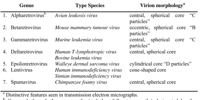
Retroviruses can be broadly divided into two main categories, simple and complex, distinguishable by the organization of their genomes (Murphy et al. 1994). All retroviruses contain three major coding domains with information for virion proteins: gag, which directs the synthesis of internal virion proteins that form the matrix, the capsid, and the nucleoprotein structures; pol, which contains the information for the reverse transcriptase and integrase enzymes; and env, from which are derived the surface and transmembrane components of the viral envelope protein. An additional, smaller, coding domain present in all retroviruses is pro, which encodes the virion protease. Simple retroviruses usually carry only this elementary information, whereas complex retroviruses code for additional regulatory proteins derived from multiply spliced messages. Retroviruses are further subdivided into seven groups defined by evolutionary relatedness, each with the taxonomic rank of genus (see Table 1). Five of these groups represent retroviruses with oncogenic potential (formerly referred to as oncoviruses), and the other two groups are the lentiviruses and the spumaviruses. All oncogenic members except the human T-cell leukemia virus–bovine leukemia virus (HTLV-BLV) genus are simple retroviruses. HTLV-BLV and the lentiviruses and spumaviruses are complex.
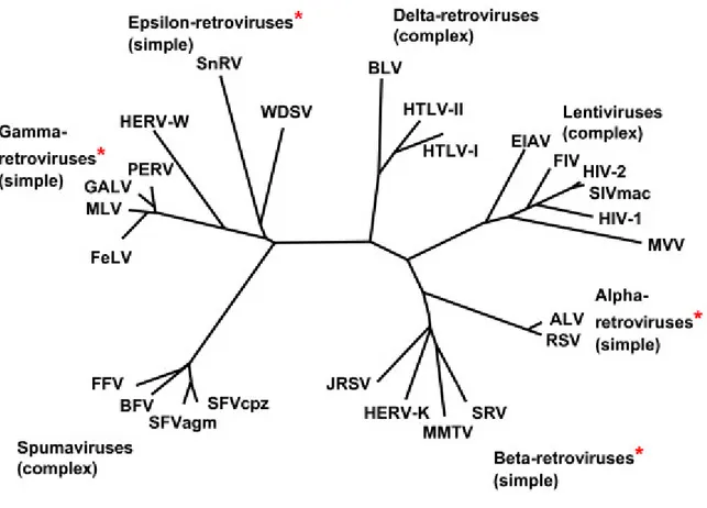
Retroviruses in different genera show little sequence similarity at the nucleotide level, as measured experimentally by standard biochemical techniques, and only limited amino acid similarity except in reverse transcriptase. In the approximately 175 amino acid residues that constitute the most conserved part of reverse transcriptase, viruses in different genera
show identity at about one third to two thirds of the residues (see Figure 1). Generally, viruses within a genus show identity at more than two thirds of these residues. However, there are exceptions to these rules, reflecting the imperfections of the classification system, restricted sampling of existing retroviruses, and perhaps unknown evolutionary constraints in some genera. For example, there are some members of the lentiviral genus with less similarity in the reverse transcriptase sequence than suggested by these approximations. HTLV-1 and HTLV-2 both belong to the genus Retrovirinae HTLV-BLV, and have sequence homology of 65%. In recent studies, the terms "HTLV-3" and "HTLV-4" have been used to describe recently characterized viruses (Mahieux R. and Gessain A., 2005; Calattini S. et al., 2006). These viruses were discovered in 2005 in rural Cameroon, and were apparently transmitted from monkeys to hunters of monkeys through bites and scratches. HTLV-3 is similar to STLV-3 (Simian T-lymphotropic virus 3), but HTLV-4 does not resemble any known virus. It is not yet known how much further transmission has occurred among humans, or whether the viruses can cause disease. The use of these names can cause some confusion, because the name HTLV-3 was the former name of HIV in early AIDS literature, but has since fallen out of use. Also, the name HTLV-4 has been used to describe HIV-2. The human imunodeficiency virus type 1 (HIV-1) is currently included in the group of Lentivirus. Although HTLV-1 and HIV-1 have many similarities in the mechanisms of transmission, including the target cell (lymphocytes CD4 +), and in the mechanisms of viral replication and messengers expression through multiple splicing events, they have a low homology of sequence. HTLV-1 causes also the immortalization of infected lymphocytes, while HIV-1 is cytopathic, which determines the death of infected cells (Del Mistro A. et al., 1986). Also, in vivo HIV is associated with stages of viraemia, while infection with HTLV-1 is mainly characterized by the absence or almost of viraemia.
Genus Type Species Virion morphologya
1. Alpharetrovirusb 2. Betaretrovirus 3. Gammaretrovirus 4. Deltaretrovirus 5. Epsilonretrovirus 6. Lentivirus 7. Spumavirus
Avian leukosis virus
Mouse mammary tumour virus Murine leukemia virus
Human T-lymphotropic virus Bovine leukemia virus
Walleye dermal sarcoma virus Human immunodeficiency virus Simian immunodeficiency virus Chimpanzee foamy virus
central, spherical core “C particles”
eccentric, spherical core “B particles”
central, spherical core “C particles”
central, spherical core
cylindrical core “D particles” cone-shaped core
central, spherical core
a Distinctive features seen in transmission electron micrographs.
b Groups 1 through 5 are presently (and, hopefully, temporarily) designated by the
awkward descriptive terms listed in the table, awaiting the proposal of more succinct appellations by the International Committee on taxonomy of viruses.
Figure 1.Phylogeny of Retroviruses: genera that include endogenous genomes are marked
1.2 HTLV-1
HTLV-1 is an acronym for the human T-cell lymphotropic virus type 1, also called the Adult T-cell lymphoma virus type 1. HTLV was discovered in 1977 in Japan. The virus was first isolated by Drs. Bernard Poiesz and Francis Ruscetti and their co-workers in the laboratory of Robert C. Gallo at the NCI (Poiesz B.J. et al., 1980;1981). HTLV was the first identified human retrovirus (Johnson J.M. et al., 2001; Shuh M. and Beilke M., 2005). HTLV-1 infects 20 million people worldwide where the majority of infected individuals are asymptomatic carriers of the virus (Verdonck K. et al., 2007). However, in a small percentage of infected people this agent has also been demonstrated to be the aetiological agent in patients with adult T-cell leukemia/lymphoma (ATLL; Poiesz B.J. et al., 1980) and a chronic, progressive neurologic disease termed HTLV-1 associated myelopathy/tropical spastic paraparesis (HAM/TSP; Gessain A. et al., 1985; Osame M. et
al., 1987).
1.3 HTLV-1 BIOLOGY 1.3.1 Epidemiology
Over the course of nearly 25 years, the epidemiology of human T-lymphotropic virus type 1 (HTLV-1) has progressively developed. The geographic distribution of the virus has been defined, although some puzzles persist such as the high prevalence in southwestern Japan but low prevalence in neighboring regions of Korea, China and eastern Russia, and seemingly isolated pockets of infection in Iran. Although the exact number of HTLV-1 seropositive individuals in the world is not known, it is estimated that about 15–20 millions persons live with HTLV infection worldwide (de The G. and Kazanji M., 1996). The seroprevalence rates differ, according to geographic area, the socio-demographic composition of the population studied and individual risk behaviors. For example,
prevalence rates as high as 37.0% were reported in small, selected populations from some areas in southwestern Japan (Yamaguchi K., 1994; Mueller N. et al., 1996) as compared to very low prevalence rates (0.0039%) among French blood donors (Courouce A.M. et al., 1993). However, information on prevalence rates from representative samples of the general population is rare. Most data on HTLV-1 prevalence rates are from studies in generally low-risk blood donors or selected population groups (pregnant women, neurological or hematological patients, relatives of infected individuals, specific native and sometimes isolated population groups, intravenous drug users (IDU) and sex workers) that are certainly not representative of the general population (Mueller N., 1991; Ferreira O.C.
et al., 1997; Manns A. et al., 1999). For non endemic geographic areas such as Europe and
North America, HTLV-1 infection is mainly found in immigrants from endemic areas, their offspring and sexual contacts, among sex workers and IDU. For blood donors in North America and Europe, seroprevalence is very low, for example, 0.01–0.03% in USA and Canada (Williams A.E et al., 1988; Murphy E.L. et al., 1991; Chiavetta J.A. et al., 2003), 0.002% in Norway (Stigum H. et al., 2000) and 0.0056% in Greece (Tseliou P.M. et
al., 2003). Data from studies of pregnant women may better reflect prevalence rates of the
general population. Prevalence rates for HTLV-1 and II were six fold higher in pregnant women than in blood donors in the United Kingdom (Taylor G.P. et al., 2005). A study on 6754 pregnant women in Salvador, Brazil found 0.84% to be seropositive (Bittencourt A.L.
Figure2: Countries with endemic HTLV-1, defined as prevalence between 1 and 5% in
some populations, are shown in dark brown. Countries with reports of low prevalence (less than 1% in some groups), due mainly to immigration from endemic areas, are shown in tan color. (from Proietti F.A. et al., 2005).
1.3.2 Receptor and transmission of HTLV-1
HTLV-1 can infect various types of cells, such as T-lymphocytes, B-lymphocytes, monocytes and fibroblasts (Koyanagi Y et al., 1993). Glucose transporter 1 (GLUT-1) has been identified as a receptor for HTLV-1 and this receptor is ubiquitously expressed on cell surfaces (Manel N et al., 2003). However, the HTLV-1 provirus is mainly detected in CD4-positive lymphocytes, with about 10% in CD8-positive T-lymphocytes (Yasunaga J.
et al., 2001). This situation possibly arises because Tax mainly induces the increase of
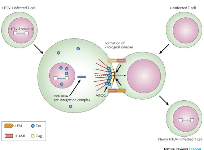
CD4-positive T-lymphocytes in vivo by enhanced proliferation and suppressed apoptosis. In HTLV-1-infected individuals, no virions are detected in the serum. In addition, the infectivity of free virions is very poor compared with that of infected cells. These findings suggest that HTLV-1 is spread by cell-to-cell transmission, rather than by free virions. In
vitro analyses of HTLV-1-infected cells revealed that HTLV-1-infected cells form
"virological synapses" with uninfected cells. Contact between an infected and a target cell induces the accumulation of the viral proteins Gag and Env, viral RNA and microtubules, and the viral complex subsequently transfers into the target cell (Igakura T. et al., 2003;see Figure 3).
HTLV-1 is mainly transmitted via three routes: 1) mother-to-infant transmission (mainly through breast feeding) (Hino S., 2003); 2) sexual transmission (mainly from male-to-female); and 3) parenteral transmission (blood transfusion or intravenous drug use) (Blattner W.A. and Gallo R.C., 1994). In either route, HTLV-1-infected cells are essential for transmission. This was supported by the findings that fresh frozen plasma from carriers did not cause transmission (Okochi K. et al., 1984) and freeze-thawing of breast milk reduced vertical transmission (Ando Y. et al., 2004).
Figure 3: This figure illustrates the cell–cell contact required to create a virological
synapse through which the viral genome is transmitted from one cell to another. The roles played by lymphocyte function-associated antigen 1 (LFA1), and intercellular adhesion molecule 1 (ICAM1) in forming cell–cell contact are shown. Tax contributes to the formation of a microtubule organizing centre (MTOC) (from Matsuoka M. et al., 2007).
1.3.3 Structure and genomic expression regulation
The HTLV-1 provirus has a similar structure to other retroviruses: a long terminal repeat (LTR) at both ends and internal sequences such as the gag, pol and env genes. A characteristic of HTLV-1 is the presence of the pX region, which exists between env and the 3'-LTR. This region encodes several accessory genes, which include the tax, rex, p12,
p21, p30, p13 and HBZ genes (see Figure 4). Among these, the tax gene plays central roles
in viral gene transcription, viral replication and the proliferation of HTLV-1-infected cells. Tax enhances viral gene transcription from the 5'-LTR via interaction with cyclic AMP responsive element binding protein (CREB). Tax also interacts with cellular factors and activates transcriptional pathways, such as NF-κB, AP-1 and SRF (Yoshida M., 2001; Franchini G. et al., 2003; Jeang KT et al., 2004; Azran I. et al., 2004). For example, activation of NF-κB induces the transcription of various cytokines and their receptor genes, as well as anti-apoptotic genes such as bcl-xL and survivin (Tsukahara T et al., 1999; Nicot C et al., 2000; Kawakami H et al., 2005). The activation of NF-κB has been demonstrated to be critical for tumour genesis both in vitro and in vivo (Yamaoka S et al., 1996; Portis T
et al., 2001). On the other hand, Tax variant without activation of NF-κB has also been
reported to immortalize primary T-lymphocytes in vitro (Rosin O et al., 1998), suggesting that mechanisms of immortalization are complex. In addition to NF-κB, activation of other transcriptional pathways such as CREB by Tax should be implicated in the immortalization and leukemogenesis.
Tax also interferes with the functions of p53, p16 and MAD1 (Suzuki T et al., 1999). These interactions enable HTLV-1-infected cells to escape from apoptosis, and also induce genetic instability. Although inactivation of p53 function by Tax is reported to be mediated by p300/CBP (Van Orden, K et al., 1999; Ariumi, Y et al., 2000) or NF-κB activation (Pise-Masison, CA et al., 2000), Tax can still repress p53's activity in spite of loss of
p300/CBP binding or in cells lacking NF-κB activation, indicating the mechanism of p53 inactivation by Tax needs further investigation (Geiger TR et al., 2008).
Although Tax promotes the proliferation of infected cells, it is also the major target of cytotoxic T-lymphocytes (CTLs) in vivo. Moreover, excess expression of Tax protein is considered to be harmful to infected cells. Therefore, HTLV-1 has redundant mechanisms to suppress Tax expression. Rex binds to Rex-responsive element (RxRE) in the U3 and R regions of the 3'-LTR, and enhances the transport of the unspliced gag/pol and the singly spliced env transcripts. By this mechanism, double-spliced tax/rex mRNA decreases, resulting in suppressed expression of Tax. On the other hand, p30 binds to tax/rex transcripts, and retains them in the nucleus (Nicot C. et al., 2004). The HBZ gene is encoded by the complementary strand of HTLV-1, and contains a leucine zipper domain. HBZ directly interacts with c-Jun or JunB (Basbous, J et al., 2003), or enhances their degradation (Matsumoto, J et al., 2005), resulting in the suppression of Tax-mediated viral transcription from the LTR.
Transforming growth factor-β (TGF-β) is an inhibitory cytokine that plays important roles in development, the immune system and oncogenesis. Since TGF-β generally suppresses the growth of tumour cells, most tumour cells acquire escape mechanisms that inhibit TGF-β signalling, including mutations in its receptor and in the Smad molecules that transduces the signal from the receptor. Tax has also been reported to inhibit TGF-β signalling by binding to Smad2, 3 and 4 or CBP/p300 (Mori N. et al., 2001; Lee D.K. et
al., 2002). Inhibition of TGF-β signalling enables HTLV-1-infected cells to escape
TGF-β-mediated growth inhibition.
ATL cells have been reported to show remarkable chromosomal abnormalities (Kamada N.
et al., 1992), which should be implicated in the disease progression. Tax has been reported
controls the mitotic checkpoint. This functional hindrance of MAD1 by Tax protein causes chromosomal instability, suggesting the involvement of this mechanism in oncogenesis. Recently, Tax has been reported to interact with Cdc20 and activate Cdc20-associated anaphase-promoting complex, an E3 ubiquitin ligase that controls the metaphase-to-anaphase transition, thereby resulting in mitotic abnormalities (Endo K. et al., 2002). In contrast to HTLV-1, HTLV-2 promotes the proliferation of CD8-positive T-lymphocytes in vivo. Although it was first discovered in a patient with variant hairy cell leukemia, HTLV-2 is less likely to have oncogenic properties since there is no obvious association between HTLV-2 infections and cancers. Regardless of the homology of their
tax sequences, the oncogenic potential of Tax1 (HTLV-1 Tax) is more prominent than that
of Tax2 (HTLV-2 Tax). The most striking difference is that Tax2 lacks the binding motif at C-terminal end to PDZ domain proteins, while Tax 1 retains it. When the PDZ domain of Tax1 is added to Tax2, the latter acquires oncogenic properties in the rat fibroblast cell line Rat-1, indicating that this domain is responsible for the transforming activity of HTLV-1 (Hirata A. et al., 2004).
To understand the pleiotropic actions of Tax protein more clearly, transcriptome analyses are essential. The transcriptional changes induced by Tax expression have been studied using DNA microarrays, which revealed that Tax upregulated the expression of the mixed-lineage kinase MLK3. MLK3 is involved in NF-κB activation by Tax as well as NIK and MEKK1 (Ng P.W. et al., 2001). In addition to transcriptional changes, Tax is also well known to interact with cellular proteins and impair or alter their functions. For example, proteomic analyses of Tax-associated complexes showed that Tax could interact with cellular proteins, including the active forms of small GTPases, such as Cdc42, RhoA and Rac1, which should be implicated in the migration, invasion and adhesion of T-cells, as well as in the activation of the JNK pathway (Wu K et al., 2004).
Many questions pertaining to in vivo tax and virus expressionare currently unanswered. Bangham and their coworkers used deuterated glucose for labelingpatients' lymphocytes to check the in vivo effect of tax/HTLVexpression on lymphocyte population dynamics. He sobserved increasedproliferation of CD4+CD45RO+ and CD8+CD45RO+ T-lymphocytes in HTLV-1–infected persons compared with controls, with the additional production of 1012 lymphocytes per year in the former.The in vivo proliferation rate of CD4+CD45RO+ cells correlated with tax expression (measured ex vivo), supporting a physiologic role (directly or indirectly) for this viral oncoprotein in the proliferation of infected cells (Asquith B. et al., 2007).
Figure 4: A scheme of the HTLV-1 genome, alternatively spliced mRNAs, and putative
proteins encoded by each mRNA is shown. ORFs are indicated by boxes (from Nicot C. et
1.4 DISORDER ASSOCIATED WITH HTLV-1
Although still not fully clarified the molecular mechanisms that occur after infection to determine the onset of various diseases associated with HTLV-1, it seems that alteration of cellular genes and tissue damage induced by the immune response to the virus could play an important role in the pathogenesis of the HTLV-1 associated diseases. As previously described, the ATLL is a T-cell leukemia while the TSP / HAM is a neurological disorder characterized by destruction of cells of the central nervous system and inflammatory processes.
1.4.1 Adult T-cell leukaemia/lymphoma (ATLL)
Adult T-cell leukaemia/lymphoma (ATLL) is a mature T-cell neoplasm of post-thymic lymphocytes aetiologically linked to the humanT-cell lymphotropic virus, HTLV-1, and with a distinct geographicaldistribution (Matutes E., 2007). The disease manifests with leukaemia in greater than two thirds of patients, while the remaining patients have a lymphomatous form. ATLL affects almost exclusively adults and is extremely rare in children, although a few cases in childhood have been described (Pombo de Oliveira M.S.
et al., 1990).The median age is around the mid-60s and there is no gender prevalence.
Familial ATLL has been documented in Japan, USA and in England (Yamaguchi K. et
al., 1985; Matutes E. et al., 1995); it is unknown whether a genetic predispositionplays a
role in the development of the disease in such cases.In addition, ATLL may coexist or follow other HTLV-1 inducednon-neoplastic diseases such as tropical spastic paraparesis (Cartier L. et al., 1995).Depending on the disease manifestations, ATLL is classifiedinto several forms: acute, chronic, smouldering and lymphoma (Shimoyama M., 1991). The most common form of presentationis acute, which is seen in around 65% of patients. This is characterised by the presence of systemic symptoms, organomegaly, in particular
lymphadenopathy, and a leukaemic picture. Hypercalcaemia with or without lytic bone lesions is present in half of these patientsand it may develop during disease progression in another third.Skin lesions are seen in close to a half of these patients.The chronic form is characterised by lymphocytosis which may be stable for months or even years, skin manifestations, no organomegaly or small volume lymphadenopathy, absence of hypercalcaemiaand normal or only slightly raised lactate dehydrogenase (LDH)(less than twice the upper normal limit value). Patients with smouldering ATLL are usually asymptomatic or manifest skin rashes that respond to topical steroids and/or lung infiltrates; unlikethe chronic form, the white blood cell count is normal and thereis no lymphocytosis and less than 3% atypical circulating lymphocytes. Less than a third of patients carry lymphoma with noevidence of blood involvement. There are several reports documenting disease progression to the acute form in patients with chronic and smouldering ATLL. Haemophagocytic syndrome as the first sign of transformation has been described in smouldering ATLL (Aouba A. et al., 2004).In addition to the symptoms related to the neoplastic condition, patients with ATLL are immunocompromised and develop opportunistic infections that complicate the disease course and make its managementmore difficult. Infestation by Strongyloides stercoralis isfrequent and may be severe and fatal. This association and the fact that a third of the patients with strongyloidiasis andpositive serology for HTLV-1 have a clonal integration of theprovirus in their lymphocytes, have led to the hypothesis that this parasite plays a role in the development of ATLL in healthycarriers (Yamaguchi K. et al., 1987) .
The immunophenotype of the ATLL cell is that of an activatedmature T-lymphocyte. The
cells express CD2 and CD5 and oftenare CD7 negative; CD3 and T-cell receptor (TCR)-β may be down-regulated (weak expression or even negative in the cell membrane); however, the cells express these proteins in their cytoplasm. The most common
immunophenotypic profile isCD4+CD8–, but cases in which cells co-express these two markers or express only CD8 have been described; it has beensuggested that CD4+CD8+ cases have a worse clinical course (Tamura K. et al., 1985). The most characteristic immunophenotypic feature of the neoplasticcell is the strong expression of the alpha chain of the interleukin-2(IL-2) receptor recognised by the monoclonal antibody CD25 (Matutes E., 2007).Such expression is distinctive but not unique to ATLL; it maybe seen in other T-cell malignancies, such as T-prolymphocytic leukaemia (T-PLL) and SS (Shyongen Syndrome), although it is expressed at a lower density in these diseases. Other T-cell activation markers suchas CD38 and HLA-DR are variably expressed and natural killer (NK) cell markers are, as a rule, negative.The diagnosis of ATLL can be established by integrating the clinical and laboratory features. The morphology of the circulating neoplastic lymphocytes and the immunophenotype are very characteristicof the disease. Histology of the lymph node is not needed fordiagnosis in patients with a leukaemic form of ATLL in whom cytology and immunophenotyping are typical of the disease and in whom serum antibodies to HTLV-1 are demonstrable. The diagnosis of the pure lymphomatous forms is more problematic as the histological features of ATLL overlap with those of other T-cell lymphomas. In both leukaemic and lymphomatous forms, HTLV-1 serology isa mandatory investigation. The differential diagnosis includes other mature T-cell malignancies, essentially the cerebriform variant of T-prolymphocytic leukemia (T-PLL), mycosis fungoides and Sjögren syndrome (SS), other peripheralT-cell lymphomas, not specified, and rarely Hodgkin disease and angioimmunoblastic T-cell lymphoma. Patients with smoulderingATLL should be distinguished from healthy carriers of the virus.
The prognosis of ATLL is poor, with a median survival of lessthan one year for the acute and lymphoma forms. The projected4-year survival is estimated to be around 5% for these
two clinicalvariants. Chronic and smouldering ATLL fare better, with a projected4-year survival of 26.9% and 62% respectively (Shimoyama M., 1992). Clinical form, age, performance status, elevation of LDH, high β2-microglobulin,high serum level of CD25, high serum neuron-specific enolase, the presence of hypercalcaemia and a high proliferative rateare the main prognostic factors (Sadamori N. et al., 1995; Nishimura S. et
al., 1995). In the chronic form of ATLL, the proliferative rate of the neoplastic cells
measuredby Ki-67 expression, albumin levels and LDH appear to distinguishsubgroups with favourable and unfavourable prognosis (Shirono K. et al., 1994).
1.4.2 Tropical spastic paraparesis/HTLV-1 associated myelopathy (TSP/HAM)
Tropical spastic paraparesis/HTLV-1 associated myelopathy (TSP/HAM), caused by human T lymphotropic virus type 1 (HTLV-1), has an incidence of 1 case per 100 HTLV-1 infected carriers in highly endemic populations (Kaplan J.E. et al., 1990). The major histopathological characteristic of TSP/HAM is a chronic inflammation of the white and gray matter of the spinal cord followed by a degenerative process that preferentially affects the white matter in the lower spinal cord (Bhigjee A. et al., 1993; Iwasaki Y., 1990). TSP/HAM is characterized by a chronic slowly progressive spastic paraparesis with bladder disturbances, absent or mild sensory loss and low back pain, with seropositivity for HTLV-1 antibodies, in the absence of spinal cord compression (Gessain A. and Gout O., 1985). Despite the more usual presentation characterized by a slow progression, 21.5% of the patients may experience a rapid progression, with severe disability two years after the onset of symptoms. This phenomenon is related to older age of onset, parenteral HTLV-1 transmission route, high viral loads, and high antibody titers (Matsuzaki T. et al., 2001; Nakagawa M. et al., 1995; Toro C. et al., 2003).
the disease. Initially, up to 5 years after onset, the leptomeninges and blood vessels are infiltrated with lymphocytes that penetrate the surrounding parenchyma. Numerous inflammatory cells including both CD8 and CD4 T cells (in equal numbers), B cells, and foamy macrophages are present in damaged areas of the spinal cord parenchyma (Umehara F. et al., 1993). HLA class I and β2-microglobulin are expressed on endothelial cells and infiltrating mononuclear cells (Wu E. et al., 1993; Moore G.R. et al., 1989). HLA class II expression also occurs in the endothelial cells, microglia, and infiltrating mononuclear cells of affected lesions. Later in the disease, immunohistochemical analysis of the affected spinal cord lesions shows the predominance of CD8 T cells. Such lesions also express HLA class I antigens and have HTLV-1–specific CD8 cytotoxic T lymphocytes (CTL) (Levin M.C. et al., 1997). CD8 CTL, thought to represent functionally cytotoxic cells, are observed frequently in active chronic lesions and occasionally in inactive chronic lesions in HAM/TSP patients (Umehara F. et al., 1994). The amount of proviral DNA in HAM/TSP patients correlates with the number of CTL. Inflammatory cells and HTLV-1 proviral DNA decrease with duration of the disease. Since infiltrating CD8 cells are thought to play an important role in the development of HAM/TSP, an effort has been made to localize HTLV-1 in the central nervous system (CNS) of HAM/TSP patients and to determine which cells might serve as targets for inflammatory CD8 cells. HTLV-1 gag, pX, and pol sequences have been localized to the thoracic cord areas (Yoshioka A. et al., 1993) and are greater in areas of increased CD4 infiltration. HTLV-1 pX and env sequences have been localized to affected spinal cord (Kira J. et al., 1992; Ohara Y. et al., 1992) and HTLV-1 RNA has been localized to astrocytes (Lehky T.J. et al., 1995). Increased expression of inflammatory cytokines (Umehara F. et al., 1994) and adhesion molecules (Umehara F. et
al., 1996) occurs in the spinal cord of HAM/TSP patients with a short duration of disease.
substantially. The expression of inflammatory cytokines is also reduced over the duration of the disease and, with the exception of interferon (IFN)-γ, becomes undetectable. Collectively, these findings support the view that inflammatory T lymphocytes (and CD8 cells in particular) may play a critical role in the immunopathogenesis of HAM/TSP (Jacobson S., 2002).
1.5 THERAPY OF HTLV-1 ASSOCIATED DISEASES 1.5.1 Treatment of adult T-cell leukemia/lymphoma
The therapeutic approaches against HTLV-1 are different.
Conventional chemotherapies, such as cyclophosphamide, adriamycin, vincristine and prednisolone (CHOP), comprise the major therapeutics for acute and lymphoma type ATLL (Ishitsuka K. and Tamura K., 2008).
Several new drugs are active against ATLL. For example MST-16, a new oral inhibitor of topoisomerase II induced one complete remission and eight partial remissions in 23 ATLL patients with or without prior therapy (Ohno et al., 1993). In addition, 2’-deoxycoformycin administered alone or in combination with other cytotoxic agents, as well as cladribrine have modest activity against ATLL.
Interferon has modest effects on ATLL and HTLV-1 infected cell lines in vitro. Sporadic reports have indicated a clinical response to interferon-α, β or γ by patients with ATLL, but prospective clinical trials of interferon-α, did not confer a significant benefit upon patients with acute and lymphoma type ATLL.
AZT and interferon−α
Arsenic trioxide and interferon-α
Targeting surface molecules on ATLL cells Anti-CD25 antibody Anti-CD2 antibody Anti-CD52 antibody Anti-CD4 antibody Anti-CCR4 antibody Retinoid derivates Bortezomib
Zidovudine, 3’-azide-3’-deoxythymidine (AZT), combined with interferon-α has been suggested as a first line treatment for ATLL. The mechanism of the anti-ATLL effects induced by AZT and interferon-α therapy have not been defined. Intracellular phosphorylation changes AZT into its active form (zidovudine triphosphate; ZDV-TP), which is incorporated into viral DNA and then reverse transcriptase is inhibited via DNA chain termination (Furman P.A. et al., 1986). However, ZDV-TP is reported to be incorporated into cellular DNA in vitro. Its ability to inhibit cellular polymerases α and γ is 100-fold less potent than that to inhibit HIV reverse transcriptase (Copeland W.C. et al., 1992). Therefore, ZDV-TP probably does not directly affect cellular DNA. In fact, neither AZT nor interferon-α has direct toxicity against ATLL cell lines and against fresh ATLL cells obtained from patients who achieved complete remission after AZT and interferon-α therapy (Bazarbachi A. et al., 2000). AZT has not yet shown activity against HTLV-1 replication/expansion. Although AZT prevented HTLV-1 transmission when administered at the onset of infection in vitro, anti-HTLV-1 activity on already infected cells has not been unequivocally established (Matsushita S. et al., 1987; Macchi B. et al., 1997; Balestrieri E. et al., 2002; Garcia-Lerma J.G. et al., 2001). Despite the absence of supportive evidence, anti-retroviral agents probably do not reduce HTLV-1 provirus load in ATLL patients based on these observations.
Arsenic trioxide is widely used as a second line treatment for acute promyelocytic leukemia (APL). This agent has been suggested to induce partial differentiation by modulating promyelocytic leukemia-retinoic acid receptor α (PML-RARα) protein, and apoptosis by activating caspases against APL cells (Mistry A.R. et al., 2003). Arsenic trioxide induces G1-phase arrest and apoptosis, and thereby inhibits the growth of ATLL
cell lines and of HTLV-1 infected T-cell lines, as well as induction of apoptosis in fresh ATLL cells derived from patients (Ishitsuka K. et al., 1998; 2002). The adverse events of
arsenic trioxide in vivo are manageable and include mainly haematological, with some neurological toxicity.
The surface of ATLL cells characteristically express CD3, CD4, and CD25 (interleukin-2 receptor α). Therefore, these molecules, especially CD25 have been considered promising therapeutic targets as normal resting cells do not express CD25. Nineteen patients, with acute and chronic lymphoma, with or without prior chemotherapy were administered with murine anti-CD25 antibody, which blocks the interaction of IL-2 with high-affinity receptors but functions neither through complement-dependent cytotoxicity, nor through inducing antibody-dependent cellular cytotoxicity (Waldmann T.A. et al., 1993). One patient each with acute and chronic type ATLL achieved complete remission, and partial remission was achieved in one with acute, two with lymphoma and one with chronic type ATLL. The response duration varied from 2 to more than 60 months. Interestingly, the clinical responses were not apparently associated with previous chemotherapy and magnitude of tumour burden. No significant adverse events developed except fever following administration. Serum levels of soluble CD25 (sCD25) are high in ATLL patients, but sCD25 in the circulation did not prevent anti-CD25 from reacting with and saturating its binding sites on ATLL cells. Recent reports have described the effects of a humanized anti-CD25 monoclonal antibody daclizumab (Osborne G.E. et al., 2006), an anti-CD25 antibody fused to the truncated Pseudomonas exotoxin LMB-2 (Kreitman R.J.
et al., 2000), and an interleukin-2 fused with amino acids from the diphtheria toxin
denileukin (Di Venuti G. et al., 2003). In particular this last work report a case of human T-cell lymphotropic virus 1 (HTLV-1)-associated adult T cell leukemia/lymphoma (ATLL) in clinical remission > 1 year after therapy with denileukin diftitox and hyper-CVAD (hyperfractionated cyclophosphamide/doxorubicin/vincristine/decadron). It suggested that denileukin diftitox may affect the paracrine secretion of HTLV-1-associated
clinical manifestations of ATLL. The frequent expression of CC chemokine receptor 4 (CCR4) on ATLL cells is suggested to be a promising target, and a clinical trial is currently underway in Japan against relapsed/refractory ATLL using the chimeric anti-CCR4 antibody KM2760, which exerts anti-ATLL activity mediated by antibody dependent cellular cytotoxicity (Yoshie O. et al., 2002; Ishida T. et al., 2004).
1.5.2 Treatment of HTLV-1-associated myelopathy/Tropical Spastic Paraparesis
There is no established therapy for TSP/HAM. Clinical improvements have been reported for a number of agents in open-label studies including corticosteroid, plasmapheresis, danazol (Harrington W.J. et al., 1991), pentoxifylline (Shirabe S. et al., 1997), and interferon. With the exception of interferon-α, however, these drugs lack the quality of evidence required to merit a strong recommendation for their use in HAM/TSP. The evidence for supporting the use of corticosteroids in this patients is based on uncontrolled case series (Osame M. et al., 1987). The immunomodulatory effects of corticosteroids (Cupps T.R. et al., 1982) are the rationale base for their use in HAM/TSP. To date, data describing the real benefit of corticosteroids use for treatment of TSP/HAM in the literature are limited. Partly, this is owe to the fact that few studies have had a longer follow-up time. In a recent study it has been evaluated the clinical corticosteroid effect on TSP/HAM in a long-term follow-up (Croda M.G. et al., 2008). It has been studied 39 patients with HAM/TSP who received methyl-prednisolone treatment and after 10 years it has been observed a significant neurological improvement reaching 24,5% according to the Incapacity Status Scale (ISS) score. These results were similar to those from a study which evaluated 200 TSP/HAM patients, and in ten patients had 30% motor improvement after the use of methyl-prednisolone, and a 69% improvement among patients who used oral prednisolone and intrathecal hydrocortisone injection (Nakagawa M. et al., 1996).
Although many questions remain regarding the usefulness of corticotseroids in HAM/TSP. Instead, interferon-α demonstrated clinical benefits for subjects with TSP/HAM in a blinded trial (Izumo S. et al., 1996). The main marker of efficacy during interferon-a therapy appears to be a reduction in HTLV-1 proviral load (Saito M. et al., 2004); the reduction in proviral load was associated with changes in CD8+ T cells. Following the modest success of α therapy in HAM/TSP, long-term treatment with interferon-β was tested in a cohort of 12 patients. No significant clinical changes were observed during the trial in this cohort; among the number of biomarkers measured, significant reduction in the frequency of Tax-specific CD8+ T cells was observed along with a reduction in HTLV-1 Tax mRNA load and the spontaneous lymphoproliferation. No significant changes were observed in the HTLV proviral load. These results suggested that interferon-β therapy had an overall immune-modulatory effect in HAM/TSP as evidenced by the relative suppression of spontaneous proliferation, and demonstrated that interferon-β therapy had a profound effect on virus-specific cellular immune responses (Oh U. and Jacobson S., 2008). Regarding antiretroviral therapy, initial observational studies suggested clinical benefit and reduction of HTLV-1 proviral load with therapy including zidovudine (Gout O. et al., 1991; Sheremata W.A. et al., 1993) and lamivudine (3TC, Taylor G.P. et al., 1999). However in vitro studies which have demonstrated limited activity of 3TC against the HTLV-1 reverse transcriptase (Derse D. et al., 2001; Balestrieri E. et al., 2002), very likely due to a natural resistance of HTLV-1 reverse transcriptase to lamivudine (Toro C. et al., 2003). On one side the poor ability of anti-retroviral agents to reduce HTLV-1 virus load in vivo in patients with HAM/TSP, might be due to the preferential HTLV-1 replication mechanism through the clonal expansion of infected cells rather than through the reverse transcriptase pathway (Machuca A. et al., 2001; Taylor G.P. et al., 2006).On the other it cannot be excluded that the antiretroviral drugs, directly
translated from the experience on HIV infection are not suitable to inhibit HTLV-1 infection.
To date, proinflammatory cytokines implicated in the pathogenesis of immunomediated inflammatory diseases represent rational targets for therapy. For example, IL15 is a proinflammatory cytokine that appears to be dysregulated in HAM/TSP, and may play a pathogenic role in this disease (McInnes I.B. and Gracie J.A., 2004). A phase I/II clinical trial is under way to test the safety and provide preliminary efficacy data of a monoclonal antibody, Hu MiK-β1, that blocks IL15 signalling.
Recent work in HIV suggest that histone deacetylase inhibitors such as valproic acid (VPA) may induce expression of HIV from previously quiescent HIV-infected CD4+ T cells (Ylisastigui L. et al., 2004; Lehrman G. et al., 2005), making the virus a target of antiretroviral therapy. A single-arm, open-labelled trial tested the benefit of VPA in 19 patients with HAM/TSP. Preliminary data suggest VPA administration induced a transient increase then a drop in HTLV-1 proviral loads in HAM/TSP patients (Lezin A. et al., 2007). The authors hypothesized that VPA led to increased viral expression allowing subsequent destruction of the virus by the host immune response.
1.6 REVERSE TRANSCRIPTASE INHIBITORS
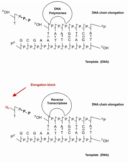
The reverse transcriptase (RT) of the viruses is a viral enzyme necessary for the catalytic formation of proviral DNA from viral RNA. This enzyme has been an active target for drug development for a number of years. Many inhibitors of viral RT which can be divided into two categories have been discovered. The first group consists of nucleoside reverse transcriptase inhibitors (NRTIs). These are competitive inhibitors, which act as DNA chain terminators. The others are the non-nucleoside reverse transcriptase inhibitors (NNRTIs). These are noncompetitive inhibitors and noncovalently bind at a site proximal but different from the site of the polymerase activity (Figure 5). Inhibitors of the latter class are particularly attractive drug candidates because their binding is highly specific to the reverse transcriptase, and therefore, they lead to less adverse side effects. A third group consist in protease inhibitors, which block the maturation of precursors of viral proteins and prevent the generation of mature viral proteins.
1.6.1 Nucleoside reverse transcriptase inhibitors (NRTIs)
NRTIs are structurally similar to the building blocks of nucleic acids (RNA, DNA) but differ from their natural analogues by the replacement of the hydroxy group in the 3′ position by another group that is unable to form the 5′ to 3′ phosphodiester linkage that is essential for DNA elongation (Stein D.S. and Moore K.H., 2001).
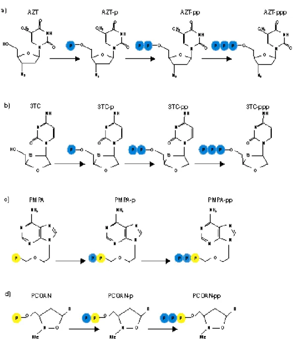
After the acyclic nucleoside analogues have been taken up by the cells, they have to be phosphorylated through three consecutive phosphorylation steps (Figure 6) before they can interact, in their triphosphate form, with their target enzyme, the viral DNA polymerase. Of crucial importance in this phosphorylation process is the first phosphorylation step which is ensured by a specific virus-encoded thymidine kinase (TK). Once that the compounds have been phosphorylated to the monophosphate, cellular kinases will afford
their further phosphorylation to the di- and triphosphate stages. In their triphosphate form, the compounds then interact as competitive inhibitors or alternate substrates with the normal substrates [2′-deoxynucleoside 5′-triphosphates (dNTPs)], and if they are incorporated into the DNA chain, they may act as chain terminators, thus preventing further chain elongation.
To work around this problem, have recently been synthesized compounds that have a phosphonate group instead of a phosphate group (ANPs, acyclic nucleoside phosphonates).
Figure 5: Mechanism of action of nucleoside and nucleotide analogues on reverse
transcriptase. In figure is reported the activities of a NRTIs, the AZT, on a cellular DNA polymerase (a) and reverse transcriptase (b).
Figure 6: Mechanism of phosphorylation of nucleoside and nucleotide analogues: a)
Acyclic nucleoside phosphonates possess a phosphonate group attached to the acyclic nucleoside moiety through a stable P—C bond. In contrast to the phosphate group (which is attached through a P—O—C bond), a phosphonate group (P—C bound) cannot be cleaved off by cellular hydrolases (esterases). Foremost among the acyclic nucleoside phosphonates that have been pursued as antiviral agents are cidofovir (HPMPC) [(S)-1-(3-hydroxy-2-phosphonylmethoxypropyl)cytosine], adefovir (PMEA) [9-(2-phosphonylmethoxyethyl)adenine], and tenofovir (PMPA) [(R)-9-(2-phosphonylmethoxypropyl)adenine] (De Clercq E., 2003).
Since the acyclic nucleoside phosphonates already contain a phosphate-mimetic group, stably attached through a P—C bond, they need only two, instead of three, phosphorylation steps to reach the active metabolite stage (De Clercq E. and Holy A., 2005). Thus, acyclic nucleoside phosphonates do not depend on the virus-induced kinase to exert their antiviral action, and, in “bypassing” the nucleoside kinase step, the acyclic nucleoside phosphonates may be expected to act against a broad range of DNA viruses, including hepadnaviruses (hepatitis B virus [HBV]) and retroviruses (human immunodeficiency virus [HIV]), i.e., all the viruses that use for their replication a DNA polymerase through which the active metabolites of the acyclic nucleoside phosphonates could enter into competition with the normal substrates (dNTPs).
1.6.2 Main nucleoside and nucleotide inhibitors used in therapy 1.6.2.1 Zidovudine
Zidovudine (3'-azido-3'-deoxythymidine, AZT) is an analogue of thymidine (Figure 7a) in which the 3’-hydroxyl group is replaced by an azido group. It is know that this compound is intracellularly phosphorylated by nucleoside and nucleotide kinase to its 5’-triphosphate, and that in this form inhibits retroviral reverse transcriptase.
The AZT remains the most common agent prescribed in the treatment of HIV infection, but it, combined with interferon-α, has been suggested as a first line treatment for ATLL. The mechanism of the anti-ATLL effects induced by AZT and interferon-α therapy have not been defined. Intracellular phosphorylation changes AZT into its active form (zidovudine triphosphate; ZDV-TP), which is incorporated into viral DNA and then reverse transcriptase is inhibited via DNA chain termination (Furman P.A. et al., 1986). However, ZDV-TP is reported to be incorporated into cellular DNA in vitro, its ability to inhibit cellular polymerases α and γ is 100-fold less potent than that to inhibit HIV reverse transcriptase (Copeland W.C. et al., 1992). Therefore, ZDV-TP probably does not directly affect cellular DNA. In fact, neither AZT nor interferon-α has direct toxicity against ATLL cell lines and against fresh ATLL cells obtained from patients who achieved complete remission after AZT and interferon-α therapy (Bazarbachi A. et al., 2000).
1.6.2.2 Lamivudine
Lamivudine (3TC), the negative enantiomer of 2'-deoxy-3'-thiacytidine (Figure 7b), is a dideoxynucleoside analogue used in combination with other agents in the treatment of human immunodeficiency virus type 1 (HIV-1) infection and as monotherapy in the treatment of hepatitis B virus (HBV) infection (Chang C.N. et al., 1992). Lamivudine undergoes anabolic phosphorylation by intracellular kinases to form lamivudine
triphosphate, the active anabolite that prevents viral replication by competitively inhibiting viral reverse transcriptase and terminating proviral DNA chain extension. The pharmacokinetics of lamivudine are similar in patients with HIV-1 or HBV infection, and in healthy volunteers. The drug is rapidly absorbed after oral administration, with maximum serum concentrations usually attained 0.5 to 1.5 hours after administration (Palumbo E., 2008).
Clinical investigations suggested that thia-2'-3'-dideoxycytidine (lamivudine; 3TC) could also be, at least partially, efficacious in thetreatment of HAM/TSP or ATLL (Machuca A. and Soriano V., 2000; Taylor G.P. et al., 1999). However, in vitrostudies have cleared up some misconceptions about using lamivudinein HTLV-1-infected individuals. In fact, we showed that lamivudinecompletely inhibited HTLV-1 infection of PBMC in vitro onlyat concentrations considered out of the range of those usedin antiviral therapy (Macchi B. et
al., 2003; Balestrieri E. et al., 2002).
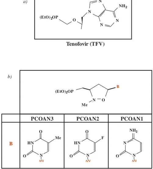
1.6.2.3 Tenofovir
The tenofovir disoproxil fumarate (TDF or bis (POC) PMEA) is a prodrug, of nucleotide tenofovir (TFV or PMPA) (Figure 8a). This is a nucleotide analogues of adenosine 5'-monophosphate, and has some efficacy against HIV RT (Cundy K.C. et al., 1998). The TDF was synthesized to increase the absorption of oral drug and to improve its cellular uptake (Robbins B.L. et al., 1998). After oral administration, the TDF is rapidly hydrolyzed in vivo in tenofovir, which is phosphorylated by cellular kinases in pharmacologically active metabolite called diphosphate tenofovir (Robbins B.L. et al., 1998). The PMPA owe its selective antiviral activity to the fact that in its diphosphorylated form, it has a higher affinity for the viral DNA polymerase than for cellular DNA polymerases α, β, γ, δ and ε. The PMPApp interact as competitive or alternate substrate
with the natural nucleotide deoxyadenosine 5'-triphosphate (ATP) for the viral DNA polymerase. The incorporation of one molecule of PMPA at the 3’ end of the growing DNA chain suffices to terminate further chain elongation (De Clercq E., 2003).
The comparison with the other NRTIs currently used for treatment of HIV infection indicates that within the class of NRTIs, the potential that tenofovir will produce adverse effects caused by drug-associated mitochondrial dysfunction is low (Birkus G. et al., 2002, Lee H. et al., 2003).
Lamivudine (3TC) HO O NH2 O N N S b) Zidovudine (AZT) O CH3 N3 O N O N O a)
Figure 7: Chemical structure of nucleoside analogues: a) Zidovudine (AZT) and b)
B PCOAN2 PCOAN3 PCOAN1 Tenofovir (TFV) B O N Me (EtO)2OP O NH2 N N F O O HN N Me O O HN N (EtO)2OP NH2 N N N N O a) b)
Figure 8: Chemical structure of nucleotide analogues: a) Tenofovir (TFV) and b)
phosphonated carbocyclic compounds of which shows the nitrogenous bases that distinguish the different PCOANs.
1.6.3 Non-nucleoside reverse transcriptase inhibitors (NNRTIs)
The nonnucleoside reverse transcriptase inhibitors (NNRTIs) are a structurally and chemically dissimilar group of antiretrovirals that are potent and highly selective inhibitors of HIV reverse transcriptase. Other retroviral reverse transcriptase enzymes, such as HIV-2, hepatitis viruses, herpes viruses, and mammalian enzyme systems, are unaffected by these compounds. Unlike the nucleoside analogs, the NNRTIs interfere with reverse transcriptase by noncompetitively binding directly to the enzyme downstream from the active catalytic site (Grob P.M. et al., 1992). The compounds are active in their native state, requiring no phosphorylation or other activity-dependent alteration. They are extensively metabolized in the liver; very little drug is excreted unchanged (Murphy R. and Montaner J., 1996).
The NNRTIs are potent antiretroviral agents that can successfully be used in appropriate triple-therapy regimens. The two main advantages of initiating therapy with an NNRTI-containing regimen are the ability to delay the use of protease inhibitors and the favourable adherence properties of NNRTIs, such as once-daily dosing. NNRTI performance in patients with advanced HIV disease has not been as impressive as that achieved with the protease inhibitors, but this may be due to suboptimal study design. Unfortunately, resistance to the NNRTIs can develop rapidly, often following a single mutation. This is particularly true if they are used as monotherapy or are added to a failing or suboptimal regimen of nucleoside analogs. Although structurally dissimilar to each other, all NNRTIs bind the reverse transcriptase enzyme in the same binding pocket. Nevirapine, efavirenz and delavirdine drugs are representative of this class.
Similarly, drugs that induce or inhibit the activity of cytochrome can have an effect on plasma concentrations of NNRTIs. The most common adverse drug reaction of NNRTIs, is the rash, observed in 35% of patients who received nevirapine (Ananworanich J. et al.,
2005). Nevirapine is also linked to serious liver toxicity (Nunez M. et al., 2001). Efavirenz is associated with the development of disturbances in the CNS (such as dizziness, hallucinations, insomnia) and fetal malformations, when administered to pregnant women (De Santis M. et al., 2002). Furthermore both nevirapine and efavirenz are associated with dyslipidemia.
1.6.5 Experimental evaluation of activity of reverse transcriptase
RT is a retroviral RNA-dependent DNA polymerase essential for retrovirus replication. Assay for RT activity are usually based on its enzymatic capacity to synthesise DNA from RNA templates. Traditional systems have relied upon incorporation of radiolabelled nucleotides for monitoring cDNA synthesis by using synthetic homopolymeric template-primers such as poly(rA)-oligo(dT) or poly(rC)-oligo(dG) (Neumuller M. et al., 1990; Lee M.H. et al., 1987). Methodological developments to improve sensitivity have included the use of non isotopic material (Urabe T. et al., 1992; Eberle J. et al., 1992). Afterwards, several PCR-based detection RT assays, called PERT (Product-Enhanced Reverse Transcriptase), have been described where RT reaction is performed on an RNA template such as the RNA from Brome Mosaic Virus or bacteriophage MS2 (Silver J. et al., 1993; Pyra H. et al., 1994). Actually PERT assay is a PCR-based RT assay that involves three steps: reverse transcription, amplification by PCR and detection of PCR products by ELISA. These PCR-based assays are several orders more sensitive than incorporation-based ones for the detection of RT activity.
Another ultrasensitive tests to evaluate RT activity is the Amp-RT assay, it utilizes a known heteropolymeric RNA template and a complementary DNA oligoprimer. The reverse-transcribed cDNA is detected by PCR amplification and Southern-blot hybridization (Yamamoto S. et al., 1996). The comparative analysis of seven retroviruses
with Amp-RT and the three conventional RT assays showed that Amp-RT was a generic with unprecedented sensitivity on both lentiviruses and oncoviruses. The high sensitivity of Amp-RT obviates the need for virus concentration which has complicated the detection of many oncoviruses by the conventional RT assays. Amp-RT was also found to be 100 and 10000 times more sensitive than antigen capture assays for HTLV-1 and HIV, respectively.
2. AIM OF STUDY
Clinical and experimental data on HTLV-1 infection have provided only partial answers that do not permit yet to define a protocol of treatment for these diseases. However, encouraging results have been obtained through the use of in vivo experimental combination of IFN-α and AZT in ATLL patients. In addition, some data from experimental procedures performed in vitro suggest the need for further studies aimed at identifying new compounds that can control the HTLV-1 life cycle. In particular the experience on HIV infection emphasized as phosphonates compounds (of which one example currently used in clinical is tenofovir) are able to check HIV infection. Data in other and our laboratory have revealed that tenofovir is active in blocking in vitro infection with HTLV-1 in different experimental models (Hill S.A. et al., 2003; Balestrieri E. et al., 2005). These findings prompted us on one hand, to further expand the study on new molecules endowed with RT inhibitory activity, on the others to develop more specific methods to test the inhibitory effect of new molecules on reverse transcriptase activity directly in patients. In line with these attempts, in a more recent work, we have shown the effects of a newly synthesized family of phosphonated nucleoside compounds, phosphonated carbocyclic 2′-oxa-3′-aza-nucleosides (PCOANs), on HTLV-1 infection in
vitro (Chiacchio U. et al., 2005; Balestrieri et al., 2008). To ascertain the anti-HTLV-1
activity of PCOANs, peripheral blood mononuclear cells from healthy donors were infected in vitro by co-culture with an HTLV-1 donor cell line in the presence of three prototype PCOAN compounds. PCOANs were able to completely inhibit HTLV-1 infection in vitro at a concentration of 1 μM, similar to what has been observed for tenofovir and azidothymidine. The mechanisms involved in the anti-HTLV-1 effects of PCOANs can mainly be ascribed to their capacity to inhibit HTLV-1 reverse transcriptase activity, as ascertained by means of a cell-free assay. Overall, these results indicate that the
family of PCOANs includes potential candidate compounds for long-lasting control of HTLV-1 infection. However, no information is available at the moment on the effects of potential anti-HTLV-1 anti-retroviral agents on RT activity in clinical isolates from HTLV-1 infected patients.
The principal aim of this study was to investigate: 1) the detectability of HTLV-1 RT activity in stimulated mononuclear cells from TSP/HAM and or ATLL HTLV-1-infected patients in comparison with chronically HTLV-1 infected cell lines, 2) the effects of RT inhibitors in vitro on reverse transcriptase activity from patients with TSP/HAM and/or ATLL, enrolled in different therapeutic protocol in comparison with those from chronically HTLV-1 infected cell lines.
3. MATERIALS AND METHODS
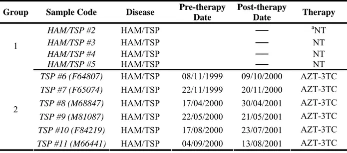
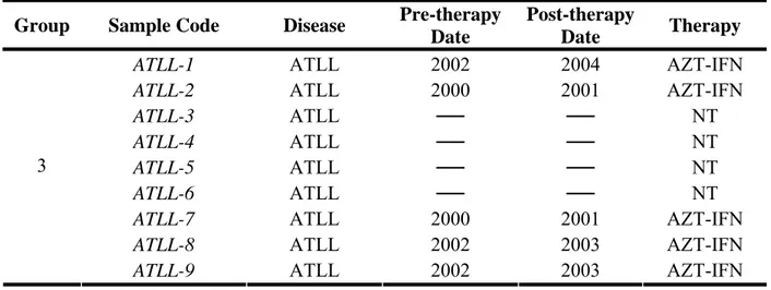
3.1 Characteristics of patients and sample collection
The patients enrolled in the study belong to two groups, represented by patients with HAM/TSP, or with ATLL diseases. Within these two groups are included both patients without therapy and a cohort of patients both pre- and post- therapy. The time of enrollment and the type of therapy are listed in the Table 3 and Table 4. In addition HAM/TSP patients failing AZT/Lamivudine protocol were enrolled in a therapy protocol encompassing treatment with TDF. PBMCs from heparinized blood of patients with HAM/TSP or ATLL were separated by Ficoll/Hypaque density gradient (Pharmacia Uppsala, Sweden), according the manufacture’s instructions, and then washed three times in phosphate buffered saline (PBS). To utilize virus isolates from patients as a source of RT for the subsequent assay, paired samples of PBMC from patients of different cohort were cultured at a concentration of 0.8x10⁶/ml in RPMI plus 15% FBS in the presence of 20 U/ml of IL-2, for at least 1 week before harvesting the supernatants. To quantify released virus in the culture medium, harvested supernatants were cleared at 1,000 g, filtered on 0,45μm and then ultracentrifuged at 30,000 g for 4 hr at 4°C (Beckman 45 TI). The presence of HTLV-1 p19 viral protein was detected in the viral lysates using a commercially available antigen capture ELISA (Retro-tek p19 antigen ELISA, Zeptometrix, Buffalo, New York). In comparison MT2 and chronic infected cells supernatants, were centrifuged and viral lysate assessed for p19 were used in the assays as positive control. The assays were performed according to the manufacturer's protocols.
3.2 Cell lines and infection of PBMCs
who were seronegative for HIV and B/C hepatitis viruses. Mononuclear cells were separated by Ficoll/Hypaque density gradient (Cederlane, Hornby, Ontario). The cells were then washed twice in RPMI-1640 medium (Gibco-Invitrogen Co., Paisley, Scotland UK). HTLV-1 infection was performed by co-culturing PBMCs with lethally irradiated (120 Cy, from a caesium gamma cell irradiator 1000, Canada Atomic Energy Ltd., Canada) MT-2 (irrMT-2) cells at a ratio of 5 PBMCs to 1 virus-donor cell. MT-2 is chronically HTLV-1-infected cell lines, derived from cord blood mononuclear cells exposed to HTLV-1 from leukemic patients. PBMC/irrMT-2 co-cultures were maintained at 0.8x10⁶ cells/ml in RPMI-1640 medium (Gibco) supplemented with 12% FBS, glutamine, penicillin-streptomycin (Gibco-Invitrogen; hereinafter referred as complete medium, CM) and 20 U/ml of recombinant interleukin 2 (IL-2; PROLEUKIN, Chiron Co., Holland).
In addition supernatant from few chronically infected cell lines was tested in the RT inhibitory assay. These cell lines have been generated either through limiting dilution of in toto PBMC infected with irradiated MT-2 donor cell line and from CD4+ and/or CD8+ T lymphocytes separated through immunomagnetic beads, and then infected by co-culture with irradiated MT-2 cells. The characteristics of these HTLV-1 chronic infected cells were listed in the Table 3.
Group Sample Code Disease Pre-therapy Date Post-therapy Date Therapy HAM/TSP #2 HAM/TSP ── aNT HAM/TSP #3 HAM/TSP ── NT HAM/TSP #4 HAM/TSP ── NT 1 HAM/TSP #5 HAM/TSP ── NT TSP #6 (F64807) HAM/TSP 08/11/1999 09/10/2000 AZT-3TC TSP #7 (F65074) HAM/TSP 22/11/1999 20/11/2000 AZT-3TC TSP #8 (M68847) HAM/TSP 17/04/2000 30/04/2001 AZT-3TC TSP #9 (M81087) HAM/TSP 22/05/2000 21/05/2001 AZT-3TC TSP #10 (F84219) HAM/TSP 17/08/2000 23/07/2001 AZT-3TC 2 TSP #11 (M66441) HAM/TSP 04/09/2000 13/08/2001 AZT-3TC
Group Sample Code Disease Pre-therapy Date
Post-therapy
Date Therapy
ATLL-1 ATLL 2002 2004 AZT-IFN
ATLL-2 ATLL 2000 2001 AZT-IFN
ATLL-3 ATLL ── ── NT
ATLL-4 ATLL ── ── NT
ATLL-5 ATLL ── ── NT
ATLL-6 ATLL ── ── NT
ATLL-7 ATLL 2000 2001 AZT-IFN
ATLL-8 ATLL 2002 2003 AZT-IFN
3
ATLL-9 ATLL 2002 2003 AZT-IFN
Sample Code weeks in colture IL-2 dependent CD4/MT2 6 + CD4/MT2 14 + B7(CD4/MT2) 44 + CD8/MT2 14 + PBMC/MT2 24 + MT2 ── -
3.3 Assayed compounds
Three licensed reverse transcriptase inhibitors, zidovudine (AZT), lamivudine (3TC) and tenofovir (TVF) and three experimental prototype compounds PCOANs referred to as, PCOAN1, PCOAN2 and PCOAN3 were used. The acyclic phosphonate tenofovir (kindly provided by Dr. Jan Balzarini, Rega Institute for Medical Research, Leuven, Belgium) and the NRTI AZT (Wellcome Research Laboratories, Beckenham, England, UK), were used as reference compounds. The PCOANs are a family of newly synthesized compounds which belong to a group of cyclic nucleoside phosphonates, with the furanose ring replaced by an N,O-heterocyclic ring (Chiacchio U. et al., 2003-2007). PCOANs were dissolved in DMSO and maintained at stock concentrations of 200 mM at –20 °C. Tenofovir and AZT were dissolved in culture medium without serum and maintained at stock concentrations of 100 mM at –20 °C. The compounds were diluted in culture medium without serum at concentrations appropriate to obtain the final usage concentrations just prior to their use.
3.4 RNA extraction
RNA isolation was performed using Nucleo Spin RNA kit and, to remove possible DNA contamination, RNA was treated with RNase free Dnase, aaccording to the manufacturer’s instructions (Machenery-Nagel, Dueren, Germany). The quantity and the quality of all RNA preparations were assessed by gel electrophoresis and OD260/OD280 ratios.
3.5 Genomic DNA extraction and pol region amplification
At 4 weeks p.i., cells from cultures were harvested and centrifuged on a density gradient to eliminate debris and dead cells. Cells were then incubated with proteinase K at 37°C for two hours, and DNA was extracted in phenol-chloroform-isoamylalcohol, according to
standard procedures. Five hundred nanograms of DNA was used as a template and amplified in a standard PCR mixture: 1× PCR buffer, a 0.2 mM concentration of each deoxynucleoside triphosphate (dNTP), 0.5 μM primer pair specific for the pol region of HTLV-1 (forward primer, CTGTCACAGAACTGCCGCAG-3′; reverse primer, 5′-AGCAGCAGTGGCCACTTG-3′) (Perkin Elmer, Boston, MA), and 1.25 U of Taq Gold polymerase (Promega, Madison, WI). As an internal control, the glyceraldehyde-3-phosphate dehydrogenase (GAPDH) gene was amplified using the same PCR mixture with a specific primer pair (forward primer, 5′-CCATGGAAAAAGGCTGGGG-3′; reverse primer, 5′-CAAAGTTGTCATGGATGACC-3′). Samples were subjected to 30 cycles of PCR amplification, each cycle consisting of 30 s at 94°C, 30 s at 55°C, and 45 s at 72°C on a DNA thermal cycler (Eppendorf, Hamburg, Germany). Following the final cycle, samples were incubated at 72°C for 7 min to ensure the completion of the final extension step.
3.6 RT inhibition assay
To test the sensitivity of HTLV-1 isolates from patients to RT inhibitors we set up a novel
ex vivo, cell-free HTLV-1-RT inhibition assay. This assay is a modified version of a
method we have described and utilized to screen the HTLV-1-RT-inhibitory activity of antiretroviral compounds in vitro (Chiacchio U. et al., 2005; Balestrieri E. et al., 2005-2008). The modification essentially consists of usage of isolates from patients as source of HTLV-1 RT in place of viral lysates from HTLV-1 chronically infected cell lines. As a template for reverse transcription, RNA was extracted from transfected cells ectopically expressing the glycoprotein D (gD) of herpes simplex virus type (Medici M.A. et al., 2003) and treated with RNase free DNase (see Figure 9). Compounds to which sensitivity was assayed were activated through pre-incubation with a crude extract, prepared from
phytohaemagglutinin (PHA) and interleukin-2 (IL-2) stimulated PBMCs from healthy, HIV/HBV/HCV negative donors. For preparation of the crude extract, 1x10⁶ PBMCs,
previously stimulated with PHA 2 μg/ml and IL-2 20 U/ml, for 72h in RPMI plus 20% FCS, were lysed in a buffer (50 mM Tris-HCl pH 7.4, 1 mM EDTA, 1 mM EGTA pH 7.4, 0.05% Triton-X, 150 mM NaCl, 0.25% sodium deoxycholate, 0.1% NP-40 and, freshly added, 1 mM PMSF, 15 μM DTT, 5 μg/ml leupeptin, 5 μg/ml pepstatin, 5 μg/ml aprotinin, 1 mM Na3VO4, 20 mM Na3F, all from Sigma) on ice and centrifuged at 10000g. Lysed
extracts (Figure 9) were incubated with the compounds at different concentrations for 15 minutes on ice and for successive 45 minutes at 30°C and then for 5 minutes at 95ºC to inactive the mixture. As a source of HTLV-1 RT ex vivo, amounts of viral lysates from different isolates obtained from HTLV-1 chronically infected cells, HAM and ATLL patient samples equivalent to 20 pg of p19 were utilized to obtain HTLV-1 RT preparations for each RT-PCR reaction. Total DNase-treated RNA (0,5 μg) was specifically reverse transcribed using 0.5 μM reverse primer specific for the gD gene US6
(5’ TGTCGTCATAGTGGGCCTCCAT 3’) in a reaction mix containing 1X RT buffer, 100U RNase inhibitor, 1 mM dNTP, 10 mM DTT, (all from Promega, Madison, WI) plus HTLV-1-RT preparations from patients in a volume of 10 μl. The reactions were performed in the presence or absence of activated substances, for 1h at 37 ºC. To investigate the dose-response, the assayed compounds, AZT, lamivudine, tenofovir and PCOAN1, PCOAN2, PCOAN3 were added at different concentrations. After incubation at 95 ºC for 5 minutes, 5 μl of RT reaction were used for DNA PCR in a reaction mix containing 1X Taq Gold buffer (Promega), 0.5 μM primer pair (US6 reverse, see above,
and US6 forward, 5’ AGACTTGTTGTAGGAGCATTCG 3’), 0.2 mM dNTP, 5 mM