SCUOLA NORMALE SUPERIORE – PISA
Transcription silencing and sub-nuclear positioning of
the HIV-1 provirus
Thesis submitted for the degree of Doctor Philosophiae
(Perfezionamento in Genetica Molecolare e Biotecnologie)
Candidate:
Supervisor:
Mariacarolina Dieudonné Alessandro Marcello
ABSTRACT... 7
INTRODUCTION ... 9
HIV-1: GENOME AND STRUCTURE... 9
Genome organization ... 9
Virion structure ... 11
HIV-1 LIFE CYCLE... 12
HIV-1 PROMOTER: LTR ... 18
REGULATION OF THE VIRAL GENE EXPRESSION... 20
Transcription Factors ... 20
Nuclear factor-kappa B: NF-κB... 20
NF-κB activators: ... 23
Tat trans-activator... 25
The Tat – TAR RNA interaction ... 26
Tat-mediated transcriptional activation... 27
Latency ... 34
Transcription Interference... 37
Chromatin environment... 39
Chromatin remodeling... 41
Histone modifications ... 42
Chromatin remodeling complex... 47
NUCLEAR ARCHITECTURE... 49
Chromatin ... 49
Euchromatin... 50
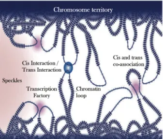
Chromosome territories... 53
Genome organization... 55
Transcription factories ... 57
NEW TECHNIQUES: IMAGING AND CHROMOSOME CONFORMATION CAPTURE... 60
Fluorescence imaging techniques... 61
MS2-based tagging of RNA ... 62
3D FISH... 65
Chromosome Conformation Capture: 3C... 66
Circular Chromosome Conformation Capture: 4C ... 68
3C-carbon copy (5C) and 4C on ChIP ... 69
RESULTS ... 71
SUB-NUCLEAR POSITIONING OF HIV-1 PROVIRUS IN LATENT CELLS... 71
Jurkat clonal cell lines... 72
HOS clonal cell lines... 73
U1 cellular model... 75
SUB NUCLEAR POSITIONING OF HIV-1 PROVIRUS IN TRANSCRIBING CELLS... 78
J-lat A1 characterization ... 79
Transcription activation... 79
Localization of HIV-1 provirus in transcribing J-lat A1... 82
RNA In situ Hybridization... 84
IDENTIFICATION OF GENOMIC SEQUENCES INTERACTING WITH HIV-1 PROVIRUS 86 MAPPING OF THE GENOMIC REGIONS BY 3D-FISH ... 93
LOCALIZATION OF HIV-1 PROVIRUS INDEPENDENT OF ITS INTERACTION WITH CH12... 99
HETEROCHROMATIN DISTRIBUTION... 100
HIV-1 INTEGRATION AND NUCLEAR PERIPHERY... 103
DISCUSSION... 106
GENE POSITIONING... 107
THE J-LAT A1 CELLULAR MODEL... 109
GENE POSITIONING AND TRANSCRIPTION ACTIVATION... 109
HIV-1 CHROMATIN CONFORMATION CHARACTERIZATION... 111
3D FISH AND HETEROCHROMATIN ASSOCIATIONS... 113
MATERIALS AND METHODS... 121
CELLS AND THEIR CHARACTERIZATION... 121
INTEGRATION SITE CONFIRMATION... 121
WESTERN BLOT ANALYSIS AND ANTIBODIES... 122
FLUORESCENT IN SITU HYBRIDIZATION (FISH)... 122
CIRCULAR CHROMATIN CONFORMATION CAPTURE (4C)... 124
FLUORESCENCE MICROSCOPY, IMMUNOFLUORESCENCE, RNA ISH... 126
Immunofluorescence ... 126
RNA in situ hybridization ... 127
RNA ISH in vivo... 128
STATISTIC ANALYSIS... 128
List of figures
Figure 1. Structure of the HIV-1 genome……….7
Figure 2. HIV-1 virion and structure of Gag polyprotein……….……8
Figure 3. HIV-1 Life cycle………..11
Figure 4. Structure of the HIV-1 viral promoter……….16
Figure 5. NF-κB pathway………20
Figure 6. P-TEFb activity…..……….….27
Figure 7. Potential transcriptional blocks in HIV-1 latency….……….………33
Figure 8. Post-transcriptional blocks of HIV-1 latency.……….……….34
Figure 9. Binding sites for the critical TFs within the HIV-1 LTR……….………..37
Figure 10. Epigenetic modifications..………...……….…..43
Figure 11. Chromatin remodeling complexes..……….….…45
Figure 12. Chromosome territories.………..………51
Figure 13. Nuclear Organization.………..….56
Figure 14. Spectral properties of variants of the GFP family………58
Figure 15. RNA detection MS2-based system ………...61
Figure 17. J-lat A1 cellular model ……….70
Figure 18. HIV24xMS2introECFPskl-IRES-TK lentiviral vector ………71
Figure 19. Localization of HIV-1 at the nuclear periphery in different cell clones …….74
Figure 20. J-Lat A1 Integration site………..……….76
Figure 21. Transcription activation in J-Lat A1 cell line………..……..77
Figure 22. Assembly of Cyclin T1 with Tat on induction of J-lat A1 cells………….……..78
Figure 23. Time course of provirus expression in J-Lat A1 cells……….78
Figure 24. Localization of HIV-1 in transcribing J-lat A1……….…….80
Figure 25. HIV-1 nascent RNA and its positioning at the nucleus in living cells………..82
Figure 26. Circular Chromosome Conformation Capture (4C) assays………..84
Figure 27. Diagram of the HIV-1 construct integrated in J-Lat A1………..85
Figure 28. 4C amplified fragments from J-lat A1 cells……….…87
Figure 29. Region of Chromosome 12 found to interact with the provirus by 3C and 4C techniques………..….88
Figure 30. 3C analysis at the site of HIV integration in J-lat A1 cells..………..….89
Figure 31. Analysis of the interaction between Chr12q12 and ChXp21……….…….92
Figure 32. Analysis of the interaction of Chr12 α-repeats with either ChXp21.1 or the provirus……….95
Figure 33. Localization of the interaction Ch12/HIV in the cell nucleus………97
Figure 34. Heterochromatin Distribution………..………..98
Figure 35. Relation between heterochromatin interaction and TPA activation………..100
Figure 36. HIV-1 integration and association with LADs………..….102
Figure 37. Schematic drawing that summarizes the concepts emerging from the experimental data……….117
Abstract
The human immunodeficiency virus (HIV-1) is a retrovirus that integrates into host cell’s chromatin for gene expression and replication. As integrated provirus HIV-1 is able to persist for long periods of time during antiretroviral therapy in quiescent memory T cells reservoirs. Understanding how these reservoirs are established, maintained, and reactivated is essential for developing methods to target and eventually eradicate HIV-1 infection.
Latency is likely established and maintained by numerous blocks at multiple steps in the HIV-1 gene expression pathway, which potentially complicates eradication strategies that aim at the purging of HIV-1 reservoirs from the infected patient. Recently, it has been proposed that the spatial distribution of genes within the nucleus contributes to transcriptional control allowing optimal gene expression as well as constitutive or regulated gene repression. Hence, the position of the provirus within the nucleus and its long-range interaction with other genomic regions could be another unexplored level of HIV-1 transcription control.
In order to gain insight in the conformation of chromatin at the site of HIV-1 integration we exploited seven different cell lines carrying a single latent provirus, which represent well-characterized models of HIV-1 latency. In the silenced state, the provirus was consistently found at the nuclear periphery. After induction of transcription the location of the transcribing provirus remained peripheral. Furthermore, in the J-lat A1 cell line, chromatin conformation studies revealed that the proviral vector is associated to a pericentromeric region of chromosome 12 (Ch12q12) located at the periphery of
the nucleus. Even though the location of the provirus did not change in transcriptionally active cells, the association between these two loci was lost. These results reveal a new mechanism of transcriptional silencing involved in HIV-1 post-transcriptional latency and reinforce the notion that gene transcription can occur also at the nuclear periphery.
Candidate's publications during the Ph.D. training:
Marcello A., Dhir, S, Dieudonné. Nuclear positional control of HIV-1 transcription in 4D. Nucleus 2010. [Epub ahead of print].
Dieudonné M, Maiuri P, Biancotto C, Knezevich A, Kula A, Lusic M, Marcello A. Transcriptional competence of the integrated HIV-1 provirus at the nuclear periphery. EMBO J. 2009 May 28. [Epub ahead of print].
Introduction
HIV-1: genome and structure
The acquired immunodeficiency syndrome (AIDS) is a life threatening disease caused by the human immunodeficiency virus type 1 (HIV-1). Following 25 years since its discovery the virus has killed more than 25 million people worldwide and it remains a major threat to public health and a challenge for drug development (Ho and Bieniasz, 2008). HIV-1 became the most studied virus in history and many years of research only partially unraveled the complexity of its life cycle. Studies on HIV-1 led to critical discoveries in antiviral drug development but also gave rise to new concepts in viral and cellular biology. However, the plasticity of viral sequences, the establishment of a latent state, and uncovered function of some auxiliary genes still remain a challenge in finding a cure for HIV-1 infection (Richman et al., 2009).
Genome organization
HIV-1 is a retrovirus, possessing an RNA genome that is replicated via a DNA intermediate. The full HIV-1 genome is encoded on one long strand of RNA and contains approximately nine thousand nucleotides. HIV-1 has several major genes encoding for structural proteins that are found in all retroviruses and several nonstructural (accessory) genes that are unique for HIV-1. The gag gene provides structural elements of the virus and the pol provides the replication enzymes. The Env gene codes for glycoproteins which are exposed on the surface of the viral envelope.
Accessory proteins (Tat, Rev, Vif, Vpr and Vpu) act at various stages of the various life cycle.
gag (Group-specific Antigen): codes for MA (p17), a matrix protein; CA (p24), the capsid protein; p6 and NC (p7) which build the nucleocapsid, and two spacer proteins (SP1,SP2).
pol (Polimerases): codes for viral enzymes such as reverse transcriptase (RT), intergrase (IN) and protease (PR).
env (Envelope): codes for gp160, the precursor to gp120 and gp41, proteins embedded in the viral envelope which enables the virus to attach to and fuse with the target cell.
tat (Trans-Activator of Transcription), rev (Regulator of Virion), nef (Negative Regulatory Factor), vif (Viral infectivity factor), vpr (Viral Protein R), vpu (Viral Protein U). The Figure 1 shows the structure of the HIV-1 genome.
Figure 1. Structure of the HIV-1 genome.
The open reading frames for various polypetides are shown as rectangles. http://bioquest.org/bioinformatics/edgridbeloit/levels.html
Virion structure
HIV-1 is composed of two copies of a single-stranded RNA enclosed by a proteic core (capsid), which is surrounded by the plasma membrane (envelope) of host-cell origin in which the viral glycoproteins are inserted. All structural proteins, which are components of the HIV-1 virion, are derived from the Gag polyprotein. Gag is co-translationally myristoylated, which allows anchoring of this polyprotein to the membrane.
Traditionally two forms of HIV-1 particles, the immature form and the mature form, have been observed. The immature virion is a roughly spherical shell of radically extended uncleaved and multimerized Gag molecules (Figure 2B). During particle maturation, viral protease (PR) is activated and cleaves Gag generating a set of new proteins termed matrix protein (MA), capsid protein (CA), nucleo capsid (NC), spacer proteins 1 and 2 (SP1 and SP2, respectively) and p6 (Figure 2A). These newly processed proteins then reassemble to form the distinct layers of the mature virion: MA remains associated with the inner viral membrane (the ‘matrix’ layer), NC coats the viral RNA genome (the ‘nucleocapsid’ layer), and CA assembles into the conical capsid that surrounds the nucleocapsid and its associated enzymes: reverse transcriptase (RT) and integrase (IN) (Figure 2C).
A
B C
A
Figure 2. HIV-1 virion and structure of Gag polyprotein.
A) Domain structure showing the localization of MA, CANTD (N-terminal domain of the capsid protein)
CACTD (C-terminal domain of the capsid protein) SP1, NC, SP2 and p6. Schematic models of the
immature (B) and mature (C) HIV-1 virions (Ganser-Pornillos et al., 2008).
HIV-1 life cycle
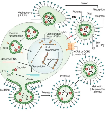
Human immunodeficiency virus (HIV-1) infectious cycle begins with the interaction of viral envelope glycoproteins gp120/gp41 with CCR4 or CXCR5 co-receptors, which are found in cells of the immune system such as CD4+ T cells and cells of the monocyte – macrophage lineage (Berger et al., 1999). CCR5 and CXCR4 co-receptors provide a critical function for virus entry. Macrophage-tropic viruses (R5 viruses) binds CCR5 and T cell tropic viruses (X4 viruses) binds CXCR4. After virus adsorption, the viral and cell membranes fuse together, and the viral 'core' is released into the cytoplasm, where the virion-associated reverse transcriptase is activated and begins synthesizing viral cDNA. The trigger for the beginning of DNA synthesis is unknown. However, it appears that initiation of reverse transcription is strictly linked to the uncoating process and it is possible that the exposure of the retrotranscription complex to the significant concentration of deoxiribonucleotides in the cytoplasmic environment is what allows reverse transcriptase to begin to act. Disassembly of the lentiviral core is characterized by dissociation of the Gag encoded capsid protein, CA (Bukrinsky et al., 1993). Finishing of reverse transcription originates the HIV-1 preintegration complex (PIC). The PIC form by integrase (IN), MA, reverse transcriptase (RT), NC, Vpr and viral retrotranscribed cDNA is delivered in to the nucleus (Farnet and Haseltine, 1991). In contrast to most retroviruses, HIV-1 is able to infect nondividing cells such as
differentiated macrophages, thus PIC needs to be translocated into the nucleus, where, the HIV-1 integrase, catalyses the integration of the viral cDNA into the host-cell genome. Integrated provirus is transcribed by the host – cell RNA polymerase II (RNAPII) producing a copy of the viral RNA genome that can be used to infect other cells (Haseltine, 1991). Transcription of the integrated viral cDNA leads to the production of genomic and messanger RNA (mRNA) molecules that are transported to the cell cytoplasm. Translation of HIV-1 mRNAs leads to the production of different proteins. Assembly and budding creates new virions through the infected cell membrane. Virus infectivity is acquired after particle maturation due to the actions of protease (Figure 3) (Monini et al., 2004).
To reach the nuclear membrane, the particles must travel through the cytoplasm. HIV-1 transport has been shown to exploit the cellular cytoskeleton. In particular, initial movements at the cell periphery occur in association with actin (Bukrinskaya et al., 1998), while subsequent translocation to the nucleus takes place along the microtubule network, likely by interaction of PIC with the dynein-dependent motor complex (McDonald et al., 2002). After reaching the nuclear envelope PIC is translocated through the nuclear pore, most likely by relying on the cellular nuclear import machinery.
Figure 3. HIV-1 Life cycle.
After infection of a cell, a nucleoprotein reverse transcription complex containing genomic viral RNA associated with virion proteins is deposited in the cytoplasm. Among the virion proteins are enzymes that catalyse the synthesis of complementary DNA (reverse transcriptase; RT) and that catalyse the integration of the viral cDNA into genomic cellular DNA (integrase). Following reverse transcription, the viral cDNA in the complex is transported to the nucleus to form the integrated provirus. The virus-encoded protein Tat and its associated cellular partner, cyclin T, promote the expression of genomic and subgenomic viral transcripts, which are exported from the nucleus by the action of the virus protein Rev and its cellular co-factor, CRM1. Genomic viral RNAs are transported to sites of virus assembly for incorporation into progeny virions. Subgenomic RNAs act as templates for structural virion proteins and regulatory/accessory proteins that perpetuate the infection and have various roles in virus–host interplay (Stevenson, 2003).
Like all retroviruses, HIV-1 integrates into the host chromatin. The process of integration of the linear viral DNA is carried out by the viral integrase protein (IN) and cellular factors like for example LEDGF/p75 (Llano et al., 2004). It has been shown that LEDGF/p75 acts as a chromatin docking factor for IN. LEDGF/p75 tethers HIV-1 integrase to chromatin, protects it from degradation, and strongly influences the genome-wide pattern of HIV-1 integration. Depleting the protein from cells and/or over-expressing its integrase-binding domain blocks viral replication (reviewed in (Poeschla, 2008)). Other proteins have been proposed as positive regulators of HIV-1 integration. HMG I (Y) dramatically stimulates integration reactions in vitro, probably by inducing changes in DNA structure (Farnet and Haseltine, 1991). Similarly, the integrase interactor 1 (Ini1), a subunit of the SWI/SNF chromatin remodelling complex, has been proposed to stimulate the in vitro DNA-joining ability of integrase (Kalpana et al., 1994).
For a long time integration of HIV-1 and more in general of all retroviruses was believed to occur randomly into the host chromatin. Recent reports have challenged this notion showing a bias for HIV-1 integration into transcriptionally active genes (Han et al., 2004). In addition, contrary to other retroviruses that integrate into promoters, HIV-1 viral genomes have been shown to reside within the introns of active host genes (Lassen et al., 2004). Once integrated into host genome, HIV-1 can undergo active transcription allowing continuous rounds of infection.
In an infectious state, the integrated provirus is actively transcribed from its promoter located in the U3 region of the HIV-1 LTR. Once the HIV-1 pre-mRNA transcript is produced, it can be spliced in alternative ways to yield three classes of mRNA in the
nucleus: unspliced mRNA (9 kb), singly spliced mRNA (4 kb) and fully spliced mRNA (2 kb). All three classes of mRNAs must be exported to the cytoplasm and translated into viral proteins for the viral life cycle to proceed. The fully spliced mRNAs, which are the first viral transcripts that appear after infection, are exported to the cytoplasm. They follow the same pathway of cellular RNA (Cullen, 1998) leading to expression of the regulatory proteins Nef, Tat and Rev. The negative factor (Nef) is a modulator of cellular signaling pathways that optimizes the cellular environment for virus replication. The transcriptional activator (Tat) up-regulates the synthesis of full length viral mRNA by elongation of RNAPII. The regulator of viral gene expression (Rev) acts at post-transcriptional level and plays a main role in Rev-dependent export of unspliced and incompletely spliced HIV-1 transcripts from the nucleus to the cytoplasm leading to the export of genomic RNA (Dorman and Lever, 2000).
After two months, in patients on Highly Active Anti-Retroviral Treatment (HAART) the plasma levels of genomic RNA falls below the limit of detection. Therefore, it was initially assumed that prolonged treatment might lead to eradication of the virus in these patients (Ho, 1998). Unfortunately, it is now clear that long-live reservoirs of HIV-1 can persist for years in the presence of HAART. This reservoir is thought to consist mainly of latently infected resting memory CD4+ T cells (Chun and Fauci, 1999) (Pierson et al., 2000). Both host transcription factors and the viral Tat trans-activator have been proposed as limiting factors for transcriptional reactivation of latent HIV-1. However, since HIV-1 is found integrated into the genome of resting memory T cells, it has been proposed that the chromatin environment at the viral integration site may play a role in the transcriptional silencing of the HIV-1 genome (Jordan et al., 2003) (Jordan et al., 2001). Indeed, integrated HIV-1 has nucleosomes positioned in its 5’ LTR that are
remodeled by deacetylase inhibitors, cytokines and Tat (Van Lint et al., 1996) (Lusic et al., 2003) (Marcello et al., 2004). Histones play an important role in regulating HIV-1 transcription since they integrate signals for repression, like the heterochromatin marker H3K9 trimethylation, Suv39H1 and HP1γ, and reactivation, like histone acetylation (Lusic et al., 2003). It is not clear for instance if also the three-dimensional (3D) nuclear architecture of the nucleus could be implicated in HIV-1 provirus regulation, as it has been proposed for cellular genes (Misteli, 2007).
Packaging of the genome RNA (gRNA) into a particle is a highly specific process that achieves selection of a single RNA species from the total capped polyadenylated mRNA in the infected cell (Kaye and Lever, 1999). Two copies of genomic RNA are encapsidated, usually linked at their 5’ end through the dimer linkage site (Darlix et al., 1990). Dimerization is associated with encapsidation (Greatorex and Lever, 1998), but dimer stabilization occurs post-capture by the Gag protein involving the NC. Viral RNAs to be specifically packaged are identified by the presence of an RNA sequence named the packaging signal (ψ) placed within the 5’leader (Berkowitz et al., 1995; McBride and Panganiban, 1996). However, the major determinants of packaging of HIV-1 reside in a short RNA segment placed between the 3’ splice donor and the Gag initiation codon (Lever et al., 1989). Deletion of the 5’leader sequence reduces the incorporation of gRNA into budding virions with little effect on other viral functions. The site of the packaging signal allows for the specificity of encapsidation that is limited to the unspliced HIV-1 mRNA.
The level of Gag may play a major role in determining the fate of the translated gRNA. Newly formed Gag molecules bind the gRNA via NC resulting in the inhibition of
translation and exposing the structural elements required to chaperone gRNA dimerization and the packaging process (DIS/SL3). Selective recognition of the gRNA is probably achieved bi Gag-NC molecules binding to SL1, SL2 and Sl3 of the Ψ signal (Brasey et al., 2003; Clever et al., 1995).
To a first approximation, all the information necessary for retroviral particle assembly resides in the Gag polypeptide. For example, Gag alone can form extracellular virus-like particles in the absence of other viral proteins (Gheysen et al., 1989), and Gag molecules can spontaneously assemble into spherical, immature virus-like particles in vitro (Campbell and Rein, 1999). All the viral proteins necessary for virion assembly and RNA genomes are transported to the plasma membranes close to lipid raft membrane domains, where the building of new virions begins. The Gag-Pol precursor binds to plasma membrane through the myristol group of the MA domain. The resulting virions bud from the plasma membrane but they are still incomplete. Their maturation is finished by viral protease (PR) that cleaves Gag and Gag-Pol. Further; Gag and Pol precursors are processed to originate the single core proteins, marix and the viral enzymes. The proteolytic activity ends when the virion is already detached from plasma membrane and results in the formation of mature infectious viruses.
HIV-1 promoter: LTR
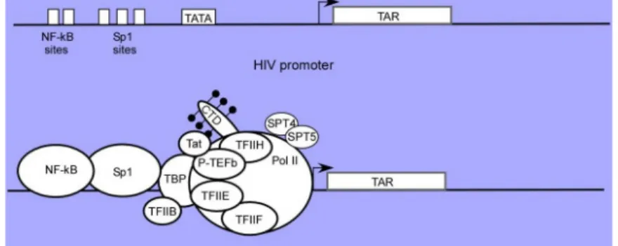
The U3 region of the HIV-1 LTR functions as the viral promoter. The viral LTR promoter has a structure typical of promoters activated by cellular RNA polymerase II. It contains several binding sites for general transcription factors. The HIV-1 promoter contains the core promoter sequence (TATA box) and enhancer elements placed
upstream and downstream of TATA box. Immediately upstream of the TATA box are two tandem NF-κB binding sites and three tandem SP–1 binding sites. Immediately downstream of the start of transcription is the transactivation response element (TAR) (Figure 4). Three tandem-binding sites for the constitutively expressed Sp1 transcription factor are necessary for basal levels of LTR-directed RNA synthesis. Mutation of individual or pairs of Sp1 sites has little, if any, effect on the basal or Tat-transactivated levels of expression (Harrich et al., 1990). However, the mutation of all three Sp1 sites markedly reduces the response to Tat (Berkhout and Jeang, 1992). Two tandemly arranged binding sites (κB sites) are recognized by the dimeric transcription factors composed of several combinations of members of the Rel/NF-κB family of polypeptides (Baeuerle and Baltimore, 1996). The predominant complex that binds to the LTR κB sites in activated cells is NF-κB (p50/p65 heterodimer). Moreover, the arrangement of the transcription factor binding sites in the LTR may vary in different HIV-1 subtypes but the specific contribution of these changes has not yet been thoroughly investigated.
Figure 4. Structure of the HIV-1 viral promoter.
The HIV-1 promoter is comprised of a series of transcription control elements including NF-kB, Sp1, TATA box, RNA initiation site and the downstream TAR RNA enhancer element. In the presence of Tat, a complex interaction between activators which include NF-kB and/or Sp1 bind to the upstream
control region and interact with transcription factors which include, but may not be limited to, TBP, TFIIH, P-TEFb and RNA Pol II. Tat and P-TEFb facilitate the binding of TBP to the complex, setting the stage for binding of other basal transcription factors and assembly of the preinitiation complex. In the initiation complex, although both TFIIH and P-TEFb are present, the Pol II CTD is phosphorylated primarily by TFIIH at Ser5 (black) (Brady and Kashanchi, 2005).
Regulation of the viral gene expression
The regulation of human immunodeficiency virus type 1 (HIV-1) gene expression involves a complex event of pathological significance, which recapitulates general concepts of cellular transcription with some peculiarities. After the integration of HIV-1 in the host genome viral transcription depends of many cellular factors and viral Tat trans-activator.
Transcription Factors
HIV-1 transcription in T lymphocytes is regulated by DNA-binding proteins which recognize sites in the regulatory region of the viral long terminal repeat (LTR). Stimulation of T cells results in increased expression of HIV-1, mediated by the inducible transcription factors (Bielinska et al., 1989). Two of the key transcription factors (TFs), upon which successful transcription depends, are NF-κB and NFAT.
Nuclear factor-kappa B: NF-κB
NF-κB is a members of the Re1 family of transcriptional activator proteins. This transcription factor is ubiquitously expressed and highly inducible. It plays an important
role in the innate/adaptive immunity, and cellular survival through the induction of genetic networks (Barnes and Karin, 1997) (Karin, 1999).
The Re1 protein family has been divided into two groups based on differences in their structures, functions, and modes of synthesis (Baeuerle and Henkel, 1994). The first group consists of p50 (NF-KB1) and p52 (NF-KB2), which are synthesized as precursor proteins of 105 (p105) and 100 (p100) kDa, respectively. Initially quiescent, this first class of proteins contains a long chain of repeats that inhibits their function. p105, is the precursor to one of NF-kB's monomers. Upon activation, the repeat chain is cleaved and P105 becomes p50. The second group generated by proteolytic processing, has a so-called Rel homology region (RHR) that contains one or more transcriptional activator region on the C-terminus side, dimerization domains and a nuclear localization signal. This group, which includes p65 (Re1A), Rel (c-Rel), RelB, and the Drosophila Rel proteins dorsal and Dif, are not synthesized as precursors.
Members of both groups of Rel proteins can form homo or heterodimers; such as, p50-p65 heterodimer (termed also NF-κB), which is the most abudant and biologically active (Huxford et al., 1998). NF-κB is a cytoplasmic complex whose nuclear translocation is controlled by its association with a family of inhibitory proteins, termed I-κBs (Baeuerle and Baltimore, 1996). Together, p50 and p65 dimerize around a 10 base pair region known as a -κB site, forming the NF-κB transcription factor. In unstimulated cells, the p65 subunit of NF-κB is retained in the cytoplasm through its interaction with inhibitor proteins. An inducing signal leads to the phosphorylation of I-κB and p105 (Henkel et al., 1993). This phosphorylation is thought to be the signal for I-κB degradation and p105 processing (to become p50), both of which generate active
NF-κB dimeric complexes that translocate to the nucleus and activate genes containing Rel protein-binding sites (κB sites) (Figure 5). In other words, activation of NF-κB results in the targeted proteolysis of I-κB, releasing NF-κB to enter the nucleus and bind to specific sequences in target promoters. The genomic actions of NF-κB are influenced by the stimulus applied and the promoter context/chromatin structure in which it binds (Tian and Brasier, 2003).
The subunit composition of the ReI complex influences its subcellular localization, transactivation potential, and mode of regulation. In addition to the heterodimer, p50/p65, homodimers of the Rel proteins exist also. The p50 homodimers do not induce transcription. They are thought to be used as post-induction repressors, competing with NF-κB following transcription activation subsequent to a viral invasion (Verma et al., 1995) (Baeuerle and Baltimore, 1996).
In the case of HIV-1, the enhancer region spanning -104 to -80 contains two consensus NF-κB binding sites that are identical with the κB site found on the promoter of the immunoglubulin (Ig) κ light-chain, also know as Ig κB site. The upstream and downstream κB sites on the HIV-1 LTR are referred to as Core II and Core I respectively (Figure 4) (Cron et al., 2000). The sequences of the κB sites and the four nucleotides spacer are highly conserved on most isolated of HIV-1 (Jeeninga et al., 2000) (Rodriguez et al., 2007).
The reactivation of silent HIV-1 provirus through NF-κB occur by the induction of multiples changes at the promotor of latent proviruses, including the recruiment of TFIIH and RNAPII (Kim et al., 2006) and changes in the local chromatin structure at
the HIV-1 LTR (Gerritsen et al., 1997) by recruiting histone acetyltransferases, such as CBP and p300 and chromatin remodeling factors (Lusic et al., 2003).
Figure 5. NF-κB pathway
Two pathways lead to NF-κB activation. In the classical NF-kB pathway, NF-kB dimers such as p50/RelA are maintained in the cytoplasm by interaction with an independent IkB molecule (often IkBa). Pro-inflammatory stimuli and genotoxic stress leads to IKKβ- and IKK-dependent phosphorylation of IκB, which results in proteasomal degradation and subsequent release of the NF-κB dimers. Activation of this pathway leads to increased transcription of genes in three functional classes. The alternative pathway works independently of IKKβ and IKK (Gilmore, 2006).
NF-κB activators:
TPA (12-O-tetradecanoyl phorbol acetate)
Phorbol is a natural, plant-derived organic compound. It is a member of the tigliane family of diterpenes. The most common phorbol ester is 12-O-tetradecanoylphorbol-13-acetate (TPA), also called phorbol-12-myristate-12-O-tetradecanoylphorbol-13-acetate (PMA), which is a potent tumor promoter able to activate the signal transduction of enzyme protein kinase C (PKC). The effects of TPA on PKC result from its similarity to one of the natural
activators of classic PKC isoforms, diacylglycerol. Therefore, TPA mimic T-cell mitogen activation and activate the NF-κB pathway.
Dinter and collaborators demonstrated that the HIV-1 enhancer is activated specifically by TPA in several non-lymphoid cell types, and that this transcriptional regulation can be reproduced in a cell-free system. In vitro transcription experiments revealed a 6-fold activation of the HIV-1 promoter in nuclear extracts prepared from TPA induced HeLa tk- cells (Dinter et al., 1987). Other studies done by Verdin, et al. have been shown that the 5' region of integrated HIV-1 DNA contains an array of precisely positioned nucleosomes. These nucleosomes define two large nucleosome-free regions corresponding to the promoter region (nt 200-452) and to the primer binding site region (nt 610-720). A nucleosome located in the R-U5 region in basal conditions was specifically disrupted following TPA or TNF-α treatment independent of transcription and independent of DNA replication (Verdin et al., 1993). Thus, TPA is used as a strong activator of HIV-1 transcription contributing to our understanding of HIV-1 regulation and ultimately AIDS pathogenesis.
Tumor Necrosis Factor: TNF
NF-κB activation is stimulated by a wide variety of both intra- and extracellular stimuli, including inflammatory cytokines, viral and bacterial products, growth factors, and prooncogenic signals (Baldwin, 2001). Among the proinflammatory cytokines, tumor necrosis factor alpha (TNFα) is one of the most potent activators of NF-κB in cell types possessing TNFα receptors (Baud and Karin, 2001). TNFα is a potent proinflammatory cytokine that plays an important role in immunity and inflammation, and in the control of cell proliferation, differentiation and apoptosis. TNFα is
produced primarily from macrophages and functions in the early phases of infection by activating and recruiting other immune cells through the production of chemokines and other proinflammatory cytokines (Tracey and Cerami, 1994). Overproduction of TNFα is also thought to contribute to the pathophysiology of several diseases including septic shock, cancer, AIDS, diabetes, and rheumatoid arthritis (Tracey and Cerami, 1994).
HIV-1 Tat protein amplifies the activity of tumor necrosis factor TNFα, stimulating HIV-1 replication through activation of NF-kB. In HeLa cells stably transfected with the HIV-1 tat gene (HeLa-tat cells), expression of the Tat protein enhanced TNFα-induced activation of NF-kappa. As we mentioned before, in an unTNFα-induced state, cellular I-kB proteins interact with NF-kB dimers to mask their nuclear location sequence, retaining the complex of NF-kB and I-kB in the cytoplasm. TNFα, like a variety of other inducers, can stimulate proteolytic degradation of I-kB by the proteasome, allowing the translocation of NF-kB into the nucleus and subsequent binding to its DNA motifs (Baeuerle and Baltimore, 1988) (Blank et al., 1992).
For TNFα-induced IKK and NF-κB activation, two TNF effector molecules are essential, the death domain kinase receptor interacting protein (RIP) and TRAF2. In response to TNFα treatment, IKK is recruited to the TNFα receptor complex through TRAF2, and its activation requires RIP (Liu, 2005).
Tat trans-activator
The trans-activator protein (Tat) is an unusual transcription factor because it interacts with a cis-acting RNA enhancer element, TAR, present at the 5’ end of all viral
transcripts. The function of Tat has been described for the first time by Sodroski et al who noted that synthesis of reporter genes placed under the control of the LTR increased 200- to 300-fold in the cells which have been previously infected by HIV-1 (Sodroski et al., 1985). They reasoned that the induction, or “transactivation” of transcription was due to the presence of a novel trans – activating factor, which they named Tat.
Transcription of the HIV-1 provirus is characterized by an early, Tat-independent and a late, Tat-dependent phase. Transcription from the HIV-1 LTR is increased several hundred-fold in the presence of Tat and the ability of Tat to activate transcription is an essential step in the HIV-1 life cycle.
The Tat – TAR RNA interaction
Tat function is mediated by the TAR RNA target element encoded within the LTR. TAR is located downstream of the initiation site for transcription (nucleotides +1 and +59). TAR RNA sequence forms a highly stable, nuclease – resistant, stem – loop structure. The stem – loop structure is located between +19 and +43 nucleotides which is the minimal sequence for transactivation. Because of its location in the R domain of the LTR, TAR is present in both viral RNA and DNA. Important structural features in the HIV-1 TAR are the stem, the pyrimidine – rich bulge, and the loop.
Tat binds TAR. Mutations that destabilize the TAR stem - loop by disturbing base – pairing abolish Tat – stimulated transcription. Tat forms a complex with TAR and recognizes the pyrimidine – rich bulge (+23 to +25) below the apex of the stem – loop. Tat interacts with the first nucleotide (U23) in the bulge and with two nucleotide pairs
on either side of bulge (G26 to C39 and A27 to U38 above the bulge, and A22 to U40 and G21 to C41 below the bulge).
Tat-mediated transcriptional activation
The interaction of Tat with TAR permits activation of HIV-1 transcription by promoting the assembly of transcriptionally active complexes at the LTR by multiple protein–protein interactions. Over the last few years, a number of cellular proteins have been reported to interact with Tat and to mediate or modulate its activity. These include general transcription factors, among which are TBP, TAFII250, TFIIB, TFIIH (Kashanchi et al., 1994) (Parada and Roeder, 1996), RNA polymerase II (Wu-Baer et al., 1995); transcription factor Sp1; the cyclin subunit of the positive transcription elongation factor complex (P-TEFb), cyclin T1, and different transcriptional co-activators that possess histone acetyltransferase activity (Figure 4). These interactions with P-TEFb and HATs appear to be of particular importance in the transcriptional activation of the viral promoter.
P-TEFb
Transcription elongation is up regulated by the positive transcription elongation factor b (P-TEFb), which consists of one component of the cyclin T family and of the cyclin-dependent kinase Cdk9. P-TEFb enables transition from abortive to productive transcription elongation by phosphorylating CTD in RNAPII and negative transcription elongation factors.
Although the HIV-1 LTR contains DNA binding sites for several transcription factors, in the absence of Tat, there is no expression of viral genes. However, there is
production of short, non-polyadenylated RNAs that include TAR RNA. The instability of synthesizing full-length viral transcripts is caused by the low processivity of RNAPII, which is overcome by P-TEFb. Thus, P-TEFb is crucial for efficient transcriptional elongation of HIV-1.
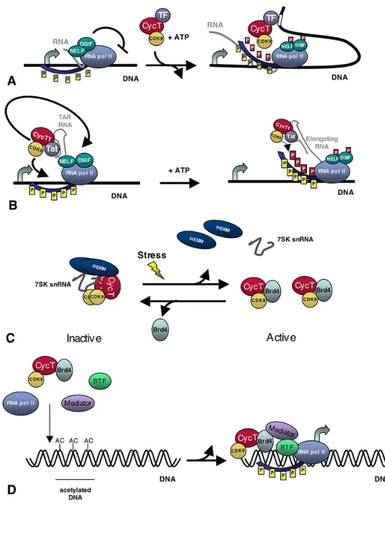
During the pre-initiation of HIV-1 transcription the CTD of RNA polymerase II (RNAPII) is phosphorylated by CDK-7, a component of TFIIH initiation factor (Figure 6A). CTD phosphorylation is an early step associated with the clearance of the promoter. Following clearance of the promoter, the phosphorylated RNAPII is able to transcribe through the TAR region. When the TAR RNA stem-loop structure is synthesized, the Tat protein binds to it and by interacting with cyclin T1, activates P-TEFb (Figure 6A). Then, CDK9 is recruited in close proximity to RNAPII and N-TEF, which become hyperphosphorylated allowing productive elongation (Figure 6B) and, as recently reported, also stimulating subsequent rounds of transcription complex assembly at the HIV-1 promoter and re-initiation (Raha et al., 2005). This molecular event is associated with increased transcriptional processivity (Figure 6B). The formation of the P-TEFb-TAR-Tat tripartite complex is an essential step towards the assembly of the processive RNAPII machinery at the LTR promoter (Bieniasz et al., 1998)
Recently have been indentified “hot-spot” genes that are frequently targeted in latent but not productively HIV-1 infected cells. One of these genes is the gene encoding bromodomain containing protein 4 (BDR4), which binds to the P-TEFb complex (Bisgrove et al., 2007). In many cell types P-TEFb exists in two complexes: large and small. P-TEFb is sequestered in an inactive ribonucleoprotein large complex which
additionally contains HEXIM1, heterogeneous nuclear ribonucleoproteins (hnRNPs) and the 7SK small nuclear RNA (snRNA) (Figure 6C). In response to stimuli such as stress, UV light or actinomycin D 7SK RNA, as well as HEXIM proteins and hnRNPs are released and P-TEFb is activated forming the small complex (Figure 6D) (Barrandon et al., 2007) (Nguyen et al., 2001). A fraction of this free P-TEFb is bound to BDR4, which may tether P-TEFb to actively transcribe genes. In vitro experiments have shown that the partitioning of P-TEFb between the active and inactive complexes changes rapidly in response to stress signals, which disrupt the 7SK ribonucleoprotein complex and promote the formation of BRD4/P-TEFb complexes (Figure 6D).
In vitro experiments demonstrated that HIV-1 Tat protein competes with Hexim1 for binding to the cyclin T and that Tat expression releases P-TEFb from sequestration by Hexim1 and 7SK. Thus, Tat can access the active P-TEFb complex by competing with Hexim1 and can substitute for Hexim1 in the absence of stress signals. The carboxyl terminus of BRD4 mediates its interaction with TEFb and targets the site within P-TEFb bound by Hexim1 and Tat. The HIV-1 transactivator Tat and BRD4 competed for binding to P-TEFb, and the carboxyl-terminal BRD4 peptide suppressed HIV-1 transcriptional activation by Tat at low concentrations (Bisgrove et al., 2007).
Figure 6. P-TEFb activity.
(A) P-TEFb is recruited near paused RNAPII via specific transcription factors and/or activators (TF), such as myc or NF-kappaB. P-TEFb then phosphorylates negative elongation factors, NELF and DSIF, as well as the C-terminal domain of RNAPII (red P), which allows for productive elongation (yellow P
represent phosphorylation by TFIIH). Both DSIF and NELF remain bound to RNAPII and may play other roles during elongation. (B) Tat recruits P-TEFb to the TAR RNA structure that forms at the 5' end of the nascent HIV RNA. Then, CDK9 phosphorylates RNAPII as well as NELF and DSIF to allow
for productive elongation (C) 7sk/HEXIM binds and sequesters P-TEFb in an inactive form in the nucleus. Stress signals, induce release of P-TEFb from it's inhibitory complex. Release of active P-TEFb allows for its association with Brd4 and perhaps other transcription factors which target P-TEFb to specific gene targets. (D) Brd4 recruits active P-TEFb to acetylated DNA during transcription initiation. The Mediator may also facilitate recruitment of P-TEFb via its association with Brd4 (Marshall and Grana, 2006).
Histone Acetyltransferase: HATs
HATs are enzymes that acetylate histone proteins by transferring an acetyl coenzyme A (acetyl-CoA) onto the ε-amino group of specific lysine residues present in the amino-terminal tails of each of the core histones, resulting in the neutralization of a single positive charge (Allfrey et al., 1964). Acetylation has long been linked with events both in chromatin synthesis and assembly, as well as the establishment of transcriptionally active chromatin. Acetylation is an energy-intensive, dynamic phenomenon, the steady-state balance of which is mediated by the opposing activities of HATs and deacetylase enzymes. Each of these enzymes generally belongs to one of two categories (Brownell and Allis, 1996) (Garcea and Alberts, 1980). Type A are located in nucleus, this kind of HATs are involved in the acetylation of all four nucleosomal histones and have long been thought to promote transcription related acetylation. In contrast B type HATs, located at the cytoplasm, are believed to have an housekeeping role in the cell, acetylating newly synthesized free histones in the cytoplasm for transport into the nucleus, where they may be deacetylated and incorporated in to chromatin (Allis et al., 1985) (Guo et al., 1995).
The LTR acts as a very strong promoter when analyzed as naked DNA in vitro, but when it is integrated into the cellular genome is almost silent. Therefore, chromatin
conformation imposes inhibition onto the integrated promoter. Experiments performed both in vivo (Verdin et al., 1993) (El Kharroubi et al., 1998) and in vitro using the HIV-1 promoter reconstituted into chromatin (Sheridan et al., 1997) have shown that, independent from the integration site, nucleosomes at the 5’ LTR are precisely positioned with respect to the cis-acting regulatory elements. In the transcriptionally silent provirus, these nucleosomes define two large nucleosome-free areas, one associated with the promoter/enhancer in the U3 region and the other spanning the primer-binding site immediately downstream of the 5’ LTR. These two open regions are separated by a single nucleosome called nuc-1 that is specifically and rapidly destabilized during transcriptional activation. Complexes containing HAT activity facilitate transcriptional activation by modulating nucleosomal repression of specific promoters. This event destabilizes the histone-DNA interaction. The role of acetylation in the processing and deposition of displaced histones, could be related with not only the formation of nucleosomes from newly synthesized histones but also reconstitution of nucleosome core particles, including the reassembly of the higher order structure of chromatin (Csordas, 1990).
The HAT proteins responsible for the TAR-dependent Tat transactivation include the transcriptional co-activators p300, the highly homologous cAMP-response element binding protein (CREB) binding protein (CBP), the p300/CBP-associated factor (P/CAF), the general control non-derepressible-5 (GCN5) factor, the TIP60 protein, and the general transcription factor TAFII250. Lusic et al. has observed acetylation of histones H3 and H4 at distinct nucleosomal regions and the recruitment of the above mentioned HATs to the viral LTR promoter upon HIV-1 activation either with Tat or phorbol esters (Lusic et al., 2003).
The Tat-TAR-P-TEFb interaction increases the processivity of RNAPol II giving support that Tat plays role in transcription elongation. The initiation model quickly lost support when Kao et al. reported that in the absence of Tat the majority of RNA polymerases initiating transcription stall near the promoter (Kao et al., 1987). On the other hand it is also clear that optimal Tat transactivation of HIV-1 gene expression requires upstream transcription co-factors. Along these lines it has been reported that Tat physically interacts with the pre-initiation complex including transcription factors such as Sp1 (Jeang et al., 1993), TATA binding protein (TBP) (Majello et al., 1998), cylinE/cdk2 (Deng et al., 2002), TFIIH, Tip60 (Kamine et al., 1996) and RNAPII (Cujec et al., 1997). The evidence of Tat interaction with histone acetyltransferases supports the role of Tat in chromatin remodeling before the onset of viral transcription. It is possible therefore that Tat facilitates chromatin remodeling, pre-initiation complex assembly and transcription elongation in a sequential manner that leads to high levels of HIV-1 transcription. All of these activities of Tat are probably exerted by the transient formation of large subnuclear complexes at the site of HIV-1 transcription and must therefore be spatially and temporally regulated (Molle et al., 2007). It has been demonstrated (Marcello et al., 2003) that cyclin T1 and p300 interact with the promyleocytic leukemia (PML) protein within specific subnuclear compartments that are coincident with PML nuclear bodies. This observation suggests that PML bodies could regulate Tat activity by modulating the availability of essential factors to the transcriptional machinery.
Latency
The major problem in curing AIDS is a reservoir of memory CD4+T cells, in which HIV-1 resides in a latent form, allowing the virus avoid host immune responses and antiretroviral drugs. Because latency represents a barrier to curing HIV-1 infection, it is crucial to understand the molecular mechanisms which are involved. It is becoming clear that HIV-1 latency is a complex multifactorial phenomenon that ultimately results from the profound differences between resting and activated CD4+T cells (Lassen et al., 2004) (Marcello, 2006).
There are two forms of HIV-1 latency, in the first one, full length viral DNA molecules; the final product of reverse transcription, remain unintegrated in resting CD4*T cells. In this case, when the infected cells are activated by signals from the microenvironment of the lymphoid tissues this extrachromosomal viral DNA integrates into the host genome and produces viral progeny (Bukrinsky et al., 1991) (Zack et al., 1990). Even though preintegration latency is quantitatively dominant in untreated individuals, this labile form of latency decays rapidly when the new infection of resting CD4*T cells is halted with HAART (Pierson et al., 2002) (Zack et al., 1990), making it less of a clinical concern comparing to the stable post integration form. The post integration latency, which represent the second kind, occur when infected CD4*T cells lymphoblast revert to a resting memory state after the integration of the viral genome into the host genome.
Activation of gene expression relies on the combined activity of a series of cellular factors that respond to different external stimuli, and on the function of the viral regulatory protein Tat. In consequence transcriptional activation is a result of both chromatin remodeling and the recruitment of elongation competent RNA polymerase
II complexes onto the integrated promoter, two events that require the coordinate, but transient assembly of different protein complexes. Today many mechanisms playing roles in the regulation of viral expression are well described and they involve interplay between host cell and HIV-1 proteins. Between the most important are:
Latency is achieved and maintained by several mechanisms that predominantly operate at transcription level (Lassen et al., 2004) (Contreras et al., 2006) such as:
Heterochromatin formation at the site of provirus integration, which can allow or not the accessibility of the integrated provirus to the transcriptional machinery (Jordan et al., 2001) (Jordan et al., 2003) (du Chene et al., 2007).
The absence in resting CD4+T cells of the active forms of the host transcription factors (TFs), that are necessary for HIV-1 gene expression (Nabel and Baltimore, 1987) (Marcello et al., 2003) (Karn, 1999).
Transcription interference (TI) from the endogenous gene harboring the provirus (Han et al., 2008) (Lenasi et al., 2008).
The presence of transcriptional repressors (He and Margolis, 2002)
The premature termination of HIV-1 transcripts due to the absence of the viral protein Tat and tat associated host factors (Kao et al., 1987).
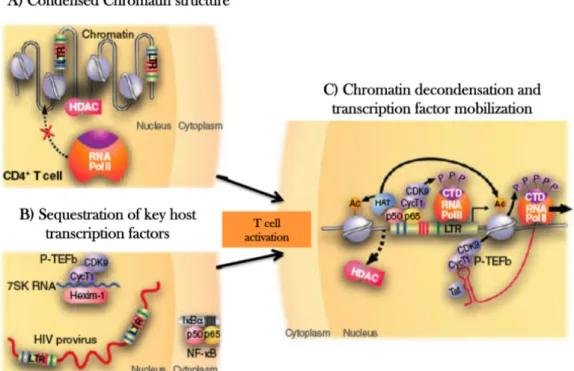
All these mechanisms show a general picture of the potential transcriptional blocks in HIV-1 latency (Figure 7).
Figure 7. Potential transcriptional blocks in HIV-1 latency.
A) Proviral latency is maintained in part, by the action of several transcriptions factor that recruit HDCAs and other complexes to the HIV-1 long terminal repeat (LRT) promoter, which results in histones modifications within chromatin at the HIV-1 promoter that limit the ability of RNA polymerase to initiate transcription. B) Key cellular factors that are required for robust HIV-1 transcription, such as NF-κB or the P-TEFb-cyclin complex, are sequester in resting CD4+Tcells by cellular regulatory complexes (e.g. inhibitor of nuclear factor κB (IkB)). Release and mobilization of these factors is required for proviral expression. C) When histones acetyltransferases (HATs) substitute the effects of HDACs, coactivators such as NF-κB cab recruit RNA polymerases complexes. Production of Tat allows the recruiment of P-TEFb, mediating and explosive increase in transcription and the escape of provirus from latency (Richman et al., 2009).
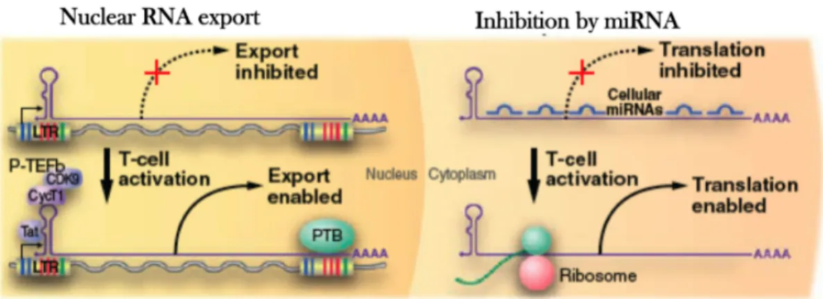
In addition, there are also mechanisms that block post-transcriptional activity such RNA interference that possibly binds as miRNA to HIV-1 RNA which is in turn not translated into viral proteins (Figure 8) (Lassen et al., 2006) (Huang et al., 2007).
Figure 8. Post-transcriptional blocks of HIV-1 latency
In the left panel (nucleus), the initial wave of Tat production may be further restricted by inefficient export of multiple spliced HIV-1 mRNAs, relieved upon cellular activation by enhanced ecpression of PTB. In the right panel (cytoplasm) cellular miRNAs that bind HIV-1 mRNAs may also restrict translation of early expressed HIV-1 mRNAs and so reduce tat production (Richman et al., 2009).
Transcription Interference
Integration of HIV-1 in the host genome could lead to occurrences of a phenomenon called transcriptional interference (TI), where an ongoing transcription from a host promoter would prevent pre-initiation complex assembly on the 5’LTR, thus interfering with the viral transcription or viceversa. (Lenasi et al., 2008) (Han et al., 2004).
TI is a direct consequence of the nature of HIV-1 integration sites in vivo. There are several potential molecular mechanisms for TI. Transcriptional activation of the
upstream promoter reduces transcription from the downstream promoter. Typically, this occurs when upstream transcription fails to terminate, so that the polymerase ‘reads through’ into the downstream gene, thereby interfering with initiation at the downstream promoter (Lassen et al., 2004). Footprinting studies showed that upstream read-through transcription complexes impaired the binding of Sp1 to the downstream HIV-1 promoter. This promoter occlusion mechanism has been directly demonstrated in pol I transcription systems (Henderson et al., 1989) and the effect could be blocked by the insertion of a strong transcriptional terminator between the two promoters. In addition, TI affects the gene expression from the HIV-1 long terminal repeat (LTR) in a system in which tandem HIV-1 promoters were integrated into the genome of HeLa cells (Greger et al., 1998), altered levels of transcription can result from changes in supercoiling caused by the transcription of linked genes (Dunaway and Ostrander, 1993), also in the case of two adjacent genes, competition for trans-acting factors might lead to TI. The spreading of epigenetic changes, such as methylation or histone modification, might also be involved (Eszterhas et al., 2002). For HIV-1, promoter occlusion remains a probable mechanism for TI, given that the observed distance between the HIV-1 LTR and the relevant host gene promoter is, on average, 30 kb (2– 429 kb) (Han et al., 2004).
Because HIV-1 genomes are found predominantly in the introns of active host genes both in activated and resting CD4+T cells, it is likely that the virus has evolved to function in this environment. The orientation of the HIV-1 genomes with respect to the host genes is not biased (Han et al., 2004). However, strong TI can occur between two adjacent transcription units, oriented in convergent, divergent or in tandem order (Eszterhas et al., 2002).
There are no known transcriptional terminators that would protect the HIV-1 LTR, and HIV-1 sequences can be found in the primary transcripts of host genes (Han et al., 2004). Therefore, TI is a virtually inevitable consequence of the nature of HIV-1 integration sites and might contribute to 1 latency. In activated CD4+T cells, HIV-1 gene expression might be efficient because the concentration of crucial host transcription factors is high enough to overcome TI. In consequence, TI might become more important as the cell reverts to a resting state and crucial transcription factors become limiting. However, it has been also observed coo expression of the endogenous genes and HIV-1 at the same loci.
Chromatin environment
The levels of viral expression are influenced by the chromatin environment of cellular DNA at the integration site of the provirus. Recently the first in vivo analysis of integration sites (Han et al., 2004) demonstrate that in purified resting CD4+T cells from patients on HAART, the cell population in which latent HIV-1 has been most extensively characterized in vivo, viral integration into transcriptional units was strongly favored (93%) (Han et al., 2004). In contrast, data presented by Lewinski, using latent cells generated in vitro, suggest that three chromosomal features correlated with inducible expression: centromeric heterochromatin, gene deserts, and highly active host transcription units, all these situations may contribute to the latent population (Lewinski et al., 2005).
Even in the case that actively transcribed genes could favor HIV-1 integration, there are several factors that could form repressive chromatin structure on the HIV-1 LTR which
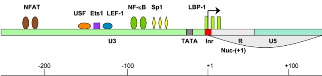
nucleosomes are found in the HIV-1 LTR. Nuc-(0) spans the region from positions −415 to −255 and Nuc-(+1) from positions +1 to +155 with respect to the transcription start site of HIV-1 genome. The region covered by and between these two nucleosomes contains recognition elements for the sequence-specific host TFs (Steger and Workman, 1997). The most important ones, for transcription of the HIV-1 genome are: NF-κB, nuclear factor of activated T cell (NFAT), upstream stimulating factor (USF), Ets1 (a protein that binds ETS - a winged helix-turn-helix domain), lymphoid enhancer binding factor-1 (LEF-1), stimulatory protein 1 (Sp1) and leader binding protein-1 (LBP-1) (Figure 9). The availability of certain cellular proteins directs the binding of chromatin remodeling complexes and consequently the formation of the preinitiation complex.
Figure 9. Binding sites for the critical TFs within the HIV-1 LTR
LTR is composed of three regions: 3′ untranslated region (U3) (green), transcription regulatory region (R) (grey) and 5′ untranslated region (U5) (blue). The following host TFs which bind to the HIV-1 LTR are designated: NFAT (brown), USF (orange), Ets1 (purple), LEF-1 (blue), NF-kB (green), Sp1 (yellow) and LBP-1 (yellow-green). TATA box and initiator element are marked in grey and red, respectively. Nucleosome 1 location is from +1 to +155 (Nuc-(+1)). Arrow marks the transcription start site (Contreras et al. 2009).
NF-κB plays a crucial role in the repression/de-repression of local chromatin structure surrounding the HIV-1 LTR. When the levels of free NF-κB in the cell are too low, the
same binding sites are occupied by p50:p50 homodimer which binds the histone deacetylase 1 (HDAC1). HDAC1 promotes deacetylation of surrounding histones, compaction of chromatin and consequently suppression of gene expression (Zhong et al., 2002). Williams et al. demonstrated that the recruitment of NF-κB to the HIV-1 LTR relieves this repressive chromatin environment by removing HDAC1 (Williams et al., 2006b). In this situation, HATs bind to the LTR and cause histone acetylation inducing the relaxation of chromatin structure and activation of gene transcription (Kurdistani and Grunstein, 2003).
Chromatin remodeling
The importance of chromatin structure in the regulation of eukaryotic gene transcription has become much more widely accepted over the past few years. It has been clear for a decade that histones contribute to the regulation of transcription both in vitro and in vivo (Clark-Adams et al., 1988) (Han and Grunstein, 1988). More recent studies have led to the striking observation that several proteins complexes involved in transcription regulation, can function, at least in part by modifying histones or altering chromatin structure (Armstrong and Emerson, 1998) (Kingston and Narlikar, 1999). While it is clear that many of these proteins complexes have functions in addition to chromatin modification, they illustrate the importance of chromatin structure as a part of transcription regulation mechanism.
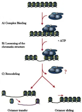
The most widely characterized chromatin-modifying complexes studied to date can be classified in two major groups, based on their modes of action as follows: (i) histones acetyltransferase (HAT) and histone deacetylase (HDCA) complexes, which regulate
the transcriptional activity of genes by determining the level of acetylation of the amino terminal domais of nucleosomal histones associated with them, and (ii) ATP dependent complexes, which use the energy of ATP hydrolysis to locally disrupt or alter the association of histones with DNA.
Histone modifications
The flexible N-terminal tails of the four histones (H2A, H2B, H3 and H4) undergo a range of post-translational modifications, including acetylation, methylation, phosphorylation and ubiquitination. Histone modifications are indicators of active or repressed chromatin, and the proposed “histone code” hypothesis suggest that combination of specific histone modifications create a complex, functional hierarchy for chromatin regulation. Specific histone modifications are responsible for the compartmentalization of the genome into distinct domains, such as transcriptionally silent heterochromatin and transcriptionally active euchromatin (Martin and Zhang, 2005). The ability of the histone code to dictate the chromatin environment allows it to regulate nuclear processes, such as replication, transcription, DNA repair, and chromosome condensation (Kouzarides, 2007).
Next to DNA methylation, histone acetylation and histone methylation are the most well-characterized epigenetic marks. Euchromatin is characterized by a high level of histone acetylation, which is mediated by histone acetyl transferases (HATs). Conversely, histone deacetylases (HDACs) have the ability to remove this epigenetic mark, which leads to transcriptional repression (Bartova et al., 2008). At least two general and distinct roles have been proposed for the acetylation of the amino-terminal histone tails. The first is based on circumstantial evidence that has for some time linked
acetylation with events that apparently require the attenuation of DNA-histone contacts within chromatin. The second proposed role suggests that acetylation affects the interaction of the amino-terminal tails with non-histone chromatin proteins that in turn modulate chromatin structure. A natural presumption of the first proposal is that acetylation weakens histone-DNA interactions within nucleosomes and thus affects directly the higher-order chromatin structure and ultimately leads to chromatin de-repression (Csordas, 1990). Indeed, hyperacetylation has long been considered a 'hallmark' of transcriptionally active chromatin (Turner and O'Neill, 1995) (Wolffe, 1994) and a wealth of mostly correlative evidence strongly supports the notion that acetylation potentiates transcriptional activity in chromatin (Hebbes et al., 1988). Numerous in vivo and in vitro experiments have indicated that transcription of some genes is influenced by the acetylation of particular lysine residues in specific histones (Thompson et al., 1994) (Durrin et al., 1991) and, conversely, that the transcriptional silencing of specific loci is associated with reduced nucleosomal acetylation.
Moreover, the number of methyl groups added to a single lysine or arginine residue can vary. Lysine residues can be mono-, di-, or trimethylated, and arginine residues can be monomethylated and symmetrically or asymmetrically dimethylated. Importantly, the precise methylation status (mono-, di-, or trimethylation) can influence the transcriptional status of genes (Schneider et al., 2004). These modifications could directly affect the structure of chromatin; e.g., by neutralizing the positive charge of histones. Notably different methylated states of the same amino acid residue provide additional hierarchical levels of regulation to epigenetic inheritance of chromatin domains. For example, acetylation of histones H3 an H4, and H3 methylation at Lys4 (H3 Lys4-Me), have been predominantly correlated with euchromatin and gene activity,
whereas methylation at Lys9 (H3 Lys9-Me) correlates with transcriptional silent chromatin (Litt et al., 2001). H3 Lys4 dimethylation (H3 Lys4-diMe) is associated with permissive chromatin that is either active or potentially active, and H3 Lys4 trimethylation (H3 Lys4-triMe) is linked with transcriptional activity. Conversely, H3 Lys9 di- and trimethylation (H3/K9-diMe and H3/K9-triMe) mark facultative and constitutive heterochromatin, respectively in mammals. Methylated H3/K9 can be “read out” by the Heterochromatin Protein 1 (HP1), a structural component of condensed chromatin that specifically recognizes and binds to the methylated form of H3/K9 (Bannister et al., 2001) (Lachner et al., 2001). Lack of K9 methylation in heterochromatin can affect the heterochromatin organization. Lanchner suggest that; HP1 itself interacts with the enzyme that methylates H3/K9, forming a positive feedback loop that would allow heterochromatin to spread over large chromosomal regions until further spreading is prevented by a boundary element. Furthermore, active promoters and enhancers are found to be associated with H3/K4 methylation and H3/K9 monomethylation. Moreover, active promoters can be identified by the presence of further activating marks that are linked with transcriptional elongation downstream in the transcribed region of the gene (Barski et al., 2007), in addition, at genes that are not regulated at the level of transcription initiation (polymerase II can be found at these genes) but rather elongation, H3/K4 methylation is enriched at the promoter. Thus, some activating marks are not only found on genes that are transcribed, but also on genes that are poised for transcription. Additionally, modifications considered to be repressing, such as H3/K9 dimethylation, can be found not only in heterochromatin but also on certain active genes and repressive and activated marks as H3/K27 trimethylation and H3/K4 trimethylation, respectively, can
coexist (Azuara et al., 2006) (Bernstein et al., 2006). All these findings suggest that the histone “code” is more complex than initially expected and that not single modifications, but rather the combination of modifications is an indicator for the transcriptional state.
Changes in chromatin structure that include modifications of histones may also have a role in the positioning of chromosomes. In experiments done in yeast, by Taddei and collaborators, the treatment of cells with trichostatin A, an inhibitor of histone deacetylases that increases the acetylation level of histones, results in large-scale movement of centromeric and pericentromeric chromatin to the nuclear periphery. After drug removal, these changes in localization are rapidly reversed (Taddei et al., 2001).
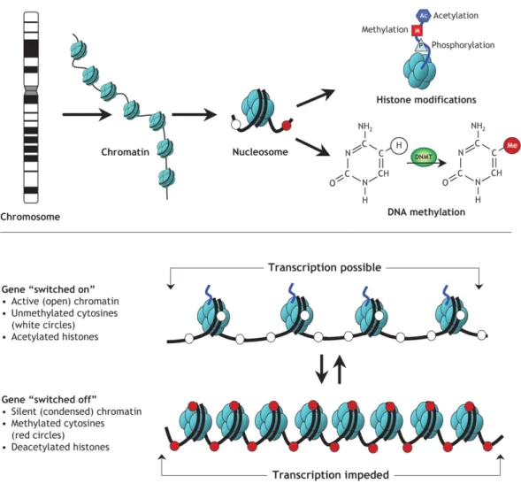
Figure 10. Epigenetic modifications.
Strands of DNA are wrapped around histone octamers, forming nucleosomes, which to be organized into chromatin, the building block of a chromosome. Reversible and site-specific histone modifications occur at multiple sites through acetylation, methylation and phosphorylation. DNA methylation occurs at 5-position of cytosine residues in a reaction catalyzed by DNA methyltransferases (DNMTs). Together, these modifications provide a unique epigenetic signature that regulates chromatin organization and gene expression. (B) Schematic of the reversible changes in chromatin organization that influence gene expression: genes are expressed (switched on) when the chromatin is open (active), and they are inactivated (switched off) when the chromatin is condensed (silent). White circles = unmethylated cytosines; red circles = methylated cytosines (http://cnx.org/content/m26565/latest/).