Epilepsy in cerebrovascular diseases: Review of
experimental and clinical data with meta-analysis of risk
factors
*
†‡
1Edoardo Ferlazzo, *
†
1Sara Gasparini,
§Ettore Beghi, †Chiara Sueri, ¶Emilio Russo,
¶Antonio Leo, *‡Angelo Labate, *‡Antonio Gambardella, #Vincenzo Belcastro,
**Pasquale Striano,
††Maurizio Paciaroni, ‡‡Laura Rosa Pisani, and *†‡Umberto Aguglia,
2
on behalf of Epilepsy Study Group of the Italian Neurological Society
Epilepsia, 57(8):1205–1214, 2016 doi: 10.1111/epi.13448 Dr. Edoardo Ferlazzo is assistant professor of neurology at Magna Graecia University of Catanzaro, Italy. Dr. Sara Gasparini is Research Associate at Magna Graecia University of Catanzaro, Italy.S
UMMARYObjective:Seizures may occur in close temporal association with a stroke or after a variable interval. Moreover, epilepsy is often encountered in patients with leukoaraio-sis. Although early post-stroke seizures have been studied extensively, less attention has been paid to post-stroke epilepsy (PSE) and to epilepsy associated with leukoaraio-sis (EAL). The aim of this paper is to review data concerning pathophysiology, progno-sis, and treatment of PSE and EAL.
Methods:We performed an extensive literature search to identify experimental and clinical articles on PSE and EAL. We also conducted a systematic review of risk factors for PSE and EAL among eligible studies.
Results:PSE is caused by enhanced neuronal excitability within and near the scar. The role played by white matter changes in EAL remains to be elucidated. Meta-analysis showed that cortical involvement (odds ratio [OR] 3.71, 95% confidence interval [CI] 2.34–5.90, p < 0.001), cerebral hemorrhage (OR 2.41, 95% CI 1.57–3.70, p < 0.001),
and early seizures (OR 4.43, 95% CI 2.36–8.32, p < 0.001) are associated with an
increased risk of PSE. As regards EAL, no prospective, population-based studies evalu-ated the role of different variables on seizure risk. Studies about the management of PSE are limited. PSE is generally well controlled by drugs. Data about risk factors, prognosis, and treatment of EAL are lacking.
Significance:Pathophysiology and risk factors are well defined for PSE but need to be elucidated for EAL. Management of PSE and EAL relies on the clinician’s judgment and should be tailored on an individual basis.
KEY WORDS:Animal models, Antiepileptic drugs, Leukoaraiosis, Seizures, Stroke.
Seizures may occur at the time of or in close temporal association with stroke (acute symptomatic, provoked, situation-related, or early seizures [ES]) or after a variable interval (several days/years) following stroke (late or
remote symptomatic seizures or post-stroke epilepsy; PSE). These two conditions have different pathophysiologic mechanisms: transient cellular biochemical dysfunctions for ES, and gliotic scarring with persistent changes in
neuronal excitability for PSE.1Moreover, these two condi-tions carry a different prognosis in terms of long-term (10 years) risk of seizure recurrence: 20% after a single ES and 60% after the first remote seizure.2These findings are at the basis of the new practical clinical definition of epi-lepsy, intended as a disease occurring even after a single unprovoked seizure if risk of recurrence within the next 10 years is at least 60%.3Although stroke-related ES have been extensively studied both in humans and in experimen-tal models, less attention has been paid to PSE. This is prob-ably due to the lack of collaboration between stroke physicians and epileptologists and led to focus on studies on clinical characteristics of strokes rather than on electroclini-cal features of seizures in PSE.4Moreover, a detailed analy-sis of the literature shows relevant differences in terms of definition, pathophysiology, and outcome of PSE. Seizures may also occur in patients with small vessel diseases (SVDs). SVDs have received little attention until recently, when modern brain imaging technologies allowed detecting small deep infarcts and white matter rarefaction (i.e., leukoaraiosis).4,5 Leukoaraiosis is often observed in adult patients with otherwise unexplained new-onset epilepsy (epilepsy associated with leukoaraiosis, EAL), but its epileptogenic role is still matter of debate.4,5SVD is com-monly caused by arteriolosclerosis or by more rare hetero-geneous conditions.5
This paper is a comprehensive review of the literature on experimental models, epidemiology and treatment of epilepsies associated with cerebrovascular diseases,
including PSE defined as remote symptomatic seizures fol-lowing a stroke, and EAL defined as epilepsy associated with leukoaraiosis due to arteriolosclerosis. To identify pre-dictors of epilepsy, we also performed a systematic review on risk factors of PSE and EAL. The aim of this review is to encourage both clinical and experimental research on this topic.
Methods
Literature review and search strategySee Supporting information, Data S1.
Systematic review and meta-analysis for risk factors of post-stroke epilepsy and epilepsy associated with leukoaraiosis
Inclusion criteria for PSE were the following: distinction between early and late post-stroke seizures; precise stroke timing; evaluation of at least one of seven predetermined variables as potential risk factors (age, sex, cortical involve-ment, primary hemorrhage or hemorrhagic transformation of ischemic stroke, infarct extension or severity, treatment with intravenous tissue plasminogen activator [tPA], occur-rence of ES, and treatment of ES). Exclusion criteria were the following: follow-up duration of<12 months and sam-ple size of<50 patients. According to the practical defini-tion of epilepsy,3 we considered a single late post-stroke seizure as equivalent to PSE. We followed the guidelines of the International League Against Epilepsy (ILAE),6which define seizures occurring within the first 7 days after stroke as ES and seizures occurring afterwards as PSE. Specific sub-analyses were planned to take into account differences in time-span for defining ES, study design, and population. As for EAL, the only inclusion criterion was the evaluation of at least one of five pre- determined variables as potential risk factors (age, sex, systemic hypertension, microbleeds at magnetic resonance imaging [MRI] studies, grading of leukoaraiosis on brain computed tomography [CT] or MRI studies), and the only exclusion criterion was sample size of <50 patients.
We used odds ratios (ORs) and 95% confidence intervals (CIs) to compare proportions of incident seizures between the exposed and the nonexposed groups. Heterogeneity
Key Points
•
We review data concerning pathophysiology, progno-sis, and treatment of epilepsies associated with cere-brovascular diseases•
Meta-analysis showed that cortical involvement, cere-bral hemorrhage, and early seizures are associated with an increased risk of post-stroke epilepsy•
Pathophysiology and risk factors for epilepsy associ-ated with leukoaraiosis remain to be elucidassoci-atedAccepted May 30, 2016; Early View publication July 6, 2016.
*Department of Medical and Surgical Sciences, Magna Graecia University, Catanzaro, Italy; †Regional Epilepsy Centre, Bianchi-Melacrino-Morelli
Hospital, Reggio Calabria, Italy; ‡Institute of Molecular Bioimaging and Physiology of the National Research Council (IBFM-CNR), Catanzaro,
Italy; §Laboratory of Neurological Disorders, IRCCS – Mario Negri Institute for Pharmacological Research Via La Masa, Milan, Italy; ¶Science of
Health Department, School of Medicine, Magna Græcia University of Catanzaro, Viale Europa, Catanzaro, Italy; #Neurology Unit, Department of
Neurosciences, S. Anna Hospital, Ravona, Como, Italy; **Department of Neurosciences, Rehabilitation, Ophthalmology, Genetics, Maternal and Child
Health, G. Gaslini Institute, University of Genova, Genova, Italy; ††Stroke Unit and Division of Cardiovascular Medicine, University of Perugia, Perugia,
Italy; and ‡‡IRCCS Centro Neurolesi Bonino-Pulejo, Messina, Italy
Address correspondence to Umberto Aguglia, Magna Graecia University of Catanzaro, Catanzaro, Italy and Regional Epilepsy Centre, Riuniti Hospital, Reggio Calabria, Italy. E-mail: [email protected] Postal address: Via G. Melacrino, 21, 89125, Reggio Calabria, Italy.
1
These authors contributed equally.
2See Appendix A for complete list.
Wiley Periodicals, Inc.
among studies was quantitatively assessed with the I2index (presence of heterogeneity when I2≥ 50%). Both fixed and random-effect models were used. Meta-analysis was per-formed with Comprehensive Meta Analysis, version 3.3.070.
Results and Discussion
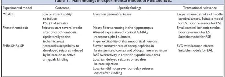
Pathophysiology: experimental and clinical data Post-stroke epilepsyEpileptogenesis of post-stroke scar is due to different mechanisms including changes in membrane properties, deafferentation, selective neuronal loss, and collateral sprouting leading to neuronal hyperexcitability and hyper-synchrony.1Most of the experimental data are obtained by two animal models, namely mechanical middle cerebral artery occlusion (MCAO) and photothrombosis. MCAO leads to cortico-subcortical stroke. However, this animal model develops ES but rarely PSE (Table 1).7In the pho-tothrombosis model, vessel occlusion with cortical stroke is induced by injection of fresh rose bengal dye activated in the brain through an external light beam: seizures start sev-eral weeks after vessel damage but mechanisms of epilepto-genesis are still controversial (Table 1).8,9 Cortical hyperexcitability of perilesional areas after stroke has been confirmed in humans by transcranial magnetic stimulation (TMS) studies.10In a series of patients with stroke involving primary motor cortex, Kessler et al.10 found a significant decrease of the TMS-induced silent period (SP) duration in either the arm or the leg on the affected side as compared to the corresponding unaffected limb in 5 of 6 PSE-patients, and in none of 76 controls without PSE. Kim et al.11found larger amplitude of TMS-induced motor-evoked potentials and intracortical facilitation in the affected compared to
unaffected hemispheres in 18 PSE patients, but not in 18 stroke patients without epilepsy.
Epilepsy associated with leukoaraiosis
Leukoaraiosis consists of white matter rarefaction due to SVD causing small deep infarcts. White matter rarefaction and SVD are observed in spontaneous hypertensive rats (SHRs) and their stroke-prone substrain, which represent useful models to assess hypertensive brain damage and related stroke (Table 1).12 The role of hypertension in modulating seizure susceptibility has been evaluated in different experimental studies. Accumulated evidence sup-ports the concept that there is an independent renin-angio-tensin system in the central nervous system.13Angiotensin peptides seem to operate through close interaction with different neurotransmitter systems including gamma-aminobutyric acid and adenosine.13 Tchekalarova et al.14 found that SHRs exhibited higher susceptibility than con-trol rats to kainate-induced status epilepticus, and that losartan (an AT1 receptor antagonist) delayed the onset of kainate-induced seizures in SHRs but not in control rats. Of interest, upregulation of AT1 and AT2 receptors has been found in hippocampus of patients who underwent anteromesial temporal lobectomy for seizure control, sug-gesting the renin-angiotensin system participation in the pathophysiologic mechanisms of underlying epileptogene-sis in these patients.15A few clinical studies evaluated the role of systemic hypertension as independent risk factor for seizures. In a case–control study including 521 subjects (227 patients with first unprovoked seizures and 294 controls) Ng et al.16found that history of hypertension is an independent risk factor (OR= 1.57) for new-onset unprovoked seizures. In a population-based case–control study including 145 incident cases of first unprovoked sei-zure and 290 controls, Hesdorffer et al.17found that severe
Table 1. Main findings in experimental models of PSE and EAL
Experimental model Outcome Specific findings Translational relevance MCAO Low or absent ability
to induce PSE (1 of 26 rats)
Gliosis in penumbral tissue Large ischemic stroke of middle cerebral artery. Suitable model for ES. Poor relevance for PSE Photothrombosis Seizures start several weeks
after photothrombosis (ipsilaterally to the ischemic area)
Mossy fiber sprouting in the hippocampus Small cortical ischemic stroke. Poor relevance for ES. Suitable model for PSE Altered expression of cortical GABAA
receptor alpha1 subunits
Hyperexcitability of thalamocortical neurons SHRs SHRs-SP Increased susceptibility to
developed seizures induced by kainate or selective amygdala kindling
Slower turnover rate of norepinephrine in brain stem and cortex and of dopamine in striatum
SVD with lacunar infarcts. Suitable models for EAL RAS overactivity in anterior hypothalamic area
Losartan delayed seizures onset after kainate injection
Losartan did not prevent or delay seizures onset after kindling
ES, early seizures; GABA,c-aminobutyric acid; MCAO, middle cerebral artery occlusion; PSE, post-stroke epilepsy; RAS, renin-angiotensin system; SHRs, spontaneous hypertensive rats; SHRs-SP, spontaneous hypertensive rats-stroke prone; SVD, small vessel disease.
uncontrolled hypertension was associated with an 11-fold increased risk of unprovoked seizure. Hypertension (esti-mated through left ventricular hypertrophy) as an indepen-dent risk factor (OR 2.9) for epilepsy was found in a cross-sectional, case–control study by Li et al.18including 4,944 subjects (65 of whom had epilepsy). However, this study was based on prevalent cases, thus limiting the inference on the pathogenic role of hypertension in these 65 patients. A few clinical studies evaluated the relation-ship between leukoaraiosis, SVD, and epilepsy. Maxwell et al.19 retrospectively evaluated 105 patients with late-onset epilepsy in comparison with 105 controls. They found that cerebrovascular disease, including severe SVD, was significantly more prevalent in patients with late-onset epilepsy than controls. However, about half of the patients included in these two studies did not undergo brain MRI, thus limiting the possibility to generalize the results. Okroglic et al.20retrospectively studied clinical symptoms and cerebrovascular risk factors in a cohort of 223 consec-utive patients with SVD, and found that about 20% of patients presented with “stroke symptoms and seizures,” especially in the presence of a high lesion load in frontal and occipitoparietal lobes. The authors speculate that some subcortical white matter lesions may also affect U-fibers, leading to a higher propensity for seizures. De Reuck and Van Maele21 investigated the amount of cognitive decline in 292 patients with leukoaraiosis, with and without sei-zures (n= 44 and 248, respectively), and found a higher proportion and severity of cognitive decline in patients with seizures. These authors speculated that an underlying neurodegenerative disease, rather than leukoaraiosis, might cause epilepsy in those patients. Gasparini et al.4analyzed the anatomical-electroclinical correlations of patients diag-nosed with PSE as compared to patients with EAL. This study included 117 patients (58 with PSE, 59 with EAL). All patients had undergone brain MRI. These two groups significantly differed in terms of seizure-onset localization: whereas in PSE patients the epileptogenic focus was coher-ent with the vascular lesions (see below), in EAL-paticoher-ents temporal lobe epilepsy predominated. However, the possi-bility of a mere incidental relationship between the two phenomena (epilepsy and leukoaraiosis) should be consid-ered, since leukoaraiosis is a common finding in the age group studied (advanced adulthood, mean age: 66.3 years) and temporal lobe epilepsy is the most frequent focal epi-lepsy at all ages. Therefore, it is still unclear whether leukoaraiosis can be considered as a cause of epilepsy, and the role of different variables, that is, cortical microin-farcts, needs to be elucidated. Because of very small size (<1 mm), most cortical microinfarcts are below the lower limit of spatial resolution (about 1 mm) for 1.5 T or 3 T MRI currently used in clinical practice.22The use of higher field (7 T) MRI may be a promising method to visualize sub-millimetric microinfarcts and to understand their role in EAL.
Systematic review and meta-analysis of risk factors for PSE
PubMed search retrieved 197 results, Scopus search retrieved 28 results, and Google Scholar retrieved 191 results. Of those, 371 were excluded on the basis of the title/ abstract or as duplicate findings, 20 did not fulfill inclusion criteria, and 5 fulfilled exclusion criteria (Fig. 1). One addi-tional paper was selected by searching references of original articles. Thus, the meta-analysis was based on 21 studies (Table 2) (for references, see Data S2) evaluating 21,548 stroke patients. Of these, 1,411 had PSE corresponding to a global prevalence of 4.7% in cohort studies (Table 2). Included papers differed in study design, history of seizures, population, and time-span defining ES (Table 2). The diag-nosis of stroke was made on the basis of clinical findings and brain imaging (CT or MRI) in all studies. Because age and infarct extension or severity were not reported in a con-sistent way across studies, meta-analysis could be per-formed on four variables (sex, cortical involvement, hemorrhage, and ES).
Analysis on cortical involvement
Six studies were included in this analysis. I2value was 0. Results are shown in Figure 2; cortical involvement was a predictor of PSE, bearing an almost fourfold risk of epilepsy (OR 3.71, 95% CI 2.34–5.90, p < 0.001). Sub-analyses con-firmed that cortical involvement was a predictor of PSE (Table S1).
Figure 1.
Flowchart showing inclusion and exclusion criteria of studies evalu-ated for meta-analysis of risk factors for PSE.
Analysis on hemorrhagic component
Fifteen studies were included in this analysis. I2 value was 84%. Results are shown in Figure 3. Presence of hemorrhage was a predictor of PSE, with a twofold risk of seizures (OR 2.41, 95% CI 1.57–3.69, p < 0.001). Sub-analyses confirmed that hemorrhage was a predictor of PSE, with two exceptions (Table S2). Indeed, when con-sidering only retrospective cohort studies or case–control studies, the association between hemorrhage and PSE was not statistically significant (OR 2.04, 95% CI 0.84–4.9, p = 0.12 for retrospective cohort studies; odds ratio 6.55, 95%CI 0.89–48.3, p = 0.07 for case–control studies; Table S2).
Analysis on occurrence of early seizures
Two studies were included in this analysis. I2value was 0. Results are shown in Figure 4; ES were a predictor, bearing a fourfold risk of PSE (OR 4.43, 95% CI 2.36–8.32, p < 0.001).
Analysis on sex
Thirteen studies were included in this analysis. I2value was 16%. Results are shown in Figure S1; sex was not a pre-dictor of PSE (OR 0.943 for female sex, 95% CI 0.79–1.13, p = 0.52). Sub-analyses confirmed that sex was not a pre-dictor of PSE (Table S3).
Variables not included in meta-analysis
The extreme variability in measurements of age (continu-ous variable in some studies, discrete variable with different cut-offs in other studies) and of size or severity of strokes prevented the use of these variables in the meta-analysis. Age was evaluated in 12 of 20 studies and younger age was associated with increased risk of PSE in 3 of these 12 studies (Table 2). Size or severity of stroke was evaluated in 12/20 studies and were associated with increased risk of PSE in 9 of these 12 studies (Table 2). Intravenous treatment with tPA was evaluated in a single study.23The authors found that PSE was more frequent among patients treated with thrombolysis compared to standard medical therapy (20.6 vs. 10.7%, p = 0.02), but the effect was lost in multivariate analysis.
Similarly, a single study evaluated the effects of early sei-zure treatment on PSE development.24In that study, starting an early antiepileptic treatment did not protect from later seizures (adjusted p at multivariable analysis = 0.170). Limits, main findings, novelty
Even with limitations due to the fairly poor quality of the included studies (about half of the studies were retrospective or did not use brain MRI), our meta-analy-sis indicates that cortical involvement, hemorrhage, and ES are associated with a higher risk of PSE; gender was not a predictor for PSE. Data about the effect of intra-venous tPA administration or of treatment of ES on
development of PSE were insufficient. In the only pub-lished meta-analysis on PSE,25 hemorrhage was not sig-nificantly associated with PSE. However, this meta-analysis25 excluded studies on children and included four studies that did not clearly distinguish between ES and PSE; finally, it is unclear how differences in the defini-tion of ES were treated.
Systematic review of risk factors for EAL
PubMed search retrieved 85 results, Scopus search retrieved 6 results and Google Scholar search retrieved no results. Of 91 results, 90 were excluded because they were not pertinent and one for a small sample size. Only one ret-rospective cohort study evaluated risk factors for epileptic seizures in patients with leukoaraiosis.26 This study included 242 subjects with previous lacunar stroke with or without leukoaraiosis; of these, 37 had seizures. Sex, sys-temic hypertension, and severity of white matter changes were not associated with an increased risk of seizures. Moreover, the authors found no differences in other vascu-lar risk factors, distribution and frequency of the lacunes, and outcome between the two groups. They concluded that there was no evidence that seizures were directly induced by lacunar infarcts and hypothesized that the occurrence of seizures in subcortical strokes could be explained by indi-rect or subtle cortical involvement. Of note, MRI was per-formed in about 50% of subjects; therefore, the number of vascular lesions could be underestimated. Moreover, this study was conducted in a selected population (patients admitted to hospital with clinical complaints in keeping with lacunar stroke) and grading of leukoaraiosis was not reliable.
To date, evidence of risk factors for EAL are insufficient. The role of hypertension and other possible determinants of epilepsy in patients with SVD need to be further evaluated.
Prognosis and treatment of PSE and EAL General considerations
PSE mainly affects the elderly; hence, physicians should consider the peculiar pharmacodynamics and pharmacoki-netics of antiepileptic drugs (AEDs) in this population, as well as the coexistence of different pathologies and, there-fore, interactions with concomitant treatments. Moreover, the elderly are more vulnerable to the adverse effects of drugs and the titration of the AEDs must take into account their narrow therapeutic range (“start slow and go slow”).27 In general, PSE has a good prognosis, being well controlled by AEDs. Indeed, an observational study by Semah et al.28 evaluated seizure frequency during 1–7 years in a cohort of 1,148 outpatients with focal epilepsy and MRI evidence of brain abnormalities. These authors found that the 36 patients with PSE had the best prognosis, 54% being seizure free. Moreover, in a prospective study by Stephen et al.29 evalu-ating 550 patients with focal epilepsy aged 16 years or
Table 2. Characteristics of studies included in the meta-analysis o n PSE Study Prevalence of PSE, % Stroke type Study design Population/ No. of included subjects
Pre-existing epilepsy excluded
Acute/remote seizures cut off Main predictors of PSE < 48 h ≤ 1 week (ILAE-defined ES) 2– 4 weeks Un specified Cortical involvement Exten sion/ Sever ity Hemor- rhage Female gender Early seizures Treatment of early seiz ures Young er age Intra -venous tPA Olsen et al. (1987 ) 41 9 Ischemic and hemorrhagic P A/77 Yes x + NE NE 0 N E N E N E N E Sung et al. (1989) 42 4.6 Hemor rhagic R A/1,402 No x + 0N A N E N E N E N E N E Kotila and Waltim o (1992 ) 43 17 All types R A/200 No x 0 NE ++ NE NE NE NE Heuts-Van Raak et al. (1996) 44 13 Ischemic P A/322 No x N E + 0 0 NE NE 0 N E Burn et al. (1997) 45 7 All types P A/675 No x N E ++ NE NE NE 0 N E Awada et al. (1999 ) 46 NA Ischemic C A/371 No x ++ NA NE NE NE 0 N E Teasell et al. (1999) 47 7.8 All types P A/549 Yes x N E N E + NE NE NE NE NE Berges et al. (2000) 48 4.9 Unspecified R A/3,205 Yes x N E + 0N E N E N E N E N E Lossius et al. (2002) 49 2.5 All types P A/472 No x 0 + 0 0 NE NE 0 N E Lamy et al. (2003) 50 5.5 Ischemic P A/581 No x + 0 + 0N E N E 0 N E Benbir et al. (2006) 51 3.6 All types P A/1,428 Yes x N E N E + 0 (ischemic stroke) NE NE NE NE Chen et al. (2012) 52 NA All types C A/4,126 Yes x N E N E + NE NE NE NE NE Elwan et al. (2012) 53 NA Ischemic and hemorrhagic C A/80 Yes x ++ 0 0 NE NE 0 N E Morais e t al. (2012) 54 29 Ischemic and hemorrhagic R Ch/65 Yes x + NE 0 0 NE NE NE NE Graham et al. (2013 ) 55 6.4 All types P A/3,310 Yes x ++ + 0N E N E + NE Jungehulsing et al. (2013 ) 56 8.2 All types P A/1,020 Yes x N E 0 0 0 NE NE 0 N E Roivainen et al. (2013 ) 57 10 Ischemic R A/978 Yes x ++ + 0 + NE 0 N E Wang et al. (2013) 58 11 Unspecified R A/2,094 Yes x + NE 0 0 NE NE 0 N E Keller et al. (2015) 23 14 Ischemic R A/302 Yes x + (indirect measures) NA 0 N E N E 0 0 Kopyta et al. (2015) 59 13 Ischemic P Ch/78 No x N E + NE 0 N E N E + NE Serafini et al. (2015) 24 3.1 All types P A/782 No x + NE 0 0 + (hemorrhage only ) 0 + NE A, adults; C, case –control study; Ch, children; ES, early seizures; P, cohort study, prospective; R, coho rt study, retrospective; + ,positive association with epilepsy; ,negati ve association with epilepsy; 0, no association; NE, not evaluated ;NA, not applica ble. References from 41 to 59 are list ed in Data S1.
older, who were treated with AEDs with a follow-up of at least 2 years, 46 (30.6%) had PSE and 31 (67%) of 46 were seizure free, most of whom (68%) were taking one AED.
Evidence supporting the administration of AEDs for pri-mary prevention of seizures after a cerebrovascular injury is insufficient. A prospective randomized, double-blind, pla-cebo-controlled trial by Gilad et al.30 evaluating the effi-cacy of sodium valproate (VPA) in primary prevention of seizures in 72 patients with spontaneous cerebral hemor-rhage followed for 1 year, showed no significant differ-ences in terms of outcome, with PSE occurring in 16.6% of patients treated with VPA and 11.1% with placebo. No stud-ies evaluated prognosis and treatment of EAL.
Trials comparing the efficacy of different AEDs
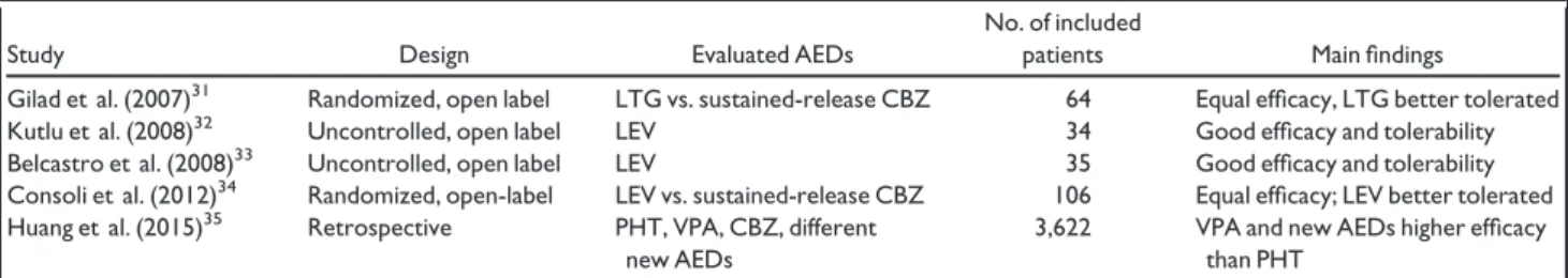
A few prospective studies have compared the efficacy of different AEDs in patients with PSE (Table 3). Gilad et al.31 conducted a randomized noncontrolled trial com-paring lamotrigine (LTG) and sustained-release carba-mazepine (CBZ) in patients >18 years with both ES and PSE, and found that LTG was more effective, albeit non-significantly (seizure freedom: 72% on LTG vs. 44% on CBZ, p = 0.06), and better tolerated (withdrawal rate due to adverse events: 3% on LTG and 31% on CBZ, p = 0.02). Two prospective open-label studies evaluated levetiracetam (LEV) monotherapy in elderly patients with new-onset PSE.32,33Both studies reported good tolerability and efficacy profile for LEV with about 80% reaching sei-zure freedom. The discontinuation rate due to adverse effects (namely drowsiness) was 14–21%. In a multicenter randomized open-label trial, Consoli et al.34compared the efficacy of monotherapy with either LEV or sustained-release CBZ in patients with PSE. No relevant differences on seizure control were found between the two groups (94% of patients seizure free in LEV group and 85% in CBZ group, p = 0.08). However, LEV was better tolerated (31% of patients experienced side effects with LEV and 39% with CBZ, p = 0.02). Finally, in a retrospective
observational study, Huang et al.35 evaluated efficacy of phenytoin (PHT), valproate (VPA), CBZ, and new AEDs (oxcarbazepine [OXC], vigabatrin, tiagabine, LTG, TPM, gabapentin [GBP], LEV, and pregabalin) on seizure control in 3,622 patients with PSE. They found that patients treated with VPA and new AEDs have better seizure control than those using PHT, as demonstrated by the lower risks of emergency room visits and hospitalizations (Table 3).
Some other trials compared efficacy of different AEDs in patients with epilepsy due to various etiologies including cerebrovascular diseases. In a double-blind randomized trial carried out by Brodie et al.,36LTG and immediate-release CBZ were tested during 24 weeks in 150 patients aged ≥65 years with new-onset epilepsy. Thirty percent of patients in the LTG group and 38% in the CBZ group had a previous cerebrovascular accident. Efficacy was similar (>60% of patients reaching seizure freedom in both groups), but LTG was better tolerated as compared to CBZ, with a significantly higher retention rate at the end of the study (71% in LTG group, 42% in CBZ group, p< 0.001). Rowan et al.37randomized 593 epileptic patients aged 60 years or older to one of three treatment arms: LTG, immediate-release CBZ, or GBP. CBZ was less tolerated than GBP and LTG (withdrawal for adverse events: LTG 12.1%, GBP 21.6%, and CBZ 31%; p= 0.001), although the three drugs had similar efficacy, with 53.3% of patients being seizure free at 12 months (51.4% on LTG, 47.4% on GBP, 64.3% on CBZ; p= 0.09). In that study, 30% of subjects had a cerebral infarction and 16% had arteriosclerosis. The SANAD (Standard and New Antiepileptic Drugs) study38 was an unblinded randomized controlled trial that compared (Arm A) the efficacy and tolerability of LTG, CBZ (either immediate or controlled release), GBP, TPM, and OXC in patients with new-onset focal epilepsy in different age groups. Among 1,721 enrolled, 108 (6.3%) had history of stroke. Authors found comparable efficacy between LTG and CBZ (expressed as time to treatment failure) with lesser effectiveness for GBP. LTG and TPM were, respectively,
Figure 2.
Forest plot showing pooled odds ratios for cortical involvement. Epilepsia ILAE
the best and less-tolerated drugs (45% of patients reported adverse events while on LTG and 53% while on TPM). The limits of this study consist in its unblinded design, which
might have led clinicians to biases in their therapeutic choices, and in the administration of CBZ at higher than therapeutic doses, thus obtaining more frequent side effects
Figure 3.
Forest plot showing pooled odds ratios for cerebral hemorrhage. Epilepsia ILAE
Figure 4.
Forest plot showing pooled odds ratios for early seizures. Epilepsia ILAE
Table 3. Summary of the main studies evaluating antiepileptic drugs in patients with PSE
Study Design Evaluated AEDs
No. of included
patients Main findings
Gilad et al. (2007)31 Randomized, open label LTG vs. sustained-release CBZ 64 Equal efficacy, LTG better tolerated Kutlu et al. (2008)32 Uncontrolled, open label LEV 34 Good efficacy and tolerability Belcastro et al. (2008)33 Uncontrolled, open label LEV 35 Good efficacy and tolerability Consoli et al. (2012)34 Randomized, open-label LEV vs. sustained-release CBZ 106 Equal efficacy; LEV better tolerated
Huang et al. (2015)35 Retrospective PHT, VPA, CBZ, different
new AEDs
3,622 VPA and new AEDs higher efficacy than PHT
with discontinuation of the drug. Moreover, the number of patients undergoing treatment with immediate release or controlled release formulation of CBZ was unspecified. Saetre et al.39conducted a multicenter, double-blind study on the effectiveness and tolerability of LTG and controlled release CBZ in 185 patients≥65 years with new-onset epi-lepsy, most of whom had symptomatic epilepsy. Although no data on specific etiologies were reported, hypertension (42% of the randomized cohort) and cerebrovascular disease (25%) were frequent in this sample. The authors showed that the two drugs had similar profiles, with a slightly higher efficacy for CBZ (63% seizure-free patients in LTG group, 76% in CBZ group, p= 0.07) and a slightly better tolerabil-ity for LTG (adverse events leading to withdrawal in 14% of LTG group and 25% of subjects in CBZ group, p = 0.08). Finally, a randomized, double-blind trial comparing sus-tained-release CBZ, LTG, and LEV as the initial therapy in 359 elderly patients with new-onset focal epilepsy, most of whom had cerebrovascular lesions, found comparable effi-cacy among the three drugs, but the retention rate for LEV was significantly higher than for sustained-release CBZ (61.5% vs. 45.8%, p = 0.02), and similar to LTG (55.6%).40
Conclusions
Although early post-stroke seizures have been studied extensively, less attention has been paid to PSE and to EAL. Experimental models of PSE show that epileptogenesis is linked to alteration of neuronal excitability in different cor-tical areas, as confirmed by TMS studies in humans. The pathophysiology of EAL is largely unknown. Data from experimental and clinical studies suggest that systemic hypertension and leukoaraiosis may modulate seizure sus-ceptibility, possibly with a contribution of cerebral renin-angiotensin system. However, coexistence of cortical microinfarcts or other adjunctive factors may also play a role. Further animal and human studies are needed to clearly define the role of risk factors, including systemic hyperten-sion, in the development of EAL and to explain whether leukoaraiosis represents a mere incidental radiologic find-ing or plays a role in the pathogenesis of epilepsy. More-over, the role of SVD and leukoaraiosis in patients with epilepsy has not yet been clarified. From the clinical point of view, our meta-analysis showed that cortical involve-ment, hemorrhage, and ES are associated with a higher risk PSE, whereas gender does not seem to play a role. Gender, systemic hypertension, and severity of white matter changes need to be evaluated as risk factors for EAL in prospective, population-based studies. Results from clinical studies on PSE suggest that seizure-free rate after AED treatment is high. Among AEDs, sustained-release CBZ, LTG, and LEV showed good efficacy, with a better tolerability for LTG and LEV over sustained-release CBZ. Conversely, no data are available on optimal treatment for EAL. Management PSE
and EAL relies on the clinician’s judgment and should be tailored on individual basis.
Disclosure
None of the authors has any conflict of interest to disclose. We confirm
that we have read the journal’s position on issues involved in ethical
publi-cation and affirm that this report is consistent with those guidelines.
Appendix A Members of the
Epilepsy Study Group of the
Italian Neurological Society
Aloisi Paolo, Arcudi Luciano, Benna Paolo, Bianchi Amedeo, Bogliun Graziella, Buttinelli Carla, Campostrini Roberto, Cantello Roberto, Cianci Vittoria, Consoli Domenico, Coppola Antonietta, Costanzo Erminio, De Falco Fabrizio Antonio, De Maria Giovanni, Elia Maurizio, Franceschetti Silvana, Galimberti Carlo Andrea, Giallonardo Anna Teresa, Gigli Gian Luigi, Iannacchero Rosario, Iudice Alfonso, La Neve Angela, Latella Maria Adele, Le Piane Emilio, Magaudda Adriana, Marciani Maria Grazia, Mas-chio Marta, Mecarelli Oriano, Michelucci Roberto, Minicucci Fabio, Moglia Arrigo, Monti Fabrizio, Mumoli Laura, Musolino Rosa, Nozzoli Cecilia, Paciello Nicola, Paladin Francesco, Palumbo Pasquale, Pisani Francesco, Primavera Alberto, Rocchi Raffaele, Sartucci Ferdinando, Sasa-nelli Francesco, Silvestri Rosalia, Sofia Vito, Specchio Luigi M., Striano Salvatore, Tinuper Paolo, Villani Flavio, Zaccara Gaetano.
References
1. Menon B, Shorvon SD. Ischaemic stroke in adults and epilepsy.
Epi-lepsy Res 2009;87:1–11.
2. Hesdorffer DC, Benn EKT, Cascino GD, et al. Is a first acute symp-tomatic seizure epilepsy? Mortality and risk for recurrent seizure.
Epilepsia 2009;50:1102–1108.
3. Fisher RS, Acevedo C, Arzimanoglou A, et al. A practical clinical
def-inition of epilepsy. Epilepsia 2014;55:475–482.
4. Gasparini S, Ferlazzo E, Beghi E, et al. Epilepsy associated with Leukoaraiosis mainly affects temporal lobe: a casual or causal
relation-ship? Epilepsy Res 2015;109:1–8.
5. Pantoni L. Cerebral small vessel disease: from pathogenesis and
clini-cal characteristics to therapetic challenges. Lancet Neurol 2010;9:689–
701.
6. Guidelines for epidemiologic studies on epilepsy. Commission on Epi-demiology and Prognosis, International League Against Epilepsy.
Epilepsia 1993;34:592–596.
7. Cuomo O, Rispoli V, Leo A, et al. The antiepileptic drug levetiracetam suppresses non-convulsive seizure activity and reduces ischemic brain damage in rats subjected to permanent middle cerebral artery occlu-sion. PLoS ONE 2013;8:e80852.
8. Watson BD, Dietrich WD, Busto R, et al. Induction of reproducible brain infarction by photochemically initiated thrombosis. Ann Neurol
1985;17:497–504.
9. Karhunen H, Bezvenyuk Z, Nissinen J, et al. Epileptogenesis after
cor-tical photothrombotic brain lesion in rats. Neuroscience 2007;48:314–
324.
10. Kessler KR, Schnitzler A, Classen J, et al. Reduced inhibition within primary motor cortex in patients with poststroke focal motor seizures.
Neurology 2002;59:1028–1033.
11. Kim JH, Lee HW, Cohen LG, et al. Motor cortical excitability in
patients with poststroke epilepsy. Epilepsia 2008;49:117–124.
12. Kaiser D, Weise G, M€oller K, et al. Spontaneous white matter damage,
cognitive decline and neuroinflammation in middle-aged hypertensive rats: an animal model of early-stage cerebral small vessel disease. Acta
Neuropathol Commun 2014;2:169.
13. Tchekalarova J, Georgiev V. Angiotensin peptides modulatory system: how is it implicated n the control of seizure susceptibility? Life Sci
14. Tchekalarova J, Loyens E, Smolders I. Effects of AT1 receptor antago-nism on kainate-induced seizures and concomitant changes in hip-pocampal extracellular noradrenaline, serotonin, and dopamine levels in Wistar-Kyoto and spontaneously hypertensive rats. Epilepsy Behav
2015;46:66–71.
15. Arganaraz GA, Konno AC, Perosa SR, et al. The renin-angiotensin system is upregulated in the cortex and hippocampus of patients with temporal lobe epilepsy related to mesial temporal sclerosis. Epilepsia
2008;49:1348–1357.
16. Ng SK, Hauser WA, Brust JC, et al. Hypertension and the risk of
new-onset unprovoked seizures. Neurology 1993;43:425–428.
17. Hesdorffer DC, Hauser WA, Annegers JF, et al. Severe, uncontrolled
hypertension and adult-onset seizures: a case–control study in
Roche-ster, Minnesota. Epilepsia 1996;37:736–741.
18. Li X, Breteler MM, deBruyne MC, et al. Vascular determinants of
epi-lepsy: the Rotterdam Study. Epilepsia 1997;38:216–1220.
19. Maxwell H, Hanby M, Parkes LM, et al. Prevalence and
subtypes of radiological cerebrovascular disease in late-onset iso-lated seizures and epilepsy. Clin Neurol Neurosurg 2013;115: 591–596.
20. Okroglic S, Widmann CN, Urbach H, et al. Clinical symptoms and risk factors in cerebral microangiopathy patients. PloS ONE 2013;8: e53455.
21. De Reuck J, Van Maele G. Cognitive impairment and seizures in
patients with lacunar strokes. Eur Neurol 2009;61:159–163.
22. Smith EE, Schneider JA, Wardlaw JM, et al. Cerebral microinfarcts:
the invisible lesions. Lancet Neurol 2012;11:272–282.
23. Keller L, Hobohm C, Zeynalova S, et al. Does treatment with t-PA increase the risk of developing epilepsy after stroke? J Neurol
2015;262:2364–2372.
24. Serafini A, Gigli GL, Gregoraci G, et al. Are early seizures predictive of epilepsy after a stroke? Results of a population-based study
Neu-roepidemiology 2015;45:50–58.
25. Zhang C, Wang X, Wang Y, et al. Risk factors for post-stroke seizures:
a systematic review and meta-analysis. Epilepsy Res 2014;108:1806–
1816.
26. De Reuck J, Nagy E, Van Maele G. Seizures and epilepsy in patients
with lacunar strokes. J Neurol Sci 2007;263:75–78.
27. Ferlazzo E, Sueri C, Gasparini S, et al. Challenges in the pharmacolog-ical management of epilepsy and its causes in the elderly. Pharmacol
Res 2016;106:21–26.
28. Semah F, Picot MC, Adam C, et al. Is the underlying cause of epilepsy
a major prognostic factor for recurrence? Neurology 1998;51:1256–
1262.
29. Stephen LJ, Kwan P, Brodie MJ. Does the cause of localisation-related epilepsy influence the response to antiepileptic drug treatment?
Epilep-sia 2001;42:357–362.
30. Gilad R, Boaz M, Dabby R, et al. Are post intracerebral hemorrhage
seizures prevented by antiepileptic treatment? Epilepsy Res
2011;95:227–231.
31. Gilad R, Sadeh M, Rapoport A, et al. Monotherapy of lamotrigine ver-sus carbamazepine in patients with poststroke seizure. Clin
Neurophar-macol 2007;30:189–195.
32. Kutlu G, Gomceli YB, Unal Y, et al. Levetiracetam monotherapy for
late post-stroke seizures in the elderly. Epilepsy Behav 2008;13:542–
544.
33. Belcastro V, Costa C, Galletti F, et al. Levetiracetam in newly diag-nosed late-onset post-stroke seizures: a prospective observational
study. Epilepsy Res 2008;82:223–226.
34. Consoli D, Bosco D, Postorino P, et al. Levetiracetam versus carba-mazepine in patients with late poststroke seizures: a multicenter prospective randomized open-label study (EpIC Project). Cerebrovasc
Dis 2012;34:282–289.
35. Huang YH, Chi NF, Kuan YC, et al. Efficacy of phenytoin, valproic acid, carbamazepine and new antiepileptic drugs on control of
late-onset post-stroke epilepsy in Taiwan. Eur J Neurol 2015;22:1459–
1468.
36. Brodie MJ, Overstall PW, Giorgi L. Multicenter, double-blind, ran-domised comparison between lamotrigine and carbamazepine in elderly patients with newly diagnosed epilepsy. Epilepsy Res
1999;37:81–87.
37. Rowan AJ, Ramsay RE, Collins JF, et al. New onset geriatric epilepsy: a randomized study of gabapentin, lamotrigine, and carbamazepine.
Neurology 2005;64:1868–1873.
38. Marson AG, Al-Kharusi AM, Alwaidh M, et al. SANAD study of effectiveness of carbamazepine, gabapentin, lamotrigine, oxcar-bazepine, or topiramate for treatment of partial epilepsy: an unblinded
randomized controlled trial. Lancet 2007;369:1000–1015.
39. Saetre E, Perucca E, Isoj€arvi J, et al. An international multicenter
ran-domized double-blind controlled trial of lamotrigine and sustained-release carbamazepine in the treatment of newly diagnosed epilepsy in
the elderly. Epilepsia 2007;48:1292–1302.
40. Werhahn KJ, Trinka E, Dobesberger J, et al. A randomized, double-blind comparison of antiepileptic drug treatment in the elderly with
new-onset focal epilepsy. Epilepsia 2015;56:450–459.
Supporting Information
Additional Supporting Information may be found in the online version of this article:Figure S1. Forest plot showing pooled odds ratios for sex.
Table S1. Risk of PSE by stroke site (cortical vs. subcortical).
Table S2.Risk of PSE by stroke type (hemorrhage vs. infarction).
Table S3.Risk of PSE by sex (women vs. men). Data S1. Search strategy for review.