International Journal of
Molecular Sciences
Article
Seeking for Non-Zinc-Binding MMP-2 Inhibitors:
Synthesis, Biological Evaluation and Molecular
Modelling Studies
Alessandra Ammazzalorso1,†, Barbara De Filippis1,†, Cristina Campestre1, Antonio Laghezza2,
Alessandro Marrone1, Rosa Amoroso1, Paolo Tortorella2and Mariangela Agamennone1,*
1 Dipartimento di Farmacia, Università “G. d’Annunzio” Chieti, Via dei Vestini 31, 66100 Chieti, Italy; [email protected] (A.A.); [email protected] (B.D.F.);
[email protected] (C.C.); [email protected] (A.M.); [email protected] (R.A.) 2 Dipartimento di Farmacia-Scienze del Farmaco, Università “A. Moro” Bari, Via Orabona 4, 70125 Bari, Italy;
[email protected] (A.L.); [email protected] (P.T.)
* Correspondence: [email protected]; Tel.: +39-0871-355-4585 † These authors contributed equally to the work.
Academic Editors: Claudiu T. Supuran and Koji Sode
Received: 29 July 2016; Accepted: 14 October 2016; Published: 22 October 2016
Abstract:Matrix metalloproteinases (MMPs) are an important family of zinc-containing enzymes
with a central role in many physiological and pathological processes. Although several MMP inhibitors have been synthesized over the years, none reached the market because of off-target effects, due to the presence of a zinc binding group in the inhibitor structure. To overcome this problem non-zinc-binding inhibitors (NZIs) have been recently designed. In a previous article, a virtual screening campaign identified some hydroxynaphtyridine and hydroxyquinoline as MMP-2 non-zinc-binding inhibitors. In the present work, simplified analogues of previously-identified hits have been synthesized and tested in enzyme inhibition assays. Docking and molecular dynamics studies were carried out to rationalize the activity data.
Keywords: MMPs; inhibitors; molecular dynamics; docking; quinoline; isoquinoline; ureas;
non-zinc-binding group
1. Introduction
Matrix metalloproteinases are calcium and zinc-containing proteases (24 in humans) involved in the hydrolysis of the extracellular matrix (ECM) components and, therefore, responsible for tissue remodeling in physiological conditions [1]. Unbalanced matrix metalloproteinase (MMP) activity has been observed in many pathologies, such as cancer, neurodegeneration, cardiovascular diseases and others [2–11]; for this reason, they represent a well-studied therapeutic target. More recently, it has been demonstrated that MMP involvement in several diseases can be due to the hydrolysis of substrates other than ECM components, in particular chemokines involved in inflammation and immune response [1,12]. Ever since the first ligands had been disclosed almost 30 years ago, several MMP inhibitors (MMPIs) have been discovered, but none reached the market because of relevant side effects emerging in long-term treatment (musculoskeletal syndrome (MSS)) [13]. Traditional MMPIs are constituted by a functional group binding the zinc ion (zinc binding group (ZBG)) and chemical functions mimicking the substrate and interacting with the MMP subsites, in particular with the hydrophobic S1’ site. The ZBG is represented in most of the cases by the hydroxamate function, that binds very efficiently in the MMP active site, but can possibly bind other divalent cations and cause an unselective inhibition of other metallo-enzymes.
Int. J. Mol. Sci. 2016, 17, 1768 2 of 19
Another important issue of MMP inhibition is represented by the unselective inhibition of the studied MMPIs. In fact, clinically-tested compounds (e.g., batimastat and marimastat) were unselective MMP inhibitors not discriminating among different isoforms; while, during the years, the diverse role played by each MMP has been better understood [14]. In particular, in anticancer therapy, it has been defined that some MMPs can be considered therapeutic targets (MMP-1, MMP-2, MMP-7), while some others are classified as anti-targets (MMP-8) [15].
These issues prompted the search for alternative approaches to the MMP inhibition [16], such as the substitution of the ZBG with less potent, more selective zinc binders [17–22], the use of MMP-directed antibody [23] and the design of non-zinc binding inhibitors (NZIs) [24]. These ligands, commonly reported as third-generation MMPIs, have been developed for MMP-13 [25], MMP-8 [26] and MMP-12 [27] and can reach a better selectivity by missing the ZBG. Recently, we attempted to identify MMP-2 NZIs characterized by enhanced interactions with the S1’ pocket [28]. Our interest toward MMP-2 selective inhibition is due to the demonstrated role of MMP-2 in tumors. MMP-2, also called gelatinase A, is constitutively expressed and contributes to tissue homeostasis. Even if poorly inducible, MMP-2 has been found overexpressed in several tumors playing a role in both tumorigenesis and cancer progression [29].
MMP-8 and MMP-13 are classified as collagenases because they mainly degrade collagen; MMP-13 is able to degrade the components of the basal membrane and is involved in tumor dissemination and progression. On the contrary, MMP-8 has a confirmed protective role in tumor progression, and its inhibition should be avoided [29]. The final aim of our study is, therefore, to identify selective NZIs blocking MMP-2 and MMP-13 activity, while sparing the antitarget MMP-8.
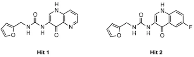
In the previous work, we moved from the analysis of the superposition between MMP-2 and MMP-8 in complex with a non-zinc-binding ligand that highlighted the possibility to design inhibitors tailored to (potential) additional interactions in the MMP-2 S1’ site. The following virtual screening campaign afforded one hit, the hydroxynaphtyridine derivative Hit 1, binding MMP-2 at a micromolar concentration (IC50= 5 µM (MMP-2), 75 µM (MMP-8) and 36 µM (MMP-13)). An analogue testing
allowed identifying also hydroxyquinoline compounds (i.e., Hit 2, IC50= 15 µM (MMP-2), 105 µM
(MMP-8) and 5 µM (MMP-13)), almost equally active than the prior hit (Figure1).
Int. J. Mol. Sci. 2016, 17, 1768 2 of 19
Another important issue of MMP inhibition is represented by the unselective inhibition of the studied MMPIs. In fact, clinically-tested compounds (e.g., batimastat and marimastat) were unselective MMP inhibitors not discriminating among different isoforms; while, during the years, the diverse role played by each MMP has been better understood [6]. In particular, in anticancer therapy, it has been defined that some MMPs can be considered therapeutic targets (MMP-1, MMP-2, MMP-7), while some others are classified as anti-targets (MMP-8) [7].
These issues prompted the search for alternative approaches to the MMP inhibition [8], such as the substitution of the ZBG with less potent, more selective zinc binders [9,18–22], the use of MMP-directed antibody [23] and the design of non-zinc binding inhibitors (NZIs) [24]. These ligands, commonly reported as third-generation MMPIs, have been developed for MMP-13 [25], MMP-8 [26] and MMP-12 [27] and can reach a better selectivity by missing the ZBG. Recently, we attempted to identify MMP-2 NZIs characterized by enhanced interactions with the S1’ pocket [28]. Our interest toward MMP-2 selective inhibition is due to the demonstrated role of MMP-2 in tumors. MMP-2, also called gelatinase A, is constitutively expressed and contributes to tissue homeostasis. Even if poorly inducible, MMP-2 has been found overexpressed in several tumors playing a role in both tumorigenesis and cancer progression [29].
MMP-8 and MMP-13 are classified as collagenases because they mainly degrade collagen; MMP-13 is able to degrade the components of the basal membrane and is involved in tumor dissemination and progression. On the contrary, MMP-8 has a confirmed protective role in tumor progression, and its inhibition should be avoided [29]. The final aim of our study is, therefore, to identify selective NZIs blocking MMP-2 and MMP-13 activity, while sparing the antitarget MMP-8.
In the previous work, we moved from the analysis of the superposition between MMP-2 and MMP-8 in complex with a non-zinc-binding ligand that highlighted the possibility to design inhibitors tailored to (potential) additional interactions in the MMP-2 S1’ site. The following virtual screening campaign afforded one hit, the hydroxynaphtyridine derivative Hit 1, binding MMP-2 at a micromolar concentration (IC50 = 5 µM (MMP-2), 75 µM (MMP-8) and 36 µM (MMP-13)). An
analogue testing allowed identifying also hydroxyquinoline compounds (i.e., Hit 2, IC50 = 15 µM
(MMP-2), 105 µM (MMP-8) and 5 µM (MMP-13)), almost equally active than the prior hit (Figure 1).
Figure 1. Chemical structures of Hits 1 and 2.
Moving from these data, in the present work we investigated the structural determinants essential for the inhibition of MMP-2 by non-zinc-binding ligands. Activity data and docking studies of hydroxynaphtyridine/hydroxyquinoline derivatives suggested the role of relevant structural features:
• the furyl/aryl group that provides the π–π stacking with His201 (MMP-2 numbering); • the CH2 spacer between the furyl/aryl group and the urea function;
• the urea function that provides H-bonds in the S1’ site with NH of Thr227 and CO of Leu218.
Starting from these evidences, we described the synthesis and the biological evaluation of new MMP inhibitors, based on the combination of three structural portions in which the lead compound could be ideally divided, in order to verify their importance for the selective inhibition of MMP isoforms. In all compounds, the furyl group was replaced by a more common benzyl function. In a first series of compounds, we kept the benzyl-ureidic scaffold unaltered, by modifying the hydroxyquinoline portion with heterocycles (quinoline, isoquinoline, indole, indazole), aromatic rings (naphthalene, benzene, pyridine), alkyl chains or saturated heterocycles (piperidine, piperazine, pyrrolidine) (Figure 2; Table 1).
Figure 1.Chemical structures of Hits 1 and 2.
Moving from these data, in the present work we investigated the structural determinants essential for the inhibition of MMP-2 by non-zinc-binding ligands. Activity data and docking studies of hydroxynaphtyridine/hydroxyquinoline derivatives suggested the role of relevant structural features:
• the furyl/aryl group that provides the π–π stacking with His201 (MMP-2 numbering);
• the CH2spacer between the furyl/aryl group and the urea function;
• the urea function that provides H-bonds in the S1’ site with NH of Thr227 and CO of Leu218. Starting from these evidences, we described the synthesis and the biological evaluation of new MMP inhibitors, based on the combination of three structural portions in which the lead compound could be ideally divided, in order to verify their importance for the selective inhibition of MMP isoforms. In all compounds, the furyl group was replaced by a more common benzyl function. In a first series of compounds, we kept the benzyl-ureidic scaffold unaltered, by modifying the hydroxyquinoline portion with heterocycles (quinoline, isoquinoline, indole, indazole), aromatic rings (naphthalene, benzene, pyridine), alkyl chains or saturated heterocycles (piperidine, piperazine, pyrrolidine) (Figure2; Table1).
Int. J. Mol. Sci. 2016, 17, 1768 3 of 19
Int. J. Mol. Sci. 2016, 17, 1768 3 of 19
Figure 2. Structural modifications performed on Hit 2. Table 1. Urea derivatives 1a–α.
ID R ID R 1a N 1n 1b N 1o 1c N 1p N 1d N 1q N 1e N 1r 1f N 1s HO 1g N 1t OCH3 OCH3 1h N 1u H3CO OCH3 1i N 1v 1j N H 1w 1k N H 1x N H NH R O
Figure 2.Structural modifications performed on Hit 2.
Table 1.Urea derivatives 1a–α.
Int. J. Mol. Sci. 2016, 17, 1768 3 of 19
Figure 2. Structural modifications performed on Hit 2. Table 1. Urea derivatives 1a–α.
ID R ID R 1a N 1n 1b N 1o 1c N 1p N 1d N 1q N 1e N 1r 1f N 1s HO 1g N 1t OCH3 OCH3 1h N 1u H3CO OCH3 1i N 1v 1j N H 1w 1k N H 1x N H NH R O ID R ID R 1a
Int. J. Mol. Sci. 2016, 17, 1768 3 of 19
Figure 2. Structural modifications performed on Hit 2. Table 1. Urea derivatives 1a–α.
ID R ID R 1a N 1n 1b N 1o 1c N 1p N 1d N 1q N 1e N 1r 1f N 1s HO 1g N 1t OCH3 OCH3 1h N 1u H3CO OCH3 1i N 1v 1j N H 1w 1k N H 1x N H NH R O 1n
Int. J. Mol. Sci. 2016, 17, 1768 3 of 19
Figure 2. Structural modifications performed on Hit 2. Table 1. Urea derivatives 1a–α.
ID R ID R 1a N 1n 1b N 1o 1c N 1p N 1d N 1q N 1e N 1r 1f N 1s HO 1g N 1t OCH3 OCH3 1h N 1u H3CO OCH3 1i N 1v 1j N H 1w 1k N H 1x N H NH R O 1b
Int. J. Mol. Sci. 2016, 17, 1768 3 of 19
Figure 2. Structural modifications performed on Hit 2. Table 1. Urea derivatives 1a–α.
ID R ID R 1a N 1n 1b N 1o 1c N 1p N 1d N 1q N 1e N 1r 1f N 1s HO 1g N 1t OCH3 OCH3 1h N 1u H3CO OCH3 1i N 1v 1j N H 1w 1k N H 1x N H NH R O 1o
Int. J. Mol. Sci. 2016, 17, 1768 3 of 19
Figure 2. Structural modifications performed on Hit 2. Table 1. Urea derivatives 1a–α.
ID R ID R 1a N 1n 1b N 1o 1c N 1p N 1d N 1q N 1e N 1r 1f N 1s HO 1g N 1t OCH3 OCH3 1h N 1u H3CO OCH3 1i N 1v 1j N H 1w 1k N H 1x N H NH R O 1c
Int. J. Mol. Sci. 2016, 17, 1768 3 of 19
Figure 2. Structural modifications performed on Hit 2. Table 1. Urea derivatives 1a–α.
ID R ID R 1a N 1n 1b N 1o 1c N 1p N 1d N 1q N 1e N 1r 1f N 1s HO 1g N 1t OCH3 OCH3 1h N 1u H3CO OCH3 1i N 1v 1j N H 1w 1k N H 1x N H NH R O 1p
Int. J. Mol. Sci. 2016, 17, 1768 3 of 19
Figure 2. Structural modifications performed on Hit 2. Table 1. Urea derivatives 1a–α.
ID R ID R 1a N 1n 1b N 1o 1c N 1p N 1d N 1q N 1e N 1r 1f N 1s HO 1g N 1t OCH3 OCH3 1h N 1u H3CO OCH3 1i N 1v 1j N H 1w 1k N H 1x N H NH R O 1d
Int. J. Mol. Sci. 2016, 17, 1768 3 of 19
Figure 2. Structural modifications performed on Hit 2. Table 1. Urea derivatives 1a–α.
ID R ID R 1a N 1n 1b N 1o 1c N 1p N 1d N 1q N 1e N 1r 1f N 1s HO 1g N 1t OCH3 OCH3 1h N 1u H3CO OCH3 1i N 1v 1j N H 1w 1k N H 1x N H NH R O 1q
Int. J. Mol. Sci. 2016, 17, 1768 3 of 19
Figure 2. Structural modifications performed on Hit 2. Table 1. Urea derivatives 1a–α.
ID R ID R 1a N 1n 1b N 1o 1c N 1p N 1d N 1q N 1e N 1r 1f N 1s HO 1g N 1t OCH3 OCH3 1h N 1u H3CO OCH3 1i N 1v 1j N H 1w 1k N H 1x N H NH R O 1e
Int. J. Mol. Sci. 2016, 17, 1768 3 of 19
Figure 2. Structural modifications performed on Hit 2. Table 1. Urea derivatives 1a–α.
ID R ID R 1a N 1n 1b N 1o 1c N 1p N 1d N 1q N 1e N 1r 1f N 1s HO 1g N 1t OCH3 OCH3 1h N 1u H3CO OCH3 1i N 1v 1j N H 1w 1k N H 1x N H NH R O 1r
Int. J. Mol. Sci. 2016, 17, 1768 3 of 19
Figure 2. Structural modifications performed on Hit 2. Table 1. Urea derivatives 1a–α.
ID R ID R 1a N 1n 1b N 1o 1c N 1p N 1d N 1q N 1e N 1r 1f N 1s HO 1g N 1t OCH3 OCH3 1h N 1u H3CO OCH3 1i N 1v 1j N H 1w 1k N H 1x N H NH R O 1f
Int. J. Mol. Sci. 2016, 17, 1768 3 of 19
Figure 2. Structural modifications performed on Hit 2. Table 1. Urea derivatives 1a–α.
ID R ID R 1a N 1n 1b N 1o 1c N 1p N 1d N 1q N 1e N 1r 1f N 1s HO 1g N 1t OCH3 OCH3 1h N 1u H3CO OCH3 1i N 1v 1j N H 1w 1k N H 1x N H NH R O 1s
Int. J. Mol. Sci. 2016, 17, 1768 3 of 19
Figure 2. Structural modifications performed on Hit 2. Table 1. Urea derivatives 1a–α.
ID R ID R 1a N 1n 1b N 1o 1c N 1p N 1d N 1q N 1e N 1r 1f N 1s HO 1g N 1t OCH3 OCH3 1h N 1u H3CO OCH3 1i N 1v 1j N H 1w 1k N H 1x N H NH R O 1g
Int. J. Mol. Sci. 2016, 17, 1768 3 of 19
Figure 2. Structural modifications performed on Hit 2. Table 1. Urea derivatives 1a–α.
ID R ID R 1a N 1n 1b N 1o 1c N 1p N 1d N 1q N 1e N 1r 1f N 1s HO 1g N 1t OCH3 OCH3 1h N 1u H3CO OCH3 1i N 1v 1j N H 1w 1k N H 1x N H NH R O 1t
Int. J. Mol. Sci. 2016, 17, 1768 3 of 19
Figure 2. Structural modifications performed on Hit 2. Table 1. Urea derivatives 1a–α.
ID R ID R 1a N 1n 1b N 1o 1c N 1p N 1d N 1q N 1e N 1r 1f N 1s HO 1g N 1t OCH3 OCH3 1h N 1u H3CO OCH3 1i N 1v 1j N H 1w 1k N H 1x N H NH R O 1h
Int. J. Mol. Sci. 2016, 17, 1768 3 of 19
Figure 2. Structural modifications performed on Hit 2. Table 1. Urea derivatives 1a–α.
ID R ID R 1a N 1n 1b N 1o 1c N 1p N 1d N 1q N 1e N 1r 1f N 1s HO 1g N 1t OCH3 OCH3 1h N 1u H3CO OCH3 1i N 1v 1j N H 1w 1k N H 1x N H NH R O 1u
Int. J. Mol. Sci. 2016, 17, 1768 3 of 19
Figure 2. Structural modifications performed on Hit 2. Table 1. Urea derivatives 1a–α.
ID R ID R 1a N 1n 1b N 1o 1c N 1p N 1d N 1q N 1e N 1r 1f N 1s HO 1g N 1t OCH3 OCH3 1h N 1u H3CO OCH3 1i N 1v 1j N H 1w 1k N H 1x N H NH R O 1i
Int. J. Mol. Sci. 2016, 17, 1768 3 of 19
Figure 2. Structural modifications performed on Hit 2. Table 1. Urea derivatives 1a–α.
ID R ID R 1a N 1n 1b N 1o 1c N 1p N 1d N 1q N 1e N 1r 1f N 1s HO 1g N 1t OCH3 OCH3 1h N 1u H3CO OCH3 1i N 1v 1j N H 1w 1k N H 1x N H NH R O 1v
Int. J. Mol. Sci. 2016, 17, 1768 3 of 19
Figure 2. Structural modifications performed on Hit 2. Table 1. Urea derivatives 1a–α.
ID R ID R 1a N 1n 1b N 1o 1c N 1p N 1d N 1q N 1e N 1r 1f N 1s HO 1g N 1t OCH3 OCH3 1h N 1u H3CO OCH3 1i N 1v 1j N H 1w 1k N H 1x N H NH R O 1j
Int. J. Mol. Sci. 2016, 17, 1768 3 of 19
Figure 2. Structural modifications performed on Hit 2. Table 1. Urea derivatives 1a–α.
ID R ID R 1a N 1n 1b N 1o 1c N 1p N 1d N 1q N 1e N 1r 1f N 1s HO 1g N 1t OCH3 OCH3 1h N 1u H3CO OCH3 1i N 1v 1j N H 1w 1k N H 1x N H NH R O 1w
Int. J. Mol. Sci. 2016, 17, 1768 3 of 19
Figure 2. Structural modifications performed on Hit 2. Table 1. Urea derivatives 1a–α.
ID R ID R 1a N 1n 1b N 1o 1c N 1p N 1d N 1q N 1e N 1r 1f N 1s HO 1g N 1t OCH3 OCH3 1h N 1u H3CO OCH3 1i N 1v 1j N H 1w 1k N H 1x N H NH R O 1k
Int. J. Mol. Sci. 2016, 17, 1768 3 of 19
Figure 2. Structural modifications performed on Hit 2. Table 1. Urea derivatives 1a–α.
ID R ID R 1a N 1n 1b N 1o 1c N 1p N 1d N 1q N 1e N 1r 1f N 1s HO 1g N 1t OCH3 OCH3 1h N 1u H3CO OCH3 1i N 1v 1j N H 1w 1k N H 1x N H NH R O 1x
Int. J. Mol. Sci. 2016, 17, 1768 3 of 19
Figure 2. Structural modifications performed on Hit 2. Table 1. Urea derivatives 1a–α.
ID R ID R 1a N 1n 1b N 1o 1c N 1p N 1d N 1q N 1e N 1r 1f N 1s HO 1g N 1t OCH3 OCH3 1h N 1u H3CO OCH3 1i N 1v 1j N H 1w 1k N H 1x N H NH R O 1l
Int. J. Mol. Sci. 2016, 17, 1768 4 of 19
1l NN H 1y N 1m NN H 1z N N H N O N
A second group of derivatives was obtained starting from the isoquinolinyl derivative 1h, by replacing the ureidic linker with an amide or sulfonamide group (2a–e; Figure 3; Table 2). Finally, novel ureidic derivatives were designed by adding para substituents on the benzyl ring, with the aim to explore the effect of the polar functional group, potentially able to interact with residues at the top of the S1’ site (5 and 7; Figure 3; Table 3).
Figure 3. From isoquinolinyl urea 1h to novel derivatives 2a–e and 5, 7.
To provide some rational explanation of the activity data, structure-based computational approaches were applied to the study of the binding process.
Table 2. Isoquinoline amide and sulfonamide derivatives 2a–2e.
Cpd Linker n 2a NHCO 1 2b NHCO 2 2c CONH 1 2d CONH 2 2e NHSO2 -
Table 3. Amide and sulfonamide derivatives of isoquinolinyl urea 5 and 7.
Cpd X 5 SO2 7 CO N HN N H O 1h N Linker amide inverse amide sulfonamide 2a-e N HN N H O 5,7 N H X amide sulfonamide n N Linker n N HN N H O N H X 1y
Int. J. Mol. Sci. 2016, 17, 1768 4 of 19
1l NN H 1y N 1m NN H 1z N N H N O N
A second group of derivatives was obtained starting from the isoquinolinyl derivative 1h, by replacing the ureidic linker with an amide or sulfonamide group (2a–e; Figure 3; Table 2). Finally, novel ureidic derivatives were designed by adding para substituents on the benzyl ring, with the aim to explore the effect of the polar functional group, potentially able to interact with residues at the top of the S1’ site (5 and 7; Figure 3; Table 3).
Figure 3. From isoquinolinyl urea 1h to novel derivatives 2a–e and 5, 7.
To provide some rational explanation of the activity data, structure-based computational approaches were applied to the study of the binding process.
Table 2. Isoquinoline amide and sulfonamide derivatives 2a–2e.
Cpd Linker n 2a NHCO 1 2b NHCO 2 2c CONH 1 2d CONH 2 2e NHSO2 -
Table 3. Amide and sulfonamide derivatives of isoquinolinyl urea 5 and 7.
Cpd X 5 SO2 7 CO N HN N H O 1h N Linker amide inverse amide sulfonamide 2a-e N HN N H O 5,7 N H X amide sulfonamide n N Linker n N HN N H O N H X
Int. J. Mol. Sci. 2016, 17, 1768 4 of 19
Table 1. Cont.
Int. J. Mol. Sci. 2016, 17, 1768 3 of 19
Figure 2. Structural modifications performed on Hit 2. Table 1. Urea derivatives 1a–α.
ID R ID R 1a N 1n 1b N 1o 1c N 1p N 1d N 1q N 1e N 1r 1f N 1s HO 1g N 1t OCH3 OCH3 1h N 1u H3CO OCH3 1i N 1v 1j N H 1w 1k N H 1x N H NH R O ID R ID R 1m
Int. J. Mol. Sci. 2016, 17, 1768 4 of 19
1l NN H 1y N 1m NN H 1z N N H N O N
A second group of derivatives was obtained starting from the isoquinolinyl derivative 1h, by replacing the ureidic linker with an amide or sulfonamide group (2a–e; Figure 3; Table 2). Finally, novel ureidic derivatives were designed by adding para substituents on the benzyl ring, with the aim to explore the effect of the polar functional group, potentially able to interact with residues at the top of the S1’ site (5 and 7; Figure 3; Table 3).
Figure 3. From isoquinolinyl urea 1h to novel derivatives 2a–e and 5, 7.
To provide some rational explanation of the activity data, structure-based computational approaches were applied to the study of the binding process.
Table 2. Isoquinoline amide and sulfonamide derivatives 2a–2e.
Cpd Linker n 2a NHCO 1 2b NHCO 2 2c CONH 1 2d CONH 2 2e NHSO2 -
Table 3. Amide and sulfonamide derivatives of isoquinolinyl urea 5 and 7.
Cpd X 5 SO2 7 CO N HN N H O 1h N Linker amide inverse amide sulfonamide 2a-e N HN N H O 5,7 N H X amide sulfonamide n N Linker n N HN N H O N H X 1z
Int. J. Mol. Sci. 2016, 17, 1768 4 of 19
1l NN H 1y N 1m NN H 1z N N H N O N
A second group of derivatives was obtained starting from the isoquinolinyl derivative 1h, by replacing the ureidic linker with an amide or sulfonamide group (2a–e; Figure 3; Table 2). Finally, novel ureidic derivatives were designed by adding para substituents on the benzyl ring, with the aim to explore the effect of the polar functional group, potentially able to interact with residues at the top of the S1’ site (5 and 7; Figure 3; Table 3).
Figure 3. From isoquinolinyl urea 1h to novel derivatives 2a–e and 5, 7.
To provide some rational explanation of the activity data, structure-based computational approaches were applied to the study of the binding process.
Table 2. Isoquinoline amide and sulfonamide derivatives 2a–2e.
Cpd Linker n 2a NHCO 1 2b NHCO 2 2c CONH 1 2d CONH 2 2e NHSO2 -
Table 3. Amide and sulfonamide derivatives of isoquinolinyl urea 5 and 7.
Cpd X 5 SO2 7 CO N HN N H O 1h N Linker amide inverse amide sulfonamide 2a-e N HN N H O 5,7 N H X amide sulfonamide n N Linker n N HN N H O N H X 1α
Int. J. Mol. Sci. 2016, 17, 1768 4 of 19
1l NN H 1y N 1m NN H 1z N N H N O N
A second group of derivatives was obtained starting from the isoquinolinyl derivative 1h, by replacing the ureidic linker with an amide or sulfonamide group (2a–e; Figure 3; Table 2). Finally, novel ureidic derivatives were designed by adding para substituents on the benzyl ring, with the aim to explore the effect of the polar functional group, potentially able to interact with residues at the top of the S1’ site (5 and 7; Figure 3; Table 3).
Figure 3. From isoquinolinyl urea 1h to novel derivatives 2a–e and 5, 7.
To provide some rational explanation of the activity data, structure-based computational approaches were applied to the study of the binding process.
Table 2. Isoquinoline amide and sulfonamide derivatives 2a–2e.
Cpd Linker n 2a NHCO 1 2b NHCO 2 2c CONH 1 2d CONH 2 2e NHSO2 -
Table 3. Amide and sulfonamide derivatives of isoquinolinyl urea 5 and 7.
Cpd X 5 SO2 7 CO N HN N H O 1h N Linker amide inverse amide sulfonamide 2a-e N HN N H O 5,7 N H X amide sulfonamide n N Linker n N HN N H O N H X
A second group of derivatives was obtained starting from the isoquinolinyl derivative 1h, by replacing the ureidic linker with an amide or sulfonamide group (2a–e; Figure3; Table2). Finally, novel ureidic derivatives were designed by adding para substituents on the benzyl ring, with the aim to explore the effect of the polar functional group, potentially able to interact with residues at the top of the S1’ site (5 and 7; Figure3; Table3).
Int. J. Mol. Sci. 2016, 17, 1768 4 of 19
1l NN H 1y N 1m NN H 1z N N H N O N
A second group of derivatives was obtained starting from the isoquinolinyl derivative 1h, by replacing the ureidic linker with an amide or sulfonamide group (2a–e; Figure 3; Table 2). Finally, novel ureidic derivatives were designed by adding para substituents on the benzyl ring, with the aim to explore the effect of the polar functional group, potentially able to interact with residues at the top of the S1’ site (5 and 7; Figure 3; Table 3).
Figure 3. From isoquinolinyl urea 1h to novel derivatives 2a–e and 5, 7.
To provide some rational explanation of the activity data, structure-based computational approaches were applied to the study of the binding process.
Table 2. Isoquinoline amide and sulfonamide derivatives 2a–2e.
Cpd Linker n 2a NHCO 1 2b NHCO 2 2c CONH 1 2d CONH 2 2e NHSO2 -
Table 3. Amide and sulfonamide derivatives of isoquinolinyl urea 5 and 7.
Cpd X 5 SO2 7 CO N HN N H O 1h N Linker amide inverse amide sulfonamide 2a-e N HN N H O 5,7 N H X amide sulfonamide n N Linker n N HN N H O N H X
Figure 3.From isoquinolinyl urea 1h to novel derivatives 2a–e and 5, 7.
To provide some rational explanation of the activity data, structure-based computational approaches were applied to the study of the binding process.
Table 2.Isoquinoline amide and sulfonamide derivatives 2a–2e.
Int. J. Mol. Sci. 2016, 17, 1768 4 of 19
1l NN H 1y N 1m NN H 1z N N H N O N
A second group of derivatives was obtained starting from the isoquinolinyl derivative 1h, by replacing the ureidic linker with an amide or sulfonamide group (2a–e; Figure 3; Table 2). Finally, novel ureidic derivatives were designed by adding para substituents on the benzyl ring, with the aim to explore the effect of the polar functional group, potentially able to interact with residues at the top of the S1’ site (5 and 7; Figure 3; Table 3).
Figure 3. From isoquinolinyl urea 1h to novel derivatives 2a–e and 5, 7.
To provide some rational explanation of the activity data, structure-based computational approaches were applied to the study of the binding process.
Table 2. Isoquinoline amide and sulfonamide derivatives 2a–2e.
Cpd Linker n 2a NHCO 1 2b NHCO 2 2c CONH 1 2d CONH 2 2e NHSO2 -
Table 3. Amide and sulfonamide derivatives of isoquinolinyl urea 5 and 7.
Cpd X 5 SO2 7 CO N HN N H O 1h N Linker amide inverse amide sulfonamide 2a-e N HN N H O 5,7 N H X amide sulfonamide n N Linker n N HN N H O N H X Cpd Linker n 2a NHCO 1 2b NHCO 2 2c CONH 1 2d CONH 2 2e NHSO2
-Table 3.Amide and sulfonamide derivatives of isoquinolinyl urea 5 and 7.
Int. J. Mol. Sci. 2016, 17, 1768 4 of 19
1l NN H 1y N 1m NN H 1z N N H N O N
A second group of derivatives was obtained starting from the isoquinolinyl derivative 1h, by replacing the ureidic linker with an amide or sulfonamide group (2a–e; Figure 3; Table 2). Finally, novel ureidic derivatives were designed by adding para substituents on the benzyl ring, with the aim to explore the effect of the polar functional group, potentially able to interact with residues at the top of the S1’ site (5 and 7; Figure 3; Table 3).
Figure 3. From isoquinolinyl urea 1h to novel derivatives 2a–e and 5, 7.
To provide some rational explanation of the activity data, structure-based computational approaches were applied to the study of the binding process.
Table 2. Isoquinoline amide and sulfonamide derivatives 2a–2e.
Cpd Linker n 2a NHCO 1 2b NHCO 2 2c CONH 1 2d CONH 2 2e NHSO2 -
Table 3. Amide and sulfonamide derivatives of isoquinolinyl urea 5 and 7.
Cpd X 5 SO2 7 CO N HN N H O 1h N Linker amide inverse amide sulfonamide 2a-e N HN N H O 5,7 N H X amide sulfonamide n N Linker n N HN N H O N H X Cpd X 5 SO2 7 CO
Int. J. Mol. Sci. 2016, 17, 1768 5 of 19
2. Results
The present work moves from a previous study that identified MMP-2 non-zinc-binding hits (NZIs) [28]. As already stated, in this study, structural modifications were applied to these NZIs to probe the structural features determining the binding at and the selective inhibition of MMP-2, one of the most relevant MMPs in anticancer therapy. In this line, the final goal of our research project was the identification of NZIs particularly suitable for anticancer therapy, being selective toward target MMPs (such as MMP-2 and MMP-13) while sparing anti-target MMPs (such as MMP-8).
2.1. Chemistry
Urea derivatives 1a–α (Table1) were prepared by adding benzyl isocyanate to the corresponding amino derivatives in methanol, or chloroform, or dichloromethane, stirring the reaction at room or refluxing temperature for 1–24 h [30,31]. The synthesis is depicted in Scheme1.
Int. J. Mol. Sci. 2016, 17, 1768 5 of 19
2. Results
The present work moves from a previous study that identified MMP-2 non-zinc-binding hits (NZIs) [28]. As already stated, in this study, structural modifications were applied to these NZIs to probe the structural features determining the binding at and the selective inhibition of MMP-2, one of the most relevant MMPs in anticancer therapy. In this line, the final goal of our research project was the identification of NZIs particularly suitable for anticancer therapy, being selective toward target MMPs (such as MMP-2 and MMP-13) while sparing anti-target MMPs (such as MMP-8).
2.1. Chemistry
Urea derivatives 1a–α (Table 1) were prepared by adding benzyl isocyanate to the corresponding amino derivatives in methanol, or chloroform, or dichloromethane, stirring the reaction at room or refluxing temperature for 1–24 h [30,31].The synthesis is depicted in Scheme 1.
NCO N H NH R O (a) or (b) 1a-α
Scheme 1. Reagents and conditions: benzyl isocyanate (1 equnivalent (eq)), amine, or aniline (1 eq),
MeOH (Method a), or CHCl3, or dichloromethane (DCM) (Method b), room temperature (r.t.) or
refluxing, 1–24 h, 41%–99%.
The synthesis of amides 2a,b (Table 2) was carried out by adding to a solution of 5-aminoisoquinoline and triethylamine (TEA), a solution of suitable phenylalkyl-chloride in dichloromethane at room temperature, while stirring at r.t. for 2–24 h (Scheme 2) [32]. Amides 2c,d were obtained by coupling 5-carboxyisoquinoline with benzylamine (for 2c) or 2-phenylethylamine (for 2d), in the presence of 1-hydroxybenzotriazole (HOBt), N,N-dicyclohexylcarbodiimide (DCC) and N-methylmorpholine (NMM), in DMF at 0 °C (Scheme 3).
N NH N 1-2 O NH2 2a-b
Scheme 2. Reagents and conditions: 5-aminoisoquinoline (1 eq), phenylalkyl-chloride (1 eq), TEA
(1.5 eq), dry DCM, N2, r.t., 2–24 h, 55%–63%; n = 1, 2a; n = 2, 2b.
N COOH N H N O n 2c-d
Scheme 3. Reagents and conditions: 5-carboxyisoquinoline (1 eq), amine (1 eq),
1-hydroxybenzotriazole (HOBt) (1 eq), N,N-dicyclohexylcarbodiimide (DCC) (1 eq),
N-methylmorpholine (NMM) (1 eq), DMF, 0 °C, r.t., 24 h, 58%–70%.
Scheme 1.Reagents and conditions: benzyl isocyanate (1 equnivalent (eq)), amine, or aniline (1 eq), MeOH (Method a), or CHCl3, or dichloromethane (DCM) (Method b), room temperature (r.t.) or refluxing, 1–24 h, 41%–99%.
The synthesis of amides 2a,b (Table 2) was carried out by adding to a solution of 5-aminoisoquinoline and triethylamine (TEA), a solution of suitable phenylalkyl-chloride in dichloromethane at room temperature, while stirring at r.t. for 2–24 h (Scheme2) [32]. Amides 2c,d were obtained by coupling 5-carboxyisoquinoline with benzylamine (for 2c) or 2-phenylethylamine (for 2d), in the presence of 1-hydroxybenzotriazole (HOBt), N,N-dicyclohexylcarbodiimide (DCC) and N-methylmorpholine (NMM), in DMF at 0◦C (Scheme3).
Int. J. Mol. Sci. 2016, 17, 1768 5 of 19
2. Results
The present work moves from a previous study that identified MMP-2 non-zinc-binding hits (NZIs) [28]. As already stated, in this study, structural modifications were applied to these NZIs to probe the structural features determining the binding at and the selective inhibition of MMP-2, one of the most relevant MMPs in anticancer therapy. In this line, the final goal of our research project was the identification of NZIs particularly suitable for anticancer therapy, being selective toward target MMPs (such as MMP-2 and MMP-13) while sparing anti-target MMPs (such as MMP-8).
2.1. Chemistry
Urea derivatives 1a–α (Table 1) were prepared by adding benzyl isocyanate to the corresponding amino derivatives in methanol, or chloroform, or dichloromethane, stirring the reaction at room or refluxing temperature for 1–24 h [30,31].The synthesis is depicted in Scheme 1.
NCO N H NH R O (a) or (b) 1a-α
Scheme 1. Reagents and conditions: benzyl isocyanate (1 equnivalent (eq)), amine, or aniline (1 eq),
MeOH (Method a), or CHCl3, or dichloromethane (DCM) (Method b), room temperature (r.t.) or
refluxing, 1–24 h, 41%–99%.
The synthesis of amides 2a,b (Table 2) was carried out by adding to a solution of 5-aminoisoquinoline and triethylamine (TEA), a solution of suitable phenylalkyl-chloride in dichloromethane at room temperature, while stirring at r.t. for 2–24 h (Scheme 2) [32]. Amides 2c,d were obtained by coupling 5-carboxyisoquinoline with benzylamine (for 2c) or 2-phenylethylamine (for 2d), in the presence of 1-hydroxybenzotriazole (HOBt), N,N-dicyclohexylcarbodiimide (DCC) and N-methylmorpholine (NMM), in DMF at 0 °C (Scheme 3).
N NH N 1-2 O NH2 2a-b
Scheme 2. Reagents and conditions: 5-aminoisoquinoline (1 eq), phenylalkyl-chloride (1 eq), TEA
(1.5 eq), dry DCM, N2, r.t., 2–24 h, 55%–63%; n = 1, 2a; n = 2, 2b.
N COOH N H N O n 2c-d
Scheme 3. Reagents and conditions: 5-carboxyisoquinoline (1 eq), amine (1 eq),
1-hydroxybenzotriazole (HOBt) (1 eq), N,N-dicyclohexylcarbodiimide (DCC) (1 eq),
N-methylmorpholine (NMM) (1 eq), DMF, 0 °C, r.t., 24 h, 58%–70%.
Scheme 2. Reagents and conditions: 5-aminoisoquinoline (1 eq), phenylalkyl-chloride (1 eq), TEA (1.5 eq), dry DCM, N2, r.t., 2–24 h, 55%–63%; n = 1, 2a; n = 2, 2b.
Int. J. Mol. Sci. 2016, 17, 1768 5 of 19
2. Results
The present work moves from a previous study that identified MMP-2 non-zinc-binding hits (NZIs) [28]. As already stated, in this study, structural modifications were applied to these NZIs to probe the structural features determining the binding at and the selective inhibition of MMP-2, one of the most relevant MMPs in anticancer therapy. In this line, the final goal of our research project was the identification of NZIs particularly suitable for anticancer therapy, being selective toward target MMPs (such as MMP-2 and MMP-13) while sparing anti-target MMPs (such as MMP-8).
2.1. Chemistry
Urea derivatives 1a–α (Table 1) were prepared by adding benzyl isocyanate to the corresponding amino derivatives in methanol, or chloroform, or dichloromethane, stirring the reaction at room or refluxing temperature for 1–24 h [30,31].The synthesis is depicted in Scheme 1.
NCO N H NH R O (a) or (b) 1a-α
Scheme 1. Reagents and conditions: benzyl isocyanate (1 equnivalent (eq)), amine, or aniline (1 eq),
MeOH (Method a), or CHCl3, or dichloromethane (DCM) (Method b), room temperature (r.t.) or
refluxing, 1–24 h, 41%–99%.
The synthesis of amides 2a,b (Table 2) was carried out by adding to a solution of 5-aminoisoquinoline and triethylamine (TEA), a solution of suitable phenylalkyl-chloride in dichloromethane at room temperature, while stirring at r.t. for 2–24 h (Scheme 2) [32]. Amides 2c,d were obtained by coupling 5-carboxyisoquinoline with benzylamine (for 2c) or 2-phenylethylamine (for 2d), in the presence of 1-hydroxybenzotriazole (HOBt), N,N-dicyclohexylcarbodiimide (DCC) and N-methylmorpholine (NMM), in DMF at 0 °C (Scheme 3).
N NH N 1-2 O NH2 2a-b
Scheme 2. Reagents and conditions: 5-aminoisoquinoline (1 eq), phenylalkyl-chloride (1 eq), TEA
(1.5 eq), dry DCM, N2, r.t., 2–24 h, 55%–63%; n = 1, 2a; n = 2, 2b.
N COOH N H N O n 2c-d
Scheme 3. Reagents and conditions: 5-carboxyisoquinoline (1 eq), amine (1 eq),
1-hydroxybenzotriazole (HOBt) (1 eq), N,N-dicyclohexylcarbodiimide (DCC) (1 eq),
N-methylmorpholine (NMM) (1 eq), DMF, 0 °C, r.t., 24 h, 58%–70%.
Scheme 3.Reagents and conditions: 5-carboxyisoquinoline (1 eq), amine (1 eq), 1-hydroxybenzotriazole (HOBt) (1 eq), N,N-dicyclohexylcarbodiimide (DCC) (1 eq), N-methylmorpholine (NMM) (1 eq), DMF, 0◦C, r.t., 24 h, 58%–70%.
Int. J. Mol. Sci. 2016, 17, 1768 6 of 19
The sulfonamide 2e was easily obtained by the reaction of 5-aminoisoquinoline with phenylsulfonyl chloride in pyridine at room temperature according to the literature (Scheme4) [33,34].
Int. J. Mol. Sci. 2016, 17, 1768 6 of 19
The sulfonamide 2e was easily obtained by the reaction of 5-aminoisoquinoline with phenylsulfonyl chloride in pyridine at room temperature according to the literature (Scheme 4) [33,34]. NH2 N NH N S 2e O O
Scheme 4. Reagents and conditions: 5-aminoisoquinoline (1 eq), phenylsulfonyl chloride (1.5 eq),
pyridine, 0 °C, 3 h, 61%.
The synthetic route to derivatives 5 and 7 (Table 3) is illustrated in Scheme 5. The 4-aminobenzylamine was monoprotected by tert-butyloxycarbonyl (Boc) in THF, at room temperature; the intermediate 3 was then reacted with phenylsulfonyl chloride, in pyridine at 0 °C. The resulting sulfonamide 4 was deprotected in acidic conditions, followed by a treatment with triphosgene and 5-aminoisoquinoline, in the presence of TEA. In this step, the intermediate isocyanate, formed by treatment with triphosgene, was not isolated, but it was reacted in situ to obtain the desired urea 5.
The amide derivative 7 was obtained by starting from intermediate 3, with the same synthetic procedure.
Scheme 5. Reagents and conditions: (a) 4-aminobenzylamine (1 eq), di-tert-butyl-dicarbonate (Boc2O)
(0.8 eq), THF, r.t., 2 h, 66%; (b) phenylsulfonyl chloride (1.2 eq), pyridine, 0 °C, r.t., 24 h, 86%; (c) 4 N HCl, dioxane, r.t., 4 h; (d) (i) TEA (1 eq), triphosgene (0.5 eq), toluene, reflux, 4 h; (ii) 5-aminoisoquinoline (1 eq), DCM, 0 °C, r.t., 18 h, 45%–48%; (e) benzoyl chloride (1.2 eq), pyridine, 0 °C, r.t., 24 h, 72%.
2.2. Biochemical Assays
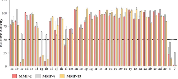
The synthesized compounds were submitted to MMP inhibition assays. A dose/response preliminary screening was performed at 100 µM by registering the residual enzyme activity on MMP-2, MMP-8 and MMP-13 (Figure 4). Compounds showing a percentage of inhibition higher than 50% were submitted to IC50 calculation. Activity data are reported in Table 4.
NH2 H2N NH2 N H Boc N H N H Boc SO O N H N H OSO HN O N a b c,d N H N H Boc N H N H HN O N 3 4 5 6 7 O O e c,d
Scheme 4. Reagents and conditions: 5-aminoisoquinoline (1 eq), phenylsulfonyl chloride (1.5 eq), pyridine, 0◦C, 3 h, 61%.
The synthetic route to derivatives 5 and 7 (Table 3) is illustrated in Scheme 5. The 4-aminobenzylamine was monoprotected by tert-butyloxycarbonyl (Boc) in THF, at room temperature; the intermediate 3 was then reacted with phenylsulfonyl chloride, in pyridine at 0◦C. The resulting sulfonamide 4 was deprotected in acidic conditions, followed by a treatment with triphosgene and 5-aminoisoquinoline, in the presence of TEA. In this step, the intermediate isocyanate, formed by treatment with triphosgene, was not isolated, but it was reacted in situ to obtain the desired urea 5.
The amide derivative 7 was obtained by starting from intermediate 3, with the same synthetic procedure.
Int. J. Mol. Sci. 2016, 17, 1768 6 of 19
The sulfonamide 2e was easily obtained by the reaction of 5-aminoisoquinoline with phenylsulfonyl chloride in pyridine at room temperature according to the literature (Scheme 4) [33,34]. NH2 N NH N S 2e O O
Scheme 4. Reagents and conditions: 5-aminoisoquinoline (1 eq), phenylsulfonyl chloride (1.5 eq),
pyridine, 0 °C, 3 h, 61%.
The synthetic route to derivatives 5 and 7 (Table 3) is illustrated in Scheme 5. The 4-aminobenzylamine was monoprotected by tert-butyloxycarbonyl (Boc) in THF, at room temperature; the intermediate 3 was then reacted with phenylsulfonyl chloride, in pyridine at 0 °C. The resulting sulfonamide 4 was deprotected in acidic conditions, followed by a treatment with triphosgene and 5-aminoisoquinoline, in the presence of TEA. In this step, the intermediate isocyanate, formed by treatment with triphosgene, was not isolated, but it was reacted in situ to obtain the desired urea 5.
The amide derivative 7 was obtained by starting from intermediate 3, with the same synthetic procedure.
Scheme 5. Reagents and conditions: (a) 4-aminobenzylamine (1 eq), di-tert-butyl-dicarbonate (Boc2O)
(0.8 eq), THF, r.t., 2 h, 66%; (b) phenylsulfonyl chloride (1.2 eq), pyridine, 0 °C, r.t., 24 h, 86%; (c) 4 N HCl, dioxane, r.t., 4 h; (d) (i) TEA (1 eq), triphosgene (0.5 eq), toluene, reflux, 4 h; (ii) 5-aminoisoquinoline (1 eq), DCM, 0 °C, r.t., 18 h, 45%–48%; (e) benzoyl chloride (1.2 eq), pyridine, 0 °C, r.t., 24 h, 72%.
2.2. Biochemical Assays
The synthesized compounds were submitted to MMP inhibition assays. A dose/response preliminary screening was performed at 100 µM by registering the residual enzyme activity on MMP-2, MMP-8 and MMP-13 (Figure 4). Compounds showing a percentage of inhibition higher than 50% were submitted to IC50 calculation. Activity data are reported in Table 4.
NH2 H2N NH2 N H Boc N H N H Boc SO O N H N H OSO HN O N a b c,d N H N H Boc N H N H HN O N 3 4 5 6 7 O O e c,d
Scheme 5.Reagents and conditions: (a) 4-aminobenzylamine (1 eq), di-tert-butyl-dicarbonate (Boc2O) (0.8 eq), THF, r.t., 2 h, 66%; (b) phenylsulfonyl chloride (1.2 eq), pyridine, 0◦C, r.t., 24 h, 86%; (c) 4 N HCl, dioxane, r.t., 4 h; (d) (i) TEA (1 eq), triphosgene (0.5 eq), toluene, reflux, 4 h; (ii) 5-aminoisoquinoline (1 eq), DCM, 0◦C, r.t., 18 h, 45%–48%; (e) benzoyl chloride (1.2 eq), pyridine, 0◦C, r.t., 24 h, 72%.
2.2. Biochemical Assays
The synthesized compounds were submitted to MMP inhibition assays. A dose/response preliminary screening was performed at 100 µM by registering the residual enzyme activity on MMP-2, MMP-8 and MMP-13 (Figure4). Compounds showing a percentage of inhibition higher than 50% were submitted to IC50calculation. Activity data are reported in Table4.
Int. J. Mol. Sci. 2016, 17, 1768 7 of 19
Int. J. Mol. Sci. 2016, 17, 1768 7 of 19
Figure 4. Residual enzyme activity on MMP-2, MMP-8 and MMP-13 of Compounds 1a–α, 2a–e, 5 and 7 at 100 µM.
Table 4. Activity data (IC50, µM) on MMP-2, MMP-8 and MMP-13.
ID MMP-2 MMP-8 MMP-13 1c 55 ± 1 >100 58 ± 1 1g 74 ± 3 >100 73 ± 11 1h 15 ± 4 >100 14 ± 4 1l 86 ± 2 >100 98 ± 2 5 54 ± 7 88 ± 5 55 ± 7 7 7.4 ± 0.8 48.5 ± 1.6 6.6 ± 0.4
The 1h resulted the most active compound among the first two series of compounds (1a–α,
2a–e), while tight analogues, such as 1i, were inactive. The active compounds showed an interesting
selectivity with respect to MMP-8.
The inhibition assays allowed to evidencing in detail the role played by specific structural features characterizing the set of NZIs: (i) the hydroxyl group on the quinoline ring (Hit 2) does not lead to detectable improvement of MMP inhibition; (ii) either urea → amide simplification or decreasing the bicycle hindrance led to inactive or less active compounds; (iii) the presence of nitrogen on the bicyclic ring is a requirement for activity, while only definite positions in the quinoline moiety led to active ligands.
In the attempt to improve inhibition activity, we extended the molecular structure of these inhibitors, to tentatively approach the residues on the top of the S1’ site, which are usually involved in binding with H-bond acceptor groups (generally the sulfonamide oxygen atoms) [35]. To this aim,
p-phenylsulfonylamide or p-phenylamide were introduced on the benzyl group to yield Compounds 5 and 7, respectively (Table 4). These compounds proved to be active toward all of the assayed MMPs.
Compared to 1h, Compound 7 presented an improved inhibition potency toward MMP-2 and MMP-13, although with a reduced selectivity toward MMP-8.
In order to elucidate the inhibition assay outcomes based on the structure of MMP-NZI complexes, computational studies were carried out by using either docking or molecular dynamics procedures.
2.3. Docking
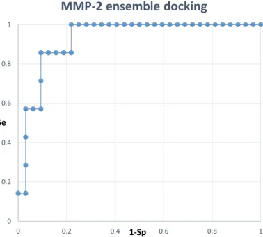
Docking calculations of the synthesized ligands in the MMP-2, MMP-8 and MMP-13 catalytic site were performed using Glide. Receptor structures were selected through a cross-docking study (Supplementary Materials), and docking calculations were conducted at both standard precision (SP) and extra precision (XP) settings, with and without a water molecule on the zinc ion. Above all, the
Figure 4.Residual enzyme activity on MMP-2, MMP-8 and MMP-13 of Compounds 1a–α, 2a–e, 5 and
7at 100 µM.
Table 4.Activity data (IC50, µM) on MMP-2, MMP-8 and MMP-13.
ID MMP-2 MMP-8 MMP-13 1c 55±1 >100 58±1 1g 74±3 >100 73±11 1h 15±4 >100 14±4 1l 86±2 >100 98±2 5 54±7 88±5 55±7 7 7.4±0.8 48.5±1.6 6.6±0.4
The 1h resulted the most active compound among the first two series of compounds (1a–α, 2a–e), while tight analogues, such as 1i, were inactive. The active compounds showed an interesting selectivity with respect to MMP-8.
The inhibition assays allowed to evidencing in detail the role played by specific structural features characterizing the set of NZIs: (i) the hydroxyl group on the quinoline ring (Hit 2) does not lead to detectable improvement of MMP inhibition; (ii) either urea→amide simplification or decreasing the bicycle hindrance led to inactive or less active compounds; (iii) the presence of nitrogen on the bicyclic ring is a requirement for activity, while only definite positions in the quinoline moiety led to active ligands.
In the attempt to improve inhibition activity, we extended the molecular structure of these inhibitors, to tentatively approach the residues on the top of the S1’ site, which are usually involved in binding with H-bond acceptor groups (generally the sulfonamide oxygen atoms) [35]. To this aim, p-phenylsulfonylamide or p-phenylamide were introduced on the benzyl group to yield Compounds 5 and 7, respectively (Table4). These compounds proved to be active toward all of the assayed MMPs. Compared to 1h, Compound 7 presented an improved inhibition potency toward MMP-2 and MMP-13, although with a reduced selectivity toward MMP-8.
In order to elucidate the inhibition assay outcomes based on the structure of MMP-NZI complexes, computational studies were carried out by using either docking or molecular dynamics procedures. 2.3. Docking
Docking calculations of the synthesized ligands in the MMP-2, MMP-8 and MMP-13 catalytic site were performed using Glide. Receptor structures were selected through a cross-docking study (Supplementary Materials), and docking calculations were conducted at both standard precision (SP) and extra precision (XP) settings, with and without a water molecule on the zinc ion. Above all, the
Int. J. Mol. Sci. 2016, 17, 1768 8 of 19
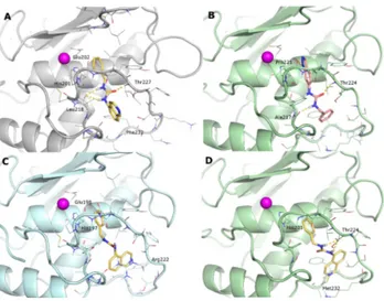
binding mode of the synthesized ligands is similar to the one described for the Hits 1 and 2. As an example, the docked pose of 1h in the MMP-2 binding site showed that the benzyl ring stacks on the His201 imidazole ring; the urea NHs form H-bonds with Leu218 CO; while the carbonyl oxygen is connected to the Thr227 NH. The isoquinoline ring is mainly solvent-exposed and only partially buried by hydrophobic residues in the lower part of the S1’ site (Leu218, Phe232) (Figure5A). A similar pattern of interactions was found in all MMP-2-NZI docking complexes, including both active and inactive ligands. The binding of indazole derivatives (1l and 1m) is inverted in all receptors with the heterocycle NH H-bonded to Pro221 CO (Figure5B).
In the MMP-8 active site, the presence of the Arg222 side chain hinders the S1’ site and limits the positioning of the bulky isoquinoline ring, giving a reason for the observed selectivity (Figure5C).
On the contrary, the larger specificity site of MMP-13 allows easily accommodating the studied ligands in a binding mode similar to that of well-known NZIs [25]: the benzyl ring forms a π–π stacking with His201; the urea NHs are engaged in H-bonding with Thr224 CO; while the isoquinoline N forms a H-bond with the Met232 NH (Figure5D).
Int. J. Mol. Sci. 2016, 17, 1768 8 of 19
binding mode of the synthesized ligands is similar to the one described for the Hits 1 and 2. As an example, the docked pose of 1h in the MMP-2 binding site showed that the benzyl ring stacks on the His201 imidazole ring; the urea NHs form H-bonds with Leu218 CO; while the carbonyl oxygen is connected to the Thr227 NH. The isoquinoline ring is mainly solvent-exposed and only partially buried by hydrophobic residues in the lower part of the S1’ site (Leu218, Phe232) (Figure 5A). A similar pattern of interactions was found in all MMP-2-NZI docking complexes, including both active and inactive ligands. The binding of indazole derivatives (1l and 1m) is inverted in all receptors with the heterocycle NH H-bonded to Pro221 CO (Figure 5B).
In the MMP-8 active site, the presence of the Arg222 side chain hinders the S1’ site and limits the positioning of the bulky isoquinoline ring, giving a reason for the observed selectivity (Figure 5C).
On the contrary, the larger specificity site of MMP-13 allows easily accommodating the studied ligands in a binding mode similar to that of well-known NZIs [25]: the benzyl ring forms a π–π stacking with His201; the urea NHs are engaged in H-bonding with Thr224 CO; while the isoquinoline N forms a H-bond with the Met232 NH (Figure 5D).
Figure 5. Docking poses of: (A) 1h in the MMP-2; (B) 1m in the MMP-13; (C) 1h in the MMP-8; and
(D) 1h in the MMP-13. MMPs are represented as grey (MMP-2), pale cyan (MMP-8) and pale green (MMP-13) cartoons. The zinc ion is a purple sphere; most relevant residues are represented as lines and ligands as sticks. H-bonds are depicted as yellow dashed lines.
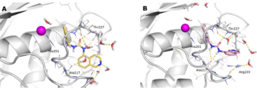
Additional interactions were evidenced by the docking of larger ligands (5 and 7): in MMP-2, these compounds interact with Leu164 and Ala165 NH, while in MMP-13, they occupy the S1’ site with a banana-shape, typical of MMP-13 NZIs (Figure 6).
Figure 6. Docking poses of: (A) ligand 5 in the MMP-2; (B) and ligand 7 in the MMP-13. MMPs are
represented as grey (MMP-2), and pale green (MMP-13) cartoons. The zinc ion is a purple sphere; most relevant residues are represented as lines and ligands as sticks. H-bonds are depicted as yellow dashed lined.
Figure 5.Docking poses of: (A) 1h in the MMP-2; (B) 1m in the MMP-13; (C) 1h in the MMP-8; and (D) 1h in the MMP-13. MMPs are represented as grey (MMP-2), pale cyan (MMP-8) and pale green (MMP-13) cartoons. The zinc ion is a purple sphere; most relevant residues are represented as lines and ligands as sticks. H-bonds are depicted as yellow dashed lines.
Additional interactions were evidenced by the docking of larger ligands (5 and 7): in MMP-2, these compounds interact with Leu164 and Ala165 NH, while in MMP-13, they occupy the S1’ site with a banana-shape, typical of MMP-13 NZIs (Figure6).
Int. J. Mol. Sci. 2016, 17, 1768 8 of 19
binding mode of the synthesized ligands is similar to the one described for the Hits 1 and 2. As an example, the docked pose of 1h in the MMP-2 binding site showed that the benzyl ring stacks on the His201 imidazole ring; the urea NHs form H-bonds with Leu218 CO; while the carbonyl oxygen is connected to the Thr227 NH. The isoquinoline ring is mainly solvent-exposed and only partially buried by hydrophobic residues in the lower part of the S1’ site (Leu218, Phe232) (Figure 5A). A similar pattern of interactions was found in all MMP-2-NZI docking complexes, including both active and inactive ligands. The binding of indazole derivatives (1l and 1m) is inverted in all receptors with the heterocycle NH H-bonded to Pro221 CO (Figure 5B).
In the MMP-8 active site, the presence of the Arg222 side chain hinders the S1’ site and limits the positioning of the bulky isoquinoline ring, giving a reason for the observed selectivity (Figure 5C).
On the contrary, the larger specificity site of MMP-13 allows easily accommodating the studied ligands in a binding mode similar to that of well-known NZIs [25]: the benzyl ring forms a π–π stacking with His201; the urea NHs are engaged in H-bonding with Thr224 CO; while the isoquinoline N forms a H-bond with the Met232 NH (Figure 5D).
Figure 5. Docking poses of: (A) 1h in the MMP-2; (B) 1m in the MMP-13; (C) 1h in the MMP-8; and
(D) 1h in the MMP-13. MMPs are represented as grey (MMP-2), pale cyan (MMP-8) and pale green (MMP-13) cartoons. The zinc ion is a purple sphere; most relevant residues are represented as lines and ligands as sticks. H-bonds are depicted as yellow dashed lines.
Additional interactions were evidenced by the docking of larger ligands (5 and 7): in MMP-2, these compounds interact with Leu164 and Ala165 NH, while in MMP-13, they occupy the S1’ site with a banana-shape, typical of MMP-13 NZIs (Figure 6).
Figure 6. Docking poses of: (A) ligand 5 in the MMP-2; (B) and ligand 7 in the MMP-13. MMPs are
represented as grey (MMP-2), and pale green (MMP-13) cartoons. The zinc ion is a purple sphere; most relevant residues are represented as lines and ligands as sticks. H-bonds are depicted as yellow dashed lined.
Figure 6.Docking poses of: (A) ligand 5 in the MMP-2; (B) and ligand 7 in the MMP-13. MMPs are represented as grey (MMP-2), and pale green (MMP-13) cartoons. The zinc ion is a purple sphere; most relevant residues are represented as lines and ligands as sticks. H-bonds are depicted as yellow dashed lined.
Int. J. Mol. Sci. 2016, 17, 1768 9 of 19
To have a better comprehension of the described SAR, the surface of the S1’ site of the studied MMPs has been represented, showing the different shapes of the three hydrophobic pockets (Figure7).
Int. J. Mol. Sci. 2016, 17, 1768 9 of 19
To have a better comprehension of the described SAR, the surface of the S1’ site of the studied MMPs has been represented, showing the different shapes of the three hydrophobic pockets (Figure 7).
Figure 7. The molecular surface of the S1’ site of, (A) MMP-2 (pale yellow), (B) MMP-8 (pale cyan)
and (C) MMP-13 (pale green) is depicted. MMP-2 has a tunnel-like S1’ site, while the MMP-8 and MMP-13 S1’ loop moves, opening an accessory pocket called the S1’* site. However, the MMP-13 site is much larger, the MMP-8 site being hindered by the Arg222 side chain.
From a quantitative point of view, no correlation between the experimental activity and scoring values can be provided by docking calculations. Moreover, the different potency observed for close analogues, i.e., the 1h and 1i ligands, cannot be easily explained based on their similar binding mode. In the latter example, it is not clear the role played by the nitrogen atom in the inhibition process, because it seems to not appreciably interact with the enzyme in the docking models of either 1h or 1i bound complexes. Thus, molecular dynamics (MD) simulations were carried out to include the effects of induced fit and the interaction with explicit water molecules into the binding model. In particular, we focused on MMP-2, as it is an important therapeutic target, and just a few NZIs have been identified so far [28,36,37]. The binding of the above-mentioned 1h and 1i ligands was studied, being representative of the case of highly similar ligands characterized by highly different inhibition activities.
2.4. Molecular Dynamics
The docking complexes of 1h and 1i were subjected to molecular dynamics simulations as reported in the Materials and Methods section. In spite of the high similarity in the ligand structures and starting poses, the 1h and 1i bound complexes required about 100 and 10 ns, respectively, to gain trajectory stabilization (Figure S1). On the one hand, the starting (obtained by docking) pose of 1i was essentially maintained and, thus, induced a softer adaptation of the MMP-2 conformation. The 1i binding pose observed in the stabilized trajectory (last 90 ns) is characterized by essentially the same interactions with MMP-2 detected in the corresponding docking pose (Figure 8B), although the presence of explicit waters allowed improving the description of the 1i binding domain.
Figure 8. Binding poses of 1h (A) and 1i (B) obtained from the last 90 ns of the MD trajectory.
MMP-2 is represented as a grey cartoon and ligands as sticks. H-bonds are depicted as yellow dashed lines.
Figure 7.The molecular surface of the S1’ site of, (A) MMP-2 (pale yellow), (B) MMP-8 (pale cyan) and (C) MMP-13 (pale green) is depicted. MMP-2 has a tunnel-like S1’ site, while the MMP-8 and MMP-13 S1’ loop moves, opening an accessory pocket called the S1’* site. However, the MMP-13 site is much larger, the MMP-8 site being hindered by the Arg222 side chain.
From a quantitative point of view, no correlation between the experimental activity and scoring values can be provided by docking calculations. Moreover, the different potency observed for close analogues, i.e., the 1h and 1i ligands, cannot be easily explained based on their similar binding mode. In the latter example, it is not clear the role played by the nitrogen atom in the inhibition process, because it seems to not appreciably interact with the enzyme in the docking models of either 1h or 1i bound complexes. Thus, molecular dynamics (MD) simulations were carried out to include the effects of induced fit and the interaction with explicit water molecules into the binding model. In particular, we focused on MMP-2, as it is an important therapeutic target, and just a few NZIs have been identified so far [28,36,37]. The binding of the above-mentioned 1h and 1i ligands was studied, being representative of the case of highly similar ligands characterized by highly different inhibition activities.
2.4. Molecular Dynamics
The docking complexes of 1h and 1i were subjected to molecular dynamics simulations as reported in the Materials and Methods section. In spite of the high similarity in the ligand structures and starting poses, the 1h and 1i bound complexes required about 100 and 10 ns, respectively, to gain trajectory stabilization (Figure S1). On the one hand, the starting (obtained by docking) pose of 1i was essentially maintained and, thus, induced a softer adaptation of the MMP-2 conformation. The 1i binding pose observed in the stabilized trajectory (last 90 ns) is characterized by essentially the same interactions with MMP-2 detected in the corresponding docking pose (Figure8B), although the presence of explicit waters allowed improving the description of the 1i binding domain.
Int. J. Mol. Sci. 2016, 17, 1768 9 of 19
To have a better comprehension of the described SAR, the surface of the S1’ site of the studied MMPs has been represented, showing the different shapes of the three hydrophobic pockets (Figure 7).
Figure 7. The molecular surface of the S1’ site of, (A) MMP-2 (pale yellow), (B) MMP-8 (pale cyan)
and (C) MMP-13 (pale green) is depicted. MMP-2 has a tunnel-like S1’ site, while the MMP-8 and MMP-13 S1’ loop moves, opening an accessory pocket called the S1’* site. However, the MMP-13 site is much larger, the MMP-8 site being hindered by the Arg222 side chain.
From a quantitative point of view, no correlation between the experimental activity and scoring values can be provided by docking calculations. Moreover, the different potency observed for close analogues, i.e., the 1h and 1i ligands, cannot be easily explained based on their similar binding mode. In the latter example, it is not clear the role played by the nitrogen atom in the inhibition process, because it seems to not appreciably interact with the enzyme in the docking models of either 1h or 1i bound complexes. Thus, molecular dynamics (MD) simulations were carried out to include the effects of induced fit and the interaction with explicit water molecules into the binding model. In particular, we focused on MMP-2, as it is an important therapeutic target, and just a few NZIs have been identified so far [28,36,37]. The binding of the above-mentioned 1h and 1i ligands was studied, being representative of the case of highly similar ligands characterized by highly different inhibition activities.
2.4. Molecular Dynamics
The docking complexes of 1h and 1i were subjected to molecular dynamics simulations as reported in the Materials and Methods section. In spite of the high similarity in the ligand structures and starting poses, the 1h and 1i bound complexes required about 100 and 10 ns, respectively, to gain trajectory stabilization (Figure S1). On the one hand, the starting (obtained by docking) pose of 1i was essentially maintained and, thus, induced a softer adaptation of the MMP-2 conformation. The 1i binding pose observed in the stabilized trajectory (last 90 ns) is characterized by essentially the same interactions with MMP-2 detected in the corresponding docking pose (Figure 8B), although the presence of explicit waters allowed improving the description of the 1i binding domain.
Figure 8. Binding poses of 1h (A) and 1i (B) obtained from the last 90 ns of the MD trajectory.
MMP-2 is represented as a grey cartoon and ligands as sticks. H-bonds are depicted as yellow dashed lines.
Figure 8.Binding poses of 1h (A) and 1i (B) obtained from the last 90 ns of the MD trajectory. MMP-2 is represented as a grey cartoon and ligands as sticks. H-bonds are depicted as yellow dashed lines.