1 Facoltà di Farmacia e Medicina – Dipartimento di Medicina Molecolare
Tesi di Dottorato di ricerca – XXXII Ciclo
p38α, the β-catenin chromatin associated kinase, as promising target
in colorectal cancer stem cells for personalized therapy
Dottorando: Relatore: Dott.sa Martina Lepore Signorile Chiar.mo Prof. Gianluca Canettieri
Supervisore: Chiar.mo Prof. Cristiano Simone
2 Summary
List of abbreviations... 4
Abstract ... 8
Introduction ... 10
1.1 The genetic-epigenetic interplay in Cancer ... 10
1.2 Colorectal cancer ... 12
1.2.1 Genetic alteration and progression of CRC ... 17
1.3 c-Myc oncogene: regulation and function ... 20
1.4 Cancer stem cell in CRC initiation and progression ... 24
1.5 The MAPK signaling: mechanism and functions in CRC ... 27
1.5.1 p38α MAPK and chemoresistance in CRC ... 34
1.6 Aim of the study ... 37
Material and Methods ... 39
2.1 Cell culture and reagents ... 39
2.2 Quantitative real-time PCR ... 39
2.3 RNA interference ... 40
2.4 Proliferation assays... 40
2.5 Nuclear/cytoplasmic fractionation and Immunoblot analysis ... 41
2.6 Immunofluorescence staining... 41
2.7 Immunoprecipitation ... 42
2.8 Serum and LiCl stimulation ... 42
2.9 Chromatin immunoprecipitation (ChIP) ... 42
2.10 Microscopic quantification of viability and cell death ... 43
2.11 Morphological evaluation... 43 2.12 Karyotype protocol ... 43 2.13 Mutation analysis ... 44 2.14 In vivo studies ... 45 1.15 Statistical Analysis ... 45 Results ... 46 3.1 Results 1 ... 46
3.1.1 p38α and ERK in c-Myc regulation ... 48
3
3.2 Results 2 ... 64
3.2.1 p38α and β-catenin localization in normal and CRC cells ... 65
3.2.2 Functional interactions between p38 and the APC/β-catenin complex ... 67
3.2.3 Characterization of p38α in CRC-SC models ... 73
3.2.4 p38α: a new member of the β-catenin complex in CRC-SC models ... 80
Discussion ... 87
Conclusion and perspectives ... 93
4
List of abbreviations
3MA, 3-methyladenine; 5-FU, 5-fluorouracil;
AKT, Rac-alpha serine/threonine-protein kinase;òl AOM, azoxy-methan;
AP-1, activator protein1;
APC, adenomatous polyposis coli; ATCC, american type culture collection; ATF2, activating transcription factor 2; ATP, adenosine triphosphate;
BAF60c, Brg1-brd associated factor subunit 60c; BAX, Bcl-2-associated X protein;
BER, base-excision DNA repair mechanism; BHLH, basic helix-loop-helix;
BMP, bone morphogenetic protein;
BRAF, serine/threonin-protein kinase B-raf; BRR, Bannayan-Riley-Ruvalcaba;
c-MET, c-mesenchymal epithelial transition factor; CD44, cluster of differentiation 44;
CD133, cluster of differentiation; Cdc25, cell division control protein 25; CDDP, cisplatin;
CHX, cycloheximide;
CIMP, CpG island methylator phenotype; CIMP1, CpG island methylator phenotype 1; CIMP2, CpG island methylator phenotype 2; CIN, chromosomal instability;
Cis, cisplatin;
CKI, casein kinase I;
c-Myc; myelocitomatosis virus; COX-2, cyclooxygenase-2; CRC, Colorectal Cancer;
5 CRC-SC, colorectal cancer stem cell;
CR-SC, colorectal stem cell; CSC, Cancer Stem Cell; DCC, deleted in colorectal; DFG, Asp-Phe-Gly motif;
DMEM, Dulbecco’s modified eagle medium; DMSO, dimethyl sulfoxide;
EDTA, ethylene diamine tetracetic acid; EGF, epidermal growth factor;
EGFR, epidermal growth factor receptor; ENU, ethylnitrosourea;
ERK1/2, extracellular-signal-regulated kinases 1/2; FAP, familial adenomatous polyposis;
FBS, fetal bovine serum;
FOLFIRI, folinic acid-fluorouracil-irinotecan; FOLFOX, folinic acid-fluorouracil-oxaliplatin; FoxO3A, forkhead transcription factor FKHR-L1;
GADD45α, growth arrest and DNA-damage inducible 45 alpha; GSK3β, glycogen synthase kinase 3 beta;
HDAC, histone deacetylase;
HER3, receptor tyrosine protein kinase erB3; HIF1α, hypoxia-inducible factor 1 alpha;
HNPCC, hereditary non-polyposis colorectal cancer; HSP27, heat shock protein 27;
JNK, c-Jun N-terminal kinase;
JNK1-3, c-Jun N-terminal kinase 1-3; JNKIP2, JNK-interacting protein 2; JP, Juvenile Polyposis;
KLF4, kruppel-like factor4;
KRAS, V-Ki ras2 Kirsten rat sarcoma viral oncogene homolog; LC3I-II, light chain 3 I/II;
LEF/TCF lymphoid enhancer factor/T cell factor;
6 LKB1/STK11, liver kinase B1/serine-threonine kinase 11;
LOH, loss of heterozygosity; LZ, leucin zipper;
MAP1LC3, Microtubule-associated proteins 1A/1B light chain 3B; MAP2K, mitogen-activated protein kinase kinase;
MAPK, mitogen-activated protein kinase;
MAPK14-11-12-13 , mitogen-activated protein kinase 14-11-12-13; MAPK5, mitogen-activated protein kinase 5;
MAPKAP-2/3, mitogen-activated protein kinase-activated protein kinase 2; MAPKK, mitogen-activated protein kinase kinase;
MAPKKK, mitogen-activated protein kinase kinase kinase; MDM2, mouse double minute 2 homolog;
MEF-2, myocyte enhancer factor 2;
MEK1/2, mitogen/extracellular signal-regulated kinase 1/2; MEKK1-4, mitogen-activated protein kinase kinase kinase 1-4; MIN, microsatellite instability;
Min, multiple intestinal neoplasia;
MK2, MAP kinase-activated protein kinase 2;
MKK3/6, mitogen-activated protein kinase kinase 3/6; MKK3Be, mitogen-activated protein kinase kinase 3Be; MKK4/7, mitogen-activated protein kinase kinase 4/7; MMP1, matrix metalloproteinase 1;
MMP13, matrix metalloproteinase 13; MMP3, matrix metalloproteinase 3; MMR, mismatch repair system;
MNK1/2, MAP kinase-interacting serine/thereonine protein; MSC, mesenchimal stem cell;
MSH2, MutS protein homolog 2; MSH6, MutS protein homolog 6; MSI, microsatellite instability;
MSK1/2, mitogen-and stress-activated protein kinase 1/2; MYOD, myogenin D;
7 p21Cip1, cycline -dependent kinase inhibitor 1;
PBS, phosphate buffered saline; PDI, protein disulfide-isomerase; P-gp, P-glycoprotein;
PI3K, phosphoinositide 3-kinase;
PI3K class I, phosphoinositide 3-kinase class I; PI3K class III, phosphoinositide 3-kinase class III;
PI3KCA, phosphatidylinositol-4,5-bisphosphate-3 kinase, catalytic subunit alpha; PJ, Peutz-Jeghers;
PP2A, protein phosphatase 2A;
PRAK, p38-regulated/activated protein kinase; PTEN, phosphatase and tensin homologue; RAF, Rapidly Accelerated Fibrosarcoma;
RAF-1, Raf proto-oncogene serine/thereonine- protein kinase; RAS, Rat Sarcoma;
ROS, reactive oxygen species; RTK, receptor tyrosine kinase; S62, serine 62;
SIRT1, NAD-dependent deacetylase sirtuin 1; SMAD4, SMAD family member 4;
SOX9, sex determining region Y T58, threonine 58;
TBP, TATA binding protein; TCF4, transcription factor 4; TCF7, transcription factor 7;
TGF-β, transforming growth factor beta; TP53, tumor protein p53;
Ub, ubiquitination;
VEGF, vascular endothelial growth factor; Wnt, Wingless intergrate;
WRE, wnt responsive element;
8
Abstract
Colorectal cancer (CRC) is the third most frequent malignancy, but the second cause of death for tumor in the western population. Only 14% of patients with advanced and metastatic disease survive five years from diagnosis. Recently, it has been shown that tumor relapse and chemoresistance depend on a small population of cells, called cancer stem cells (CSCs). Current evidence indicates that the Wnt cascade is the main driver in controlling CSC fate; the key player in this pathway is β-catenin, a cytoplasmic protein whose stability is regulated by the so-called “destruction complex”. During carcinogenesis, the increasing amount of β-catenin resulting from APC inactivation translocates into the nucleus, causing the transcriptional activation of several mitogenic genes, including c-Myc. c-Myc is one of the most important factors involved in CRC initiation and progression; indeed, it functions as a link connecting malignancy with stemness. During colorectal carcinogenesis, c-Myc is maintained upregulated through β-catenin-mediated transcriptional activation and ERK-mediated post-translational stabilization. Our data showed that p38α, a kinase involved in CRC metabolism and survival, contributes to both mechanisms. Previous reports in other tissues provided evidence that Wnt3a can activate p38, and the p38 pathway feeds into the canonical Wnt/β-catenin pathway at least at the level of GSK3β. Our findings also highlighted that CRC cells and colorectal cancer stem cells (CRC-SCs) have higher levels of activated p38 than their normal counterparts, and experiments using kinase-specific inhibitors revealed that these cells are “addicted” to p38 activity. Importantly, we found that p38α co-immunoprecipitates with β-catenin in both normal and cancer cells; however, these proteins are confined to the cytoplasm in colonocytes, while they significantly occupy discrete nuclear regions in CRC cells, CRC-SCs, and in vivo models. These data were further corroborated by the inhibitory effect of p38α blockade on several β-catenin-responsive genes (i.e. c-Myc, cyclin D1/2, survivin, and others). This functional interaction was further characterized by chromatin immunoprecipitation experiments, which demonstrated that p38α is a chromatin-associated β-catenin kinase required for the transcriptional induction of several Wnt target genes, including c-Myc. Additionally, we demonstrated that p38α, like ERK, stabilizes c-Myc protein levels by preventing its ubiquitination. The finding that the phenotypes arising after APC loss in the intestine are fully dependent on c-Myc target gene expression suggests that c-Myc inhibition may
9 be a good target for chemoprevention in CRC. These considerations underline the relevance of molecular profiling and preclinical investigation in order to achieve more efficient and accurate therapies. Indeed, our study identifies p38α as a promising therapeutic target acting directly on c-Myc and CRC-SCs, which are thought to be responsible for tumor proliferation, metastatic dissemination, and chemoresistance.
10
Introduction
1.1 The genetic-epigenetic interplay in Cancer
Cancer is a set of more than 100 different diseases in which abnormal cells divide in an uncontrolled way and can invade other tissues (metastasize). Carcinogenesis is driven by the accumulation of different types of alterations, which affect the structure and the function of the genome. Primarily, it has been described as a consequence of the accumulation of genetic mutations. This view has now been expanded to include epigenetic changes. Genetic and epigenetic alterations allow cells to lose all physiological processes and mechanisms of control which regulate the homeostatic balance between cell proliferation and cell death (Hanahan D and Weinberg RA, 2011). Studying the complexity of cancer, ten biological capabilities were identified as “Hallmarks of cancer”, as common principles acquired during the multistep development of human tumors. These characteristics, that distinguish cancer cells from normal cells, include: self-sufficient cell division, insensitivity to signals to stop cell division, resisting cell death, limitless reproductive potential, creating their own blood supply, ability to invade other organs, ability to survive with little oxygen, evading the immune system, genome instability and inflammation. Epigenetic mechanisms contribute to each hallmark by modulating chromatin structure and factors involved in this regulation through tumor suppressor silencing, oncogene activation by repurposed enhancers, or cell fate transitions (Figure 1).
11 Figure 1. The hallmarks of cancer. Ten functional capabilities are acquired in the multistep carcinogenesis that leads to human cancer. The order in which they are acquired and the importance of their contributions appears to vary across the spectrum of human cancers. (Adapted from Hanahan D and Weinberg RA, 2011).
12
1.2 Colorectal cancer
Colorectal cancer (CRC) is the main malignant tumor affecting the gastrointestinal tract and it is the third most common cancer in both men and women (Haggar FA and Boushey RP, 2009; Bhandari A, Woodhouse M, and Gupta S, 2017). Worldwide, about 2 million people each year will develop CRC (WORLD HEALTH ORGANIZATION -2018- https://www.who.int/news-room/fact-sheets/detail/cancer)
with a substantial variation in the five-year relative survival rate in relation to the stage of disease at diagnosis. 93% of patients diagnosed with the earliest stage of disease survived five-years from diagnosis compared to only 14% of those with advanced disease which had spread to other parts of the body at diagnosis (Figure 2).
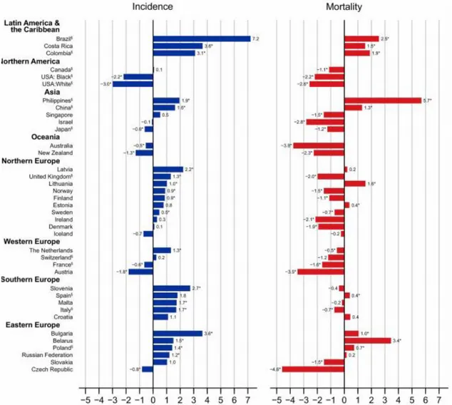
Figure 2. Average annual percentage change of colorectal cancer incidence and mortality in the most recent period (10 years). (Adapted from Arnold M et al, 2016).
13 Every year in our country, about 27,000 people suffer from CRC, and over half of them die due to illness (data from AIRTUM -Associazione Italiana Registro Tumori 2017-). The risk of CRC increases with age, indeed the median age at diagnosis is 68 in men and 72 in women. Furthermore males are affected slightly more often than females (Figure 3).
Figure 3. Age-specific incidence of CRC in men and women. (Adapted from Cancer Research: bowel cancer incidence statistics, 2017).
Approximately 5% of men and women will be diagnosed with CRC in their lifetime. People with a first-degree relative (parent, sibling or child) who has been diagnosed with CRC have 2 to 3 times the risk of developing the disease compared to people without this family history, depending on the age at diagnosis and the number of affected relatives. Risk is also increased among people with a first or second-degree relative diagnosed with adenomas. Also, hereditary factors, such as Lynch syndrome (HNPCC), familial adenomatous polyposis (FAP) and rare hamartomatous polyposis such as the Peutz-Jeghers (PJ) syndrome, the juvenile polyposis (JP) syndrome, the Cowden syndrome and the Bannayan-Riley-Ruvalcaba (BRR) syndrome increase CRC risk (Figure 4).
14 Figure 4. CRC at a glance. The risk of CRC during lifetime is approximately 5%. People older than 50 years old are at the highest risk for CRC (90% of cases occur in people aged 50+) and therefore should receive regular screenings. This is particularly important if a first degree family’s medical record shows history of polyps and colon cancer which could double or even triple the risk of developing the disease. (From Gastrointestinal Specialists, 2018).
Aside from age and family history, many of the known risk factors for CRC are behaviors traditionally associated with high-income countries, such as a sedentary lifestyle, western diet, and smoking. The prevalence of these factors is reflected in the substantial variation in CRC incidence worldwide, which is highest in developed countries and lowest in sub-Saharan Africa (Arnold M et al, 2016).
The slow course of growth from precancerous polyp to invasive cancer provides a unique opportunity for the prevention and early detection of CRC. Screening can prevent cancer through the detection and removal of precancerous growths and can detect cancer at an early stage, when treatment is usually more successful. As a result, screening such as fecal occult blood test and colonoscopy, reduce CRC mortality both by the decreasing incidence of disease and by increasing the likelihood of survival. Surgery is the most common treatment for CRC which has not spread. Chemotherapy, alone for colon cancer or in combination with radiation for rectal cancer, is given before (neoadjuvant) or after (adjuvant) surgery to most patients whose cancer has penetrated the bowel wall deeply or spread to lymph nodes. For metastatic CRC treatment typically include chemotherapy with FOLFIRI (5-Fluorouracil or capecitabine and irinotecan) or
15 FOLFOX (oxaliplatin and irinotecan) and/or targeted therapy. Indeed, the core of the chemotherapy regimens is 5-FU, a fluorinated pyrimidine which inhibits DNA synthesis, with the addition of calcium leucovorin increasing the response rate (Zhang ZG et al, 1992; Petrelli N et al, 1989). Other compounds used in systemic chemotherapy are Capecitabine, an orally administered form of 5-FU, Oxaliplatin, a DNA replication inhibitor, and Irinotecan, a topoisomerase inhibitor. All these compounds were developed to preferentially target cancer cells due to their higher growth rate compared to normal cells. However, as several normal cell types in the human body display high proliferation levels, and because these chemical compounds target every cell in the organism, the side effects associated with the therapies are of great relevance. Among these the most common are: stomatitis, hand-foot syndrome, diarrhea, neutropenia, nausea, alopecia, and sensory neuropathy. Moreover, the chemotherapy affects apoptosis by inducing DNA damage response, but gene mutations at apoptotic and/or anti-apoptotic loci cause the acquisition of chemoresistance.
Due to the requirement for more selective pharmaceutical compounds, a class of humanized antibodies targeting growth and angiogenic factors were developed and are now available for clinical trials. According to ClinicalTrials.gov (https://clinicaltrials.gov/ct2/results?cond=colorectal+cancer&term=&cntry=&state=&city=&di st=) 1850 studies on CRC are currently ongoing. Recently, Bevacizumab, a monoclonal antibodies targeting vascular endothelial growth factor (VEGF), and Cetuximab or Panitumumab, two chimeric monoclonal antibody targeting epidermal growth factor receptor (EGFR) used in KRAS wild type tumors, have entered routine clinical practice in metastatic CRC (Goldberg R M, Montagut C, Wainberg R A, Ronga P, Audhuy F, Taieb J, Stintzing S, Siena S, Santini D, 2018). In combination with chemotherapy, they offer higher survival advantages compared to chemotherapy alone. The major limitation is the limited presence of the targeted antigen on the cancer cell’s surface or in the cancer microenvironment. For instance, VEGF is over-expressed in 50% of CRC and the addition of Bevacizumab to normal chemotherapy led to a 10% improvement in the response rate (44,8% versus 34,8%). Nonetheless, rare but serious side effects have been observed using Bevacizumab. Clinical trials are now directed at evaluating new drug combinations, treatment schedules, and new ways of diagnostic imaging to enhance tumor regression, increasing overall survival, and improving the quality of life for patients. Indeed, today, there is a new trial ongoing, which combined MEK and
16 MDM2 inhibition in order to induce apoptosis in RAS/BRAF-mutant and TP53 wild-type CRC models. Clinicians are using new inhibitors, such as Trametinib, an orally bioavailable inhibitor of MEK/ERK kinases with potential antineoplastic activity. Trametinib specifically binds to and inhibits MEK 1 and 2, resulting in an inhibition of growth factor-mediated cell signaling and cellular proliferation in various cancers (Flaherty KT et al, 2012). Finally, the main purpose of another in-progress clinical trial is to evaluate the safety of the drug Prexasertib (cell cycle checkpoint kinase 1 and 2 inhibitor) in combination with Ralimetinib, p38α inhibitor, in participants with advanced or metastatic CRC (Bence A L, McNeely S C, and Beckmann R P, 2017). However, other therapeutic targets are being awaited to develop new strategies, which are expected to prove more effective, more specific and possibly associated with fewer side effects.
17 1.2.1 Genetic alteration and progression of CRC
CRC arises from adenomas, which gradually progress through an increase in size, dysplasia and the acquisition of villous morphology. At the beginning, the proliferation is induced by the loss or inactivation of the familial adenomatous polyposis (APC) gene on chromosome 5 or by activating a mutation of β-catenin on chromosome 3. One of these hyperproliferating cells may then give rise, by clonal expansion, to a small adenoma. Subsequently, mutation in the RAS pathway (KRAS 50% or BRAF 11%) appears to occur and through clonal expansion produces a larger and more dysplastic adenoma. After which, usually happens allelic deletions of chromosome 18q and 17q with the loss of SMAD4 and p53 genes. Tumors continue to progress once carcinomas have formed, and the accumulated loss of suppressor genes on additional chromosomes correlates with the ability of carcinomas to metastasize and cause death (Fearon ER and Vogelstein B, 1990; Kinzler KW and Vogelstein B, 1996; Kinzler KW and Vogelstein B, 1996). The evolution from benign growth to invasive stages often occurs over 10 to 20 years. This gives rise to dynamic processes whose genetic contributions interconnect with each other and with the environment. The adenoma-carcinoma sequence is characterized by the lack of allelic balance at several chromosomal loci (APC 5q, DCC/SMAD4 18q and p53 17q), and chromosomal amplification and translocation which together contribute to tumor aneuploidy and chromosomal instability (CIN). This canonical pathway happens in 85% of sporadic CRCs.
The remaining sporadic cases, about 15%, are microsatellite instability (MSI) phenotypes, mostly due to mismatch (MMR) and base-excision (BER) DNA repair mechanism impairments, giving rise to frameshift or base-pair substitutions in short tandem-repeat sequences. This is the case for mutations in various genes (MLH1, MSH2, MSH6) often associated with CpG methylation-silenced promoter regions (MLH1) and in genes (MINT1, MINT2, MINT3) involved in NOTCH signaling. Interestingly, together with methylation most of the defects in MMR or BER genes specifically induce cancer initiation. The HNPCC syndrome is linked to mutations in MMR genes, which are inherited in a dominant pattern and cause multiple primary CRCs, acceleration of tumor progression in the case of somatic mutations, as well as other tumors (Markowitz SD and Bertagnolli MM, 2009).
18 As well as CIN- or MSI-associated CRCs, several low-penetrant mutations have been observed, inducing rare syndromes. Germline mutations of the LKB1/STK11 tumor suppressor gene on chromosome 19p, driven by LOH or by missense mutations and frameshifts, have been shown to cause the rare PJ syndrome (Howe JR et al, 1998). Additionally, mutations in the tumor suppressor PTEN, which are frequently observed in tumors, are linked to the Cowden syndrome and the BRR syndrome, a spectrum of conditions collectively known as PTEN hamartoma tumor syndromes.
Epigenetic alterations in CRCs are widely reported, mainly the gene promoter DNA methylation. Classification of CRCs according to DNA methylation status has identified a subset of tumors with extensive epigenetic instability, characterized by the concordant promoter hypermethylation (Toyota M et al, 2000). The existence of a CpG island methylator phenotype (CIMP) and its correlation with clinicopathologic features has been confirmed extensively by the use of high-throughput techniques (Estecio MR et al, 2007; Weisenberger DJ et al, 2006). Typical high-level CIMP (CIMP high, CIMP1) CRCs are associated with microsatellite instability through epigenetic silencing of mismatch repair gene MLH1. They often have a BRAF mutation, and they occur predominantly in the proximal colon. Low-level CIMP (CIMP low, CIMP2) has been characterized by DNA methylation of a limited group of genes and mutation of KRAS (Shen L et al, 2007) (Figure 5).
Figure 5. Colorectal tumorigenesis. Schematic representation of hereditary and sporadic predisposition. (Adapted from Kinzler W and Vogelstein B. Cell, 87: 159-170; 1996).
19 Activation of the Wnt pathway has been linked to CRC since the recognition that abnormalities of chromosome 5q were early events in the carcinogenic process for sporadic and hereditary (FAP) tumors. Encoded at 5q is the APC gene, the protein product of which is a key component of the β-catenin destruction complex, together with GSK3β, Axin and CK1. About 90% of sporadic CRC show high levels of nuclear β-catenin resulting from mutations in the APC gene (90%) or β-catenin itself (5%) (Jeong W-J, Ro E J and Choi K-Y, 2018). Stabilized β-catenin exerts its oncogenic role by activating the transcription of many regulatory genes, such as c-Myc (Figure 6).
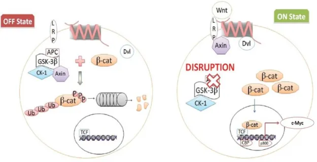
Figure 6. The Wnt canonical pathway. Wnt ligand binds to G-protein coupled receptors called Frizzled and initiates intracellular signaling cascade leading to nuclear accumulation of β-catenin and activation of target genes, which are mainly involved in proliferation. In the absence of Wnt, cellular β-catenin is phosphorylated by GSK3β and with the destruction complex catalyzed its degradation. High level of nuclear β-catenin results from mutation in the APC protein or β-catenin itself, leading to costitutive activation of the Wnt pathway, loss of normal cellular architecture and neoplastic conversion. (Adapted From Kazi M, Trivedi T, Kobawala T, Ghosh N, 2016).
20
1.3 c-Myc oncogene: regulation and function
The c-Myc proto-oncogene was identified for the first time as the cellular homolog to the viral oncogene (v-myc) of the avian myelocytomatosis retrovirus (Pelengaris S and Khan M, 2013).
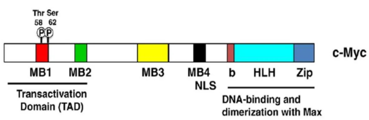
c-Myc protein belongs to the Myc family of transcription factors, which also includes n-Myc and l-n-Myc genes. c-n-Myc as transcription factor contains a basic helix-loop-helix (bHLH) and leucine zipper (LZ) motives. Through its bHLH DNA-binding motif, c-Myc interacts with the E-box sequence, a specific DNA sequence (CACGTG), while the leucine zipper TF-binding motif allows the dimerization with another bHLH transcription factor called Max (Figure 7).
Figure 7. Structure of the c-Myc protein. TAD, transcriptional activation domain; Thr-58 and Ser-62, regulatory phosphorylation sites; MB1-4, the evolutionary conserved Myc boxes 1–4; NLS, nuclear localization signal; bHLHZip, basic region/helix–loop–helix/leucine zipper. (Adapted from Larsson LG and Henriksson MA, 2010)
Surprisingly, Samson et al, in 2008 found that after APC loss, nuclear β-catenin was not sufficient to induce any phenotypes in the absence of c-Myc, indeed over half of Wnt target significantly induced after APC loss were no longer up-regulated in the absence of c-Myc. These data indicate that the phenotypes acquired after APC loss in the intestine are fully dependent on c-Myc target gene expression (Wilkins JA and Sansom OJ, 2008).
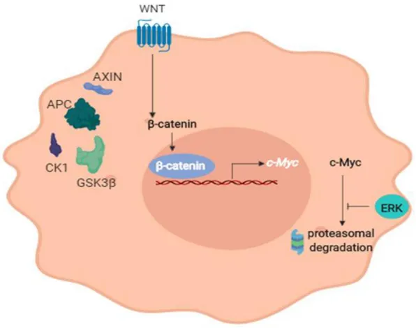
21 c-Myc is one of the most important factors implied at the beginning and during the progression of CRC, indeed it is found overexpressed in up to 70-80% of CRCs. Considering its role in intestinal tumorigenesis, it is important to underline that it is maintained upregulated in CRC by two different mechanisms. On the one hand there is the transcriptional activation by β-catenin due to APC inactivation, on the other hand there is the post-translational stabilization due to serine 62 (S62) phosphorylation mediated by ERK, a downstream kinase at KRAS, which represents the second mutation in the classic adenoma-carcinoma sequence.
Indeed the stability of c-Myc protein is controlled by phosphorylation at two specific sites: S62 and threonine 58 (T58). Phosphorylation at T58 but not at S62 making c-Myc prone to the subsequent proteasome degradation (Sears R, 2004) (Figure 8).
Figure 8. The upregulation of c-Myc in CRC. c-Myc is maintened upregulated in CRC by two different mechanism. On the one hand there is the transcriptional activation by β-catenin due to APC inactivation, on the other hand there is the post-translational stabilization due to serine 62 phosphorylation mediated by ERK.
22 c-Myc deregulation is implicated in many pathological conditions, including genomic instability, uncontrolled cell proliferation, immortalization, escape from immune cells, angiogenesis and metastasis (Dang C V et al, 2006). Moreover, c-Myc can act on control points of gene expression programs that determine the cancer stem cells (CSCs) features. Indeed, c-Myc is highly expressed in these cells and required for maintaining their phenotypes, so its functions as a link connecting malignancy and ‘stemness’ (Zhang H et al, 2019). It has been shown that c-Myc is indispensable for the self-renewal properties of colorectal cancer stem cells (CRC-SCs), since its ablation reduces the formation of tumorspheres (Sanchita R and Adhip P N M, 2012). Invasive and migratory abilities are critical features of CRC-SCs, which promote the establishment of a functional phenotype associated with tumor aggressiveness. It has been demonstrated that c-Myc plays a fundamental role in invasion and migration of CRC-SCs since its knockdown inhibits these two features. Moreover, c-Myc is considered the core of CRC-SCs chemoresistance mechanism. Indeed it was reported that c-Myc expression in surviving tumor cells (CRC-SCs), increases after treatment with platinum-based chemotherapy (Walker T L et al, 1996). Furthermore, it has been shown that c-Myc induces a high expression of the ATP-binding cassette and multidrug resistance protein, since its silencing decreases the expression of ABCG2 and ABCB5 and sensitizes CRC-SCs to chemotherapy-induced cytotoxicity (Zhang HL et al, 2019). Once tumor cells become resistant to conventional therapy such as cisplatin, they naturally develops resistance to many other chemotherapeutic agents leading to multi-drug cross-resistance. Since c-Myc protects tumor genomes from therapeutic DNA damaging drugs, it could be a promising target to overcome chemoresistance in various types of cancers, including CRC (Elbadawy M, Usui T, Yamawaki H and Sasaki K, 2019) (Figure 9).
23 Figure 9. Cellular processes controlled by c-Myc during normal conditions and during tumorigenesis. c-Myc is a key regulator of many biological activities including cell growth and division (regulation of chromatin modification and components of the biosynthetic machinery); cell-cycle progression (modulation of cyclins, dependent kinases, cyclin-dependent kinase inhibitors and phosphatases); apoptosis (p53 cyclin-dependent or incyclin-dependent mechanisms); cell differentiation (downregulation of growth arrest genes); cell metabolism (glycolysis, amino acid biosynthesis and transport, synthesis of macromolecules and DNA metabolism); angiogenesis (upregulation of VEGF); cell adhesion and motility (control of expression of integrins). Deregulation of Myc may result in apoptosis, genomic instability, uncontrolled cell proliferation, escape from immune surveillance, growth factor independence, and immortalization. (From Vita M and Henriksson M, 2006).
24
1.4 Cancer stem cell in CRC initiation and progression
It is assumed that the tumor is a heterogeneous entity produced by the proliferation of tumor-initiating cells. So many similarities have been widely assessed between these undifferentiated cells and normal stem cells that the name of “cancer stem cells” (CSC) has been given to the tumor-initiating cells (Saulnier N et al, 2009).
Self-renewal and multi-lineage differentiation are the main features of stem cells and these two peculiarities in addition to the tumorigenic potential typical of cancer cells, produced the three main parameters that outline the profile of CSC. Specifically, stem cells can undergo asymmetric division by which a cell can self-renew producing one daughter cell that preserves stemness and another cell that has the potential to differentiate during cell division. Multi-lineage differentiation is defined as the ability of a stem cell to give rise to many specialized cell types. It happens in the intestinal crypt where a colorectal stem cell (CR-SC) should be able to produce several cell types, which include enterocytes, goblet cells, paneth cells, and enteroendocrine cells (Abetov D et al, 2015). The tumorigenic potential expresses itself through tumor initiation and recurrence. Indeed, following the growth of the primary tumor, CSCs are implicated in the metastasis program through dormancy, re-initiation, and escape of immune surveillance. This can be achieved by Wnt signaling that can re-initiate cell cycle progression in dormant CSCs by upregulating c-Myc expression. Accordingly, stable c-Myc knockdown decreases colony formation on primary and secondary organs (Wolfer A et al, 2010). Moreover, CSCs are generally resistant to chemotherapy because of the enhancement of active ABC transporters that rapidly efflux chemotherapeutic agents. Indeed, indiscriminate use of chemotherapeutic agents may select the growth of CSCs that can survive this treatment, with the result that they then encounter less competition for resources. In support of this concept, CD133+ cells (a marker for CSC) were enriched when xenografts derived from CRC-SCs were exposed to oxaliplatin (Dallas NA et al, 2009). Additionally, CSC can tolerate radiotherapy upregulating multiple pathways of DNA damage response (Vila PM et al, 2017). Thus, CSCs are thought to be responsible for cancer relapse and to contribute to tumor chemo and radioresistance (Figure 10).
25 Figure 10. The role of cancer stem cells. Normal tissues arise from a central stem cell that grows and differentiates to create progenitor and mature cell populations. Cancer stem cells arise by means of a mutation in normal stem cells or progenitor cells and subsequently grow and differentiate to create primary tumors. Like normal stem cells, cancer stem cells can self-renew, give rise to heterogeneous populations of daughter cells, and proliferate extensively to form tumor at the end. During chemotherapy and radiotherapy, the majority of cells in a primary tumor may be destroyed; however, if the cancer stem cells are not eradicated, the tumor may regrow and cause a relapses.( Adapted from Wang Z, Yang H, Wang, X, Wang L, Cheng Y, Zhang Y and Tu Y, 2016).
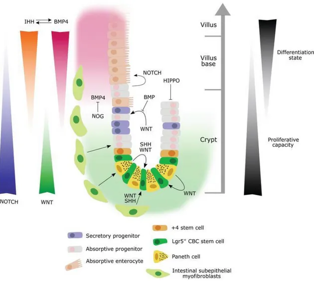
Usually, CRC-SCs have the same molecular signaling profile as normal stem cells (Abetov D et al, 2015). Indeed, it is important to underline how the microenvironment of the intestinal crypt plays a key role in the initiation and maintenance of CRC. CRC-SCs in the crypt receive Wnt factors that stimulate the undifferentiated state, obtain NOTCH ligands from neighboring cells required to block differentiation, and receive many mitogenic stimuli that activate the epidermal growth factor receptor (EGFR). Also, the niche defend CRC-SCs from cytostatic signals derived from bone-morphogenetic protein (BMP) and the transforming growth factor β (TGFβ) pathways (Batlle E and Clevers H, 2017). In this signal network, the Wnt pathway plays the most
26 important role in maintaining the CRC-SC crypt population at a proliferative state. When the pathway is over-activated, crypts enlarge; when the pathway is blocked they disappear. c-Myc, as the main Wnt target, plays a major role in this switch by direct repression of the p21 promoter. Indeed declined expression of c-Myc results in the transcription of p21, which in turn triggers G1 arrest (Elbadawy M, Usui T, Yamawaki H and Sasaki K, 2019). Finally, Muncan et al. have found that genetic ablation of c-Myc resulted in rapid loss of crypts and decreased cell numbers in crypts that remained (Muncan V et al, 2006) (Figure 11).
Figure 11. Schematic representation of the major signaling pathways involved in CSC biology. A gradient of BMP and Hh signaling, with relatively high activity in the villus and less activity within the crypt, regulates cell renewal and lineage specification. Wnt and Notch signaling gradients in the opposite direction (highest expression at the crypt base) play an important role in maintaining the stem cell compartment. (From Gleizes A, Cavailles V and Lapierre M, 2018).
27
1.5 The MAPK signaling: mechanism and functions in CRC
As mentioned above c-Myc is upregulated in intestinal tumorigenesis by transcriptional activation of β-catenin and the post-translational stabilization mediated by ERK.
The importance of ERK activation in the intestinal tumorigenesis was highlighted in a work carried out by Sung Hee Lee and colleagues, by the use of the APCMin/+ model. The APCMin/+ mouse was identified in 1990 from an ethylnitrosourea (ENU) mutagenesis screen in C57Bl/6J mice, which represents an animal model of FAP. The genetic basis for the intestinal phenotype is a T-to-A transversion at nucleotide 2,549 of the mouse APC gene that truncates the APC protein at amino acid 850. These mice develop multiple intestinal neoplasia (Min) after they spontaneously lose the heterozygous wild-type APC allele in intestinal epithelial cells, and consequently die by the time they reach 6 months of age (Moser AR et al, 1990). Sung Hee Lee and colleagues revealed that the activation of ERK in these cells is essential for tumor growth in APCMin/+ mice via the stabilization of c-Myc protein (Lee SH et al, 2010). In CRC, the MEK/ERK signaling pathway has been frequently found to be overactive due to activating mutations in the upstream kinases RAS (50%) and BRAF (11%). This pathway is activated by a variety of different stimuli, many of which promote cell proliferation and inhibit anti-proliferative and pro-apoptotic responses. This pathway is being widely studied as a promising pharmacological target.
ERK belongs to MAPKs family, which is composed of protein kinases whose function and regulation have been conserved during evolution from unicellular organism such as brewers’ yeast to complex organisms including humans (Widmann C et al, 1999). MAPKs are expressed in all cell types and regulate a variety of physiological processes such as cell growth, metabolism, differentiation, and cell death. MAPK signaling is over-activated in cancer, especially in CRC.
To date, six distinct groups of MAPKs have been characterized in mammals: the ERK 1/2, ERK 3/4, ERK5, ERK 7/8 family, the Jun N-terminal kinases JNK 1/2/3 and the p38 MAPKs p38α/β/γ/δ (Kyriakis JM and Avruch J, 2001-2012).
It is becoming increasingly clear that the pathways rather than the individual genes appear to govern the course of tumorigenesis. A paradigm of this view is represented by the p38 pathway, which is involved in proliferation, differentiation, metabolism and cell death (Hui L et al, 2004; Simone C et al, 2004; Grossi et al, 2014).
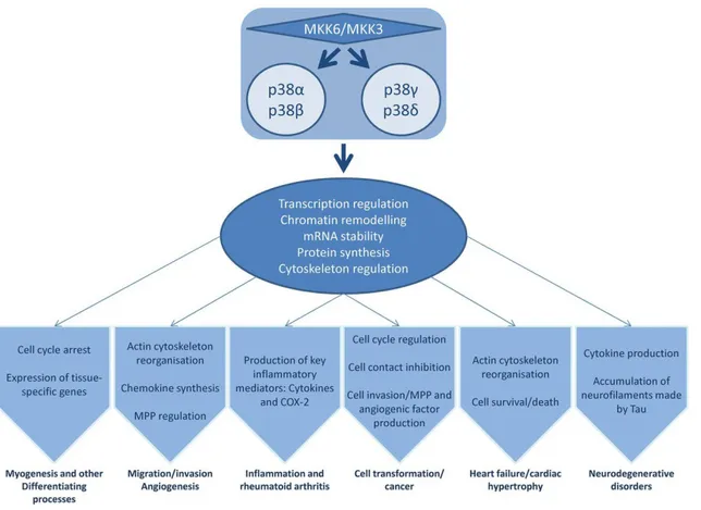
28 In mammals, four genes have been identified that encode p38 MAPKs: MAPK14 (p38α), MAPK11 (p38β), MAPK12 (p38γ) and MAPK13 (p38δ). p38α and p38β are closely related proteins that might have overlapping functions. While p38α and p38β expression is ubiquitous, the expression of p38γ and δ is only related to some specific tissue, e.g. muscle expresses p38γ and p38δ is identified in skin, small intestine and kidney (Cuadrado A and Nebreda AR, 2010; Cuenda A and Rousseau S, 2007). In addition, the differentiation status of the cell controls the expression profile of p38 isoforms. For example, undifferentiated intestinal epithelial cells show the α, β and γ isoforms, while differentiated cells express the α, γ and δ isoforms (Vachon PH et al, 2002; Comes F et al, 2007) (Figure 12).
Figure 12. p38 MAPK family regulate multiple cellular processes and physiological functions. Deregulation of p38 MAPKs pathway lead to the development of several pathological conditions. (Adapted from Oeztuerk-Winder F and Ventura JJ, Biochemical Journal, 2012).
The strict regulation of life/death signals by p38 MAPKs can result in opposite molecular functions during tumor growth. Currently, it is assumed that p38 acts as
29 tumor suppressor in normal cells at the onset of cellular transformation, subsequently after the acquisition of the malignant phenotype p38 acts as an oncogene (Dolado I et al, 2007; Hui L et al, 2007). Several studies, taking advantage of mouse models deficient in p38 or its upstream activators (such as MAPKKs, GADD45α, MAPK5), demonstrated higher susceptibility to tumor development (Ventura JJ et al, 2007; Brancho D et al, 2003; Bulavin DV et al, 2004; Tront JS et al, 2006).
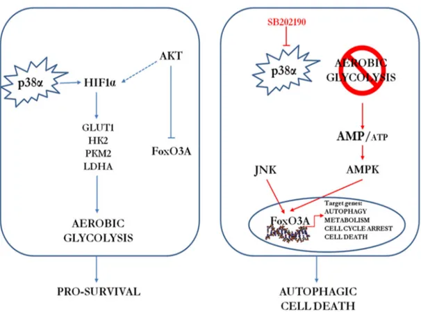
However, after the acquisition of the malignant phenotype, p38 is involved in sustaining tumor growth in many types of cancer including follicular lymphoma, lung, thyroid, colorectal, ovarian (Matrone A et al, 2010; Grossi V and Simone C, 2012)and breast carcinomas, as well as gliomas and head and neck squamous cell carcinomas (Junttila MR et al, 2007; Bakin AV et al, 2002; Hsieh YH et al, 2007; Kim MS et al, 2003). In these cases, depending on the type and stage of the tumor, cancer cells with pro-tumorigenic activation of the p38 MAPK pathway may have a selective advantage. The different p38 MAPK isoforms have been shown to have redundant, specific or even opposite functions depending on the cell type involved and the nature of the stimulus. The p38α signaling pathway shows the typical kinase cascade of the MAPK family, which result in the regulation of several cellular functions (Oeztuerk-Winder F and Ventura J-J, 2012). The potential pro-tumorigenic role of p38α signaling is based on correlations with bad prognosis in cancer, and there is evidence that this pathway may contribute to the survival or proliferation of cancer cell lines from different origins, including breast (Chen L et al, 2009), colorectal (Chiacchiera F and Simone C, 2009), prostate (Ricote M et al, 2006)or skin (Schindler EM et al, 2009). In addition, p38α is involved in cancer cell metabolism by driving HIF1α stabilization (Emerling BM et al, 2005), in chondrosarcoma cell proliferation (Halawani D et al, 2004) and tumor dormancy (Adam AP et al, 2009). Typical features of cancer aggressiveness, such as migration and invasion, are also positively regulated by p38 MAPK activation in breast, head and neck squamous and hepatocellular carcinomas (Junttila MR et al, 2007; Bakin AV et al, 2002; Hsieh YH et al, 2007; Kim MS et al, 2003). Importantly, in our lab, we also detected aberrant activation of p38α in high-grade CRC biopsies (Chiacchiera F et al, 2012) and inflammatory bowel disease-associated human CRC specimens (Simone C, unpublished results). Our lab has previously reported that p38α is required for CRC cell proliferation and survival and that its genetic depletion or the pharmacological blockade of its kinase activity induces growth arrest, autophagy and cell death in a
cell-30 type-specific manner (Comes F et al, 2007; Simone C, 2007; Madia F et al, 2012). Studies indicate that the autophagy response to p38α blockade initially represents a survival pathway, while prolonged inactivation of the kinase leads to cell death (Comes F et al, 2007; Chiacchiera F et al, 2009). In vivo, pharmacological blockade of p38α has both a cytostatic and cytotoxic effect on colorectal neoplasms, and is associated with nuclear enrichment of FoxO3A and expression of its target genes p21 and PTEN (Chiacchiera F et al, 2009) (Figure 13).
Figure 13. Schematic representation of the p38α involvement in CRC. (a) p38α is involved in the regulation of key metabolic cascades in CRC, sustaining HIF1α protein expression and the transcription of HIF1α target genes, such as GLUT1, HK2, PKM2 and LDHA. (b) p38α blockade causes a significant decrease in the intracellular levels of ATP, which correlates with a time-dependent reduction of HIF1α protein levels and the consequent decrease in HIF1α target gene expression and phospho-activation of AMPK. AMPK activity is required for the nuclear accumulation of FoxO3A and the subsequent activation of FoxO3A target genes involved in autophagy, metabolism, cell cycle arrest and cell death, leading to autophagic cell death in CRC in vitro and in vivo. PI3K/Akt and JNK kinases regulate the nuclear/cytoplasmic shuttling of FoxO3A proteins by phosphorylation. (Adapted from Grossi et al, 2014).
31 Recently, we demonstrated the existence of p38α and ERK crosstalk which is crucial for CRC therapy response. Indeed, p38α inhibition by SB202190 upregulates HER3, one of the receptor tyrosine kinases (RTK) of the EGF pathway and this effect is dependent upon the activity of FoxO3A and its cofactor Sirt1 (Brunet A et al, 2004). Concomitant MEK inhibition triggers Bax dependent apoptosis, and in fact the combined inhibition of p38α and MEK1 efficiently reduces the volume of xenografted orthotopic tumors and colitis-associated (mice treated with AOM/DSS) tumors in vivo (Chiacchiera F, et al 2012). Sorafenib is reported to potently inhibit nine protein kinases, including BRAF, the kinase upstream of MEK/ERK. It has also been shown to target the DFG-out conformational state, which is the inactive state of p38α kinase in vitro. In our lab was tested the association of Sorafenib, a type II kinase inhibitor, with SB202190, a type I inhibitor, which acts on p38α in DFG-in conformational state. The results indicated that simultaneous inhibition of p38α DFG-in and -out conformations and BRAF leads to a synergistic increase of the apoptotic response in CRC cells (Grossi V et al, 2012). It is quite evident that p38 MAPK plays a critical role in several aspects of cancer biology including cell survival, cell death, cell differentiation/dedifferentiation, apoptosis, checkpoint control, overcoming dependence on addictive oncogenic pathways, drug resistance, cell migration, invasion and metastasis (Koul H et al, 2013). p38 MAPK is relatively inactive in its non-phosphorylated form and becomes rapidly activated by phosphorylation of two residues Thr-Gly-Tyr motifs (Zhang F et al, 1994; Wilson KP et al, 1996). Phosphorylated p38 proteins can activate a variety of kinases, including MNK1, MNK2, MSK1, PRAK, MAPKAPK2 and MAPKAPK3, which are involved in controlling cytoplasmic and/or nuclear signaling networks and response to cytokines, growth factors, toxins and pharmacological drugs. Intriguingly, p38α can be considered a prototype of chromatin-associated kinase. Indeed, it is able to associate with and phosphorylate many transcription factors and it can recruit subunits of the SWI/SNF ATP-dependent remodeling complexes directly on DNA, thereby modulating chromatin structure and transcription. For instance, p38α phosphorylates MEF2 and stimulates the heterodimerization between MyoD/E47, inducing MyoD-dependent transcription (Simone C et al, 2004). Moreover, it has been proved that p38α phosphorylates BAF60c on threonine 229 promoting chromatin remodeling and assembly of the transcriptome for initiation of the transcription of MyoD target genes (Forcales S et al, 2012). Also, p38α phosphorylates MSK1, which in turn can
32 phosphorylates serine 10 on histone 3, inducing a chromatin modification permissive for transcriptional activation (Soloaga A et al, 2003). Additionally, p38α can physically interact with RNA Polymerase II and promote the elongation step of the transcription process (Alepuz P M et al, 2003). Finally, p38 can phosphorylate the TATA-binding protein (TBP) component of the TFIID transcription factor complex and enhance its binding to the TATA box (Biggs J R et al, 1998). In recent years, an emerging role has also been established for the p38-hsp27 pathway. Indeed, it seems that p38 promotes survival in hypoxic and serum-starved CRC-SCs (Lin SP et al, 2012), and mediates CSC drug resistance to oxaliplatin and anti-angiogenic agents (Chen SF et al, 2012). Moreover, p38 inhibited cells showed significantly reduced expression of CSC markers and sphere-forming ability in head and neck squamous cell carcinoma (Shomereeta R et al, 2018). In addition, the activation of p38 stabilizes Nanog and Klf4 mRNA through increased inactivating phosphorylation of RNA binding protein ZFP36L1, and promotes specification of the breast cancer stem cells phenotype (Haiquan L et al, 2018). Furthermore, breast cancer stem cells are resistant to pulsed proton beams by the upregulation of p38 and ERK pathway, indeed MAPK inhibitors could overcome this type of radio-resistance (Myung-Hwan J and Jeong Chan P, 2015). Moreover, it has been shown that p38 modulated tobacco smoke-stimulated hepatic CSC-like properties, as evidenced by the findings that long term tobacco smoke exposure activated p38, and that tobacco smoke-induced stemness was abolished by p38 inhibition (Chunfeng X et al, 2019). Additionally, it has been demonstrated that p38 activates AP-1 and upregulates MMP-2/9 and VEGF expression in melanoma cancer stem cells (CD133+ cells), promoting cell proliferation and angiogenesis (Kumar D et al, 2016). It would also seem that the activation of p38 mediated by Wnt4 is a critical step in the enhancement of the osteogenic differentiation in mesenchymal stem cells (MSCs) (Chang J et al, 2007). Indeed, Wnts have been reported to be capable of activating p38. Recently, it was shown that p38 MAPK is transiently activated upon Wnt3a stimulation and this activation is dependent on both G-protein and Dishevelleds in totipotent mouse F9 teratocarcinoma cells (Bikkavilli RK et al, 2008). Conversely, it seems that the p38 pathway feeds into the canonical Wnt pathway at least at the level of GSK3β, a key regulatory enzyme in the cytoplasmic destruction of β-catenin. Indeed, Thornton and colleagues demonstrated that p38 can phosphorylate GSK3β at Threonine 390, inactivating GSK3β kinase activity (Thornton TM et al, 2008).
33 Understanding these molecular mechanisms might be crucial knowledge when it comes to designing new therapeutic strategies to fight chemoresistance and improve treatment response in cancer patients.
34 1.5.1 p38α MAPK and chemoresistance in CRC
Cancer cells can develop chemoresistance in the course of chemotherapy due to the alteration of signaling pathways during tumorigenesis. Following drug exposure, some clones within the cancer tissue can reprogram the expression of a specific set of genes leading to overactive and/or suppressed signaling networks. These adaptive changes may ultimately favor survival by desensitizing cells to drug-induced death signals. This mechanism, can result in patients suffering from recurrent tumors originating from resistant clones (Benson VS et al, 2008; Katano K et al, 2002; Ferry KV et al, 2000; Roux PP et al, 2004).
The occurrence of chemoresistance is responsible for the limited success of various drugs in many cancers. For example, cisplatin, a chemotherapeutic agent frequently used against CRC, has been shown to induce cell death rates of up to 70% in the first phase of therapy. However, over time this rate gradually decreases to 15% due to the existence of unresponsive (chemoresistant) cells during chronic chemotherapy exposure (Rabik CA et al, 2007; Ozols RF et al, 1991; Marin JJ et al, 2012).
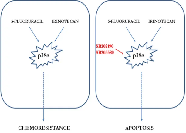
Thus, studies focusing on specific resistance mechanisms with the aim of finding new therapeutic strategies directed against specific targets have become increasingly desirable to improve patient’s survival. p38α might well be one of these targets, since it has been shown to be involved in several chemoresistance mechanisms. Indeed, response to cisplatin treatment is potentiated upon p38α inhibition, resulting in ROS-dependent upregulation of the JNK pathway in cancer cells, including CRC. In vivo, p38α inhibition cooperates with cisplatin treatment to reduce the size and malignancy of xenografted breast tumors in mice (Pereira L et al, 2013). Moreover, it has been shown that p38α signaling is activated in cisplatin-treated CRC cells, and p38α genetic ablation or pharmacological blockade sensitizes chemoresistant cells to cisplatin. Furthermore, p38α inhibition showed an additive effect with cisplatin in chemosensitive cells and co-treatment induced Bax-dependent apoptosis in both sensitive and resistant cells in vitro and xenograft CRC mouse models (Germani et al, 2014). Using p38α inhibitors together with common chemotherapeutics has also given promising results in different experimental setups. Co-treatment with SB202190 and the chemotherapy drug irinotecan appears to sensitize chemoresistant CRC cells to chemotherapy thus
35 supporting the important role for p38α in controlling chemoresistance (Paillas S et al, 2011).
CRC cells treated with p38α inhibitors together with 5-fluorouracil which is the backbone of chemotherapy in CRC, showed increased caspase activity and sensitivity to chemotherapy due to increased BAX expression (Yang SY et al, 2011).
Finally, chemoresistant cells may either show specific resistance to a particular drug or display multidrug resistance. P-glycoprotein (P-gp) functions as a drug efflux pump and it is involved in the efflux of some chemotherapeutic agents like cisplatin, vincristine, and doxorubicin outside of cancer cells. When high p38α activity is observed in chemoresistant cells, P-gp expression is often upregulated and chemoresistance can be reversed by SB203580 treatment (Veneroni S et al, 1994; Barancik M et al, 2001) (Figure 14).
Figure 14. p38α and chemoresistance in CRC. (a) p38α is a mediator of resistance to irinotecan and 5-FU and its activation correlates with impaired response to therapy in CRC patients. (b) Pharmacological inhibition combined with chemotherapeutic agents decreases viability of cancer cells and increases apoptotic cell death. (Adapted from Grossi et al, 2014).
36 As a result of the correlation with metastatic potential, cell migration in advanced tumors is another important issue to overcome in cancer therapy. Inhibition of p38α by SB203580 has also been reported to be an effective sensitizer reducing cell migration/invasion and enhancing sensitivity to etoposide treatment through Cox-2 downregulation in neuroblastomas (Limami Y et al, 2011).
These studies show that chemoresistant CRC cells may induce a p38α-mediated resistance mechanism to support survival or to delay the ongoing cell death processes.
37
1.6 Aim of the study
Tumor development and progression is a multistep process characterized by the progressive alteration of multiple molecular pathways that control the fine balance between proliferation, differentiation and cell death. These anomalies selectively favor the loss of anti-proliferative and pro-apoptotic checkpoints, the acquisition of a metabolic profile capable of sustaining a high proliferation rate and the ability to invade surrounding tissues and migrate. In this scenario, some of the signaling pathways involved in preventing tumor development in healthy cells can acquire a tumor-supporting function in cancer cells, thus contributing to the maintenance of the malignant phenotype. Despite its tumor-suppressive role in healthy cells of various tissues, p38α is required to maintain CRC metabolism, as its inhibition leads to activation of autophagy, cell death, and tumor growth reduction both in vitro and in vivo (Comes F et al, 2007; Chiacchiera F et al, 2009). Moreover, recent studies identified p38α as a mediator of resistance to irinotecan and impaired response to FOLFIRI therapy, while its inhibition sensitized resistant CRC cells to irinotecan and 5-fluorouracil. Recently, it has been shown that tumor relapse and chemoresistance are dependent on a small cell population called cancer stem cells (CSCs). The gene expression programs that determine CSC features are controlled by c-Myc, which function as a link connecting malignancy and stemness. Unfortunately, finding small-molecule or biologic inhibitors of c-Myc has proved difficult because c-Myc is localized within the nucleus and does not have a deep surface binding pocket. Therefore, considering the role of ERK exercises on c-Myc, the proposed study model involves investigating the crosstalk between p38α and ERK and their function on c-Myc stabilization (Chapter 1). Also, the study plans to investigate the possible role of p38α as chromatin-associated kinase with β-catenin, the well known c-Myc transcription factor (Chapter 2). Finally, in order to find new potential therapeutic strategies for the purpose of targeting CSCs and bulk tumor population we finally investigated the role of p38α in CRC-SCs model (Chapter 3).
Since p38α is a promising therapeutical target directly placed on chromatin, the idea is to use Ralimetinib, p38α inhibitor, in current clinical trials for inflammatory disease and cancer, to inhibit the uncontrolled cellular proliferation of both stem and differentiated
38 cells prompted by c-Myc and other Wnt target genes, preventing metastatic dissemination and overcoming chemoresistance (Figure 15).
Figure 15. c-Myc is one of the most important factor in CRC. Considering the role of ERK exercises on c-Myc, the proposed study model involves investigating the crosstalk between p38α and ERK and their function on c-Myc stabilization (Chapter 1). Also, the study want to investigate the possible role of p38α as chromatin associated kinase with β-catenin, the well known c-Myc transcription factor (Chapter 2) and in the interest to find new potential therapeutic strategies aimed at selectively target CSCs and bulk tumor population we finally investigate the role of p38α in CRC-SC model (Chapter 3). The study advice the use of Ralimetinib (p38α inhibitor) in order to impair cellular proliferation of CSCs and differentiated cells, reduce metastatic dissemination and overcome chemoresistance.
39
Material and Methods
2.1 Cell culture and reagents
HCEC-1CT, HCT116, HT29 and Caco2 were purchased from ATCC. HCEC-1CT cells were maintained in Colo-UP medium supplemented with 1% of antibiotic. HCT116 and HT29 were maintained in DMEM supplemented with 10% fetal bovine serum (FBS) and 1% antibiotic. Caco-2 cells were maintained in DMEM supplemented with 20% FBS and 1% antibiotic. CRC-SC cells (511, G605, DA13 and ME59) were kindly provided by Prof. Giorgio Stassi from consenting patients. CSC cells were maintained in DMEM/F12 ADVANCED (cod. #12634-028) supplemented with 1% L-Glutamine (cod.25030081), 1% antibiotic, 0.6% glucose solution at 45% (cod. SIGMA G8769), B27 (cod. thermo 12587010), N2 (cod. Gibco 17502-048), 10 ng/ml bFGF (cod. Sigma F0291), 20ng/ml EGF (cod. Sigma E9644). The cells were routinely propagated under standard conditions.
SB202190 (5-10µM), PD988059 (5-20 µM), 3-Methyladenin (10 µM), trypan, Bafylomicin (B1793) and Wnt3a (H17001) were from Sigma Aldrich. Ralimetinib (LY22228820) (1-10 µM) was kindly provided by Eli Lilly and Company. Trametinib (GSK1120212) (25nm) and PRI-724 (S8262) were from Selleckchem, cycloheximide (239763), MG132 (474790) and TWS119 (361554) were all from Calbiochem.
2.2 Quantitative real-time PCR
Total RNA was isolated by TRIzol reagent (Sigma) following manufacturer’s instructions. To avoid possible DNA contamination, RNA was treated with DNase-1 (Ambion). RNA purity was checked by spectrophotometer and RNA integrity by examination on agarose gel electrophoresis. cDNA was synthesized retro-transcribing 1μg of RNA total in 100μl using iScript cDNA synthesis kit Biorad and following the manufacturer’s instructions. Real-time PCR primers were designed using Primer Express software. PCR assays were performed in 96 well optical reaction plates using the QuantStudio 3 machine (Thermo Fisher Scientific). PCR assay were conducted in
40 triplicate wells for each sample. Baseline values of amplification plots were set automatically and threshold values were kept constant to obtain normalized cycle times and linear regression data. The following reaction mixture per well was used: 5μl SYBR Green PCR Master Mix (Biorad), 1,2μl primer at the final concentration of 100nM, 2,3μl RNAse free water, 1,5μl cDNA.
For all experiments the following PCR conditions were used: denaturation at 95°C for 30seconds, followed by 40 cycles at 95°C for 15 seconds then at 60°C for 30 seconds. Quantitative normalization of cDNA in each sample was performed using β-actin as an internal control. Relative quantification was done using the ddCT method. Primer sequences are available on request.
2.3 RNA interference
For RNA interference, cells were transfected with 100nM Select Silencer siRNA against p38α and MEK (all from Ambion by life technologies) using Hiperfect Transfection reagent from Qiagen. On target plus control siRNA (Thermo Scientific) was used as control sequences.
2.4 Proliferation assays
Proliferation assays were conducted using the WST-1 reagent (Roche) as the manufacturer’s instructions. Briefly, cells were seeded into 96-well plates one day before treatment. After 12h, 24h, 36h drugs (or DMSO) exposure, 10μl of the Cell Proliferation Reagent WST-1 was added to each well and incubated at 37°C in a humidified incubator for 30min-1h-2h. The absorbance was measured on a microplate reader (SPECTROstar OMEGA) at 450-655nm. Each assay was performed in 6 replicates and the experiment was repeated three times. The proliferation index was calculated as the ratio of WST-1 absorbance of treated cells at the indicated time point (12h-24h-36h) to the WST-1 absorbance of the same experimental group at 0h.
41
2.5 Nuclear/cytoplasmic fractionation and Immunoblot analysis
Nuclear/cytoplasmic fractionation was carried out using the Nuclear extraction kit from Abcam (ab113474). Immunoblot cells were collected and homogenized in lysis buffer (50mM Tris-HCl ph 7.4, 5mM EDTA, 250mM NaCl and 0.1% Triton x-100) supplemented with protease and phosphatase inhibitors. 20-40 μg of protein extracts from each sample were denatured in 5x Laemmli sample buffer and used for immunoblot analysis. Immunoblot were performed using Anti-βactin (#3700S), anti-c-Myc (#9402s), anti-p44/42 MAPK (Erk1/2) (#9102), anti-phospho p44/42 MAPK (Erk1/2) (Thr202/tyr204) (#9106S), anti-LC3 A/B (NK12741S), anti-p38 MAPK (#9212), anti-p38α MAPK (#9228S), anti-phospho-p38 MAPK(Thr180/Tyr182) (#9211), anti-MEK (#9122S), anti-phospho-MEK (#9154S), Anti-lamin (#12586S), Keratin20 (BK13063S), Anti-phospho-MAPKAPK-2 (Thr334) (BK3316S), anti-PDI (#2446S), anti-βcatenin (#9562S), anti-Ubiquitin (#3936), anti-APC (#2504) all from cell signaling technologies-CST and anti-HSP90α/β sc-13119 from Santa cruz. HRPO-conjugated secondary antibodies were used (GE Healthcare) and the signal was revealed using the ECL-plus chemiluminescence reagent (GE Healthcare) as per manufacturer’s instructions. The densitometric evaluations were carried out both by ImageJ software and ImageLab software.
2.6 Immunofluorescence staining
Cells were seeded on glass coverslips and after 12h were treated as indicated. At the end of the treatment, cells were fixed in 4% paraformaldehyde and permeabilized using 0,01–0,1% Triton X-100. Coverslips were incubated with the indicated primary antibodies (LC3 A/B). Secondary antibodies were Alexa Fluor 488 from Invitrogen; nuclei were counterstained using DAPI (Sigma). Slides were sealed using Vectashield mounting medium (Vector Laboratories). Images were acquired using a Zeiss fluorescence microscopy.
42
2.7 Immunoprecipitation
Cells were collected and homogenized in lysis buffer (50 mM Tris-HCl pH 7,4; 5 mM EDTA; 250 mM NaCl and 1% Triton X-100) supplemented with protease and phosphatase inhibitors. The coupling was performed between magnetic beads and antibody (p38α #8690 and βcatenin #9562S) in T-PBS for 30 min. Cell lysates were immunoprecipitated with antibody-beads or (IgG control) on the wheel for 45 min. Immunoprecipitated proteins were extensively washed with lysis buffer, resuspended in Laemmli buffer, separated on polyacrylamide gel and transferred to nitrocellulose membranes, after which precipitated proteins were subjected to immunoblot analysis.
2.8 Serum and LiCl stimulation
Cells were synchronized in the cell cycle by culturing theme for 48 hours without serum. Then, the medium was replaced with ones containing serum and/or 10mM LiCl for 4h.
2.9 Chromatin immunoprecipitation (ChIP)
ChIP assays were performed using the MAGnify Chromatin Immunoprecipitation System (Life Technologies) according to the manufacturer’s instructions. IgG antibodies were included in the kit and 1 μg of anti-β-catenin, anti-p38α antibodies (CST) was used for each assay. PCR was performed on “input” DNA of different samples and equivalent amounts of immunoprecipitated DNA were amplified by real-time PCR as described in the “Quantitative Real-real-time PCR” method section. Quantification was done using the Input % method. The set of primers used for ChIP allows the amplification of target promoter regions including β-catenin binding sites (sequences are available upon request).
43
2.10 Microscopic quantification of viability and cell death
Cell viability and cell death of the reported cell lines were scored by counting. The supernatants (containing dead/floating cells) were collected, and the remaining adherent cells were detached by Trypsin/EDTA (Sigma). Cell pellets were resuspended in 1X PBS and 10μl were mixed with an equal volume of 0.01% trypan blue solution. Viable cells (unstained, trypan blue negative cells) and dead cells (stained, trypan blue positive cells) were counted with a phase-contrast microscope. The percentages of viable and dead cells were calculated. The data shown in the results section are representative of 3 or more independent sets of experiments.
2.11 Morphological evaluation
Numerous slides with a monolayer cells were either rapidly fixed in 95% ethyl alcohol for a minimum of 15 min. Slides were examined by optic microscopy.
2.12 Karyotype protocol
Colorectal cancer stem cells were seeded the day prior to the experiment at a high density. After 24h was added 10ug/ml of colcemid, which can collapse mitotic spindles and prevent the completion of mitosis, to each dish and mix gently. Incubated at 37 °C, 5 % CO2 for 90minutes. Transfer medium to centrifuge tubes from the cell culture
dishes. Further, centrifuge at 2000rpm for 5 min at room temperature. Discard the supernatant and leave 0.5 ml medium to mix the pellet gently. Resuspend the pellet in 5-7 ml 35-7 °C hypotonic solution (KCl 56%) and mix thoroughly. Incubate in a water bath at 37 °C for 10 min. Neutralize the hypotonic solution with 1 ml of methanol: glacial acetic acid (3:1) fixative. Centrifuge at 2000rpm for 5 min at room temperature. Discard the supernatant and leave 0.5 ml solution to mix the pellet gently. Resuspend the pellet in 5 ml cold fixative (drop by drop and flick the tube between drops to prevent cell clumping) to wash the pellet. Centrifuge at 2000rpm for 5 min at room temperature.
44 Discard the supernatant and leave 0.5 ml solution to mix the pellet gently. Resuspend the pellet in 5 ml cold fixative. Put the centrifuge tube on ice at least 60 min. Centrifuge at 2000rpm for 5 min at room temperature. Discard the supernatant and leave 0.5 ml solution to mix the pellet gently. Resuspend the pellet in 5 ml cold fixative. Repeat the washing steps other two times until the supernatant is clear and the pellet become white. After the final centrifugation, suspend the cells in a few drops of cold fixative to give a slightly opaque suspension. Drop 1-2 drops onto wet and clean slides (before that, the slides have to be rinsed by 1-2 drops of cold fixative). Dry the slides at room temperature. Observe the chromosomes with the microscope (Olympus DP71 with 200x magnification). Count the number of chromosomes about 50 cells.
2.13 Mutation analysis
Genomic DNA was purified from colorectal cancer stem cells according to manufacturer’s instructions (Genomic DNA purification kit, ThermoFisher Scientific, K0512). The gene coding region were screened for mutations. promoter sequences were analyzed for mutations using a PCR-direct sequencing method. Sequencing and capillary electrophoresis were performed on the Applied Biosystems® 3130 Genetic Analyzer (ThermoFisher Scientific, Waltham). Mutations and polymorphisms were confirmed in independently amplified PCR products.
MSI analysis was performed with a reference panel of five fluorescent dye-labeled microsatellite primers (NR-21, BAT-25, MONO-27, NR-24, BAT-26) using the MSI Analysis System kit, Version_1.2 (Promega). Amplified fragments were detected by loading the PCR products for capillary electrophoresis using the ABI Prism 3500 Genetic Analyser and the POP-4 polymer (both from Applied Biosystems, Foster City, California, USA) according to manufacturer’s instructions (Promega). MSI status was determined upon analysis with GeneMapper software, Version_4.1 (Applied-Biosystems).