Scuola Normale Superiore_Pisa
Regulation of TMEM16A Alternative Splicing
Thesis Submitted for the Degree
of Doctor of Philosophiae inLife Sciences
Candidate: Ifeoma Ubby
Supervisor: Prof. Franco Pagani, M.D. Ph.D
Pisa, 2011/2012
Keep moving forward…
TABLE OF CONTENTS
Table of Contents ………..………... 3
Abstract………..………..………... 5
Introduction .………..…………...…….………..………..………..….. 7
Gene Expression and RNA Splicing… ……...………..………..………. 8
Signal for Splicing Specificity ………..……….………... 13
Splice Site Sequence………..……….……….. 13
Splicing Regulatory Elements ……….………… 15
Proteins involved in splicing regulation……….……….……..…….. 19
Intron/Exon architecture……….……….………….... 28
Alternative Splicing ……….………..………... 31
Intragenic Alternative Splicing events and Alternative Splicing Coordination……….…………. 35
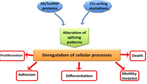
Alternative Splicing and Cancer………..……….. 37
Cis-‐acting mutation in cancer………. 38
HnRNP and SR proteins in cancer ………... 39
Splicing and apoptosis ………. 42
Alternative Splicing cancer genes………. 46
Alternative Splicing isoforms associated with metastasis and invasion………. 46
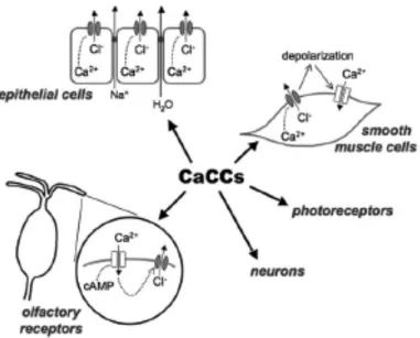
Anoctamin/TMEM16 family members are Ca2+-‐activated Cl-‐ channels……….……... 49
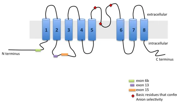
TMEM16A……….………..……….. 54
The other TMEM16 proteins ……….……….. 59
TMEM16A in cancer………... 61
Aim of the project ………..…... 64
Results………..… 65
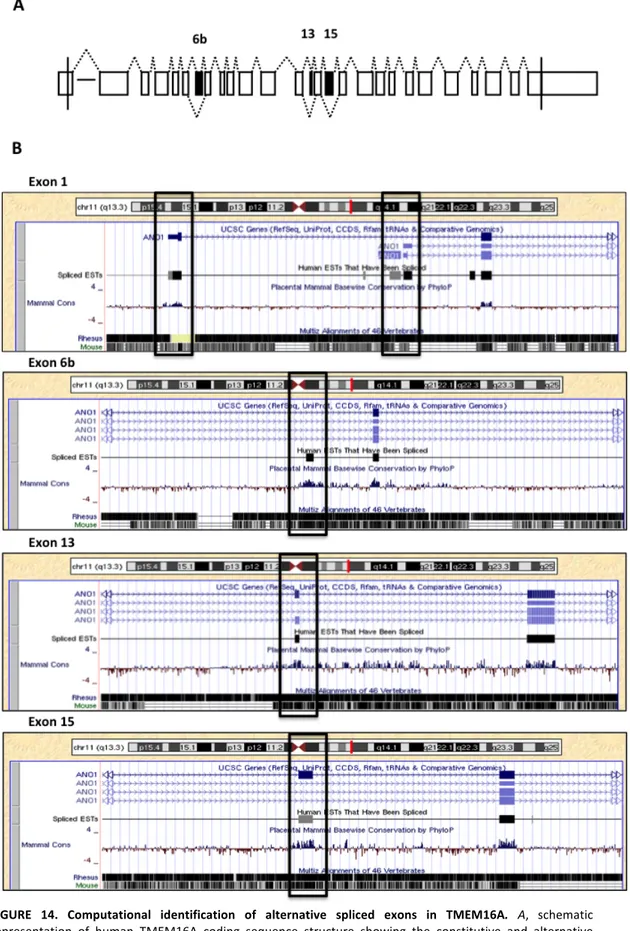
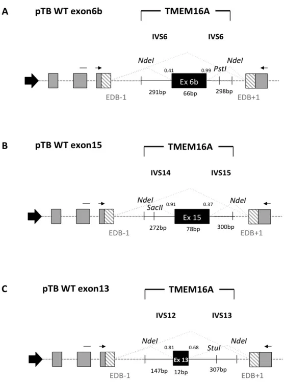
Identification of Alternative Splicing events in TMEM16A and evaluation of the splicing pattern in normal adult human tissues………..……….……. 65 Analysis of TMEM16A exon 6b in minigene splicing regulation…...………..…… 69
Effect of splicing factors on TMEM16A exon 6b splicing……… 71
Identification of enhancers and silencers sequences that affect splicing of TMEM16A exon 6b……….……… 75 Overexpression of splicing factors that induce exon 6b inclusion do not affect splicing in the absence of the 16bp long GAA-‐rich ESE……… 80 Most of the splicing inhibitory factors do not affect splicing in the ESSs deletion exon 6b minigenes……….……….. 80 Role of the GAA-‐rich ESE in TMEM16A exon 6b……….……….………..…... 88
Effect of SRSF9, TRA2B, PTB1 and RbFOX1 overexpression on TMEM16A MUT2 exon 6b minigene………..……….…….…….
92
Analysis of TRA2B binding specifity against WT, MUT2-‐A, MUT2-‐E, MUT2-‐G,
MUT2-‐H and MUT2-‐I mutants……….……….……….
94
Analysis of TMEM16A exon 15 in minigene splicing regulation……….………….. 96
Effect of splicing factors families on TMEM16A exon 15 splicing ……….... 96
Identification of enhancers and silencers sequences that affect splicing of TMEM16A exon 15 ……… 99 Analysis of TMEM16A exon 13 in minigene splicing regulation……….. 111
Analysis of TMEM16A mRNA isoforms in human tissues….……… 115
Analysis of TMEM16A mRNA isoforms in mouse tissues….……… 123
Alternative splicing of TMEM16A in human breast cancer tissues..……….... 129
Discussion ………...………... 137
Conclusion and Future Directions………...………... 152
Materials and Methods ………... 154
References ……….………... 172
Abbreviations……… 199
Appendix ……….……….. 202
Oligonucleotides List ……….……….……… 202
Figures and Tables List ……….……….……… 205
ABSTRACT
TMEM16A/Anoctamin1 is a novel calcium-‐activated chloride channel involved in neuronal and cardiac excitation, vascular tone, pain perception and olfactory and sensory signal transduction and GI tract motility. It is also associated to diverse type of cancer including breast cancer malignancy. Alternative splicing (AS) of exons 6b, 13 and 15 generates functionally distinct TMEM16A isoforms with different electrophysiological properties. To study their splicing regulation, I performed in minigene system a systematic analysis of exonic and intronic regulatory elements followed by co-‐transfection of a panel of splicing regulatory factors. Analysis of TMEM16A pre-‐mRNA splicing supports a model in which each exon is regulated by different cis-‐ and trans-‐acting elements. Exon 6b inclusion is regulated primarily by SRSF9 and TRA2B, through a unique GAA-‐rich ESE element. Exon 15 is enhanced only by TIA1 and FOX1 and this effect is mediated by downstream intronic sequences. On the other hand, the small exon 13, included in most human tissues, was mainly skipped in the minigene and only FOX1 and U2AF65 enhanced its inclusion.
To understand if there is any preferential association between three AS exons, I have evaluated TMEM16A isoforms using a long range RT-‐PCR assay that amplifies transcripts across the AS events. Coordination between distant alternative spliced exons in the same gene has been suggested to be an important mechanism to regulate gene expression but very few genes have been studied in detail. I observed that the selection of exons 6b and 15 is preferentially coordinated in several human normal tissues: mature transcripts that predominantly include exon 6b tend to exclude exon 15. Unexpectedly, this coordination was not conserved in mouse tissues. This was mainly due to the fact that exon 15 was largely and predominantly excluded in the mouse, a fact that suggest a peculiar evolutionary conservation of AS in this gene.
To explore if changes in splicing coordination of the two major AS events are associated to cancer development I evaluated normal mammalian tissue and corresponding breast tumors of the same cohort, obtained from surgical excision
(n=18). The distribution of individual AS events did not change between normal and tumor tissues. However, the TMEM16A splicing coordination increased significantly in tumors. Indeed, the splicing coordination was present in 50% of normal mammalian breast tissues and in 84% in tumors. In conclusion this study identifies several cis-‐acting elements and trans-‐acting factors involved in the regulation of TMEM16A Alternative Splicing and provides evidence of its intragenic splicing coordination. The increase of TMEM16A splicing coordination observed in breast tumor, might represent a common event in genes with multiple AS events.
INTRODUCTION
The genetic information is stored in DNA, which is transferred from one generation to the next. During the life of a cell, this DNA information is regained as RNA. Whereas DNA is chemically very stable and therefore well suited to archive the genetic information, RNA is chemically more reactive, and thus unstable. Therefore, with the exception of RNA viruses, RNA does not store the genetic information but rather acts as an intermediate between DNA and proteins.
However, RNA does not simply copy the genetic information, as the primary RNA transcript generated from DNA undergoes processing. Most human polymerase II transcripts contain exonic sequences that are finally exported into the cytoplasm, whereas the intervening sequences (introns) remain in the nucleus. The removal of the introns and the joining of the exons is known as pre-‐ mRNA splicing (Berget and Sharp 1977). Almost all human protein-‐coding genes undergo alternative splicing (AS) (Pan, Shai et al. 2008); this means that, depending on the cellular conditions, an alternative exon can be either included or removed from the final messenger RNA (mRNA).
RNA is more than just a copy of the genetic code: RNA can “interpret” the genetic information depending on environmental cues that the cell receives. Alternative splicing is a central mechanism in this interpretation process, as it allows the expression of selected parts of gene.
Due to its role as a flexible “interpreter”, AS strongly enhances the number of proteins that can be encoded by the genome. For example, by combining one exon out of four alternatively spliced regions that contain 12, 48, 33, and 2 alternative exons each, the Drosophila Dscam gene can generate 38016 protein isoforms (Celotto and Graveley 2001). Alternative splicing can thus generate from a single gene a number of protein isoforms that is larger than the total number of protein coding genes. The ability to change the output of the genetic information depending on cellular states, and the ability to expand the information content of the genome, makes AS a central element in gene expression.
Gene Expression and RNA Splicing
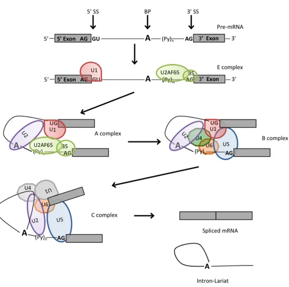
In higher eukaryotes, splicing constitutes a critical mode for the regulation of gene expression at the level of RNA processing (Black 2003). The large majority of eukaryotic protein coding genes are transcribed as precursors of messenger RNAs (pre-‐mRNAs), in which exons are separated from each other by intervening regions of non-‐protein coding information (introns), which have to be correctly spliced out to produce a mature mRNA. Splicing of pre-‐mRNAs occurs in a two-‐ step reaction (figure 1A). In the first step, the 2'OH of a specific branch-‐point nucleotide performs a nucleophilic attack on the first nucleotide of the intron at the 5' splice site forming the lariat intermediate. In the second step, the 3'OH of the released 5' exon performs a nucleophilic attack at the last nucleotide of the intron at the 3' splice site thus joining the exons and releasing the intron lariat (Black 2003).
Signals that specify exon–intron junctions are located at the termini of introns. cis-‐Acting nucleic-‐acid elements are: located at the 5’spice site (5’SS AG/GURAGU), the branch point (BP) sequence (CURAY), the polypyrimidine (Py) tract (a run of polypyrimidines located between the 3’ splice site and the branch point), and the 3’ splice site (3’SS, YAG). These four canonical intronic sequences are recognized by specific components of the spliceosome or associated splicing factors. Indeed, the 5’SS, BP and 3’SS of nuclear pre-‐mRNA introns are defined by very short consensus sequences that, in metazoans, are very poorly conserved (figure 1B). Unlikely most other enzymes, the spliceosome does not have a preformed active site; on the contrary, the catalytic center must be assembled anew on each pre-‐mRNA intron in a stepwise interaction of five uridine-‐rich (U-‐ rich) small nuclear ribonucleoproteins particles (snRNPs) called U1, U2, U4/U6 and U5, and a large number of protein splicing factors (Kramer 1996), and participate in several RNA-‐RNA and RNA-‐protein interactions. Human spliceosome also interact with a large number (from 150 to 300) of non-‐snRNP proteins, including heterogeneous nuclear RNPs (hnRNPs) and serine-‐arginine (SR) proteins (Wahl, Will et al. 2009).
Figure 1. Schematic representation of the two step splicing pathway of nuclear pre-‐mRNA. A, two
successive phosphoester transfer reactions. In the first step, a specific adenine nucleotide in the intron sequence (indicated in bold) attacks the 5ʹ′ splice site and cuts the sugar-‐phosphate backbone of the RNA at this point. The cut 5ʹ′ end of the intron becomes covalently linked to the adenine nucleotide, thereby creating a loop in the RNA molecule. The released free 3ʹ′-‐OH end of the exon sequence then reacts with the start of the next exon sequence, joining the two exons together and releasing the intron sequence in the shape of a
lariat. The two exon sequences thereby become joined into a continuous coding sequence; the released
intron sequence is then degraded i. B, two exons are separated by an intron. Between them are reported the consensus sequences present within an intron. The arrows indicate the position of the 5’ (GU) and 3’ (AG) splice sites and the branch point (A), shown in bold, the polypirimidine-‐tract is represented by (Yn). There are several conserved nucleotides near the sequences surrounding the intron-‐exon junctions that act as essential splicing signals. N means any base. To note only the universally conserved nucleotides are the dinucleotide cores of the 5’ and 3’ splice together with the branch point (A) showed 100% of frecquency of occurrence.
Each snRNA have a U-‐rich RNA molecule and a variable number of associated proteins, which make up more than two-‐thirds of its mass. The U snRNPs undergo a complex maturation process before becoming functional in the spliceosome. All U snRNAs contain a so-‐called “Sm” binding site that serves as a binding platform for Sm proteins. Sm proteins (B/B', D1, D2, D3, E, F and G) are common factors of U1, U2, U4 and U5, which bound to a conserved Sm motif on the each snRNA. Besides Sm and Sm-‐like proteins, other particle-‐specific proteins associate with snRNAs. The number of specific associated proteins vary greatly among the U snRNAs, from only 3 for U1 snRNP to more than 20 for U2 snRNP (Stanek and Neugebauer 2006). The association of the U snRNP-‐specific proteins is believed to happen in the nucleus since it is known that import of some of them (U1A and U1C for U1 snRNP; U2A' and U2B''for U2 snRNP) occurs independently of snRNA nuclear import ((Feeney and Zieve 1990); (Jantsch and Gall 1992); (Kambach and Mattaj 1994)).
The RNA and protein component of the snRNPs play critical roles in splice site recognition and spliceosome assembly follows a carefully orchestrated stepwise pathway (Will and Luhrmann 2001). The basic steps of the splicing cycle, whereby a single intron is removed from the pre-‐mRNA being processed, are shown in figure 2. Initially, the U1snRNP interacts with the 5’SS of the pre-‐mRNA to form the so-‐called E complex (E –early-‐ complex or commitment complex). U1 snRNP-‐associated proteins U1-‐70k and U1C stabilize this transient interaction. The U2 Auxiliary Factor snRNP (U2AF65/35) identifies the AG dinucleotide at the 3’ intron/exon junction together with the polypyrimidine tract (Py)n. Mutual
stabilization of contacts with the U2AF bound to the 3' SS and the upstream U1 snRNP at the 5' SS can be mediated by members of the serine/arginine-‐rich (SR) protein family, generating the A complex (figure 2). The establishment of multiple weak interactions from the 3' SS to the 5' SS defines an exon, and constitutes the commitment step towards the splicing pathway ((Robberson, Cote et al. 1990); (Berget 1995)). The transition from A to B complex is marked by the ATP-‐ dependent addition of the U4/U6 and U5 snRNP, preassembled in U4/U6.U5 tri-‐ snRNP. At this level all snRNPs are present, but the spliceosome is catalytically inactive and requires a conformational and compositional rearrangement to come
active and to promote the first transesterification step of splicing. During spliceosome activation, U1 and U4 are destabilized or removed, leading to a B* complex (B activated complex) (Turner, Norman et al. 2004). Eight evolutionarily conserved DExD/H-‐type RNA-‐dependent ATPase/helicases act at specific steps of the splicing cycle to catalyze RNA-‐RNA rearrangements and RNP remodelling events (Valadkhan, Mohammadi et al. 2009). The C complex is then formed, and the spliceosome undergoes the first catalytic step. Subsequently additional rearrangements in the RNPs network are necessary prior to undergo the second transesterification reaction ((Wahl, Will et al. 2009); (Konarska, Vilardell et al. 2006)). Complex C catalyzes the second step, after which the spliceosome dissociates. The mRNA product is then released, while the excised intron remains bound to U2, U5 and U6. Finally, these snRNPs also dissociate and the next round of splicing can then take place.
Figure 2. Basic exon recognition and spliceosome assembly. The spliceosome assembles onto the pre-‐mRNA
in a stepwise manner. A consensus sequences that define exon/intron boundaries. Exons are noted by grey
boxes, and introns by lines. Consensus nucleotides are indicated above the line, and the term for sequence is
shown below. 5’ss, splice site; bp, the branch point; 3’ss, splice site; Py, polypyrimidine tract. In the E complex, U1 binds the 5ʹ′ splice site, U2AF65 binds the polypyrimidine tract, and U2AF35 binds the 3ʹ′ splice site. U2AF65 recruits U2 to the branch point, forming the A complex. The B complex forms when the U4/U6-‐ U5 tri-‐snRNP joins, and then rearragement of the components forms the C complex. U4/U6 base pairing is destabilized, allowing U6 to displace U1 at the 5ʹ′ splice site and base pair with U2. U1 and U4 are displaced from the complex. U5 interacts with the 5ʹ′ and 3ʹ′ splice sites, bridging the exons. It is in the final complex where the two sequential transesterification reactions occur. The end result is the fully spliced mRNA and the intron lariat, which is debranched and degraded.
Signals for Splicing Specificity
Splice Site Sequence
The excision of the introns from a pre-‐mRNA and the joining of the exons are directed by special consensus sequences at the intron/exon junctions called splice sites.
The 5’SS, also referred to as the donor site, is defined as a single sequence element, with a length of nine nucleotides (nt) (figure 3A). In mammals only the first two bases of the intron GU are universally conserved although they are not the only important bases (Aebi, Hornig et al. 1987). Indeed, this site follows a degenerate consensus sequence YAG/GURAGU (where Y is a pyrimidine, R is A or G) (Sun and Chasin 2000), that base pairs with U1 snRNA in the early spliceosomal E complex.
At the other end of the intron, the 3’ splice site region, also referred to as the acceptor site, has three conserved sequence elements: the branch point (BP), the polypyrimidine tract (Py)n or PPT, and a terminal AG at the extreme 3’ end of
the intron, as figure 3B shows (Reed 1989). The BP is characterized by the presence of a conserved “A” surrounded by a highly degenerated motif YNYURAY (Y=pyrimidine and R= purine) (Reed and Maniatis 1985). It is commonly found about 18-‐40 nucleotides upstream of the AG dinucleotide ((Ruskin, Krainer et al. 1984); (Reed and Maniatis 1985)) although some exceptions can be found hundreds of nucleotides away (Reed 1989). The recognition of the branch site involves a base-‐pairing with the U2 snRNP auxiliary factor (U2AF) in order to form the spliceosome A complex (Berglund, Chua et al. 1997). The 3’SS is functionally coupled with the PPT located between the branch site and the terminal AG at the intron/exon junction (Reed 1989). (Py)n can display a high percentage of
pyrimidines, it varies in length and distance to the BP and the AG. It is essential for efficient branch-‐point utilisation and correct AG recognition as it has been shown that progressive deletion of the polypyrimidine tract impairs splicing while elongating its length can improve its efficiency (Roscigno, Weiner et al. 1993). The terminal AG dinucleotide or acceptor site defines the 3’ border of the intron. This site is characterized by the short YAG/G sequence (Y pyrimidines; the slash is the
intron-‐exon boundary and the underlined nucleotides are conserved). Even if it is essential for the second step of splicing reaction no base-‐pairing interactions with snRNAs are involved in recognizing this sequence (Wu, Romfo et al. 1999). The sequences between the branch-‐point and the acceptor site are commonly devoid of AG dinucleotides (Gooding, Clark et al. 2006).
Figure 3. Consensus sequences of splice sites. Below the consensus sequences for each canonical signals are
shown. A, Diagram of the 5’ end of the intron, recognized initially recognized by U1 snRNP, is identified by 9 nt consensus sequences shown. B, Diagram of the 3’ splice site. The 3 elements within approx. 40 nt of the intron’s 3’ end are shown.
The role of these RNA sequence elements is of such importance that the sequence complementarity of the 5’SS to U1 snRNA and the extent of the (Py)n at
the 3’ SS are used to determine the strength of splice sites. A greater complementarity to U1 snRNA and a longer uninterrupted (Py)n is translated into
higher-‐affinity binding sites for spliceosomal components and, thus, a more efficient splice site recognition (Hertel 2008).
Splicing Regulatory Elements
The information content of 5’SS, 3’SS, and BP signals is less and less preserved with an increasing number of introns ((Lim and Burge 2001); (Irimia, Penny et al. 2007)), and in higher eukaryotes it is often insufficient to ensure correct RNA splicing. This intrinsic feature of the splice sites allows for a great degree of regulation of the splicing process in the form of alternative splicing. It has also an undesired consequence: pseudo splice sites largely outnumber real splice sites and in many cases their strength can surpass that of correct splice sites. Thus, the growing degeneracy, in turn, opens up the possibility for making alternative splice sites choices, and additional signals become necessary (Wang, Rolish et al. 2004), in particular when weak splice sites are involved.
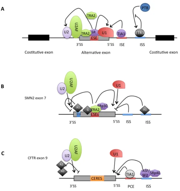
In addition to the conventional splicing signals located at exon-‐intron boundaries, other cis-‐elements in pre-‐mRNA increase exon inclusion by serving as binding sites for the assembly of multicomponent splicing enhancer complexes (Cartegni, Chew et al. 2002). These elements, localized both in exon and intron sequences, act by stimulating or inhibiting the use of specific splice sites. Depending on their location and their function, these elements are referred as exonic splicing enhancers (ESE), that facilitate the process of exon definition, thus helping exon inclusion in the mRNA ((Mardon, Sebastio et al. 1987); (Lam and Hertel 2002)). Although originally discovered in regulated exons, ESEs are today also known to be components of constitutively spliced exons ((Mayeda, Screaton et al. 1999); (Schaal and Maniatis 1999)). ESEs assist early spliceosomal complex formation by interacting with components of the splicing machinery that make up previously described the E complex (Reed 1996). This is done generally through the
serine/arginine-‐rich proteins, SRs, and SR related proteins that generally assemble on ESEs elements to promote both regulated and constitutive splicing by forming networks of interaction with each other (Blencowe 2000) and are also involved in recruitment of spliceosomal components such as U1 snRNP to the 5’SS, or U2AF to the 3’SS to the regulated splice site ((Black 2003); (Graveley 2000)). Thus, SR proteins bound to ESEs function as general activators of exon definition (Lam and Hertel 2002) (figure 4). Several algorithms have been constructed to predict splicing enhancing or silencing regions (Chasin 2007). These computational tools have been helpful in identifying putative exonic splicing enhancers (PESEs) and putative exonic splicing silencers (PESSs).
While the positive regulation of splicing is thought to occur as a result of protein-‐protein interaction strengthening the recognition of the splice site, negative regulation of splice site choice often result from the prevention of the splice site to be recognized. These negative regulatory sequences, within an exon, are called exonic splicing silencer, ESSs, and in the majority of cases are bound by particular hnRNP family proteins. Cis-‐acting regulatory elements in the intronic sequences have being studied to a lesser extent than those present in exons. Like exonic regulation, there are both positive and negative cis-‐acting sequences in the intron, so called intronic splicing enhancer (ISE) and intronic splicing silencer (ISS). Some of the best characterized ISSs include binding sites for hnRNP A1 protein (described below) ((Hui, Hung et al. 2005); (Matlin, Clark et al. 2005); (Wagner, Baraniak et al. 2005); (Kashima, Rao et al. 2007)); hnRNP I, also called PTB and PTBP1. PTB has strong RNA binding activity, and its preferred binding site is UCUU, often flanked by pyrimidines (Perez, Lin et al. 1997). Initially it was hypothesized that PTB is a positive splicing regulator, but later studies revealed that PTB is rather a splicing repressor ((Garcia-‐Blanco, Jamison et al. 1989); (Valcarcel and Gebauer 1997)). Early models of PTB repression suggested that it may compete with U2AF65 for polypirimidine tract, however regulation by PTB often requires additional PTB binding elements.
There are several elements known to act as intronic splicing enhancers, ISE’s, but the proteins that mediate their effects are less well characterized than for ESE’s. One well characterized ISE element is the triplet (GGG) which often
occurs in clusters and can enhance recognition of adjacent 5’ or 3’ splice sites ((McCullough and Berget 1997)). Intronic (CA) repeats in several cases can enhance splicing of upstream exons, probably through binding of hnRNP L protein ((Hui and Bindereif 2005); ((Hung, Heiner et al. 2008)). TIA-‐1 is an important ISE protein that binds pyrimidine-‐rich sequences that binds downstream of exon 9, of the CFTR (cystic fibrosis transmembrane conductance regulator) gene (Zuccato, Buratti et al. 2004); (Venables 2007)). Another example in which TIA-‐1 regulates splicing via ISE elements is Fas exon 6 that can be included or skipped to generate mRNA encoding, respectively, a membrane boud form of the receptor that promotes apoptosis or a soluble isoform that prevents programmed cell death. TIA-‐1 protein promotes U1 snRNP binding to the 5’ splice site of intron 6, by binding to the ISE element in the +6 to 20 position downstream of Fas exon 6, which in turn facilitate exon inclusion (Izquierdo, Majos et al. 2005).
Two RNA-‐binding proteins with family members expressed specifically in nervous system and/or muscle tissues are the muscleblind-‐like (MBNL) and CUGBP/ETR-‐like (CELF; also known as Bruno-‐like) factors ((Barreau, Paillard et al. 2006); (Pascual, Vicente et al. 2006)). These factors have been implicated in neurological disorders and in CUG trinucleotide expansion diseases such as myotonic dystrophy (Gallo and Spickett 2010). These factors are also involved in several aspects of mRNA metabolism, including alternative splicing ((Ladd, Charlet et al. 2001); (Ho, Charlet et al. 2004)). Several studies using model pre-‐mRNA substrates have demonstrated that MBNL and CELF bind intronic CUG-‐repeat and UG-‐rich elements respectively. At least some members of this family activate splicing through intronic enhancer elements ((Charlet, Logan et al. 2002); (Ho, Charlet et al. 2004)).
Another common ISE element is the hexanucletide (U)GCAUG motif that
represents highly specific recognition element for the Fox family proteins. This element strongly enhances the splicing.
Figure 4. Regulatory elements in pre-‐mRNA splicing. Schematic representation of the possible distribution of
canonical and additional splicing cis-‐acting elements. The canonical splicing signals that define the exon boundaries are relatively short and poorly conserved sequences. Only the GU at 5’ end and the AG at 3’ end together with the the branch-‐point adenosine are conserved (the consensus nucleotides are bold). The polypyrimidine tract of variable length (represented by “y”, cytosine or thymine) is reported upstream of the 3’-‐splice site. The basal components of the splicing machinery bind to the consensus sequences and promote assembly of the splicing complex. The U1 snRNP binds to the 5’-‐splice site, the U2 snRNP recognizes the branch site and the U2AF localizes to polypirimide tract and 3’splice site sequences. Additional enhancer and silencer elements in the exons (ESE; ESS) and/or introns (ISE; ISS) allow the correct splice sites to be distinguished from the many cryptic splice sites that have identical signal sequences. Trans-‐acting splicing factors can interact with enhancers and silencers and can accordingly be divided into two main groups: members of the serine arginine (SR) family of proteins and of the heterogeneous nuclear ribonucleoprotein particles (hnRNPs). In general, SR protein binding at ESE facilitates exon recognition whereas hnRNPs are inhibitory. Figure adapted from Pagani and Baralle (Pagani and Baralle 2004).
It has to be noticed that a cis-‐acting element can have overlapping functions as reported for the cystic fibrosis transmembrane conductance regulator gene (CFTR) exons 9 and 12 (Pagani, Buratti et al. 2003; Pagani, Stuani et al. 2003).
In this case, it may be more appropriate to talk about composite exonic regulatory elements of splicing (CERES) (Figure 6). Moreover, different regulatory elements can act in a cooperative fashion. The exonic UAGG motif and intronic GGGG motif overlapping the 5’ss can function cooperatively in the silencing of the
brain-‐specific cassette exon of the glutamate NMDA R1 receptor gene (Han, Yeo et al. 2005). All these different types of auxiliary cis-‐acting elements contribute significantly to recognition of the correct exons as pointed out by their known role in alternative splicing, contribute significantly to the regulation of constitutive splicing (Cartegni, Chew et al. 2002; Black 2003).
Proteins involved in splicing regulation
The two main players of splicing regulation are: proteins with arginine-‐ serine-‐rich (RS) domain, and RNA-‐binding proteins without RS domain. Within the first category, the most important family is the one of SR (serine/arginine-‐rich) proteins ((Graveley 2000); (Zhong, Wang et al. 2009)), while the second category includes many of the common heterogeneous nuclear ribonucleoproteins (hnRNPs). These proteins can act either positively or negatively depending on where they are binding. Particularly, hnRNPs and SRs have been found as components of distinct regulatory complexes with functional specificity in splicing (David and Manley 2008).
The SR proteins are nuclear phosphoproteins that are concentrated, together with most other splicing factors, in nuclear subregions termed speckels, which are believed to be sites of storage or assembly of splicing factors. It has been proposed that SR proteins and other splicing factors are recruited from the speckles to the sites of active transcription (Lamond and Spector 2003). Thus, certain SR proteins shuttle continuously between the nucleus and cytoplasm (Caceres, Screaton et al. 1998). The SR proteins have a common domain structure of one or two N-‐terminal RNA-‐recognition motifs (RRM) followed by an RS domain containing repeated arginine/serine dipeptides (Birney, Kumar et al. 1993) (figure 5).
The RNA binding and the RS domain are modular structures and they can be exchanged between different SR proteins. Differences among SR protein structures depend on the length of the RS domain and the presence or absence of the second RRM domain; when the second RRM domain is present, the sequence is often divergent from the canonical consensus sequence (Graveley 2000). In addition to SR proteins other factors involved in pre-‐mRNA metabolism can
contain a RS domain. These proteins are usually referred as SR-‐related proteins and they include both subunits of U2AF, the U1-‐70K protein and several ATPases (Boucher, Ouzounis et al. 2001). The structural organization of SR proteins suggests a model for their function. The RRM mediates sequence-‐specific binding to the mRNA, whereas the RS domain seems to be involved mainly in protein-‐ protein interactions (Graveley 2000; Cartegni, Chew et al. 2002).
Figure 5. The SR protein family. Scheme representing the level of homology among human SR proteins
(Manley and Krainer 2010), based on the result of a multiple sequence alignment using ClustalW2. Alternate names are in parentheses. Both RS and RRMs are modular domains, which means that RRMs can be exchanged between SR proteins, and may still bind to RNA in the absence of the RS domain. The RS domain can bind to a heterologous RNA-‐binding domain and be still functional.
However, a recent work has reported that the RS domains can also be involved in direct RNA contacts during splicing. A specific interaction between the RS domain and the branch point was described as promoting spliceosome formation (Shen and Green 2004). The sequence-‐specific binding to pre-‐mRNAs is crucial for the function of SR proteins in the earliest step of spliceosome assembly (Graveley 2000; Sanford, Ellis et al. 2005). The binding specificity of individual SR protein has been studied using the SELEX technique showing that the consensus
sequences recognized are highly degenerated (Liu, Zhang et al. 1998; Liu, Chew et al. 2000). Due to their involvement in splice site selection, specifically in promoting the selection of the proximal 5’SS (Mayeda and Krainer 1992; Caceres, Stamm et al. 1994) and 3’SS (Caceres, Stamm et al. 1994; Bai, Lee et al. 1999), SR proteins have been reported to be important players in regulating alternative splicing. They exert a role in promoting U2AF binding to weak 3’SS (Wu and Maniatis 1993; Zuo and Maniatis 1996) or antagonizing the activity of negative splicing factor such as hnRNP A1 (Caceres, Stamm et al. 1994) or other SR proteins (Gallego, Gattoni et al. 1997). SR proteins have been shown to promote exon inclusion when bound to a target site within exons (ESEs) (Figure 6A); however, in some cases they can act in a negative fashion. Much of initial understanding of ESEs derives from the regulation of inclusion of exon 4 in the doublesex (dsx) gene in Drosophila. The doublesex repeat element (dsxRE), in this exon, is one of the first best characterized ESE element consisting of six repeats of a 13-‐nucleotide consensus sequence and a purine-‐rich element located between repeat 5 and 6. A multisubunit complex, containing SR-‐related proteins, alternative splicing factor Transformer 2 (Tra2) and additional SR proteins assembles on dsx ESE element and once fully assembled, the dsxRE complex activates the recognition of a weak, sex specific 3’ splice site, thereby promoting exon 4 inclusion and the female differentiation pathway ((Ryner and Baker 1991); (Tian and Maniatis 1992)). TRA2B protein, can also bind a target sequence recognized only by one SR protein and regulate splicing decision (Longman, Johnstone et al. 2000). TRA2B and other related SR proteins, contains RS domain but no direct RNA-‐binding domains. The archetype of this class of proteins is the Drosophilla female-‐specific TRA protein, which activates specific splicing events in Doublesex and Fruitless RNAs ((Ring and Lis 1994); (Lopez 1998)).
The negative effect on splicing can be mediated by the binding to an intronic sequence (ISS) (Ibrahim el, Schaal et al. 2005; Buratti, Stuani et al. 2007) or by the inhibitory complex interaction property (Barnard, Li et al. 2002). Protein-‐ protein interaction screens have indicated, that through their RS domains, SR-‐ family proteins, such as SC35 and ASF/SF2 can interact with each other, with U1-‐ 70K SR related protein, and with small U2AF35 subunit of the heterodimeric
U2AF35/65 auxiliary splicing factor, which contains a short RS domain (Fu 1995). These interaction studies led to the proposal that SR family and SR related proteins facilitate splicing by forming interactions across the exons and introns. However, it was proposed that, in naturally occurring enhancers, SR proteins apparently may bind as a component of a multiprotein complex, whom components are not yet identified but may include known splicing factors as the large RS domain proteins SRm160/300, Tra2, U1 snRNP and heterogeneous ribonucleoproteins, such as hnRNPs A1 and hnRNP H. SR protein activity is regulated through phosphorylation/dephosphorylation at multiple positions within the RS domain (Stamm 2008). This phosphorylation modulates protein-‐ protein interaction, that serves as a bridge between the 5’ and 3’ splice sites across the introns and across the exons and/or between enhancers and adjacent splice site, within the spliceosome ((Tian and Maniatis 1992); (Black 2003); (Ram and Ast 2007)).
The RS domain phosphorylation is required for the translocation of the SR proteins from the cytoplasm to the nucleus and also for the recruitment of these factors from nuclear speckles (“splicing factor compartments”) to the sites of pre-‐ mRNA synthesis (Bourgeois, Lejeune et al. 2004).
The hnRNP proteins are large group of molecules identified by their association with pre-‐mRNA or hnRNAs (heterogeneous nuclear RNAs). These factors remain associated with pre-‐mRNA until its processing is completed and with mRNA during its export from nucleus to cytoplasm ((Hocine, Singer et al. 2010); (Kohler and Hurt 2007)). At least 20 hnRNPs have been identified, and are designated from hnRNP A1 to hnRNP U (Dreyfuss, Kim et al. 2002).
The structure of hnRNPs is modular, each of them are characterized by one or more RNA-‐binding domain of the RRM, KH (hnRNP K homology) domain, or RGG (arginine-‐glycine-‐glycine) boxes (Burd and Dreyfuss 1994), associated with an auxiliary domain often involved in protein-‐protein interactions (Dreyfuss, Matunis et al. 1993). For instance, the hnRNP A/B proteins contain two RNP domains at the N-‐terminus and a glycine-‐rich auxiliary domain at the carboxy end (Cartegni, Maconi et al. 1996). In a tethered function assay, the glycine-‐rich region is sufficient to induce exon skipping. The hnRNP proteins show general RNA-‐binding
specificity and individual proteins display preference for specific sequences that tend to coincide with sites of functional importance in pre-‐mRNA processing. However, hnRNP proteins generally do not bind specific sites exclusively but recognize different RNA sequences with a wide spectrum of affinities. This RNA binding ability is further modulated by cooperative protein-‐protein interactions (Dreyfuss, Matunis et al. 1993; Dreyfuss, Kim et al. 2002). hnRNP proteins participate in various nuclear events, such as transcriptional regulation ((Tomonaga and Levens 1995); (Michelotti, Michelotti et al. 1996); (Miau, Chang et al. 1998)), telomere length maintenance ((Eversole and Maizels 2000); (Fiset and Chabot 2001)), immunoglobulin gene recombination (Dempsey, Sun et al. 1999), splicing ((Ashiya and Grabowski 1997); (Chan and Black 1997); (Chou, Rooke et al. 1999); (Del Gatto-‐Konczak, Olive et al. 1999); Tange, Damgaard et al. 2001; Chen and Manley 2009), pre-‐ribosomal-‐RNA (Russell and Tollervey 1992), and 3’ end processing (Kessler, Henry et al. 1997). These proteins are also important in nucleo-‐cytoplasmic transport of mRNA (Lee, Henry et al. 1996), in mRNA localization ((Hoek, Kidd et al. 1998); (Mouland, Xu et al. 2001)), translation (Habelhah, Shah et al. 2001) and stability (Xu, Chen et al. 2001). Although many of the hnRNPs localize in the nucleus, a subset of these proteins shuttle continuously between nucleus and cytoplasm. This indicates a role of these proteins in nuclear export and in other cytoplasmic processes (Pinol-‐Roma and Dreyfuss 1992; Martinez-‐Contreras, Cloutier et al. 2007). For many of the hnRNP proteins family multiple isoforms are produced by alternative splicing processes. This diversity is further increased by post-‐translational modifications of potential physiological significance, including phosphorylation, arginine methylation and sumoylation (Dreyfuss, Kim et al. 2002; Martinez-‐Contreras, Cloutier et al. 2007).
The hnRNP proteins frequently mediate splicing repression, particularly through binding to ESS elements or by sterical interference through protein interaction (Figure 6B) ((Cartegni, Chew et al. 2002); (Fisette, Toutant et al. 2010). Nevertheless, depending on the position of the splicing regulatory elements, hnRNPs can also associate with enhancer elements to help exon inclusion (Caputi and Zahler 2002) and a generic role for hnRNP A1 and F/H proteins in intron definition has been recently proposed (Martinez-‐Contreras, Fisette et al. 2006).
Many other cases of hnRNPs antagonizing SR proteins and promoting exon skipping have been reported over the last years ((Okunola and Krainer 2009); (Rooke, Markovtsov et al. 2003); (Guil, Gattoni et al. 2003); (Singh, Androphy et al. 2004); (Dreumont, Hardy et al. 2010)). Alternative mechanisms by which hnRNP proteins promote exon skipping consist in inhibiting intron or exon definition crosstalks ((Izquierdo, Majos et al. 2005); (Sharma 2008)), due to direct competition with U2AF65 for the binding to the (Py)n ((Lin and Patton 1995);
(Matlin, Southby et al. 2007)) or looping of exons through contacts between hnRNPs flanking these exons ((Lamichhane, Daubner et al. 2010); (Oberstrass, Auweter et al. 2005)). hnRNP A1, the most studied, contains two RNA binding domains and a glycine rich auxiliary domain. HnRNP A1 has been shown to induce a shift toward the use of two competing 5’SS (Mayeda, Munroe et al. 1994) and also to affect the use of the 3’ splice site, as in the case of K-‐SAM exon in FGFR2 receptor mRNA, resulting in skipping of this exon (Del Gatto, Gesnel et al. 1996). The application of the SELEX approach has identified UAGGGA/U as the highest “winner” sequence for hnRNP A1 (Burd and Dreyfuss 1994). hnRNP A1 can interact with itself and with other hnRNPs, as well as with U2 and U4 snRNPs ((Buvoli, Cobianchi et al. 1992); (Cartegni, Maconi et al. 1996)).
Other well-‐known hnRNPs that regulate splicing include the hnRNPs H and F, hnRNP G and hnRNP L. HnRNP proteins belonging to the H group (hnRNPs F, H, H’ and 2H9) are encoded by different genes but share common binding site (GGGA) and similar structure. hnRNP H has been shown to act both as a activator and repressor of splicing. For example, hnRNP H acts as a powerful repressor in the rat β-‐tropomyosin gene by binding to the UGUGGG motif and causing exon 7 skipping in non muscle cells ((Chen, Kobayashi et al. 1999); (Chou, Rooke et al. 1999). It was also demonstrated that insertion of a UGUGGG motif in CFTR exon 9 results in lower inclusion of this exon (Pagani, Stuani et al. 2003; Buratti, Baralle et al. 2004). HnRNP G proteins, in humans, are encoded by two genes RBMY and RBMX located on chromosome Y and X respectively. The product of RBMX has been implicated in splicing regulation of two mutually exclusive exons in the α-‐ tropomyosin gene, in particular hnRNP G was shown to promote skipping of the skeletal muscle specific exon (SK) and to enhance the inclusion of the non muscle