UNIVERSITY OF MESSINA
DEPARTMENT OF VETERINARY SCIENCES
PhD in Veterinary Sciences
Coordinator: Prof. Adriana Ferlazzo Curriculum: Veterinary Clinical Sciences
The ICU patient: from reanimation to pain therapy
– personal experiences
Thesis of:
Dr. Chiara Crinò
Tutor:
Prof. Elisabetta Giudice
Cotutor:
Dr. Chiara Valtolina
2
Contents
Abstract 4
PREFACE 6
I. CARDIOVASCULAR SYSTEM
1.1 Limited fluid volume resuscitation (LFVR) in severe shock unresponsive to initial fluid challenge: a preliminary study in ten cats 9
1.1.1 Aim of the study 11
1.1.2 Materials and method 11
Case selection criteria 12
Animals 12
Procedure 12
Fluid resuscitation protocol 13
Statistical analysis 14
1.1.3 Results 14
1.1.4 Discussion 22
1.2 Clinical and therapeutic investigations on pericardial effusion in dogs 27
1.2.1 Aim of the study 29
1.2.2 Materials and method
Case selection criteria 30
Diagnostic tests 30
Pericardiocentesis 30
Statistical analysis 34
1.2.3 Results 34
1.2.4 Discussion 43
II. RESPIRATORY SYSTEM
2.1 Autologous blood transfusion (ABT) in a puppy with haemothorax due to anticoagulant rodenticide intoxication
Dicumarine rodentices intoxication 47
Autologous blood transfusion (ABT) 49
2.1.1 Case presentation 53
3 2.2 Acute Hypoxaemic Respiratory Failure With Haemoptysis in a Dog Exposed
to Copper Sulfate Powder 65
2.2.1 Case presentation 66
2.2.2 Discussion 79
III. HAEMATOLOGY
3.1 Clinical efficacy of cyclosporine in aplastic pancytopenia associated with multiple CVBDs (Canine Vector-Borne Diseases) in a dog 83
3.1.1 Case presentation 86
3.1.2 Discussion 92
3.2 Outbreak of Canine Adenovirus (CAV-1) infection in four shelter dogs in
Sicily 98
3.2.1 Cases presentation 102
3.2.2 Discussion 116
IV. PAIN EVALUATION AND ANALGESIA
4.1 Attitudes, knowledge and opinions of Italian veterinary practitioners towards pain and analgesia in dogs and cats: regional North-South scenarios 120
4.1.1 Aim of the study 129
4.1.2 Materials and method 129
Statistical analysis 132
4.1.3 Results 133
4.1.4 Discussion 151
4.2 Pain management in dogs with acute thoracolumbar intervertebral disc extrusion and degenerative lumbosacral stenosis: comparison of different analgesic protocols
Neuropathic pain 157
Acute thoracolumbar intervertebral disc extrusion 160
Degenerative Lumbosacral Stenosis (DLSS) 161
Buprenorphine 165
Tramadol 166
Gabapentin 167
4.2.1 Aim of the studies 169
4.2.2 Materials and methods
Comparison of the analgesic activity of buprenorphine and tramadol in
4
Analgesic activity of tramadol and gabapentin in dogs with Degenerative
Lumbosacral Stenosis (DLSS, cauda equina syndrome) 173
4.2.3 Results
Comparison of the analgesic activity of buprenorphine and tramadol in
dogs undergoing hemilaminectomy 177
Analgesic activity of tramadol and gabapentin in dogs with Degenerative
Lumbosacral Stenosis (DLSS, cauda equina syndrome) 181
4.2.4 Discussion 189
CONCLUSIONS 195
Acknowledgments 196
5
Abstract
This thesis wants to be a contribution to the current knowledge in the Emergency and Critical Care medicine, by focusing on different aspects varying from the reanimation to the pain therapy of ICU patients. The thesis has been organized in different Sections, regarding the cardiovascular and respiratory systems, and haematology, respectively.
The first Section, which focuses on the cardiovascular system, consists of two different clinical studies. The first describes the effect of a Limited Fluid Volume Resuscitation (LFVR) protocol in ten cats with non cardiogenic shock, unresponsive to the initial Conventional Resuscitation (CR) with isotonic crystalloids. Cats were fluid resuscitated with up to 8 mL/kg of hypertonic saline (HS: 7% NaCl) and up to 8 mL/kg of hydroxyethyl starch (HES). Each bolus was administered over 5-10 minutes and patients vital signs were re-evaluated, for additional boluses, every 5-10 minutes until stabilization. Animals were considered stable once the vital parameters (heart and respiratory rates, temperature, sensorium and quality of the pulse) were within the physiological ranges. The stabilization of the vital parameters occurred in 33±12.7 minutes (15-60 minutes) in all cats. Of the ten cats, six currently enjoy good health (6-25 month follow up).
The aim of the second study was to assess epidemiology and echocardiographic findings of pericardial effusion (PE) in canine patients and to determine the clinical usefulness and safety of a new pericardiocentesis technique, using a “fistula needle” for haemodialysis. On a database of 5304 dogs, PE was identified in 91 dogs (1.71%). The most common causes of PE were neoplasm in 33 cases (36.26%) and a severe degenerative mitral and tricuspid degenerative valve disease in 32 cases (35.16%). Echo-guided pericardiocentesis, using a “fistula needle” for haemodialysis, was performed in 28 cases (30.77%) with cardiac tamponade. No adverse effects were found in any of the patients during the following 48 hours of follow up.
The second Section, regarding the respiratory system, consists of two different case reports. The first one was about a case of severe shock and haemothorax, due to anticoagulant rodenticides poisoning, treated with autotransfusion. The second report was about a case of acute hypoxaemic respiratory failure with haemoptysis in a dog exposed to copper sulphate powder.
The third Section describes one case report of a dog with aplastic pancytopenia associated with multiple CVBDs, successfully treated with cyclosporine and aetiological treatments, and a case series of four dogs with Canine Adenovirus (CAV-1) infection, alone or together with Canine Parvovirus (CPV)
The last Section was destined to the topic of pain assessment and analgesia. The first chapter of the Section describes the findings of a survey on Italian veterinarians knowledge and attitude towards pain assessment and analgesia, with the aim of assessing if any regional variation exists between the South and the North of Italy and if analgesia is an adequately considered topic in the South of Italy. The results of the survey and of the statistical analysis, showed no significant differences between the South and North groups, except for the greater use of meloxicam, methylprednisolone and local anaesthetics in the South group, whereas buprenorphine, butorphanol and ketamine were more used in the North group.
6
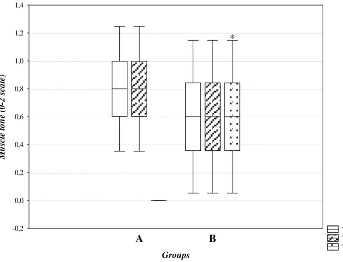
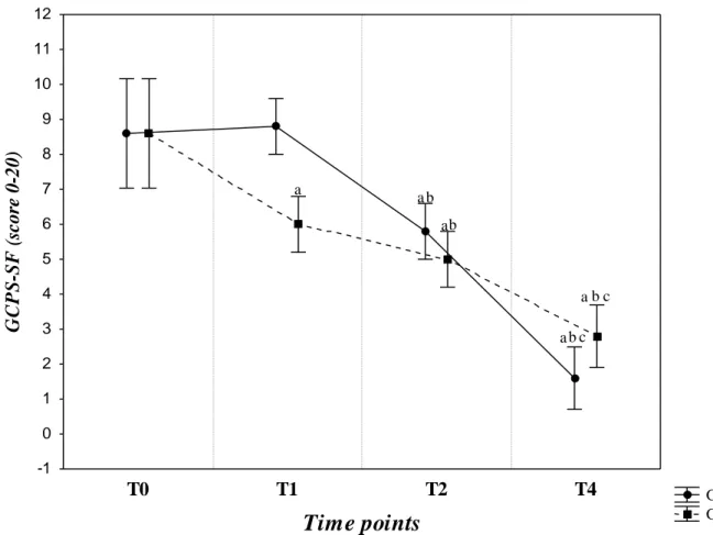
The last chapter describes two clinical studies carried out on dogs undergoing hemilaminectomy for acute thoracolumbar intervertebral disc extrusion and on dogs with Degenerative Lumbosacral Stenosis (DLSS), respectively. In both study, the Short Form of the Glasgow Composite Pain Scale (GCPS-SF) was applied to evaluate pain score. Based on the results of the first study, which compared the analgesic activity of buprenorphine and tramadol, both drugs showed a good analgesic activity. However, buprenorphine showed a faster and greater analgesic effect, compared to tramadol. None of the two molecules showed any side effect. In the second study, the analgesic conservative treatment of DLSS was started with tramadol. However, after the first week of treatment, the dogs of Group A were considered to be non-responder to tramadol. For this reason, the analgesic treatment was switched to gabapentin. After that, no statistical differences have been found between the two groups. Already after the 2nd week of treatment, GCPS scores were considerably lower in both groups, suggesting that both tramadol and gabapentin could be effective in reducing lumbosacral pain in dogs with DLSS. Moreover, the improvement of muscle tone in the group treated with gabapentin after 4 weeks of follow-up could be due to the resumption of a moderate physical activity determined by a better pain control than in dogs treated with tramadol.
Veterinary patients may be admitted to an Intensive Care Unit (ICU) because of different pathological conditions, that may represent a life-threatening hazard for the patient if not promptly and properly managed, as described in the cases reported in this thesis. For these reasons, the ICU doctor should have knowledge on different medical fields and should be able to take prompt decisions on the diagnostic tests and treatments to perform. “Time is money” but, in a critical patient, can also make the difference between life and death.
7
PREFACE
Emergency and critical care has been a growing field in small animal practice over the last 20
years. Emergency clinicians have to face with several life-threatening conditions and must be
able to take prompt decisions regarding clinical, diagnostic and surgical interventions, with
the main goal of saving patient‟s life. The appropriateness of these decisions can make the difference between life and death. This approach represents the main diversity of Emergency
which focuses on monitoring patient‟s vital parameters, preventing the onset of possible
complications and keeping the patient alive. Intensive Care medicine, instead, has the aim of
investigating the underlying clinical problem of every patient, in order to reach a definitive
diagnosis.
The definition of emergency patient refers to a patient who suffers from an acute or chronic
condition which make the clinical status of the animal unstable and may pose an immediate
risk for his/her life. This means that emergencies may vary from minor issues to patients that
are close to death. Moreover, ICU patients are often sick since a long time and are referred
from smaller practices, where the animal has already been treated.
Although nowadays, more and more specialist Emergency and Intensive Care Units (ICU)
exists, most of the emergencies are primarily seen by first line veterinary practitioners. For
this reason, it is very important that every veterinarian should have, at least, the basic
knowledge on how to deal with an emergency patient. However, although smaller practices
should be able to provide urgent stabilization, they should also be able to recognize when
complicated conditions need to be referred to a more specialised ICU.
All the staff of an Emergency Service has to be well prepared on how to approach an
emergency patient. Preparation includes taking prompt and resolute decisions, but also having
the right equipment close to hand and ready to use. All the members of the emergency team
8
are delegated to. The environment should be appropriated, with a well-equipped central
location to triage and stabilise the patient. Having a good communication both within the
team and with the owner, is also a primary important factor. Even during the following
hospitalization, sudden changes in the clinical status of critical patients can occur at any time,
and the period over which it is possible to act in order to reduce morbidity and mortality, may
be really short. For this reason, every loss of time due to the lack of organization or to the
search of the needed instruments, can result in a negative outcome.
Regarding the skills that all the members of emergency and ICU teams should have, triage
and stabilization of the major body systems are on the top of the list. The word triage refer to
the process of rapidly classifying patients on the basis of the severity of their clinical
condition, allowing patients with life-threatening conditions to be seen before patients with
less severe problems. Triage should be performed for every patient, within 5-10 minutes from
the admission and involves the collection of the first information from patient‟s history and
the evaluation of the cardiovascular, respiratory and neurological body systems. In fact,
severe life-threatening conditions involving these major body systems can suddenly
deteriorate and cause the patient‟s death.
Immediately after the triage examination, urgent empirical stabilisation (e.g. oxygen
administration and fluid therapy) has to be started according to the patient‟s need. In the
meantime, further diagnostic has to be carried out in order to identify the life-threatening
problem and to formulate a specific treatment plan.
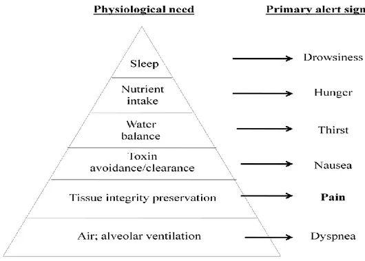
Among the conditions that require an emergency examination, severe pain is one of the most
common. Moreover, ICU patients often show various degrees of pain, which can determine
several detrimental effects on different body systems that may have a severe impact on the
outcome. Although analgesia may be postponed for a severely injured patient due to the need
for immediate lifesaving interventions, adequate pain control is ultimately essential to offset
9
family. Most of the owners take into account and appreciate the way the veterinary
practitioner approaches their animal. By being kind, sensible and respectful of the animal‟s
clinical condition, the patient may be better helped to deal with a pathologic disease and a
new environment that could result in a very stressful situation, and the owner could be more
satisfied, despite the final outcome. In this perspective, pain management has also the aim of
avoiding or, at least, reducing, animal suffering, and of ensuring animal welfare.
This thesis wants to be a contribution to the current knowledge in the Emergency and Critical
Care medicine, by focusing on different aspects varying from the fluid resuscitation to the
pain therapy of the ICU patient.
The thesis has been organized in different Sections, regarding the cardiovascular and
respiratory systems, and haematology, respectively. The last Section was destined to the topic
of pain assessment and analgesia. Every Section has been divided into different chapters, each
one reporting different studies carried out during the PhD course, regarding resuscitation fluid
therapy, management of pleural effusion, and pain management in dogs with neuropathic
pain. The last Section includes a survey on the attitude and knowledge of Italian veterinary
practitioners toward pain assessment and analgesia. Finally, case reports or case series
describing uncommon emergency conditions that have occurred in the abovementioned
10
I. CARDIOVASCULAR SYSTEM
1.1 Limited fluid volume resuscitation (LFVR) in severe shock unresponsive
to initial fluid challenge: a preliminary study in ten cats
Shock is a complex syndrome caused by a limited organ blood perfusion with a consequent
reduction in the availability of oxygen tissue level that, if not quickly handled, can determine
patient‟s death. It may be a result of many diseases and it is a common condition in critically
ill patients. Intravenous fluid resuscitation plays a primary role in the treatment of circulatory
failure in the emergency and intensive care unit settings (Cazzolli and Prittie, 2015). The
goals of fluid treatment include the restoration of plasma volume and organ perfusion, and the
rapid control of the main underlying problem. The so-called “triad of shock” (bradycardia,
hypotension and hypothermia) is a common condition in cats with hypovolemic shock. It is
assumed that in cats, unlike other species, bradycardia development instead of tachycardia in
response to a state of hypotension is due to the simultaneous activation of vagal and
sympathetic fibres from the aortic and carotid baroreceptors (Hackett, 2015; Schwartz et al.,
1973). Moreover, the adrenergic receptors become refractory to catecholamines at a body
temperature below than 37.5 °C, perpetuating both bradycardia and secondary compensatory
vasoconstriction (Brady et al., 2000).
Intravenously administration of every type of fluid determines an immediate plasma volume
expansion. However, each type of fluid owns different systemic effects that could affect
patient outcome (Myburgh and Mythen, 2013). Every type of fluid has its own safety and
toxicity profile and their pharmacologic properties can influence organ function, alter
coagulation, modulate the immune system and affect blood viscosity, red blood cell rheology
11
Conventional resuscitation (CR) consists in the administration of large volumes of isotonic
crystalloid solutions (60-90 mL/kg/h in dogs and 40-60 mL/kg/h in cats), although it is
currently recommended to start with 1/4 or 1/3 of the total volume and re-evaluate the patient
before administering additional fluids (Driessen and Brainard, 2006; Rozanski and Rondeau,
2002). Another protocol used in clinical practice, is to administer 15-20 mL/kg of isotonic
crystalloids in rapid bolus in dog and within 15 minutes in cat (for the higher risk of
pulmonary oedema caused by fluid overload), until a maximum of four boluses in dogs and
three in cats, re-evaluating the patient every fifteen minutes (Viganò, 2011). Disadvantages of
CR consist in: i) prolonged administration time; ii) rapid redistribution of isotonic crystalloids
into the interstitial space (60-80% of the administered fluid within 30-60 minutes), with the
risk of oedema formation; iii) hypothermia and iiii) risk of aggravation of any bleeding caused
by the destabilization of clots and dilution of the circulating clotting factors (Driessen and
Brainard, 2006). The positive fluid balance that could follow aggressive resuscitation with
crystalloids is associated with worsened patient outcome. The expansion of extracellular
spaces determines a damage of the endothelial surface layer so causing capillary leak and
tissue oedema (Cotton et al., 2006). Moreover, isotonic crystalloids could induce a
pro-inflammatory state, with an increase in cytokine production and endothelial cell activation,
worsening oedema formation (Cazzolli and Prittie, 2015).
Colloidal solutions have a volume-sparing effect with a decreased risk of inducing a positive
fluid balance. This type of fluid contains large molecules that cannot cross an intact vascular
barrier, so decreasing the possibility of tissue oedema formation, and has a longer plasma
half-life. However, adverse effects such as coagulation disorders, acute kidney injury (AKI),
pruritus, hepatopathies and anaphylactoid reactions have been reported as adverse effects
which could follow the administration of colloidal solutions (Cazzolli and Prittie, 2015).
These problems led to the research of alternative treatments such as the Limited Fluid Volume
12
(Hammond et al., 2014; Hammond and Holm, 2009; Rocha-e-Silva and Poli de Figueiredo.
2005; Stern, 2001; Muir and Sally, 1989). The LFVR protocols, involving the use of a
hypertonic saline solution and/or colloids, are based on the use of a smaller volume of fluid
able to restore blood volume and to solve the shock, thus minimizing the risk of interstitial
fluid extravasation and clots breaking (Hammond and Holm, 2009). The level of blood
pressure reached in LFVR is lower than in CR. A mean arterial blood pressure of 70 mmHg
or a systolic arterial blood pressure of 90 mmHg is still enough to keep perfusion of vital
organs, with reduced risk of bleeding (Hammond and Holm, 2009). Several studies on
hypovolemic shock, in animal models and humans, evaluated different protocols of
resuscitation fluid therapy and highlighted how the combination of hypertonic saline and
colloids produces best effects on the increase of mean arterial blood pressure, oxygen
saturation and cardiac output compared to isotonic crystalloids (Wang et al., 2015; Hammond
et al., 2014; Watters et al., 2006; Friedman et al., 2003; Mapstone et al., 2003; Schertel et al.,
1997; Muir and Sally, 1989). However, only a few studies have been carried out on the use of
HS solution and LVFR protocol in cats (Dupe et al., 1993; Muir and Sally, 1989).
1.1.1 Aim of the study
The aim of this study was to evaluate the effect of a LFVR protocol, with HS alone or
combined with HES administration, in cats with severe not cardiogenic shock, unresponsive
to initial CR with isotonic crystalloids.
1.1.2 Materials and method
Protocols of animal husbandry and experimentation were reviewed in accordance with the
standards recommended by the Guide for the Care and Use of Laboratory Animals and
13
of the Veterinary Department of the University of Messina (record number 009/2016). This
study was carried out in accordance with the Good Clinical Practice.
Case selection criteria
The study was carried out in the Veterinary Teaching Hospital of the University of Messina,
in the period between November 2014 and November 2016, on cats referred in the emergency
service with a red triage code for shock of various origins unresponsive to initial CR. Cats
were enrolled in the study after the informed consent has been provided by the owners.
Animals
During the study period, 60 cats in shock have been referred to the emergency service. The
selection criteria included the presence of a non-cardiogenic shock due to various causes and
the unresponsiveness to the initial CR protocol with isotonic crystalloids. Exclusion criteria
were the presence of cardiac signs (murmur, gallop rhythm, muffled heart sounds, distended
jugular and jugular pulse) and dyspnoea on physical examination, and the administration of
glucocorticoids, vasopressors and mannitol prior to or on admission.
Procedure
Shock was established on the basis of alterations of three or more of the following objective
parameters: heart rate (HR) <140 or >225 beats/minute (bpm); capillary refill time (CRT) >2
seconds; respiratory rate (RR) >40 breaths/min; rectal temperature (T°) <37.8 °C; blood
lactate > 2.5 mmol/L (laboratory range: 0.5-2.5 mmol/L). Subjective parameters (Kirby’ Rule
of 20), like pulse quality, mucous membranes colour, state of the sensorium and temperature of the extremities (Mazzaferro, 2013) were recorded, too. When it was possible, a micro
venous withdrawal was performed, in order to determine the following parameters: packed
cell volume (PCV), by means of capillary tubes for microhaematocrit; plasma total solids
(TS), by means of a refractometer; lactate, by means of a portable blood lactate meter (Lactate
14
meter (Glucocard, Menarini, Firenze, Italy). Immediately after the presentation, a continuous
supply of oxygen flow-by using an oxygen concentrator was provided to all the animals.
When needed, patients were re-warmed and the rectal temperature was recorded continuously
using a probe connected to a monitor.
Fluid resuscitation protocol
A venous (cephalic vein) access was achieved and 15-20 mL/kg of lactate Ringer's solution
(LRS; Ringer lactate solution S.A.L.F. S.p.A., Cenate Sotto, Bergamo, Italy), preheated (38
°C), were administered over 15 minutes. When a vascular access was not immediately
available, an intraosseous (femoral trochanteric fossae) catheterization was performed.
Cats that did not show any improvement of the vital parameters within 10 minutes after the
end of the initial CR with isotonic crystalloid, were selected for the administration of the
LVFR protocol.
Hypertonic saline solution (HS) was prepared from a commercial solution of 11.7% NaCl (10
mL vials, NaCl 2 mEq/mL, Galenica Senese, Monteroni D'Arbia, Siena, Italy). The dilution
was prepared by adding, under sterile conditions, 3 mL of the concentrated solution to 2 mL
of sterile water for injections, obtaining a final concentration of 7.02%. Animals could receive
up to a maximum of 8 mL/kg of HS and 8 mL/kg of hydroxyethyl starch (HES; Voluven®,
Fresenius Kabi Italy S.r.l., Isola della Scala ,Verona, Italy). Boluses of 2 mL/kg were
administered over 5-10 minutes and patients vital parameters were re-evaluated every 5-10
minutes until stabilization and, then, every 30 minutes. Additional boluses were administered
10 minutes after the end of the previous bolus, if the vital parameters were still abnormal.
Cats were resuscitated until the achievement of the desired levels of vital parameters and not
until the infusion of a fixed volume of fluid. The main purpose of assessment was the time
(minutes) until the stabilization of vital parameters.
Animals were considered stable once the vital parameters (temperature, heart rate, respiratory
15
species. In particular, animals were deemed stable afterachieving the following stabilization
endpoints: T >37.8 °C; HR >140 and <225 bpm; CRT <2 sec; RR <40 breaths/minute; full
and strong pulse; alert and bright sensorium; warm extremities. When possible, serial
micro-withdrawals for the PCV, TS, lactate and glucose levels were performed every 30 minutes
until stabilization, and 30 minutes later.
After stabilization, animals were hospitalized and underwent any other additional
investigation (blood chemistry, haematology, ultrasound and radiological diagnostic
evaluations) in order to establish the underlying problem. Additional supportive care and
treatments were at veterinary practitioner discretion.
Statistical analysis
The time until stabilization (minutes), the volume of HS and HES administered and the effect
of LFVR on heart rate (HR), rectal temperature (T) and respiratory frequency (RR) were
determined. A descriptive statistical analysis was applied for age, body weight, volume of HS
and HES administered and time until stabilization of the ten cats enrolled in the study.
One-way analysis of variance (ANOVA) for repeated measures was applied to evaluate the effect
of LFVR protocols on vital parameters (HR, T and RR) throughout the experimental period.
The parameters recorded before (T0), every 10 minutes during the first 30 minutes (T10, T20,
T30) and 60 minutes (T60) after LFVR protocol was started, were used for the statistical
analysis. When significant differences were found Bonferroni‟s post hoc comparison was
applied. Statistical significance was set at p values <0.05.
Data were analysed using the software STATISTICA 7 (Stat Soft Inc., USA, 2003).
1.1.3 Results
During the study period, ten client-owned cats met the selection criteria. Among them, nine
16
four female (two neutered), aged between 1 month and 10 years (mean±SD: 27.9±41.7
months; median: 5.5 months) and body weight from 0.26 to 5 kg (mean±SD: 2.3±1.9 kg;
median: 2.2) (Table 1).
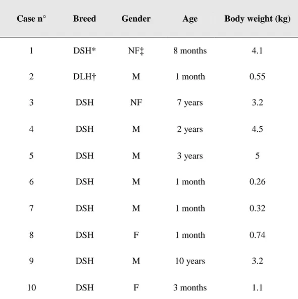
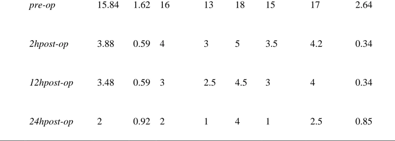
Table 1. Breed, gender, age and body weight of the cats enrolled in the study
Case n° Breed Gender Age Body weight (kg)
1 DSH* NF‡ 8 months 4.1 2 DLH† M 1 month 0.55 3 DSH NF 7 years 3.2 4 DSH M 2 years 4.5 5 DSH M 3 years 5 6 DSH M 1 month 0.26 7 DSH M 1 month 0.32 8 DSH F 1 month 0.74 9 DSH M 10 years 3.2 10 DSH F 3 months 1.1
17
On admission, all the cats were bradycardic (HR 96.6±21.9 bpm), with a weak pulse, pale to
white mucous membranes, CRT ≥3 seconds and depressed to stuporous sensorium; nine were hypothermic (T: 32.8±3.3 °C) and seven tachypnoeic (RR 50.4±22.6 breaths/minute) (Table
2). Conventional fluid resuscitation was initially started in all the cats enrolled, with no
improvement of vital parameters. Therefore, LFVR protocol was started. Conversely, HS
administration alone or together with HES determined a rapid improvement of the vital
parameters.
Cats received a mean±SD volume of 4.0±1.6 mL/kg of HS (range: 2-6 mL/kg) and 2.6±2.7
mL/kg of HES (range: 0-8 mL/kg). The administration of HS alone or together with HES
determined an improvement of the vital parameters within 33±12.7 minutes, with a range of
15-60 minutes. In six patients (60%) it was sufficient a single administration of HS as the
only solution (n=3/10) or in combination with HES (n=3/10). In the other four cases (40%) it
was necessary to use a second bolus of HS, alone (n=2/10) or in combination with HES
(n=2/10), before achieving the stabilization of the vital parameters (Table 3 and Figure 1).
Due to the severity of clinical condition and the small size of some of the patients, only in
four cats (cases 3, 4, 5 and 9) it was possible to perform venous catheterization and only in
two of them (cases 4 and 5) a blood sample suitable for the examinations was collected. In
both cats, blood parameters showed the same trend: a slight decrease in PCV (T0: 25.5±9.5;
T60: 24.0±10.0%), TS (T0: 62.5±7.5; T60: 59.0±9.0 g/L) and glucose values (T0: 12.7±4.9;
T30: 11.2±3.8; T60: 10.3±3.3 mmol/L), and a remarkable reduction in lactate values (T0:
6.4±1.6; T30: 3.4±0.7; 1.75±0.9 mmol/L), suggesting the achievement of a good perfusion
18 Table 2. Objective (rectal temperature [T°], heart rate [HR], respiratory rate [RR], and
capillary refill time [CRT]) and subjective (quality of pulse, state of sensorium and mucous
membrane colour) parameters at first clinical examination of the ten cats enrolled in the study
Case
Objective parameters Subjective parameters
T° (°C) HR (bpm) RR (breaths/min) CRT (sec) Pulse State of sensorium Mucous membrane colour
1 34 120 40 2-3 Weak Stuporous Pale
2 30 46 28 >3 Weak Stuporous White
3 35.1 116 48 >3 Weak Stuporous White
4 28.9 120 40 >3 Weak Stuporous White
5 38.8 100 100 2-3 Weak Depressed Grey
6 36.8 94 80 2-3 Weak Depressed Grey
7 31.1 84 36 >3 Weak Stuporous Pale
8 29.8 88 56 2-3 Weak Depressed Pale
9 31.3 98 36 >3 Weak Depressed White
10 33 100 40 >3 Weak Depressed Pale
Mean 32.8 96.6 50.4
±SD 3.3 21.9 22.6
19 Table 3. Type of the shock, administration route (intravenous, IV; intraosseus, IO), volume of
hypertonic saline solution (HS) and hydroxyethyl starch (HES), time until the initial and
complete stabilization, follow up and outcome of the ten cats enrolled in the study
Case Type of
shock Route
Volume of fluid (mL/kg)
Time until stabilization
(minutes) Follow up
HS HES Beginning Complete
stabilization Length Outcome
1 Distributive IO/IV 6 4 5 45 25
months Positive
2 Hypovolemic IO 6 8 5 30 22
months Positive
3 Hypovolemic IV 4 4 5 30 3 days Negative
4 Traumatic IV 4 0 5 30 20
months Positive
5 Traumatic IV 2 0 5 15 17
months Positive
6 Hypovolemic IO 2 4 5 30 5 hours Negative
7 Traumatic IO 6 0 5 60 5 days Negative
8 Traumatic IO 2 0 5 30 5 days Negative
9 Traumatic IV 4 2 5 20 9 months Positive 10 Hypovolemic IO 4 4 5 40 6 months Positive Mean 4.0 2.6 33.0 ±SD 1.6 2.7 12.7
20 Figure 1. Limited volume fluid resuscitation (LFVR) algorithm in the 10 cats in
non-cardiogenic shock enrolled in the study. LRS: Ringer‟s lactate solution; HS: hypertonic saline
21
As reported in Figure 2, the statistical analysis showed a significant effect of LFVR (p<0.001)
on T° (p<0.0001), HR (p<0.0001) and RR (p<0.001). In particular, lower values of T° were
found at T0 compared to T10 (p=0.03), T20 (p<0.0001), T30 (p<0.0001) and T60 (p<0.0001)
and at T10 compared to T30 (p=0.002) and T60 (p<0.0001). Regarding HR, the lowest values
were found at T0 respect to T10 (p<0.0001), T20 (p<0.0001), T30 (p<0.0001) and T60
(p<0.0001), at T10 respect to T20 (p=0.001), T30 (p<0.0001) and T60 (p<0.0001) and at T20
respect to T30 (p=0.01) and T60 (p<0.0001). Finally, higher values of RR were found at T0
versus T30 (p=0.01) and T60 (p<0.001). For the small number of the data collected, statistical evaluations on blood parameters were not performed.
According to the patient's medical history and post-stabilization examinations, it was possible
to establish the type and the origin of the shock in all the 10 cats: distributive,
post-anaesthesia for sterilization surgery (case 1); hypovolemic, for severe dehydration (8-10%)
due to rhinotracheitis and intestinal parasites (cases 2, 6 and 10) or renal failure and chronic
enteritis (case 3); traumatic due to car accident (cases 4, 8 and 9), falling from heights (case 7)
or aggression by dogs (case 5) (Table 3).
Of the ten cats, six (cases 1, 2, 4, 5, 9 and 10) currently enjoy good health (follow-up 6 to 25
months), whereas four died during hospitalization. Of these, one patient (case 3) died on the
third day, because of worsening of the underlying disease (chronic renal failure); another one
(case 6) died after 5 hours for pulmonary oedema due to a fault in the infusion pump and the
22 Figure 2. Effect of LFVR protocol on mean ± standard deviation (SD) of rectal temperature,
heart and respiratory rates values measured in the ten cats enrolled in the study. Relative
significances throughout the experimental period time points (T0, before LFVR protocol and
after the administration of the first bolus of Ringer Lactate solution, RLS; T10, T20, T30 and
23 1.1.4 Discussion
In the present study, the LVFR (hypertonic saline alone or associated with colloid)
determined a rapid improvement of the vital parameters in contrast to the initial bolus of CR,
suggesting a rapid improvement in blood perfusion and tissue oxygenation. No evident
adverse effects occurred in any patient during the administration of the protocol.
Limited fluid volume resuscitation with hypertonic saline and colloid solutions expands
plasma volume for 2-3 hours, quickly recalling the interstitial fluids and probably reducing
endothelial permeability (Victorino et al., 2003). In contrast, in CR with isotonic crystalloids
only 10-25% of fluids remains in the intravascular compartment after an hour from the
administration (Silverstein et al., 2005). Further theoretical benefits of LFVR can be a
reduced risk of hypothermia, damage from ischemia-reperfusion injury, interstitial oedema,
possible with conventional resuscitation and coagulopathy by dilution (Hammond et al., 1014;
Rocha-e-Silva and Poli de Figueiredo, 2005; Pascual et al., 2003). However, the real benefit
on coagulation could be argued due to the good, if not even better, expansion of blood volume
provided by LFVR.
Moreover, it has been demonstrated that the administration of HS together with dextran 70
has immunological and anti-inflammatory effects (Dubick et al., 2013) which last for 24 hours
and could be useful in the prevention of multiple organ dysfunction syndrome (MODS)
(Rizoli et al., 2006).
Only a few references reported the use of hypertonic saline solution in cats (Dupe et al., 1993;
Muir and Sally, 1989). In an experimental model of hypovolemic shock, 4 mL/Kg intravenous
administration of 7.5% NaCl solution determined positive hemodynamic effects,
characterized by a rapid normalization of blood pressure, aortic flow and cardiac contractility
and by the decreasing of peripheral resistance; the effect did not last more than 60 minutes
24
However, LFVR protocols are not free from the risk of adverse effects. The hypertonic saline
solution, besides being contraindicated in severe dehydration, can cause occasional premature
ventricular contractions, bradyarrhythmias, transient hypotension and bronchoconstriction
when administered at speeds faster than 1 mL/kg/min (Ford and Schaer, 1993; Stern, 2001). It
can also determine transient hypernatremia, with alteration of sodium level in early hours
after resuscitation, as well as affecting the osmolality and the levels of chloride, potassium
and bicarbonate (Hammond et al., 2014). After administration of HS in peripheral vessels, it
can be observed haemolysis which leads to haemoglobinuria (Muir and Sally, 1989). In
addition, the HS can rapidly elevate cardiac output and blood pressure, which together with
the increase of sodium level can increase the risk of congestive heart failure or neurological
signs in patients with hyponatremia prior to the infusion (pontine myelinolysis) (Churcher et
al., 1999). Although among the cats enrolled in this study four were clinically dehydrated, the
LFVR protocol was started after the administration of the resuscitation bolus of isotonic
crystalloids which partly improved the hydration status of the patients. Moreover, the
rehydration fluid therapy was started immediately after the reanimation protocol.
Limited fluid volume resuscitation could also facilitate bleeding in the vascular injury site for
clot breaking (re-bleeding) (Adamik et al., 2015). However, the bleeding is not a problem
related only to hypertonic fluids, since it could be caused by all types of resuscitation fluid
therapy.
In the present study, the administration of an initial bolus of crystalloid did not determine any
improvement of the vital parameters, in contrast to the next administration of HS alone or
associated with colloid. Conventional Resuscitation protocols usually consist of the
administration of repeated boluses of isotonic crystalloids. However, cats seem less able to
cope with large volumes of intravenous fluid and have been reported with a high incidence of
pulmonary oedema, especially if they are hypothermic (Adamantos and Hughes, 2015). For
25
of the critical clinical conditions of the cats enrolled in this study, it was decided to start the
LFVR as an alternative protocol. Approximately the 50% of haemodynamically unstable
human patients treated with CR are non-responders, with no increase in cardiac output
following intravenous fluid administration. This failed fluid challenge increase the risk of
endothelial surface layer damage and tissue oedema (Cazzolli and Prittie, 2015). Although
colloidal solutions have been reported with fairly common adverse effects, such as AKI and
coagulopathy, in both human and veterinary medicine, these are commonly dose- and time-
dependent. Many of the reports which describe these complications have been carried out in
patients that have undergone a long period of treatment (Cazzolli and Prittie, 2015). On the
contrary, some of the clinical trials on the use of HES within a shorter period for fluid
resuscitation in patients with hypovolemic shock, found no evidences of an increased risk of
AKI or coagulation impairment (Annane et al., 2015; Guidet et al., 2012). Moreover, in a
recent study on rats with endotoxemia induced by lipopolysaccharide administration, fluid
resuscitation with HES and hypertonic saline determined an amelioration of kidney injury
(Wang et al., 2015).
Veterinary reports showed that bolus and continuous rate infusions of 6% hetastarch solution
at moderate doses are well tolerated in feline and canine patients (Glover et al., 2014).
Moreover, the administration of HES in cats did not results in a significant increase is serum
creatinine compared with cats treated with crystalloid (Yozova et al., 2016). However, few
safety and efficacy data exist in veterinary medicine on the use of HES as a resuscitation
fluid.
In this study, the total administered volume of HES was far below (40%) the recommended
daily dosage of 20 mL/kg/day (Mizzi et al. 2011), with an administered volume of 2.6±2.7
mL/kg and a maximum of 8 mL/kg. Regarding HTS, the total volume administered to each
cat did not exceed the maximum range reported in the literature (Balakrishnan and Silverstein,
26
of the desired levels of vital signs and followed by a maintenance and correction of
dehydration fluid therapy with isotonic crystalloids, so that the administration of HS and/or
HES did not last for more than 60 minutes.
Response to treatment occurred very quickly, with the first positive effects already registered
within 5 min after infusion. In particular, the increase in rectal temperature and heart rate
occurred already after 10 min (T10), while the decrease of respiratory rate was significant
only after 30 minutes (T30), suggesting an improvement in blood perfusion and, as a
consequence, in tissue oxygenation.
In 30% of the patients a single administration of HS ± HES was sufficient, while in the other
cases it was necessary to use one additional boluses of HS ± HES before reaching the
stabilization of vital signs, always occurred within 15 and 60 minutes, even in very small
(body weight <500 g) and immature (3-4 weeks old) cats.
The survival rate to the treatment was 90% on the first day and 60% on the fifth. Considering
the severity of the clinical condition combined with the absence of response to the initial
conventional resuscitation, the survival could be considered acceptable. Moreover, the cause
of death was probably not to be attributed to a late adverse reaction to the treatment but rather
to common complications in critically ill patients (e.g., pneumonitis) or irreversible chronic
illness (chronic renal failure). In one case death was caused by infusion pump failure during
hospitalization that quickly determined a fluid overload in a very young and small cat (1
month of age, 0.26 kg body weight).
The stabilization of vital parameters was optimal and persistent in nine of the ten cats enrolled
in the study. Only in one case (case 7) it was observed a thermoregulatory control deficiency,
probably due to a traumatic neurological damage.
The limitations of this study are the lack of a control group resuscitated only with crystalloids,
the small size and the heterogeneity of the sample (cats were different for age, weight and
27
administration of the LFVR protocol. Therefore, further studies should be carried out on a
wider number of animals, in order to establish the incidence of adverse effects, such as
electrolytic abnormalities and eventual neurological signs related with an osmotic central
damage induced by HS administration, as previously observed in hyponatremic patients
(Churcher et al. 1999) and the effects of each specific LFVR protocol in order to establish if
the administration of HES is really relevant.
The results obtained in this study seem to suggest that the administration of HS with or
without HES could be used as an alternative fluid treatment in cats with severe
non-cardiogenic shock. Although the present study should be considered a preliminary
investigation due to the small number of cats enrolled, the absence of evident adverse effects,
the good results achieved in the short term and the cost reduction due to a faster stabilization
should support further investigations on LFVR effectiveness and safety in companion
28
1.2 Clinical and therapeutic investigations on pericardial effusion in dogs
Pericardial effusion (PE) is an abnormal accumulation of fluid within the pericardial space
and it represents the most common disease of the pericardium in dogs. Prevalence has been
reported to be 0.43% and it was found in approximately 7% of dogs presented with clinical
signs of cardiac disease (MacDonald et al., 2009). Most commonly, the aetiology of
pericardial effusion in dogs is neoplastic or idiopathic in origin. The most common neoplastic
causes of PE in dogs include haemangiosarcoma, heart base tumours (chemodectoma) or
mesothelioma (Stafford et al., 2004; Shaw and Rush, 2007a). The idiopathic pericardial
effusion (IPE) refers to a sterile hemorrhagic effusion, with no known aetiology. Prognosis of
dogs with PE is strongly related to the cause of the effusion. Neoplastic PE, especially due to
haemangiosarcoma, is associated with short survival times, ranging from 26 to 56 days, while
IPE usually has a good long-term prognosis, ranging from 790 to 1068 days (Weisse et al.,
2005; Stafford et al., 2004), although recurrent effusions might need a pericardiectomy in
order to control clinical signs (Fine et al., 2003). Moreover, dogs with PE due to a heart base
mass usually have a better prognosis than dogs with PE due to haemangiosarcoma (Weisse et
al., 2005; Stafford et al., 2004).
The accumulation of fluid of any type in the pericardial space reduces diastolic cardiac filling
and may lead to right heart failure; when the intra-pericardial pressure increases to a level
higher than or equal to that of the right ventricle, cardiac tamponade occurs (Humm et al.,
2009). However, patients with mild PE may not show clinical signs. The intra-pericardial
pressure depends on the amount and on the rate of fluid accumulation. For this reason, even
large volumes of effusion that accumulate slowly may result only in slight hemodynamic
abnormalities, while small but rapid accumulation of pericardial effusion, as seen in acute
29
The most common findings on general clinical examination in dogs with PE are collapse or
weakness, muffled heart sounds on heart auscultation, distended jugular veins, the presence of
a jugular pulse and pulsus paradoxus and abdominal distension due to hepatomegaly and
ascites. Vomiting has been described in association with PE in humans (Jabr, 2004). In a
recent study in veterinary medicine, Fahey et al. (2017) described the occurrence of vomiting
in the 51% of dogs with PE, especially in those with evidence of hypoperfusion.
The diagnosis of PE may be suspected based on the history, clinical examination and by the
radiographic findings of a globoid heart silhouette, pleural effusion and enlargement of the
caudal vena cava. However, echocardiography is the gold standard to confirm the diagnosis of
PE and to distinguish between the non-neoplastic and neoplastic aetiologies, as well as to
define the specific causes and locations of the neoplasm (MacDonald et al., 2009). Several
echocardiographic views of the heart, such as right parasternal short-axis and long-axis views,
left apical view and left cranial parasternal long-axis view, have to be performed in order to
obtain a complete image of the heart and to detect any eventual masses, especially the ones
localized in the right atrium (MacDonald et al., 2009). Cardiac tamponade is diagnosed when
diastolic collapse of the right atrium, right ventricle or both is found together with pericardial
effusion. In a study in 107 dogs, echocardiography showed a sensitivity and specificity of
82% and 100%, respectively, for the diagnosis of a cardiac mass in dogs with pericardial
effusion. Moreover, it showed a high sensitivity and specificity in differentiating the specific
type of cardiac mass (MacDonald et al., 2009). In this study, the two most common causes of
pericardial effusion were haemangiosarcoma (33.6%) and idiopathic pericarditis (19.6%), but
even mesothelioma (14%) and chemodectoma (12%) were fairly described. Recently, the
diaphragmatic-hepatic (DH) view of the abdominal and thoracic focused assessment with
sonography (AFAST and TFAST) has been described to be clinically helpful in detecting PE
30
Pericardiocentesis is required for the relief of cardiac tamponade and clinical sings in
emergency conditions and to carry out cytological examinations of the fluid for diagnostic
evaluations. In order to avoid the laceration of the left extramural coronary artery and to go
through the cardiac notch of the right lung lobes, pericardiocentesis is usually performed with
the dog in sternal or left lateral recumbency from the right haemithorax, in a small area where
the lungs do not cover the heart and the pericardium is just beside the thoracic wall
(Gidlewsky and Petrie, 2005). Echocardiography is helpful in finding the best intercostals
space but, if it is not available, pericardiocentesis should be performed at the level of the
cardiac notch, starting from the fourth or fifth interscostal space, dorsally to the costochondral
junction. Several types of over- or through-the-needle catheters may be used to perform
pericardiocentesis (Shaw and Rush, 2007b; Gidlewsky and Petrie, 2005). The most common
adverse events reported during or following pericardiocentesis are cardiac puncture,
arrhythmias, laceration of the tumour or the coronary artery, leading to intrapericardial
haemorrhage or cardiopulmonary arrest, with a rate of occurrence of 15% in dogs (Humm et
al., 2009). The rate of relapse of the fluid accumulation described in dogs both with or without
neoplasia is high (Stafford et al., 2004), so repeated pericardiocentesis may be required.
In cases of recurrent PE, pericardiectomy is required to achieve a definitive diagnosis and to
solve the problem of frequent relapses. Moreover, prolonged survival after pericardiectomy
has been described in dogs with IPE, while dogs with neoplastic pericardial effusion have
been reported to have shorter survival times (Stafford et al., 2004).
1.2.1 Aim of the study
The aim of this retrospective study was to evaluate the signalment, clinical signs,
echocardiographic findings and epidemiology of PE in canine patients. Secondary objectives
31
determining the clinical usefulness of a new pericardiocentesis technique, using a “fistula needle” for haemodialysis.
1.2.2 Materials and method
Case selection criteria
A database of dogs referred for specialist cardiology and echocardiographic examination from
2009 to 2016 was reviewed. The examinations were performed in a high standard veterinary
referral clinic in Sicily (Italy), on client-owned dogs, after the informed consent had been
signed by the owners.
Animal husbandry and clinical procedures were in accordance with the standards
recommended by the Guide for the Care and Use of Laboratory Animals and Directive
2010/63/EU on the protection of animals used for scientific purposes.
Diagnostic tests
All the dogs were subjected to a general physical examination, radiographs of the thorax in
dorso-ventral and right lateral recumbency and echocardiography in right parasternal
short-axis and long-short-axis views, left apical view and left cranial parasternal long-short-axis view. All the
echocardiographic examinations were performed by the same specialized operator by mean of
an ultrasound machine (Esaote My Lab 30 Gold, Genova, Italy) with a multi-frequency probe
of 2.5-3.5 MHz with sterile cover.
Pericardiocentesis
When required, an echo-guided pericardiocentesis was performed. The right lateral thorax
was clipped and aseptically prepared between the second and the eight intercostal spaces.
Patients were placed in right lateral recumbency, on an echocardiography table with a suitable
hole for performing the diagnostics procedures. Pericardiocentesis was performed between the
32
Medical Care, Bad Homburg, Germany) (Figure 1 and 2), commonly used for haemodialysis,
with a catheter of 2.5 cm provided with a lateral hole and two wings, connected to a flexible
tube 30 cm long. A large 50 mL syringe and a three-way stop cock were attached to it (Figure
3). Following the echo-guided insertion of the needle into the intercostal space and the
perforation of the pericardium, the aspiration of the effusion was performed. A sample of the
effusion was kept for physical and cytological examinations in blood tubes and stored under
refrigeration until examinations were performed. During the pericardiocentesis procedure,
continuous electrocardiogram monitoring was performed. Complete echocardiography was
repeated at the end of the procedure, in order to detect any mass or abnormality that may have
33 Figure 1. “Fistula needle” (Fresenius Medical Care, Bad Homburg, Germany) used for the
pericardiocentesis
34 Figure 3. Three-way stop cock and 50 mL syringe attached to the “fistula needle”. In the
35
Statistical analysis
A descriptive statistical analysis (percentage, mean, standard deviation, SD, and range) was
applied for the age and gender for each breed of dog enrolled in the study. Percentages have
been calculated for the incidence rate of pericardial effusion, specific cause of PE and cardiac
tamponade in the dogs enrolled in the study. Data were analyzed using a commercially
available statistical software program (STATISTICA 7, Stat Soft Inc., USA, 2003).
1.2.3 Results
The records of 5304 dogs referred for specialist cardiology and echocardiographic
examination were reviewed for this study.
Dogs were of different breeds, age, gender, type and severity of the cardiac disease. The total
number of dogs found with pericardial effusion was 91 (1.7%). Among this, 20 were female
(21.98%) and 71 were male (78.02%). The mean age at presentation was 10.63±2.90 standard
deviation (SD) years (ranging from 1 to 19 years). Breed, gender and age of the 91 dogs with
36 Table 1. Number (n) of animal, gender (female [F] and male [M]) and descriptive statistic of
ages (Mean, Standard Deviation [SD], minimum [Min] and maximum [Max] values) for each
the breed of dogs with pericardial effusion (PE) enrolled in the study
Breed N Gender Age
F (n) M (n) Mean SD Min Max
Mongrel dog 38 7 31 11.74 2.45 6 19 German shepherd 5 0 5 9.8 0.84 9 11 Beagle 5 0 5 9.75 1.89 7 11 Labrador retriever 4 0 4 7.25 3.86 2 11 Miniature Poodle 4 0 4 12 1.63 10 14 Italian mastiff 4 1 3 8.5 3.11 5 12 Yorkshire terrier 3 1 2 11.33 1.53 10 13 Pit bull 3 2 1 11 3.46 9 15 English setter 3 2 1 9.33 4.72 4 13 Pug 2 0 2 8.5 0.71 8 9 Golden retriever 2 0 2 10 0 10 10 CKCS* 2 1 1 8.5 0.71 8 9 Siberian husky 2 1 1 11.5 0.71 11 12 Irish setter 1 1 / / / 10 / Pekingese 1 / 1 / / 15 / Rottweiler 1 / 1 / / 8 / German pinscher 1 1 / / / 12 / Dogue de Bordeaux 1 1 / / / 1 / Bull mastiff 1 1 / / / 8 / Sharpei 1 1 / / / 9 / Maltese 1 / 1 / / 8 / Etna Cirneco 1 / 1 / / 15 / Dachshund 1 / 1 / / 6 / Chihuahua 1 / 1 / / 12 / Hound 1 / 1 / / 10 / Shih Tzu 1 / 1 / / 15 / Newfoundland 1 / 1 / / 8 /
37
Among the 91 dogs with PE, cardiac tamponade, with signs of obstructive shock (lethargy,
distended jugular veins, jugular pulse and pulsus paradoxus, tachycardia and weak peripheral
pulse), occurred in 37 cases (40.65%). All the patients with pericardial effusion showed
various degrees of ascites.
Radiographs of the thorax showed cardiomegaly with a globular cardiac silhouette in all the
91 dogs. On echocardiographic examination, clear evidence of a neoplasia was found in 33
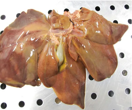
cases (36.26%); among these, 15 were localised in the right atrium (45.45%) (Figure 4), 9
were heart base masses (27.27%) (Figure 5), 5 were on the left atrium (15.15%) and 4 were in
the thorax (mediastinum and pleural space) (12.12%).
In 32 cases (35.16%) a severe mitral regurgitation together with tricuspid regurgitation was
detected as the possible cause of the PE; among these, pulmonary hypertension was found in
22 cases (68.75%). In only one case (1.10%) the PE was due to a foreign body (a lead bullet)
lodged in the interventricular septum (Figure 6). In the remaining 25 cases (27.47%) a clear
38 Figure 4. Ecocardiographic picture, in left apical view, of a right auricle neoplasia (arrow)
39 Figure 5. Ecocardiographic picture, in left apical view, of a heart base tumour (arrow)
40 Figure 6. Ecocardiographic, in right parasternal short axis view (A and B), and radiologic (C)
pictures of a foreign body (a lead bullet) stuck in the interventricular septum (arrow). In B an
enlarged ecocardigraphic picture of the foreign body
A
B
41 Table 2. Incidence of tumours, valve regurgitation, foreign body and unidentified cause:
number of dogs (n) and cardiac tamponade (CT) for each specific condition, for each breed of
dogs with pericardial effusion (PE) enrolled in the study
Breed n Aetiology
Tumours Valve
regurgitation Foreign body
Unidentified cause n CT N CT n CT N CT Mongrel dog 38 16 7 16 0 0 0 6 3 German shepherd 5 2 0 1 0 0 0 2 2 Beagle 5 2 1 2 0 0 0 1 1 Labrador retriever 4 1 0 0 0 0 0 3 2 Miniature Poodle 4 0 0 4 0 0 0 0 0 Italian mastiff 4 2 2 0 0 0 0 2 2 Yorkshire terrier 3 2 1 0 0 0 0 1 1 Pit bull 3 0 0 0 0 0 0 3 3 English setter 3 0 0 1 0 1 0 1 1 Pug 2 0 0 0 0 0 0 2 1 Golden retriever 2 1 0 0 0 0 0 1 1 CKCS* 2 0 0 2 0 0 0 0 0 Siberian husky 2 2 2 0 0 0 0 0 0 Irish setter 1 0 0 1 0 0 0 0 0 Pekingese 1 0 0 1 0 0 0 0 0 Rottweiler 1 0 0 0 0 0 0 1 1 German pinscher 1 0 0 0 0 0 0 1 1 Dogue de Bordeaux 1 1 1 0 0 0 0 0 0 Bull mastiff 1 1 1 0 0 0 0 0 0 Sharpei 1 0 0 0 0 0 0 1 1 Maltese 1 0 0 1 0 0 0 0 0 Etna Cirneco 1 0 0 1 0 0 0 0 0 Dachshund 1 0 0 1 0 0 0 0 0 Chihuahua 1 1 0 0 0 0 0 0 0 Hound 1 1 1 0 0 0 0 0 0 Shih Tzu 1 0 0 1 0 0 0 0 0 Newfoundland 1 1 1 0 0 0 0 0 0
42
No cardiac tamponade was found in any of the patients with PE due to severe mitral
regurgitation.
Echo-guided pericardiocentesis was performed in 28 cases (30.77% of dogs with PE) with
cardiac tamponade (73.68% of patients with cardiac tamponade). In all animals before and
after the execution of the pericardiocentesis, a complete echocardiographic examination was
performed. No patient required sedation or general anaesthesia. The remaining nine patients
with cardiac tamponade, for which pericardiocentesis was not performed, were euthanized
immediately after the initial diagnosis at the will of the owner, because of the severity of the
clinical condition combined with a poor prognosis.
Pericardiocentesis was carried out until the complete disappearance of the echocardiographic
signs of cardiac tamponade (normal distension of the cardiac chambers) and, at least, until
almost complete aspiration of the effusion (Figure 7). The procedure lasted in all cases no
more than 15 minutes.
In all patients that underwent pericardiocentesis, no adverse effects or complications occurred,
except for a few cases of occasional ventricular ectopies during the final stage of the
procedure, due to the contact between the tip of the needle with the free wall of the right
ventricle. All patients were discharged the same day, with an antibiotic and analgesic therapy
for 5 and 2 days, respectively. All the patients experienced an immediate improvement of
their clinical condition with gradual disappearance of pulsus paradoxus, jugular veins
distension and jugular pulse, improvement of the mentation state and of the quality of the
peripheral pulse, a normalization in heart rate, and, afterwards, reduction of the ascites. No
adverse effects or complications following pericardicentesis were noticed in any of the
43 Figure 7. Ecocardiographic pictures in right parasternal long axis view, of two dogs with
cardiac tamponade due to pericardial effusion, before (A and C) and after (B and D,
respectively) pericardiocentesis. Before pericardiocentesis (A and C), it can be noticed the
collapsed right atrium and a large amount of pericardial effusion. After pericardiocentesis (B
and D), the right atrium and ventricle are completely distended with only a slight amount of
pericardial effusion left
A
C
D
B
44 1.2.4 Discussion
Pericardial effusion is reported to be a fairly common disease in dogs due to several different
causes and the prognosis varies from good to grave depending on the aetiology of the
pericardial effusion (MacDonald et al., 2009). Most commonly, pericardial effusion in the dog
has a neoplastic aetiology or is idiopathic. Less common reported causes of PE are infectious
diseases (Ribas et al., 2015; Johnson et al., 2003; Shubitz et al., 2001; Aronson and Gregory,
1995), coagulopathies (Petrus and Henik, 1999), foreign bodies (Kolm et al., 2001), trauma
(Witt and Mathews, 2000), congenital defects (Evans and Bierry, 1980), uremic pericarditis,
left atrial rupture (Sadanaga et al., 1990), right-sided heart failure (Shaw and Rush, 2007a)
and chronic mitral degenerative valve disease (DVD), secondary to myxomatous degeneration
(Chetboul and Tissier, 2012).
The results obtained in this retrospective study showed that the most common causes of PE
were tumours and chronic DVD, such as the myxomatous degeneration of the mitral and
tricuspid valves.
Regarding heart tumours, our results are consistent with the ones found in other studies
(MacDonald et al., 2009; Stafford et al., 2004).
In literature, the second cause reported of PE is idiopathic pericarditis (Shaw and Rush,
2007a; Stafford et al., 2004) that, together with tumours, represents more than 90% of the
causes of PE. The degenerative mitral valve disease is a well-known, although unusual, cause
of PE (Mellanby et al., 2002). On the contrary, among the dogs with PE enrolled in this
retrospective study, mitral and tricuspid valves degeneration was the most common
non-neoplastic cause of PE. Therefore, according to this finding, the DVD should be considered as
a possible and not such unusual cause of PE in the dog.
Moreover, in this study patients with PE were more likely to be male (78.02%) instead of
female (21.98%). This can be due to the fact that the incidence of DVD is higher among male
45
Cardiac tamponade, a common PE complication, is a real emergency in both human and
veterinary medicine, causing a serious hemodynamic deficit, which, if not promptly treated,
can even cause the death of the patient. For this reason, diagnosis and treatment of cardiac
tamponade have to be carried out as quickly as possible in order to save the patient‟s life (Gidlewsky and Petrie, 2005).
Clinical and radiographic examinations of veterinary patients could provide a strong suspicion
of cardiac tamponade. However, echocardiography is the gold standard for the diagnosis of
PE and cardiac tamponade; moreover, it is extremely useful in the execution of
pericardiocentesis (MacDonald et al., 2009).
In this study, the use of a fistula needle for haemodialysis seemed to be an inexpensive, safe
and effective procedure in dogs with PE without adverse effects after a follow up of 48 hours.
This device, characterized by a lateral hole and a fairly large diameter (17G) but a relatively
short cannula (2.5 cm), seemed to be less traumatic than the commercial kit available for
pericardiocentesis, allowing the procedure to be carried out without the need of a sedation or
general anaesthesia that could be risky in patients with a severe hemodynamic failure. In all
the patients, the device used was able to drain almost the whole amount of pericardial
effusion, or at least the amount needed to determine the complete disappearance of the
echocardiographic signs of cardiac tamponade (normal distension of the cardiac chambers).
In a previous study, percentages of adverse effects (dysarrhythmias, cardiopulmonary arrest,
bleeding into the pericardium, ventricular tachycardia and atrial fibrillation) of 10.7% and
15.2% within 1 hour and 48 hours, respectively, after the end of the pericardiocentesis, have
been described, and the 41% of dogs died or were euthanized within the first 48 hours (Humm
et al., 2009). This can be due to the different device used, but it may also be due to the fact
that, in the mentioned study, pericardiocentesis was not echo-guided. The most common