Programme and
Book of Abstracts
Fifth Meeting of the Italian (AIC) and
Spanish Crystallographic (GE3C)
Associations (MISCA V)
2
Summary
Committees
………...3
Sponsor
………4
Conference Schedule
………...5
Abstracts
Plenary lectures
….……….………...11
Awards
……….……….………...14
Commercial presentations
……….………..23
MS1 - Crystallography out of Academia
………...27
MS2 - In situ and in operando studies of systems structural evolution……...…31
MS3 - Coordination compounds to coordination materials
………...…..37
MS4 - Crystal growth: from fundamentals to applications in nature and
technology
………..….….44
MS5 - Bio-crystallography: from Structure to Function
………..……51
MS6 - Mineral structures and mineral characterization………..…58
MS7 - Crystal forms in Pharma
……….…..…66
MS8 - Contemporary challenges in Structural Biology
………..…73
Posters of Session 1: MS1
– MS4………...………..….79
3
Committees
Steering Committee
G. Diego Gatta
AIC President Univ. MilanoFernando Lahoz
Past GE3C President CSIC, Zaragoza
Organizing Committee
Scientific Committee
Luigi Vitagliano
IBB-CNR, Napoli, Chair
Michele Saviano
IC-CNR, Bari, Chair
Filomena Sica
Univ. Federico II, Napoli
Piergiulio Cappelletti
Univ. Federico II, Napoli
Consiglia Tedesco
Univ. SalernoLuciana Esposito
IBB-CNR, NapoliRita Berisio
IBB-CNR, NapoliLuca De Luca
IBB-CNR, NapoliAlessia Ruggiero
IBB-CNR, NapoliNicole Balasco
IBB-CNR, NapoliAnnalisa Guerri
Univ. FirenzeSimona Galli
Univ. InsubriaMatteo Alvaro
Univ. PaviaGiuseppe Falini
Univ. BolognaFelipe Gándara
CSIC, MadridPilar García Orduña
CSIC, Zaragoza
Andrea Ilari
IBPM-CNR, Roma
Sol López-Andres
Univ. Complutense, Madrid
Lucia Maini
Univ. Bologna
Alfonso Martínez de la Cruz
CICBioGUNE, Bilbao
Paolo Pio Mazzeo
Univ. Parma
Marta E.G. Mosquera
4
5
Wednesday, September 4
th09:00-10:30
Registration - Centro Congressi
10:30-11:00
Welcome
Luigi Vitagliano (IBB-CNR) and Michele Saviano (IC-CNR), Organizing committee
Opening Ceremony
Pilar Gómez Sal, GE3C President G. Diego Gatta, AIC President Fernando Lahoz, Past GE3C President
Piergiulio Cappelletti, President of the “Società Italiana di Minerologia e Petrologia” Luigi Paduano, Representative of the Chemistry Dept. of Univ. “Federico II”, Napoli Lelio Mazzarella, Emeritus Professor Univ. “Federico II”, Napoli, A memory of
Attilio Immirzi, a founder of the AIC
11:00-12:00
Plenary Lecture 1
Chair: G. Diego GattaGiacomo Chiari (Getty Conservation Institute, Los Angeles) Crystallography in Art and Cultural Heritage
12:00-13:00
Misca Medal - Prize and Presentation
Chair: Pilar Gómez SalAngeles Monge (ICMM - CSIC, Madrid) About crystals and their properties
13:00-14:30
Lunch & Poster Session 1 (MS1-MS4 Part 1)
14:30-15:50
MS1 - Crystallography out of Academia
Chairs: Alessia Bacchi, Aurelio Cabeza Díaz
14.30-15.00 KN1 - Miguel Ángel García Aranda (Univ. de Malaga) Diffraction for industries, businesses and health
15.00-15.30 KN2 - Irene Bassanetti (Chiesi Farmaceutici, Parma) Crystalline form suitable for inhalation in drug discovery
Oral Presentation
15.30-15.50 O1 - Samuele Ciattini
Determining the concentration of diamond powder deposited on a textile yarn: a multitechnique approach
15:50-16:20
Coffee Break
16:20-18:40
AIC/GE3C Prizes and Presentations
Chairs: G. Diego Gatta, Pilar Gómez Sal - Aula Magna6 Development and application of crystallographic methodologies for powder diffraction 16.50-17.10 Nardelli Award - Federica Bertolotti (Univ. dell’Insubria) Total scattering methods in reciprocal space: developing crystallographic tools for nanostructured and disordered materials
17.10-17.30 XavierSolans Prize-Paul B. Klar (CzechAcademyof Sciences) Ordering patterns in mullite
17.30-17.45 Best PhD Thesis Award - Davide Comboni (Univ. di Milano) High-pressure behavior of microporous materials: crystal-fluid interactions and deformation mechanisms at the atomic scale
17.45-18.00 Best PhD Thesis Award - Luca Mazzei (Univ. di Bologna) Biochemical and structural studies on urease inhibition, a nickel-dependent virulence factor
18.00-18.15 Best PhD Thesis Award - Stefano Racioppi (Chalmers Univ. of Technology)
Electronic properties and chemical bonding in tetrazolate based coordination polymers 18.15-18.30 Fellowship for Abroad Research - Benny Danilo Belviso (IC-CNR Bari)
NSD protein family: a new promising target for epigenetic therapy of tumor diseases 18.30-18.40 Best Graduate Thesis Award - Ida Freda (Sapienza Univ. di Roma)
Engineering murine neuroglobin: structural and functional analysis of the heme sliding mechanism
18:40-19:40
Poster Session 1 (MS1-MS4 Part 2)
20:30
Welcome Party -
Centro Congressi, Via Partenope 36, NapoliThursday, September 5
th9:00-10:00
Plenary Lecture 2
Chair: Fernando LahozEnrique Espinosa (Univ. de Lorraine, Nancy)
Unraveling intensity, nature and preferential geometries of intermolecular interactions in molecular assembling
10:00-10:30
Coffee Break
10:30-11:00
Commercial Presentations
10:30-10:45 Bruker by Vernon Smith 10:45-11:00 Rigaku by Mark Benson
11:00-13:00
MS2-In
situ
and
in
operando
studies
of
systems
structural
evolution
Chairs: Rossella Arletti, Javier Gonzales-Plata11:00-11:30 KN1 - Elisa Borfecchia (Univ. di Torino)
X-ray absorption spectroscopy to track structural and chemical dynamics of metal ions in nanoporous frameworks
7 11:30-12:00 KN2 - Laura Canadiñas-Delgado (Centro Universitario de la Defensa de Zaragoza)
Neutron diffraction studies to explore low temperature structural phases of a metal-organic framework
Oral Presentations
12:00-12:20 O1 - Giorgia Confalonieri
The role of pressure and of CHA framework in the differential absorption of ethanol and water from the azeotrope solution
12:20-12:40 O2 - Davide Comboni
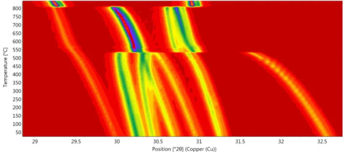
Ettringite at high pressure: structure evolution and elastic behaviour 12:40-13:00 O3 - Elisa Rodeghero
Temperature-induced phase transitions in natural metavariscite
13:00-14:30
Lunch & Poster Session 1 (MS1-MS4 - Part 3)
14:30-16:30
MS3 & MS4 - Parallel sessions
MS3 - Coordination compounds to coordination materials
- Aula MagnaChairs: Victor Antonio de la Peña O’Shea, Simona Galli
14:30-15:00 KN1 - Ana E. Platero-Prats (Autonoma Univ. of Madrid) Chasing local structures in nanoporous materials using advanced synchrotron X-ray scattering tools
15:00-15:30 KN2 - Volodymyr Bon (Technical Univ. of Dresden)
Development of in situ diffraction techniques for characterization of nanoporous materials during adsorption of gases and vapours
Oral Presentations
15:30-15:45 O1 - Celia Castillo-Blas
Metal-cation arrangement control in secondary building units of metal-organic frameworks and their translation to oxides
15:45 -16:00 O2 - Rebecca Vismara
Mixing bis(pyrazolate) ligands in Zn(II) MOFs for CO2 adsorption, affinity and selectivity optimization: a structural and textural study
16:00-16:15 O3 - Inés Ruiz Salcedo
Proton Conductivity in Sulfate-Containing Lanthanide Nitrilotris-methylphosphonates 16:15-16:30 O4 - Marco Milanesio
Synthesis and structural characterization of a Ce-containing AZO-MOF, showing a complex polymorph landascape
MS4 - Crystal growth: from fundamentals to applications in
nature and technology
- Aula AChairs: Simona Fermani, Jaime Gómez Morales
14:30-15:00 KN1 - Rocco Caliandro (IC-CNR Bari)
Protein crystallization in hydrogel composite membranes and deep eutectic solvents 15:00-15:30 KN2 - Ylenia Jabalera (Univ. de Granada)
8 Oral Presentations
15:30-15:45 O1 - Daniele Moscheni
Structure and morphology of CdSe and CdSe@CdS colloidal quantum qots from advanced X-ray scattering techniques
15:45 -16:00 O2 - Jose A. Gavira
Use and overuse of reinforced Cross-Linked Enzyme Crystals 16:00-16:15 O3 - Stefano Canossa
Ultra-stable MOFs across the microscale: novel approach for the size control of Al-frameworks
16:15-16:30 O4 - Jaime Gómez-Morales
Induced nucleation of nanocrystalline apatite on graphene nanoflakes by sitting drop vapor diffusion
16:30-17:00
Coffee Break
17:00-18:30
Poster Session 2 (MS5-MS8 Part 1)
18:30-19:15
Herculaneum Ruins: between Science and Tourism
Chair: Rita Berisio18:30-18:55 Vito Mocella, (IMM-CNR Napoli)
The quest of lost ancient literature. Synchrotron based techniques reveal the secrets of Herculaneum papyri: letters and ink composition.
18:55-19:15 Lomè Galliano (Professional Tour Guide Campania Region) Presentation of the Herculaneum Ruins
Friday, September 6
th8:50-10:00
Plenary Lecture 3
Chairs: Giuseppe Zanotti and Anna Marabotti
Martin Weik (Institut de Biologie Structurale, Grenoble) Watching proteins at work by kinetic X-ray crystallography
(The lecture will be preceded by a short talk in memory of Guido Capitani by Giuseppe Zanotti)
10:00-10:30
Coffee Break
10:30-11:00
Commercial Presentations
10:30-10:45 Malvern Panalytical by Eugenio Casini 10:45-11:00 Thermofisher by Max Maletta
11:00-13:00
MS5 & MS6 - Parallel sessions
MS5 - Bio-crystallography: from Structure to Function
- Aula MagnaChairs: Adele Di Matteo, Lourdes Infantes
11.00-11:30 KN1 - Claudia Binda (Univ. di Pavia)
Structural studies on flavoenzymes at the cross-road of drug design and cell biology 11:30-12:00 KN2 - Marian Oliva (CSIC, Madrid)
9 Oral Presentations
12:00-12:15 O1 - Giusy Tassone
Structural characterization of a synthetic mimic of the Photoactive Yellow Protein 12:15-12:30 O2 - Simona Fermani
The last gap in the structural proteome of the photosynthetic CO2 assimilation has been
filled
12:30-12:45 O3 - Mario Milani
Structural and functional characterization of TgpA, a critical protein for the viability of
Pseudomonas aeruginosa
12:45-13:00 O4 - Marta Ferraroni
Dioxygen, an unexpected Carbonic anhydrase ligand
MS6 - Mineral structures and mineral characterization - Aula A
Chairs: Fernando Camara, Laura Leon-Reina11.00-11:30 KN1 - Cristian Biagioni (Univ. di Pisa)
Advances in mineral crystal-chemistry and the role played by rare minerals Oral Presentations
11:30-11:45 O1 - Carlotta Giacobbe
When diffraction meets tomography: the ferrierite case for the improvement of mineral fibers crystallographic characterization
11:45-12:00 O2 - Matteo Ardit
Vanadium-induced color in calcium aluminates: grossite (CaAl4O7) and hibonite (CaAl12O19)
12:00-12:15 O3 - Gioacchino Tempesta
Crystal-chemical characterization of red beryl by standarless Laser Induced Breakdown Spectroscopy and Single Crystal X-Ray Diffraction: an example of validation of an innovative method for the chemical analyses of minerals
12:15-12:30 O4 - Michelangelo Polisi
Amino acids confinement and condensation in mordenite channels 12:30-12:45 O5 - Donato Belmonte
Thermodynamics, elasticity and phase stability of grossite (CaAl4O7) at high pressure and temperature
12:45-13:00 O6 - Eugenio Casini
Crystal structure and thermodynamic behavior of Bi6Te2O15: the likely structure of the rare mineral pingguite
13:00-14:00
Lunch & Poster Session 2 (MS5-MS8 Part 2)
14:00-16:00
MS7 & MS8 - Parallel sessions
MS7 - Crystal forms in Pharma - Aula A
Chairs: Duane Choquesillo-Lazarte, Paola Paoli14.00-14:30 KN1 - Jordi Benet-Buchholz (ICIQ, Tarragona)
Pharmaceutical Co-Crystals: New developments using the ‘in situ’ crystallization with a CO2-laser
14:30-15:00 KN2 - Leonardo Lo Presti (Univ. di Milano)
A crystallographic route to understand drug solubility: the case of 4-aminoquinoline antimalarials
10 Oral Presentations
15:00-15:15 O1 - Andrea Ienco
Computational Investigation on Metoprolol Anisotropic Lattice Expansion 15:15-15:30 O2 - Oleksii Shemchuk
Ionic cocrystals of the molecules of pharmaceutical interest 15:30-15:45 O3 - Lourdes Infantes
Search for multicomponent structures of Nevirapine to improve its physicochemical properties
15:45-16:00 O4 - Claudia Carraro
Designing new Cocrystals based on Essential Oil for agricultural applications
MS8 - Contemporary challenges in Structural Biology -
Aula Magna Chairs: Armando Albert, Martino Bolognesi14.00-14:30 KN1 - Juan A. Hermoso (CSIC, Madrid)
The structure of full-length human phenylalanine hydroxylase in complex with tetrahydrobiopterin
14:30-15:00 KN2 - Beatrice Vallone (Sapienza Univ. di Roma)
Key interactions for iron uptake are exploited by infectious agents as revealed by the cryo-EM structure of the Ferritin-CD71 complex
Oral Presentations
15:00-15:18 O1 - Dritan Siliqi
Shwachman-Diamond Syndrome: SBDS and EFL1 conformational characterization through Small Angle X-ray Scattering and Molecular Dynamics Simulations
15:18-15:36 O2 - Marco Mazzorana
Synergies within Life Science at Diamond: towards the future of structural biology 15:36-15:54 O3 - Daniela Caruso
Structural characterization of the aggregates formed by the members of the GADD45 family
15:54-16:00 Digital Posters
16:00-16:30
Coffee Break
16:30-18:00
General Assemblies (in parallel)
18:00-19:00
Meetings of the subgroups
19:00-19:30
Closing Ceremony
20:30
Social Dinner - Ristorante La Scialuppa
Borgo Marinari, Piazzetta Marinari 6, NapoliSaturday, September 7
th11
PL1: Crystallography in Art and Cultural Heritage
Giacomo Chiari
Gia' Chief Scientist - Getty conservation Institute – Los Angeles - California
Many applications to archaeometry and to diagnostics in the preservation of cultural heritage are based on crystallography. A deep understanding of the material is fundamental for both disciplines. Recently, noninvasive portable instruments have become common, due to (or generating) the present modus operandi that excludes the possibility of taking samples. Because of this, the imaging of various type has become fundamental. These are either based on radiation familiar to crystallographers (X-rays) or based on properties elucidated by them.
This lecture will showcase a series of crystallographic applications in the field of conservation science, from Maya Blue structure (synchrotron and neutron powder diffraction) to Tutankhamen tomb paintings, from Michelangelo’s censure panels in the Sistine chapel to a hidden face under a Rembrandt painting. The most recent tool (XRF scanners) will be compared to a procedure based on a much smaller number of measurements producing very promising results.
I hope that I will be able to highlight the unique contribution that crystallography can make to the widening field of Conservation Science in the preservation of our ever more challenging cultural heritage.
12
Enrique EspinosaLaboratoire de Cristallographie, Résonance Magnétique et Modélisations (CRM2), UMR CNRS 7036, Université de Lorraine, Nancy, France. [email protected]
Through the Hellmann-Feynman and Hohenberg-Kohn theorems,1,2 the electron density distribution (r) can be considered the conceptual bridge between the structure and the energetic properties of molecular systems. Accordingly, (r) codes essential features of molecular organization in space. Important aspects such as intensity, nature, and directionality of intermolecular interactions are at the origin of molecular assembling, and therefore of supramolecular structures with important applications in the fields of Crystal Engineering, Supramolecular Chemistry and Material Science. These aspects are not only implicitly coded on
(r) but also in its derived properties, as the Laplacian function 2(r) and the electrostatic potential (r). These three scalar functions bring complementary information to study the assembling of molecules because each of them focuses on a different electronic aspect of the molecular system. Thus, whereas (r) reflects the interatomic and intermolecular interactions taking place in the system, 2(r) and (r) point respectively the regions of the space where
(r) is locally concentrated (2(r) < 0) or depleted (2
(r) > 0) and the molecular electrophilic ((r) > 0) and nucleophilic ((r) < 0) regions.
The topological analysis of any scalar function, which is the analysis of its derivatives, provides richer information than appears from the direct observation of the scalar function itself. In the widely used QTAIM theory,3 this kind of analysis has been deeply developed for (r) to characterize many aspects that this function exhibits in atoms, molecules, and their interactions. In our work, topological developments on the (r), 2(r) and (r) scalar functions have permitted the analysis of (i) the intensity4,5 and nature6,7 of intermolecular interactions, (ii) the geometrical preferences found on molecular assembling8,9 and (iii) the influence zones of molecular electrophilic and nucleophilic sites that are at the origin of molecular recognition and molecular assembling, in particular when dealing with the paradoxical cases of anionic or cationic aggregates.10,11
[1] H. Hellmann Einführung in die Quantenchemie. Leipzig, Vienna: Deuticke, 1937.
[2] R. P. Feynman Phys. Rev., 1939, 56, 340; P. Hohenberg, W. Kohn Phys. Rev., 1964, 136, 864. [3] R. F. W. Bader, Atoms in Molecules: a Quantum Theory, Clarendon, Oxford, 1990.
[4] E. Espinosa, E. Molins, C. Lecomte Chem. Phys. Lett., 1998, 285, 170. [5] E. Espinosa, I. Alkorta, J. Elguero, E. Molins J. Chem. Phys., 2002, 117, 5529.
[6] O. Makhotkina, J. Lieffrig, O. Jeannin, M. Fourmigué, E. Aubert, E. Espinosa Cryst.Growth Des.,
2015, 15, 3464.
[7] K. Lamberts, P. Handels, U. Englert, E. Aubert, E. Espinosa CrystEngComm, 2016, 18, 3832.
[8] M. E. Brezgunova, E. Aubert, S. Dahaoui, P. Fertey, S. Lebègue, C. Jelsch, J. G. Ángyán, E. Espinosa
Cryst. Growth Des., 2012, 12, 5373.
[9] M. E. Brezgunova, J. Lieffrig, E. Aubert, S. Dahaoui, P. Fertey, S. Lebègue, J. G. Ángyán, E. Espinosa
Cryst. Growth Des., 2013, 13, 3283.
[10] I. Mata, E. Molins, I. Alkorta, E. Espinosa J. Phys. Chem. A, 2015, 119, 183. [11] I. Alkorta, I. Mata, E. Molins, E. Espinosa Chem. Eur. J, 2016, 22, 9226.
13
Institut de Biologie Structurale, Grenoble, France. [email protected]
Kinetic X-ray crystallography permits the structural characterization of macromolecular conformational changes along a reaction pathway at the atomic level of spatial resolution. After triggering the biological reaction within a macromolecular crystal, functionally relevant conformational changes are either arrested by flash-cooling the crystal, allowing characterization of the structure by conventional cryo-crystallography (intermediate trapping), or followed in real time by time-resolved crystallography at room temperature. The temporal resolution of the latter is limited to 100 ps if carried out in the form of Laue crystallography at synchrotrons. The advent of X-ray free electron lasers (XFELs) has pushed the resolution to the sub-ps regime, allowing ultrafast changes to be studied by time-resolved serial femtosecond crystallography [1].
After reviewing the current status of the field, we will illustrate the time-resolved crystallography approach with the study of ultra-fast photoswitching in a fluorescent protein from a non-fluorescent (off) to a fluorescent (on) state further to excitation by a light flash [2]. Our consortium (see author list of [2]) has employed time-resolved serial femtosecond crystallography at an XFEL to identify the transient structure of the photoswitchable fluorescent protein rsEGFP2 in its excited state, and to observe its chromophore in a twisted conformation, midway between the stable configurations of the on and off states. This observation has been confirmed by simulations and has allowed us to rationally design a mutant with a two-fold increased photoswitching quantum yield.
[1] Colletier J-P, Schiro G., Weik. M., Time-resolved serial femtosecond crystallography, towards molecular movies of biomolecules in action. X-ray Free Electron Lasers : A Revolution in Structural
Biology., Ed. Petra Fromme, Sebastien Boutet, Mark Hunter. Springer International Publishing. 2018 pp
331 - 356
[2] Coquelle N, Sliwa M, Woodhouse J, Schirò G, Adam V, Aquila A, Barends TRM, Boutet S, Byrdin M, Carbajo S, De la Mora E, Doak RB, Feliks M, Fieschi F, Foucar L, Guillon V, Hilpert M, Hunter MS, Jakobs S, Koglin JE, Kovacsova G, Lane TJ, Lévy B, Liang M, Nass K, Ridard J, Robinson JS, Roome CM, Ruckebusch C, Seaberg M, Thepaut M, Cammarata M, Demachy I, Field M, Shoeman RL, Bourgeois D, Colletier J-P, Schlichting I, Weik M. Chromophore twisting in the excited state of a photoswitchable fluorescent protein captured by time-resolved serial femtosecond crystallography.
14
MISCA MEDAL
ABOUT CRYSTALS AND THEIR PROPERTIES
Angeles Monge
Instituto de Ciencia de Materiales. Consejo Superior de Investigaciones Científicas (ICMM-CSIC) Sor Juana Ines de la Cruz, Madrid Spain . [email protected]
In this talk, Angeles Monge will show some of her contributions to the main research fields in which she has been involved, followed by recent results from her present investigation in the field of reticular chemistry: Enhancement of properties through MOFs structural design (Figure 1): materials discovery, theoretical calculations, chemical activity, and physical properties.
Figure 1. Is it possible to code and transfer selected metal ratios from MOFs to multimetal oxides with
novel, desired compositions through a simple calcination process. [1,2]
[1] Castillo-Blas, C.; de la Peña-O’Shea, V. A.; Puente-Orench, I.; de Paz, J. R.; Sáez-Puche, R.; Gutiérrez-Puebla, E.; Gándara, F.; A. Monge,. Science Advances 2017
[2] Celia Castillo-Blas, Nieves López-Salas, María C. Gutiérrez, Inés Puente-Orench, Enrique Gutiérrez-Puebla, M. Luisa Ferrer, M. Ángeles Monge, Felipe Gándara, J.Am. Chem. Soc. 2019, 141, 1766−1774.
15
MAMMI AWARD
Development and application of crystallographic methodologies
for powder diffraction
Angela Altomarea a
CNR-IC, Bari, Italy. [email protected]
Crystal structure solution by powder diffraction data, although limited to small molecules, is an attractive and still challenging topic. The software EXPO [1] occupies a relevant role in this field. Among its distinguishing features we mention: based on sound theoretical principles; supported by innovative methodologies; developed according to the most recent and advanced programming languages; mainly automatic; continuously improved in terms of computational and graphic performances.
EXPO is able to face arduous problems, with the aim of pushing more and more the limits of the structure solution process by powder.
The presentation will be mainly focused on some highlights of EXPO: the determination of crystal cell parameters of small proteins; the successful application of a clever procedure, based on a random approach, for extracting the structure factor moduli from the experimental diffraction pattern [2]; the crucial action of the structure model optimization step able to provide the correct solution starting from the model derived by Direct Methods, even approximate and very far from the true one [3]; the strategies, both methodological and computational, for attaining the global minimum and gaining execution time in the solution process by Simulated Annealing [4]; the effective combination of solving structure by both reciprocal and direct space method.
Examples of solution will be discussed by covering a quite large range of cases in terms of experimental conditions and structure complexity. The capacity of the current version of EXPO to increase the number of successful outcomes and expand the limits of feasibility of powder diffraction for solving crystal structures will be proved.
A quick presentation of QUALX [5] and OChemDb [6] software will be also given for pointing out the benefit of their use in the process of deep investigation of a polycrystalline material.
[1] A. Altomare, C. Cuocci, C. Giacovazzo, A. Moliterni, R. Rizzi, N. Corriero, A. Falcicchio J. Appl.
Cryst. 2013, 46, 1231.
[2]A. Altomare, C. Giacovazzo, A.G.G Moliterni, R. Rizzi J. Appl. Cryst. 2001, 34, 704.
[3] A. Altomare, C. Cuocci, A. Moliterni, R. Rizzi, N. Corriero, A. Falcicchio J. Appl. Cryst. 2017, 50, 1812.
[4] A. Altomare, N. Corriero, C. Cuocci, A. Falcicchio, A. Moliterni, R. Rizzi J. Appl. Cryst. 2018, 51, 505.
[5] A. Altomare, N. Corriero, C. Cuocci, A. Falcicchio, A. Moliterni, R. Rizzi J. Appl. Cryst. 2015, 48, 598.
[6] A. Altomare, N. Corriero, C. Cuocci, A. Falcicchio, A. Moliterni, R. Rizzi J. Appl. Cryst. 2018, 51, 1229.
16
NARDELLI AWARD
Total Scattering Methods in Reciprocal Space: Developing
Crystallographic Tools for Nanostructured and Disordered Materials
Federica Bertolotti
Dipartimento di Scienza e Alta Tecnologia and To.Sca.Lab., Università dell’Insubria, Como, Italy [email protected]
Nanocrystals (NCs) represent an innovative class of materials showing highly designable and tunable properties. Their size/shape dependent physico-chemical properties provide the opportunity to develop innovative functional materials for an extremely vast range of applications.[1] In spite of this exciting scenario, an exhaustive characterization of NCs is still a challenging task.
In this field, electron microscopy techniques, i.e. transmission electron microscopy (TEM), are often employed to get information of particles’ size, shape, morphology and aspect ratio. However, these methods can provide only 2D, local information on selected particles that might not be representative of the ensemble.[2]
On the other hand, conventional powder diffraction methods based on the Rietveld-like approach are able to provide structural information on the entire volume of sample irradiated by X-ray, but they are rather inadequate in the proper characterization of NCs. Indeed, for NCs, peaks broadening in the diffraction profile is the result of the coexistence of different effects, such as reduced crystalline domains, peculiar (eventually anisotropic) morphologies, surface induced lattice strains and other type of structural or compositional defects (i.e. point or planar defects, such as dislocation or stacking faults).[3]
Total scattering (TS) methods, in particular the Debye Scattering Equation (DSE)[4] based approach treats both Bragg and diffuse scattering on equal basis and allows the modeling of the TS pattern of NCs without any assumption of periodicity and order. Since the computation operates starting from real space atomistic models, structural and microstructural information can be simultaneously derived within a unified approach.[3] The modeling of the TS pattern is then performed in reciprocal space, avoiding all the issues related to the Fourier transform of the experimental data (i.e. Fourier transform truncation errors and very poor angular resolution associated to the experimental requirements).
Moreover, the possibility of modeling both the Small- and Wide-angle regions of the TS pattern, with the DSE method, taking advantage of their complementarity in terms of length scales (from the atomic to the nanometer one), offers the opportunity of detecting sophisticate nanocrystals morphologies and disentangling defects-induced peaks broadening, from the size-induced effects.
Accordingly, this method has been successfully extensively applied to a wide-range of nanomaterials, characterized by different kind of structural distortions and defectiveness.[5]
Here, experimental and modeling aspects related to the DSE approach, along with an overview of applications, from nanosized colloidal quantum dots and perovskites up to innovative engineered nanoapatites, will be presented.
Acknowledgement. Part of this work was supported by Fondazione Cariplo, HYPATIA Project: 2016-0248.
[1] Carey, G. H.; Abdelhady, A. L.; Ning, Z.; Thon, S. M.; Bakr, O. M.; Sargent, E. H. Colloidal Quantum Dot Solar Cells. Chem. Rev. 2015, 115, 12732–12763.
[2] Liu, J.; Olds, D.; Peng, R.; Yu, L.; Foo, G. S.; Qian, S.; Keum, J.; Guiton, B. S.; Wu, Z.; Page, K., Chem. Mater.
2017, 29, 5591-5604.
[3] Bertolotti, F.; Moscheni, D.; Guagliardi, A.; Masciocchi, N. Eur J Inorg Chem 2018, 3789-3803
[4] a) Debye, P., Ann. Phys. 1915, 351, (6), 809-823; Cervellino, A.; b) Frison, R.; Bertolotti, F.; Guagliardi, J. Appl.
Cryst. 2015, 48, 2026-2032.
[5] a) Moscheni, D.; Bertolotti, F.; Piveteau, L.; Protesescu, L.; Dirin, D. N.; Kovalenko, M. V.; Cervellino, A.; Pedersen, J. S.; Masciocchi, N.; Guagliardi, A. ACS Nano 2018, 12, 12558–12570; b) Bertolotti, F.; Protesescu, L.; Kovalenko, M. V.; Yakunin, S.; Cervellino, A.; Billinge, S. J. L.; Terban, M. W.; Pedersen, J. S.; Masciocchi, N.; Guagliardi, ACS Nano 2017, 11, 3819–3831; c) Bertolotti, F.; Dirin, D. N.; Ibanez, M.; Krumeich, F.; Cervellino, A.; Frison, R.; Voznyy, O.; Sargent, E. H.; Kovalenko, M. V.; Guagliardi, A.; and Masciocchi, N. Nat Mater 2016, 15, 987–94; d) Bertolotti, F.; Moscheni, D.; Migliori, A.; Zacchini, S.; Cervellino, A.; Guagliardi, A.; Masciocchi, N. Acta
17
XAVIER SOLANS PRIZE
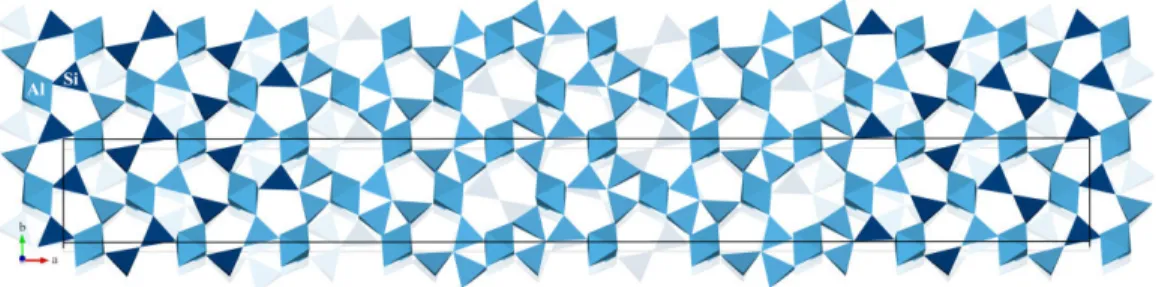
Ordering patterns in mullite
Paul B. Klar,a Iñigo Etxebarriab, Gotzon Madariagac a
Institute of Physics of the Czech Academy of Sciences, Prague, Czech Republic
b
Departamento de Física Aplicada II, Universidad del País Vasco UPV/EHU, Spain
b
Departamento de Física de la Materia Condensada, Universidad del País Vasco UPV/EHU, Spain [email protected]
Mullite (Al4+2xSi2–2xO10–x) is a well-known material with numerous high-tech applications in
different branches including the aerospace industry. Although mullite was thoroughly investigated for decades, structural research on mullite could not establish a model that involved a fundamental understanding of the ordering phenomena in mullite, namely the distribution of oxygen vacancies and Al/Si ordering. Therefore, recent computational simulations, performed to understand the properties and applications of mullite, usually assume a random distribution of vacancies and Al/Si [1]. The objective of this presentation is to explain the fundamental ordering mechanisms in mullite, based on a combination of synchrotron single crystal diffraction experiments and first-principles calculations.
Diffraction patterns of mullite reveal a characteristic diffuse scattering alongside a set of satellite reflections. The latter requires using higher-dimensional superspace crystallography for a complete structural description. Superspace model refinements confirm that mullite is mainly disordered and the modulation is caused by a specific distribution pattern of oxygen vacancies, related to Al/Si ordering and the arrangement of tetrahedra around vacancies. However, the refinements indicate that the identified ordering patterns are present to different extents, i.e. different degrees of order exist [2]. From that, and in accordance with superspace symmetry, a completely ordered model was established, suitable for different kinds of atomistic simulations. Refined modulation functions from disordered models and from DFT calculations on ordered commensurate superstructures are in very good agreement [3]. The underlying ordering patterns establish a structural model that serves as a basis for future investigations of the properties and applications of mullite.
Figure 1. Superstructure of mullite Al4.8Si1.2O9.6 (x = 0.4) with Al/Si and vacancy ordering pattern.
[1] Schneider, H. , Fischer, R. X. and Schreuer, J. J. Am. Ceram. Soc. 2015, 98, 2948–2967 [2] Klar, P. B., Etxebarria, I. & Madariaga, G. IUCrJ. 2018, 5, 497–509.
18
BEST PhD THESIS AWARD
HIGH-PRESSURE BEHAVIOR OF MICROPOROUS MATERIALS:
CRYSTAL-FLUID INTERACTIONS AND DEFORMATION
MECHANISMS AT THE ATOMIC SCALE
DAVIDE COMBONI
Dipartimento di scienze della terra "Ardito Desio", Università degli Studi di Milano
Zeolite are crystalline, hydrated aluminosilicates characterized by a tetrahedral framework of TO4 units, connected in such a way that sub-nanometric channels and cages occur. These
structural cavities host the so-called extra-framework population, which mainly consists of alkali and alkaline-earth cations and small molecules, such as H2O
1,2
. In the last decades, the scientific community showed a rising interest on the behavior of microporous and mesoporous compounds at high-pressure conditions, and on the crystal-fluid interaction phenomena occurring at extreme conditions3,4. High-pressure experiments on synthetic zeolites may pave the way for new routes of tailoring new functional materials (made by hybrid host-guest architecture), bearing a potentially relevant technological impact5. From a geological point of view, zeolites could act as a carrier for H2O and others small molecules or for monoatomic
species (e.g., CO2, CH4, H2S, He, Ar, Kr, Xe,…), and any change of the P-induced
extra-framework population (at ambient to low/high T) can have relevant geochemical and geophysical implications6. In this experimental thesis, the high-pressure behavior and the crystal-fluid interaction at the atomic scale of a selected series of natural and synthetic zeolites (i.e., AlPO4-5, leonhardite, laumontite, phillipsite) and a zeolites-like mineral (i.e.,
armstrongite) have been investigated by means of in-situ single-crystal X-ray diffraction, using “penetrating” and “non-penetrating” pressure-transmitting fluids in a diamond anvil-cell, exploring the different crystal-fluid interaction in materials with large or small channels, and with or without extra-framework population. The experiments were conducted at the Earth Science Department of Milan (ESD-MI) and at the beamlines ID15b (at ESRF, Grenoble) and P2.02 (at DESY, Hamburg). The results obtained in the framework of this experimental thesis showed that several variables govern the sorption phenomena at high pressure, among those: the “free diameters” of the framework cavities, the chemical nature and the configuration of the extra-framework population, the partial pressure of the penetrating molecule in the fluid (if mixed with other non-penetrating molecules), the rate of P-increase, the surface/volume ratio of the crystallites and the temperature at which the experiment is conducted. P-induced phenomena at the atomic scale were described on the basis of high-quality structure refinements. Geological and technological potential implications were discussed, e.g. about the H2O load carried by laumontite, leonhardite and phillipsite in relevant geologic environments
(e.g., first km of the subducted oceanic crust, oil reservoir, …), along with the potential of synthetic and natural zeolites as energy storage materials.
1
Baerlocher, C., Mccusker, L.B., and Olson, D.H. 2007, Atlas of Zeolite frAmework types.
2R.T. Pabalan and F.P. Bertetti. Rev. Mineral. Geochem. 2001, 45, 453–518. 3G.D. Gatta and Y. Lee Mineral. Mag. 2014, 78, 267–291.
4
G.D. Gatta, P. Lotti and G. Tabacchi. Phys. Chem. Miner. 2018 , 45, 115–138.
5
V. Eroshenko, R. Regis, M. Soulard, J. Patarin, J. Cr. Phys. 2002, 3, 111–119.
6
19
BEST PhD THESIS AWARD
Biochemical and structural studies on urease inhibition,
a nickel-dependent virulence factor
Luca Mazzei,a Michele Cianci,b Francesco Musiani,a Stefano Benini,c Marta Palombo,a Gábor Lente,d Stefano Ciurli a
a
Dept. of Pharmacy and Biotechnology, University of Bologna (Italy)
b
Dept. of Agricultural, Food and Environmental Sciences, Polytechnic University of Marche (Italy)
c
Bioorganic Chemistry and Bio-Crystallography Laboratory, Faculty of Science and Technology, Free University of Bolzano (Italy)
d
Dept. of General and Physical Chemistry, University of Pécs (Hungary) [email protected]
Urease is a nickel-dependent enzyme that catalyzes the rapid hydrolysis of urea, determining an overall increase of pH and causing negative consequences for human health as well as agriculture [1]. For these reasons, the scientific community has devoted intense efforts in the last several decades for the development of efficient and specific inhibitors of urease able to counteract its negative effects [2].
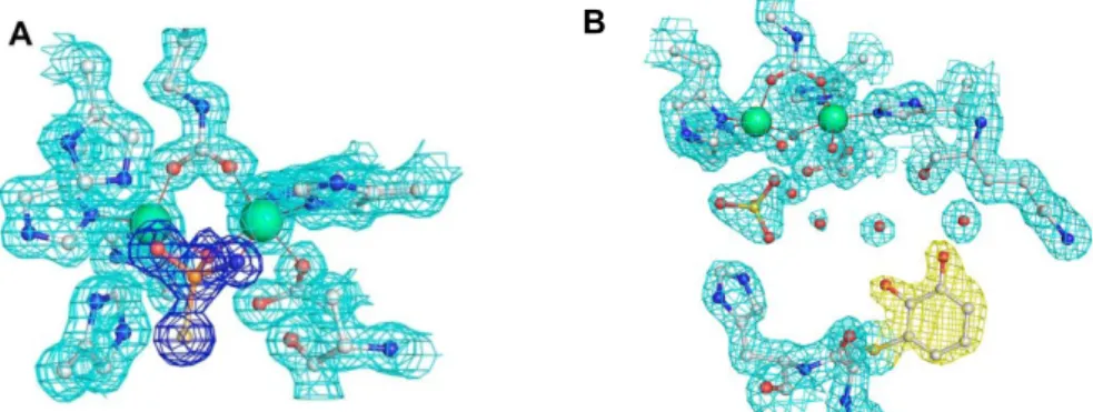
In this work, X-ray protein crystallography has been applied to urease to determine the inhibition mode of several urease inhibitors: i) fluoride [3], and ii) N-butylthiophosphotriamide (NBPT) [4] directly bind to the di-nuclear metallic center in the active site. In particular, NBPT, a commercial nitrogen stabilizer extensively used in agriculture, acts as a slow-binding inhibitor of urease, undergoing an in situ hydrolysis that generates a tetrahedral moiety blocking the active site and precluding the enzyme from further substrate hydrolysis. Two other molecules, iii) benzoquinone [5] and iv) catechol [6] have been structurally proved to act as urease inhibitors by blocking the mobile flap covering the active site of the enzyme in an open state. All the results shown in this work will be useful to develop, through a structure-based drug design procedure, novel and more efficient urease inhibitors.
Figure 1. Atomic models of the active site of urease inhibited by the hydrolyzed form of NBPT (A) and
bound to catechol (B), superimposed on the final 2Fo-Fc electron density maps.
[1] M. J. Maroney, S. Ciurli, Chem. Rev. 2014, 114, 4206.
[2] L. Mazzei, F. Musiani, S. Ciurli, in The Biological Chemistry of Nickel, RSC. 2017. [3] S. Benini, M. Cianci, L. Mazzei, S. Ciurli, J. Biol. Inorg. Chem. 2014, 19, 1243. [4] L. Mazzei, M. Cianci, U. Contaldo, F. Musiani, S. Ciurli, Biochemistry 2017, 56, 5391. [5] L. Mazzei, M. Cianci, F. Musiani, S. Ciurli, Dalton Trans. 2016, 45, 5455.
[6] L. Mazzei, M. Cianci, F. Musiani, G. Lente, M. Palombo, S. Ciurli, J. Inorg. Biochem. 2017, 166, 182. B
20
BEST PhD THESIS AWARD
Electronic properties and chemical bonding in tetrazolate based
coordination polymers
S. Racioppiabc, V. Colombob, A. Sironib, M. Andrzejewskic, R. Scatenac, P. Macchicd a
Chalmers University of Technology, Department of Chemistry and Chemical engineering, Göteborg, Sweden.
b
University of Milan, Department of Chemistry, Milan, Italy
c
University of Bern, Department of Chemistry and Biochemistry, Bern, Switzerland
d
Polytechnic of Milan, Department of Chemistry Materials and Chemical Engineering, Milan, Italy [email protected]
Materials having dielectric constant lower than silicon dioxide (≈ 4) as interlayer insulators are nowadays necessary in modern microelectronic devices to guarantee high performance and low power consumption.[1] In order to reach lower and lower dielectric constants, the research moved to porous materials, particularly to Metal Organic-Frameworks (MOFs).[2] In this context, Metal Azolate Frameworks (MAFs)[3] are chemically and thermally more stable then classic systems based on carboxylic ligands, and so, suitable for this kind of applications. In order to investigate the performances of MAFs as low dielectric constant materials, we studied CuL∙2EtOH (L=5-(4-pyridyl)tetrazolate)[4]
, a 2-dimensional system. Susceptibility and dielectric constant, of both the solvated and evacuated systems, were measured with impedance spectroscopy on pellets and computed using Crystal17 code[5]. While high resolution x-ray diffraction experiments and Hansen&Coppens multipolar models[6] were used to rationalize the interactions in this material. Impedance spectroscopy analysis demonstrated that, CuL has an extremely low value of static dielectric constant, equal to 1.90 at 105 Hz. It was proved that the synergic combination of preferential orientation with the anisotropy of the dielectric tensor promoted the insulator character of this material when prepared as a pellet.
Further, the decoration of L with one fluorine atom (LF=5-(2-fluoro-4-pyridyl)tetrazolate) produced two new isostructural coordination polymer with coinage metal cations: AgLF and CuLF. Chemical behavior under pressure of both not-porous (AgLF, CuLF) and porous (CuL∙2EtOH) systems was investigated. In spite of being isostructural at ambient conditions, Cu(I) and Ag(I) coordination polymers behaved differently at high pressure. Indeed, an opposite attitude for metallophilic interactions was revealed and confirmed by topological analysis[7]. On the other hand, negative linear compressibility (NLC) was observed for CuL∙2EtOH, due to interactions between layers induced by compression, with a consequently increment of the network’s dimensionality.
[1] W. Volksen, R. D. Miller, G. Dubois, Chem. Rev. 2010, 110, 56–110. [2] M. Usman, S. Mendiratta, K. L. Lu, ChemElectroChem 2015, 2, 786–788.
[3] J. P. Zhang, Y. B. Zhang, J. Bin Lin, X. M. Chen, Chem. Rev. 2012, 112, 1001–1033.
[4] F. Wang, R. Yu, Q. S. Zhang, Z. G. Zhao, X. Y. Wu, Y. M. Xie, L. Qin, S. C. Chen, C. Z. Lu, J. Solid
State Chem. 2009, 182, 2555–2559.
[5] R. Dovesi, V. R. Saunders, C. Roetti, R. Orlando, C. M. Zicovich-Wilson, F. Pascale, B. Civalleri, K. Doll, N. M. Harrison, I. J. Bush, et al., 2018.
[6] P. Coppens, X-Ray Charge Densities and Chemical Bonding, Oxford University Press, New York,
1997.
21
FELLOWSHIP FOR ABROAD RESEARCH
NSD protein family: a new promising target for
epigenetic therapy of tumor diseases
Benny Danilo Belviso,a Yunpeng Shen,b Benedetta Carrozzini, a Masayo Morishita, b Eric di Luccio,b RoccoCaliandroa
a
Istituto di Cristallografia, Consiglio Nazionale delle Ricerche, Bari, Italy
b
Department of genetic engineering, Kyungpook National University, Daegu, South Korea [email protected]
Epigenetics is considered a powerful weapon to fight cancer diseases, especially against the ones with poor prognostics and still incurable, such as multiple myeloma.[1] Differently from traditional chemioterapy, this therapeitic strategy is reversible, thus offering the opportunity for cancer cells to get back to physiological state. This is why proteins involved in epigenetics are today considered promising targets for cancer treatment. Among these proteins, nuclear receptor binding SET domain (NSD) proteins, a family of three histon lysine methyl transferases, are considered decisive for suppressing currently lethal cancer diseases.[2] The Institute of Crystallography and the Kyungpook National University are working together to provide structural information for NSDs, as a first step of a structure-based drug design of NSD inhibitors. By combaining Small-Angle X-ray Scattering (SAXS) and homology modeling, we obtained the first structural details regarding NSDs and new insights about NSDs stability and conformation. Based on this structural knowledge, peptido-mimetic potential NSD inhibitors have been selected by virtual screening. Moreover, an optimized protocol to express NSDs fused with T4L crystallization-chaperon has been succesfully optimized with the aim to increase crystallization chances of these crystallization-recalictrant family of proteins. Finally, the Schizosaccharomyces pombe Set7 protein, belonging to the histone-methyltransferase (HMT) family, has been characterized by X-diffraction at 2 Å resolution (PDB code 5h6z, Fig. 1).[3] Altogether, these results represent an important breakthrough in the structural study of this family of protein and pave the way for the development of NSD inhibitors.
Figure 1. Schizosaccharomyces pombe Set7 protein crystal structure
obtained at 2Å resolution (PDB code 5h6z) [1] M. Fardi, S. Solali, M.F. Haghe,Genes Dis. 2018, 5(4), 304
[2] M. Morishita, E. di Luccio, Biochim Biophys Acta, 2011, 1816(2), 158
[3] Y. Shen, D.E.H.F. Mevius, R. Caliandro, B. Carrozzini, Y. Roh, J. Kim, S. Kim, S.C. Ha, M. Morishita, E. di Luccio, Structure 2019, 27, 631
22
BEST GRADUATE THESIS AWARD
Engineering murine neuroglobin: structural and functional analysis of the
heme sliding mechanism
Ida Fredaa,b, Cécile Exertiera,b Linda Celeste Montemiglioa,b,c, Antonella Scaglionea,b,e, Gabriele Ceruttia, Giacomo Parisia,b,d, Carmelinda Savinoc, Beatrice Vallonea,b,c
aDipartimento di Scienze Biochimiche “A. Rossi Fanelli”, Università di Roma “La Sapienza”, Rome, Italy b
Istituto Pasteur - Fondazione Cenci Bolognetti, Rome, Italy,
c
Istituto di Biologia e Patologia Molecolare - CNR, Rome, Italy.
d
Department of Physiology and Cellular Biophysics, Columbia University, New York, NY, USA
e
Neuroscience Initiative, CUNY Advanced Science Research Center, New York, NY, USA. [email protected]
Neuroglobin (Ngb) is a 17 kDa heme protein, predominantly expressed in the central and peripheral nervous system. It clearly seems to be involved in neuroprotection, most likely through a role in gas/redox sensing and radical scavenging, although its mechanism of action is still not fully clarified. Even though this protein displays the canonical globin fold, it only shares less than 21% of sequence identity with myoglobin [1]. In the absence of exogenous ligands, the heme iron atom in Ngb is hexacoordinated in ferrous and ferric form by two conserved histidines, His(F8)96 proximally and His(E7)64 distally, and this feature confers to the protein peculiar binding kinetics. Indeed, binding of diatomic gaseous ligands occurs upon rupture of the His64-Fe bond, and this latter event triggers the heme sliding inside a large internal cavity, as revealed by the structure of CO-bound Ngb [2]. This movement is accompanied by the reshaping of the cavity, by the motion of helices and loops, and by the conformational change of residues adjacent to the heme, such as Phe106 which is displaced to allow the heme sliding [3].
On the basis of these observations, artificial point mutants have been produced to pinpoint the major structural determinants of ligand binding regulation in murine neuroglobin. Among them, the mutant F106A was studied to probe the role of the heme sliding by analyzing the structural features and the binding kinetics of a Ngb variant with reduced hindrance at the heme proximal side [4].
[1] Burmester T, Weich B, Reinhardt S, Hankeln T. Nature. 2000, 407, 520-523.
[2] Dewilde S, Kiger L, Burmester T, Hankeln T, Baudin-Creuza V, Aerts T, Marden MC, Caubergs R, Moens L. J Biol Chem. 2001, 276, 38949.
[3] Vallone B, Nienhaus K, Matthes A, Brunori M, Nienhaus GU. Proc Natl Acad Sci USA. 2004, 101, 17351.
[4] Exertier C, Milazzo L, Freda I, Montemiglio LC, Scaglione A, Cerutti G, Parisi G, Anselmi M, Smulevich G, Savino C, Vallone B. Sci Rep. 2019, 9:5326.
23
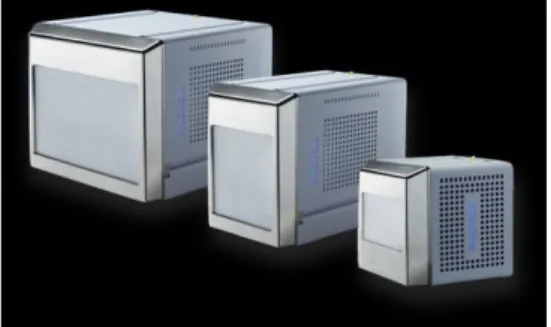
COMMERCIAL PRESENTATIONS
Photon Counting with Mixed-mode Detectors
Vernon Smith,a Roger Dursta, Vernon Smitha, Joerg Kaercherb, Bruce Beckerb, Tobias Stuerzera a
Bruker AXS GmbH, Karlsruhe, Germany, bBruker AXS Inc, Madison, WI, USA, [email protected]
Modern single-crystal X-ray diffraction relies entirely on two-dimensional pixel array detectors (PADs). Their underlying CMOS technology allows shutterless data collection which has widely eliminated experimental overhead time. Today, two different techniques are competing in the home laboratory market. HPADs have now been available for over ten years [1] and rely on being able to count individual X-ray events in the time-domain. The introduction of HPADS was a significant advance in detector technology, but several fundamental drawbacks have been extensively described in the scientific literature [2, 3].
More recently, detector developments at leading synchrotrons and at Bruker AXS have focussed attention on photon-counting using mixed-mode detection. Mixed-mode detection uses charge-integrating technology to count individual photon events in the space-domain. Mixed-mode detection retains all the benefits of the older HPAD technology but does not suffer from the same drawbacks. As a result, the reflection intensities are measured significantly more accurately [4].
Bruker AXS have introduced the first commercially available mixed-mode detector – the PHOTON III [5]. The PHOTON III series employs large monolithic sensors with no dead areas in genuine photon-counting detectors with no charge sharing or counting errors.
The technical background of mixed-mode detection will be introduced accompanied by a variety of crystallographic examples to highlight the features of the PHOTON III detectors.
Figure 1. PHOTON III Series of mixed-mode photon-counting detectors
[1] M. J. Renzi, M. W. Tate, A. Ercan, S. M. Gruner, E. Fontes, C. F. Powell, A. G. MacPhee, S. Narayanan, J. Wang and R. Cuenca, Review of Scientific Instruments , 2002, 73:3, 1621-1624.
[2] K. Mathieson, R. Bates, V. O’Shea, M.S. Passmore, M. Rahman, K.M. Smith, J. Watt, C. Whitehill,
Nuclear Instruments and Methods in Physics Research A. 2002 477, 191–197.
[3] A. Bergamaschi , R.Dinapoli, B.Henrich, I.Johnson, A.Mozzanica, X.Shi, B.Schmitt, Nuclear
Instruments and Methods in Physics Research A. 2011, 628, 238-241.
[4] F. Leonarski, S. Redford, A. Mozzanica, C. Lopez-Cuenca, E. Panepucci, K. Nass, D. Ozerov, L. Vera, V. Olieric, D. Buntschu, R. Schneider, G. Tinti, E. Froejdh, K. Diederichs, O. Bunk, B. Schmitt , M. Wang , Nature Methods, 2018, 15, 799-804.
24
Latest developments from Rigaku
Mark Benson
Rigaku Europe SE, Neu Isenburg, Germany. [email protected]
Performing data collections which are not only high quality but also efficient is a key requirement of many diffractometer users around the world, either to enable high throughput, or maximise data quality in a given time.
The new XtaLAB Synergy diffractometer line represent improvements in both hardware and software design. Advancements have been made not only in the microfocus sources but also in the goniometer and detector hardware. The sample environment has been completely redesigned allowing ample space for easier access or incorporation of specialist equipment. The combination of these enhancements leads to better data quality in shorter data collection times for both weakly and strongly diffracting crystals.
Ensuring the best possible use of the diffractometer hardware and the data it provides requires high performance software. The CrysAlisProsoftware package is under continual development in order to provide enhanced automation features, new options for sample screening, data collection and improvements to data processing algorithms.
25
Characterization of a few vanadate compounds using both the Aeris
benchtop and the Empyrean platform
Eugenio Casini,a Gwilherm Nénert,b a
Malvern PANalytical Italy, Via Cadore, 21, 20851 - Lissone, Italy
b
Malvern PANalytical, Lelyweg 1, 7602 EA Almelo, The Netherlands. [email protected]
The relation between crystal structure and properties of mineral-related materials is the key for technological benefits. MalvernPanalytical is committed to provide state-of-the-art solutions for basic and advanced studies both in ambient and non-ambient conditions (temperature, pressure, humidity, etc.). The synthesis and characterization of two new vanadate related materials is presented. AgCaVO4 is a new material related to arcanite (-K2SO4). By means of laboratory
powder X-ray data, its crystal structure has been determined [1]. This compound exhibits a distorted arcanite structure, contrary what has been previously reported for AgBVO4 (B = Mg,
Cd) compounds. Furthermore we report a new larnite related vanadate NaSrVO4 exhibiting a
complex phase diagram as a function of temperature [2]. This compound is the first report for a AIBIIXVO4 chemistry exhibiting the larnite structure. This result hints that the larnite structure
can accommodate a richer chemistry that initially foreseen.
Figure 1. HT-XRD data collected on the NaSrVO4 sample.
[1] G. Nénert; Zeitschrift fuer Kristallographie,2017, 232(10), 669 [2] G. Nénert; O’Meara, P., Degen, T.; Acta. Cryst. A., 2016, 72, s62
26
The new Thermo Scientific Krios G4 Cryo-TEM and Falcon 4:
“Bringing leading technology into the standard lab and offering a road to
success for experienced and new cryo-EM user
s”.
Max Malettaa, Marc Storms, Kristian Wadel, Steve Reyntjens a
Materials and Structural Analysis, Analytical Instruments Group, Thermo Fisher Scientific Achtseweg Noord 5651 GG Eindhoven, The Netherlands.
Structural biologists have made great progress in unraveling the structures of individual proteins and protein complexes. Much of these recent results have been made using Single Particle Analysis (SPA), which has turned into a well-accepted and almost routine technique. Continuous efforts to make cryo-EM even more mature are taken by further improving its productivity (throughput) and ease-of-use.
During this talk, we will introduce the latest generation of the award-winning Krios platform including new direct electron detector which embarks experienced, as well as new users, on their continuing mission to become more productive and improve the image quality of their 3D reconstructions. Simultaneously, the adoption of cryo-EM is further facilitated through improving the system’s usability and installability.
27
MS1 - Crystallography out of Academia
Chairs: Alessia Bacchi (Univ. di Parma)
28
MS1 - KN1: Diffraction for industries, businesses and health
Miguel A. G. Aranda,a,b Angeles G. De la Torre,a Laura León-Reina,c Marta García-Matéd aDepartamento de Química Inorgánica, Universidad de Málaga, Campus Teatinos S/N. 29071, Málaga,
Spain. [email protected]
b
ALBA synchrotron collaborator, Carrer de la Llum 2-26, E-08290 Barcelona, Spain
c
Servicios Centrales de Apoyo a la Investigación, Universidad de Málaga, 29071, Málaga, Spain
d
X-Ray Data Services, Pje Carlos Cano 7, 296200, Torremolinos, Málaga, Spain
Powder diffraction is widely used at, or requested by, industries. Even more, new regulations are including the use of powder diffraction and even its data treatment by the Rietveld method. Several examples will be provided.
I. The amount of respirable low-solubility dust particles in air in the workplace is undesirable and already subject to regulation(s). A key subgroup is respirable crystalline silica which if inhaled it increases the risk of developing serious silica-related diseases, including: (a) silicosis, (b) lung cancer; (c) chronic obstructive pulmonary disease; and (d) kidney disease [1]. OSHA (Occupational Safety and Health Administration of USA) Method ID-142 (2015) describes the collection of airborne respirable α-quartz and/or cristobalite in the breathing zone of personnel and the subsequent analysis by X-ray diffraction according to Method NIOSH-7500. In Spain there are related regulations: (1) MTA/MA-014/A11 by Insituto Nacional de Seguridad e Higiene en el Trabajo, INSHT, “Determinación de materia particulada (fracciones inhalable, torácica y respirable) en aire - método gravimétrico”; (2) MTA/MA-056/A06 by INSHT “Determinación de sílice libre cristalina (cuarzo, cristobalita, tridimita) en aire - Método del filtro de membrana / Difracción de rayos X”; and (3) Guía Técnica – Métodos de determinación de fracción respirable y sílice cristalina respirable” Departamento Técnico. Instituto Nacional de la Silicosis. Some details about our meticulous implementation will be provided but some specific details of our know-how will not be shared.
II. ASTM C1365 test method for cement Rietveld quantitative phase analysis validation procedure uses SRM 2686a reference material from NIST, which is a reference Portland clinker with reported elemental and mineralogical analyses. Laboratories and cement factories can implement ASTM C1365, by using the powder diffraction data of SRM 2686a collected with their X-ray equipments and employing their protocols but the output shall be within the ASTM C1365 limits. We were aware of problems for validating ASTM C1365 by several industries and we have thoroughly studied SRM 2686a using: (1) strictly monochromatic CuKα1 ‘flat
reflection geometry’; (2) strictly monochromatic MoKα1 ‘flat transmission geometry’; and (3)
synchrotron radiation ‘rotating capillary transmission geometry’. Our results [2] indicate that belite in SRM 2686a is composed of two polymorphs (- and α’H-) that must be included in the
analyses. The use of a unique phase for describing belite and inadequate peak shape modelling explain the problems found for implementing ASTM C1365. Polymorphism matters.
III. ALBA synchrotron and DRX unit of SCAI of University of Malaga have provided powder diffraction analyses for different industries and fields. Some examples will be sketched.
Acknowledgements. Partly supported by MinCIU research grants, BIA2014-57658 & BIA2017-82391-R Data accessibility. SRM 2686a powder diffraction data are deposited at Zenodo and they can be freely
downloaded (doi: 10.5281/zenodo.1318500). Many data are confidential (they are subjected to NDA). [1] https://www.osha.gov/dsg/topics/silicacrystalline/
29
[2] Garcia-Mate, M.; et al. “Rietveld quantitative phase analysis of SRM 2686a – A standard Portland Clinker” Cement and Concrete Research, 2019, 115, 361–366.MS1 - KN2: Crystalline form suitable for inhalation in drug discovery
Irene Bassanettia
aChiesi Farmaceutici – Corp.Precl. R&D Analytics & Early Formulations Dept., Parma, Italy
Inhalatory drug administration is a promising alternative route for chronic treatment of pulmonary diseases as it minimizes systemic side effects: in fact, it allows to localize and target the delivery with minimum systemic exposure. A successful pulmonary administration requires a harmonic interaction between the API, the excipient, the inhaler device, and the patient. Three main inhalation systems are widely used for pulmonary drugs and are clinically equivalent: nebulizers and pressurized Metered Dose Inhalers (pMDIs) are based on solution/suspension formulations, while Dry Powder Inhalers (DPIs) generate aerosol directly from solid API mixed with solid excipients or carriers. Especially for the latter the complete solid state characterization of API is the key role for an efficient deposition in the lungs: packing, size, morphology, excipient compatibility and stability could affect the aerodynamic performance and the efficacy of the drug.[1] For the formers, instead, solid state properties are essential for API solubility and primarily bioavailability: polymorphism, solvate and salt formation can have a profound impact on solubility rate thus influencing both absorption and the pharmacokinetic. As a result, the crystallization screening (polymorphs, salts and co-crystals screening) should start in earlier phases of drug discovery during lead selection or lead optimization, as a part of the Screening Cascade of tests for the selection of Candidate Drugs (CD). Moreover, due to the type of delivery chosen many constraints are introduced. Solvents used must be safe (Class III); the counterions for salts are preferably limited to ones already known for inhalation; coformers must be selected not only following the crystal engineering laws but must be either inactive ingredients or selected co-drugs tolerated in inhalation and with a synergistic effect on the targeted disease[2].
In addition, also the particle engineering technologies should be considered in the choice of a lead form for developability. To reach low airways, to be absorbed, the crystalline drug should be micronized (1-5 µm) prior to blending, using a jet mill or similar device with the risk of change polymorphic form, melting or amorphous formation due to the heating evolved during the process. Then, the crystal size, habit, shape and surface of micronized material need to be fully controlled after blending to be sure the micronized material results compatible with the chosen excipients.
The challenges to face for selecting a CD are tough: here different case studies are shown to demonstrate how crystallography is essential also for researchers out of academia.
[1Patton, J. S., & Byron, P. R. (2007). Inhaling medicines: delivering drugs to the body through the lungs.
Nature Reviews Drug Discovery, 6(1), 67-74.
[2] Huang, L.F.&Tong, WQ. (2004). Impact of solid-state properties on developability assessment of drug candidates. Advanced Drug Delivery Reviews, 56(3), 321-334.
[3] Selby, M. D., de Koning, P. D., & Roberts, D. F. (2011). A perspective on synthetic and solid-form enablement of inhalation candidates. Future medicinal chemistry, 3(13), 1679-1701.
30
MS1 - O1: Determining the concentration of diamond powder deposited on
a textile yarn: a multitechnique approach
S. Ciattini1,L. Chelazzi1,
1
Center of Structural Crystallography (CRIST). University of Florence. Via della Lastruccia 3, 50019 Sesto Fiorenitno (Firenze), Italy, [email protected]
The aim of our study was the development of an analytical method to determine the amount of diamond powder deposited on a textile yarn. This analytical need arises to certify the concentration of diamond itself in textile fabric. Due to the fact that diamond textile yarn had to be used to produce luxury and high fashion manufactures, the certification was an absolute need in the marketing of such products that are proposed with a remarkable price to the final users. The production line of the diamond wire was developed by a company of Prato (Tuscany, Italy) textile area.
The analytical issues could be pointed out as follow:
1) verification of the presence of the diamond powder on the final fabric and as consequence on the textile yarn,
2) determination of the linear concentration of diamond on the textile yarn,
3) verification of the homogeneity of the product sample, i.e. the concentration should be constant throughout the whole process production.
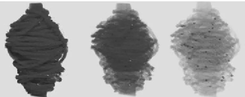
CRIST (Crystallography Center - Università di Firenze) is an academic facility with a wide range of available X-ray instrumentations, most of them dedicated to X-ray diffraction analysis[1]. Then, the first idea was to verify the presence of diamond in the final fabric through an X-ray diffraction analysis. But for the quantitive analisys a multi tecniques approach, like XRF-WD spectrometry, X-ray microtomografy[2] and of course XRD-PD, had to be used.
Figure 1. 3D reconstruction of a textile yarn used in micro tomography analysis with different transparency
References:
[1] Bruker-AXS, DIFFRACPlus TOPAS: TOPAS 4.2 Technical Reference, Bruker-AXS GmbH, Karlsruhe, Germany (2008)
[2] Feldkamp L.A., Davis L.C., Kress J.W. Practical cone-beam algorithm. J Opt Soc Am. A1:612-619, 1984