Received: April 26, 2018.
Accepted: August 30, 2018.
Pre-published: September 6, 2018.
©2019 Ferrata Storti Foundation
Material published in Haematologica is covered by copyright. All rights are reserved to the Ferrata Storti Foundation. Use of published material is allowed under the following terms and conditions:
https://creativecommons.org/licenses/by-nc/4.0/legalcode. Copies of published material are allowed for personal or inter-nal use. Sharing published material for non-commercial pur-poses is subject to the following conditions:
https://creativecommons.org/licenses/by-nc/4.0/legalcode, sect. 3. Reproducing and sharing published material for com-mercial purposes is not allowed without permission in writing from the publisher.
Correspondence:
[email protected] or
[email protected]
Haematologica
2019
Volume 104(2):312-318
doi:10.3324/haematol.2018.196055
Check the online version for the most updated
information on this article, online supplements,
and information on authorship & disclosures:
www.haematologica.org/content/104/2/312
Ferrata Storti Foundation
Introduction
The Philadelphia (Ph) chromosome derives from the t(9;22)(q34;q11) and leads to
a BCR-ABL1 rearrangement.
1The incidence of this chromosomal change in acute
lymphoblastic leukemia (ALL) increases with age, being detected in 25% of adults
and in about 50% of elderly patients.
2Prior to the advent of tyrosine kinase
inhibitors, the outcome of Ph+ ALL patients was extremely poor,
3-5and the only
T
o shed light onto the molecular basis of Philadelphia
chromo-some-positive acute lymphoblastic leukemia and to investigate
the prognostic role of additional genomic lesions, we analyzed
copy number aberrations using the Cytoscan HD Array in 116 newly
diagnosed adult patients with Philadelphia chromosome-positive acute
lymphoblastic leukemia enrolled in four different GIMEMA protocols,
all based on a chemotherapy-free induction strategy. This analysis
showed that patients with Philadelphia chromosome-positive acute
lymphoblastic leukemia carry an average of 7.8 lesions/case, with
tions outnumbering gains (88% versus 12%). The most common
dele-tions were those targeting IKZF1, PAX5 and CDKN2A/B, which were
detected in 84%, 36% and 32% of cases, respectively. Patients carrying
simultaneous deletions of IKZF1 plus CDKN2A/B and/or PAX5 had a
sig-nificantly lower disease-free survival rate (24.9% versus 43.3%; P=0.026).
The only IKZF1 isoform affecting prognosis was the dominant negative
one (P=0.003). Analysis of copy number aberrations showed that 18%
of patients harbored MEF2C deletions, which were of two types,
differ-ing in size: the longer deletions were associated with the achievement of
a complete molecular remission (P=0.05) and had a favorable impact on
disease-free survival (64.3% versus 32.1% at 36 months; P=0.031). These
findings retained statistical significance also in multivariate analysis
(P=0.057). KRAS deletions, detected in 6% of cases, were associated
with the achievement of a complete molecular remission (P=0.009).
These results indicate that in adults with Philadelphia
chromosome-pos-itive acute lymphoblastic leukemia a detailed evaluation of additional
deletions - including CDKN2A/B, PAX5, IKZF1, MEF2C and KRAS - has
prognostic implications and should be incorporated in the design of
more personalized treatment strategies.
Prognostic implications of additional genomic
lesions in adult Philadelphia
chromosome-positive acute lymphoblastic leukemia
Anna Lucia Fedullo,
1* Monica Messina,
1* Loredana Elia,
1Alfonso Piciocchi,
2Valentina Gianfelici,
1Alessia Lauretti,
1Stefano Soddu,
2Maria Cristina
Puzzolo,
1Clara Minotti,
1Felicetto Ferrara,
3Bruno Martino,
4Patrizia Chiusolo,
5Valeria Calafiore,
6Stefania Paolini,
7Marco Vignetti,
1,2Antonella Vitale,
1Anna
Guarini,
8Robin Foà
1* and Sabina Chiaretti
1*
1
Hematology, Department of Translational and Precision Medicine, Sapienza University,
Rome;
2GIMEMA Data Center, Rome;
3Division of Hematology and Stem Cell
Transplantation Unit, Cardarelli Hospital, Naples;
4Hematology Unit, Azienda Ospedaliera
Bianchi Melacrino Morelli, Reggio Calabria;
5Institute of Hematology, Catholic University,
Rome;
6Division of Hematology, AOU Policlinico, University of Catania;
7"L. and A. Seràgnoli"
Institute of Hematology, University of Bologna and
8Department of Molecular Medicine,
Sapienza University, Rome, Italy
*These authors contributed equally to this work
possibility of a cure was allogeneic stem cell
transplanta-tion (HSCT), when feasible.
6,7The introduction of tyrosine
kinase inhibitors, administered with low doses or without
chemotherapy during induction, followed by
consolida-tion chemotherapy and HSCT has markedly improved the
management and outcome of adult Ph+ ALL patients,
with survival rates at 5 years now approaching 50%.
8-17Different biological features - the type of fusion
tran-script (i.e. p190 or p210),
18the persistence and/or
reappear-ance of minimal residual disease (MRD),
19,20additional
genomic deletions (particularly IKZF1, and to a lesser
extent CDKN2A/B and PAX5
21-24) - and the presence of
mutations at relapse are associated with a worse
outcome.
25-27However, a broad and refined biological
algo-rithm that could help to optimize treatment strategies and
define better whether some patients could be spared
intensive treatment, including HSCT, has so far not been
proposed.
To this end, in the present study we investigated copy
number aberrations (CNA) in 116 newly diagnosed adult
Ph+ ALL patients to identify additional molecular lesions
with the aim of improving patients’ stratification and
management.
Methods
Experimental strategy
Bone marrow and/or peripheral blood samples from 116 patients (Table 1) with newly diagnosed Ph+ ALL enrolled in four GIMEMA (Gruppo Italiano Malattie EMatologiche dell'Adulto) trials were analyzed (Online Supplementary Table S1). The study was car-ried out in four phases (Online Supplementary Figure S1): (i) CNA analysis of 116 samples by Cytoscan; (ii) multiplex ligation-depen-dent probe amplification analysis; (iii) validation of MEF2C dele-tions by digital droplet (dd) polymerase chain reaction (PCR); and (iv) MEF2C and KRAS mutational screening.
This study was approved in the context of an Associazione
Italiana per la Ricerca sul Cancro (AIRC) project (10007) with
Institutional Review Board number 2182/16.06.2011.
Copy number aberration analysis
CNA were analyzed using CytoScan®HD Arrays (Affymetrix, Santa Clara, CA, USA) and Chromosome Analysis Suite (ChAS) software. Germline material from five paired samples was also evaluated. Recurrent deletions were validated with the Salsa MLPA P335 ALL-IKZF1 kit (MRC-Holland, Amsterdam, the Netherlands)28,29(Online Supplementary Data). Statistical analyses on clinical correlates are described in the Online Supplementary Data.
Digital droplet polymerase chain reaction assays
MEF2C deletions were validated by ddPCR using the QX200TM
Droplet DigitalTMPCR System (BioRad, Hercules, CA, USA) and QuantaSoft Analysis Pro software according to the manufacturer's instructions (Online Supplementary Data).
Mutational screening
Sanger sequencing of PCR products for MEF2C and KRAS exons (Online Supplementary Table S2) was performed with the ABI-Prism 3500 sequencer (Applied Biosystem, Life Technologies, Foster City, CA, USA) (Online Supplementary Data).
Results
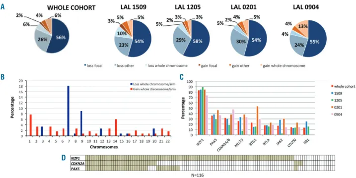
Copy number aberration analysis
CNA analysis revealed 7.8 aberrations/patient (range:
0-28), the majority being losses (88%) with only 12% gains,
both spanning from whole chromosomes to focal
lesions;
22,23,30-32no differences were recorded among trials
(Figure 1A).
Gross chromosomal lesions were found in 42% of
cases: the majority were losses of chromosome 7 (18.1%),
followed by monosomy of chromosome 9 or 9p deletion
(9%) and gain of 1q (7.7%) (Figure 1B, Online
Supplementary Table S3). Smaller deletions - limited to one
to three genes and defined as focal - were found in 56% of
cases.
The most frequently deleted region involved the 7p12
cytoband comprising IKZF1
22,23,33,34which was detected in
97 cases (84%).
PAX5 was deleted in 43 patients (36.2%), while 37
(31.9%) had deletions of CDKN2A/B. MLLT3, BTG1,
BTLA, CD200 and RB1 were deleted in 30, 27, 21, 17 and
16 cases, respectively (25.9%, 23%, 18.1%, 17.2%,
14.6%, and 13.8%) (Figure 1C).
IKZF1 deletions (
ΔIKZF1) occurred together with
CDKN2A/B and/or PAX5 deletions in 45/97 cases (46.4%)
and are defined as ΔIKZF1+CDKN2A and/or PAX5 (Figure
1D): this subset displayed similar lesions to those recently
described by Stanulla and colleagues.
35With regard to
potential interactions, we found a significant association
between IKZF1 and PAX5 deletions (P=0.01), but not with
CDKN2A.
Multiplex ligation-dependent probe amplification
con-firmed IKZF1, PAX5, CDKN2A, BTG1, EBF1, ETV6 and
RB1 lesions, and allowed evaluation of IKZF1 isoforms.
These isoforms were grouped into four classes:
24,36wild-type, dominant-negative (comprising
Δ4-7 cases, 29.8%),
haploinsufficient (including all cases harboring a deletion
that involves exon 2 - i.e.
Δ2-7, Δ2-8, Δ2-3, Δ1-3 - or the
whole gene, 57.7%) and miscellaneous (remaining cases,
11.3%).
Identification of novel lesions
CNA analysis highlighted additional genomic lesions
(Table 2, Online Supplementary Table S4). We focused in
particular on MEF2C and KRAS deletions since these had
prognostic significance (see below). MEF2C deletions
were detected in 21 patients (18.1%) and differed in size.
According to the length of intron 1-2 losses, deletions
were grouped into two categories. One category -
detect-ed in 14 cases (67% of MEF2C deletdetect-ed cases) - was
char-acterized by a larger minimal common region (6.2 Kb)
involving introns 1-2 and exon 2 (the first codifying exon),
Table 1.
Patients’ clinical features.
Patients (n=116)
Gender (male/female) 55/61
Age, years (range) 51.1 (18.9-89)
Median white cell count x 10
9/L (range) 25.4 (1.7-597)
Median hemoglobin g/dL (range) 9.6 (4-16.3)
Median platelet count x 10
9/L (range) 50 (4-333)
Fusion transcript (p190/p210/p190-210)
¥70/29/16
Complete molecular response* yes/no 17/99
defined
ΔMEF2C-long. The other category, detected in
seven patients, was smaller (5.4 Kb) and involved only
exon 2, and was called ΔMEF2C-short (Figure 2A). ddPCR
confirmed MEF2C lesions in all cases. No MEF2C
muta-tions were identified.
KRAS deletions (
ΔKRAS) were detected in seven cases
(6%); the focal lesion of KRAS started in the 5’
untranslat-ed region and enduntranslat-ed in intron 1-2, involving the first
non-codifying exon (Figure 2B). The minimal common region
consisted of 135 Kb. KRAS was not affected by mutations.
Impact of known and novel deletions on complete
molecular response achievement and disease-free
survival
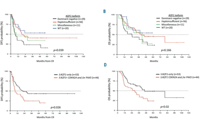
We did not find significant differences between patients
with
ΔIKZF1 and IKZF1 wild-type cases with regard to
achievement of complete molecular response (CMR) or
disease-free survival (DFS) (Online Supplementary Figure
S2). Further stratification according to IKZF1 isoforms
showed that patients with the dominant-negative isoform
had a lower DFS rate (23.3%; P=0.039) compared to
patients with the other isorforms, particularly wild-type
(53.3%; P=0.016) and haploinsufficient cases (40.3%;
P=0.015); the DFS rate of the miscellaneous group (34.1%)
did not differ significantly from that of the
dominant-neg-ative cases (Figure 3A). These differences were not
statis-tically significant in the overall survival analysis (Figure
3B).
We also investigated the outcome of ΔIKZF1+CDKN2A
and/or PAX5 cases. The CMR rate did not differ between
ΔIKZF1+CDKN2A and/or PAX5 and ΔIKZF1-only cases;
contrariwise,
ΔIKZF1+CDKN2A and/or PAX5 patients
had a significantly worse DFS than
ΔIKZF1-only cases
(43.3% versus 24.9%; P=0.026) (Figure 3C) and an inferior
overall survival (62.6% versus 40.2%; P=0.02) (Figure 3D).
The presence of
ΔMEF2C-long was associated with a
higher rate of CMR achievement (P=0.05); this effect was
not influenced by the protocol or the tyrosine kinase
inhibitor used (imatinib or dasatinib). Furthermore,
ΔMEF2C-long cases were also associated with a
signifi-Figure 1. Overall load and incidence of genetic lesions in Philadelphia chromosome-positive acute lymphoblastic leukemia.(A) Distribution of copy number aber-rations in the whole cohort and across different protocols. (B) Percentages of gross chromosomal aberaber-rations. (C) Percentages of deletions of known genes in the whole cohort (n=116) and in the different studies analyzed. (D) Heatmap of IKZF1, CDKN2A/B, and PAX5 deletions in the whole cohort.
A
B
C
D
Table 2.
Minimal common region (MCR) of identified focal lesions.
Deleted gene N. of patients (%) MCR (hg19)
FOCAD 29 (25) chr9: 20685149 - 20759956
CDK6 24 (20.7) chr7: 92456635 - 92266647
PTPRD 21 (18.1) chr9: 8153932 - 8854489
MEF2C 21 (18.1) chr5:88122179 - 88127630
BTLA 21 (18.1) chr3:112154702 - 112217769
JAK2 20 (17.2) chr9: 5123013 - 5234403
ADD3 18 (15.5) chr10: 111795029 - 111853667
SLX4IP 17 (14.6) chr20: 10417444 - 10451891
CD200 17 (14.6) chr3:112054292 - 112217769
HBS1L 16 (13.7) chr6: 135375338 - 135418257
ATP10A 14 (12) chr15: 26055568 - 26103185
TOX 8 (6.9) chr8:60028851 - 60110235
KRAS 7 (6) chr12: 25402194 - 25537468
ARHGAP24 7 (6) chr4:86493655 - 86436188
EBF1 6 (5.1) chr5: 158440156 - 158164599
LEF1 5 (4.3) chr4:109034392 - 109084557
MDM2 5 (4.3) chr12:69159076 - 69205287
TCF12 4 (3.4) chr15:57294905 - 57399047
ERG 2 (1.7) chr21:39772775 - 39788683
cantly better DFS (64.3% versus 32.1%; P=0.031) (Figure
4A) and overall survival (77.9% versus 48.4%; P=0.036)
(Figure 4B).
Lastly,
ΔKRAS was more frequently found in patients
who obtained a CMR (24% versus 3%; P=0.009), but this
finding did not have an impact on DFS.
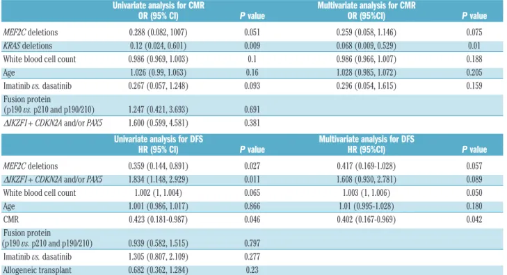
Prognostic impact of known and novel genomic lesions
in univariate and multivariate analyses
In univariate analysis, ΔMEF2C-long and ΔKRAS had an
impact on CMR achievement, while
ΔMEF2C-long and
ΔIKZF1+CDKN2A and/or PAX5 influenced DFS (Table 3).
In multivariate analysis for CMR, performed taking into
Figure 3. Survival probability curves according to IKZF1 status.(A) Disease-free survival and (B) overall survival at 36 months of patients divided according to IKZF1 isoform. (C) Disease-free survival and (D) overall survival at 36 months of ΔIKZF1-only and ΔIKZF1+ CDKN2A and/or PAX5 patients. WT: wild-type; DFS: disease-free survival; OS: overall survival; CR: complete remission.
A
B
C
D
Figure 2. Representation of ΔMEF2C and ΔKRAS. (A) Representation of ΔMEF2C for each patient. Lesions are ordered according to their size: one case had a dele-tion of the whole gene, one had a deledele-tion that involved only exon 1 spanning from intron 1-2 to the 5’ untranslated region (5’UTR), four had deledele-tions starting from intron 2-3 and ending at 5’-UTR, thereby involving both exons 2 and 1 (the latter being an untranslated exon), 13 had lesions spanning from intron 2-3 to intron 1-2, therefore involving exon 2 (the first codifying exon), with six of them harboring a longer intron 1-2 deletion. Lastly, two cases had deletions that involved only intron 1-2. The first 14 cases were considered as ΔMEF2C-long and the remaining as ΔMEF2C-short. (B) Representation of ΔKRAS for each patient. Lesions are ordered according to their size: in four cases, the deletion encompassed only KRAS itself, whereas in three it involved the short arm of chromosome 12. INTR: intron; EX: exon; 5’UTR: 5’ untranslated region.
account white blood cell count, age, tyrosine kinase
inhibitor use and the genomic lesions described above, the
only factor that retained statistical significance was
ΔKRAS (P=0.01); a trend was noted for ΔMEF2C-long
deletions (P=0.075) (Table 3).
In multivariate analysis for DFS, considering
ΔMEF2C-long,
ΔIKZF1+CDKN2A and/or PAX5, white blood cell
count and CMR as variables, the factors that had a
nega-tive impact were
ΔMEF2C-long (P=0.057) and white
blood cell count (P=0.05), while a trend towards a worse
DFS was observed for
ΔIKZF1+CDKN2A and/or PAX5
(P=0.089) (Table 3). HSCT did not affect the prognostic
role of the above-mentioned lesions.
Discussion
The management of adults with Ph+ ALL currently
relies on the use of first,
8-10,13-16second
11,12and third
37gener-ation tyrosine kinase inhibitors, either alone
9-12or in
com-bination with chemotherapy,
8,13-16,37followed - if feasible
and necessary - by HSCT. These approaches have greatly
improved the outcome of Ph+ ALL: nowadays, virtually
all patients - independent of age - achieve a complete
hematologic remission, coupled to a CMR in a variable
proportion of cases. Nonetheless, in all reported studies
the long-term outcome is in the range of 50% at 5 years;
thus, additional prognosticators capable of better
stratify-Table 3.
Summary of univariate and multivariate analyses for complete molecular response and disease-free survival for the factors identified.
Univariate analysis for CMR Multivariate analysis for CMR
OR (95% CI) P value OR (95%CI) P value
MEF2C deletions 0.288 (0.082, 1007) 0.051 0.259 (0.058, 1.146) 0.075
KRAS deletions 0.12 (0.024, 0.601) 0.009 0.068 (0.009, 0.529) 0.01
White blood cell count 0.986 (0.969, 1.003) 0.1 0.986 (0.966, 1.007) 0.188
Age 1.026 (0.99, 1.063) 0.16 1.028 (0.985, 1.072) 0.205
Imatinib vs. dasatinib 0.267 (0.057, 1.248) 0.093 0.296 (0.054, 1.615) 0.159
Fusion protein
(p190 vs. p210 and p190/210) 1.247 (0.421, 3.693) 0.691
ΔIKZF1+ CDKN2A and/or PAX5 1.600 (0.599, 4.581) 0.381
Univariate analysis for DFS Multivariate analysis for DFS
HR (95% CI) P value HR (95%CI) P value
MEF2C deletions 0.359 (0.144, 0.891) 0.027 0.417 (0.169-1.028) 0.057
ΔIKZF1+ CDKN2A and/or PAX5 1.834 (1.148, 2.929) 0.011 1.608 (0.930, 2.781) 0.089
White blood cell count 1.002 (1, 1.004) 0.065 1.003 (1, 1.006) 0.050
Age 1.001 (0.986, 1.017) 0.866 1.01 (0.995-1.028) 0.180
CMR 0.423 (0.181-0.987) 0.046 0.402 (0.167-0.969) 0.042
Fusion protein
(p190 vs. p210 and p190/210) 0.939 (0.582, 1.515) 0.797
Imatinib vs. dasatinib 1.305 (0.807, 2.109) 0.277
Allogeneic transplant 0.682 (0.362, 1.284) 0.23
CMR: complete molecular response; OR: odds ratio; 95% CI: 95% confidence interval; DFS: disease-free survival; HR: hazard ratio.
Figure 4. Survival probability curves according to MEF2C status. (A) Disease-free survival at 36 months of ΔMEF2C versus MEF2C wild-type patients. (B) Overall survival at 36 months of ΔMEF2C versus MEF2C wild-type patients. WT: wild-type; DFS: disease-free survival; OS: overall survival; CR: complete remission.
ing patients into high- and low-risk categories are urgently
needed to further optimize treatment. Moreover, another
unsolved issue is whether all eligible patients should
undergo HSCT,
7,17a procedure still associated with
short-and long-term side effects, as well as treatment-related
mortality. This is particularly important for patients who
obtain a CMR.
To address these issues we sought to identify additional
genomic lesions with prognostic significance in adult Ph+
ALL using high density Cytoscan arrays. We found that
adult Ph+ ALL patients carried an average of 7.8
aberra-tions each, with deleaberra-tions outnumbering gains, in line
with other ALL subsets.
22,30,38,39Macro-aberrations were
identified in 48% of cases and micro-aberrations in the
majority of patients: among the latter, the most frequent
was ΔIKZF1, which was detected in 84% of cases. ΔIKZF1
has been regarded as a poor prognostic marker in both
childhood and adult ALL.
34,36,39-41This finding was not,
however, confirmed in our cohort: in fact, the presence of
ΔIKZF1 alone was not associated with a worse DFS. A
fur-ther evaluation of the various IKZF1 isoforms showed
that only the dominant-negative genotype was
deleteri-ous for outcome. In addition, patients with
ΔIKZF1+CDKN2A and/or PAX5, accounting for almost
half the
ΔIKZF1 cases, experienced a significantly inferior
DFS (P=0.005) and overall survival (P=0.02), in line with
previous
reports
on
ALL
in
general.
28,29,36,39,42,43ΔIKZF1+CDKN2A and/or PAX5 also had a prognostic
impact in multivariate analysis; survival analysis was
car-ried out merging all cases enrolled in the different trials
together in order to gain statistical significance.
Recently, studies have been focused on the presence of
additional karyotypic aberrations in Ph+ ALL.
44-48These
studies have highlighted that a high percentage of Ph+ ALL
cases (60-80%) harbor additional chromosomal
abnormal-ities, with the most frequent aberrations involving
chro-mosomes 7, 9, and 14. Patients with additional
abnormali-ties, particularly loss of 9/9p and/or CDKN2A, have a
worse outcome. These results point to the importance of
screening for other molecular markers, and not only IKZF1,
in agreement with our findings on
ΔIKZF1+CDKN2A
and/or PAX5. At variance from these reports, our study
also identified novel lesions that had a favorable impact on
outcome. Among these, it is worth mentioning
ΔMEF2C,
which occurred in 18.1% of patients and was of two sizes,
a long deletion, encompassing introns 1-2 and exon 2, and
a second, smaller one, involving only exon 2. MEF2C is a
transcription factor involved in B-cell survival and
prolifer-ation whose overexpression is associated with an
unfavor-able prognosis in T-ALL and acute myeloid leukemia.
49-52In
our study, the presence of
ΔMEF2C-long was associated
with achievement of a CMR (P=0.05) and with a
signifi-cantly better DFS compared to the remaining cases
(P=0.031) also in a multivariate model; as for IKZF1
dele-tions, survival analysis was performed merging the whole
cohort because of the sample sizes.
ΔMEF2C-long was
widely distributed among cases, with no association with
white blood cell count, age, type of fusion protein or
addi-tional deletions. To our knowledge, this is the first report
that correlates
ΔMEF2C-long with prognosis in Ph+ ALL:
Martinelli et al.
40and Mullighan et al.
22,41described
ΔMEF2C
in Ph+ ALL, but did not demonstrate a correlation with
outcome. Finally,
ΔKRAS was associated with a higher rate
of CMR achievement upon induction (P=0.01), but not
with a better DFS.
In conclusion, we show that additional genetic lesions
can be found at presentation in adult Ph+ ALL patients
and that these lesions have prognostic significance, with
the
IKZF1
dominant-negative
isoform
and
ΔIKZF1+CDKN2A and/or PAX5 negatively affecting
out-come, and ΔMEF2C and ΔKRAS being instead associated
with a more favorable prognosis. Screening for these
genetic lesions should, therefore, be performed at the
time of diagnosis for a more refined prognostic
stratifica-tion, and for a more personalized and tailored
manage-ment of Ph+ ALL patients.
Acknowledgments
The authors thank Associazione Italiana per la Ricerca sul
Cancro (AIRC), Special Program Molecular Clinical
Oncology-Extension program, 5 x 1000 (10007), Milan (Italy)
for funding RF; Finanziamento per l’avvio alla ricerca 2015
(Sapienza University of Rome) for funding MM;
Finanziamento Medi Progetti Universitari 2015 for funding SC
(Sapienza University of Rome); and Fondazione Le Molinette
Onlus, Turin (Italy).
References
1. Nowell PC, Hungerford DA. Chromosome studies on normal and leukemic human leukocytes. J Natl Cancer Inst. 1960;25:85-109.
2. Chiaretti S, Vitale A, Cazzaniga G, et al. Clinico-biological features of 5202 patients with acute lymphoblastic leukemia enrolled in the Italian AIEOP and GIMEMA proto-cols and stratified in age cohorts. Haematologica. 2013;98(11):1702-1710. 3. Dombret H, Gabert J, Boiron JM, et al.
Outcome of treatment in adults Philadelphia chromosome-positive acute lymphoblastic leukemia-results of the prospective multi-center LALA-94 trial. Blood. 2002;100(7): 2357-2366.
4. Gleissner B, Gökbuget N, Bartram CR, et al. Leading prognostic relevance of the BCR-ABL translocation in adult acute B-lineage
lymphoblastic leukemia: a prospective study of the German Multicenter Trial Group and confirmed polymerase chain reaction analysis. Blood. 2002;99(5):1536-1543.
5. Pullarkat V, Slovak ML, Kopecky KJ, et al. Impact of cytogenetics on the outcome of adult acute lymphoblastic leukemia: results of Southwest Oncology Group 9400 study. Blood. 2008;111(5):2563-2572.
6. Hunault M, Harousseau JL, Delain M, et al. Better outcome of adult acute lymphoblastic leukemia after early genoidentical allogeneic bone marrow transplantation (BMT) than after late high-dose therapy and autologous BMT: a GOELAMS trial. Blood. 2004;104(10):3028-3037.
7. Patel JN, Druhan LJ. Genetic effects on hematopoietic stem cell transplant progno-sis and outcomes, more than just histocom-patibility. Biol Blood Marrow Transplant. 2017;23(8):1227-1228.
8. de Labarthe A, Rousselot P, Huguet-Rigal F, et al. Imatinib combined with induction or consolidation chemotherapy in patients with de novo Philadelphia chromosome-positive acute lymphoblastic leukemia: results of the GRAAPH-2003 study. Blood. 2007;109(4):1408-1413.
9. Vignetti M, Fazi P, Cimino G, et al. Imatinib plus steroids induces complete remissions and prolonged survival in elderly Philadelphia chromosome-positive patients with acute lymphoblastic leukemia without additional chemotherapy: results of the Gruppo Italiano Malattie Ematologiche dell'Adulto (GIMEMA) LAL0201-B proto-col. Blood. 2007;109(9):3676-3678. 10. Chiaretti S, Vitale A, Vignetti M, et al. A
sequential approach with imatinib, chemotherapy and transplant for adult Ph+ acute lymphoblastic leukemia. Final results of the GIMEMA LAL 0904 study. Haematologica. 2016;101(12):1544-1552.
11. Foà R, Vitale A, Vignetti M, et al. Dasatinib as first-line treatment for adult patients with Philadelphia chromosome-positive acute lymphoblastic leukemia. Blood. 2011;118 (25):6521-6528.
12. Chiaretti S, Vitale A, Elia L, et al. Multicenter Total Therapy GIMEMA LAL 1509 protocol for de novo adult Ph+ acute lymphoblastic leukemia (ALL) patients. Updated results and refined genetic-based prognostic stratifi-cation. Blood. 2015;126 (23):81.
13. Bassan R, Rossi G, Pogliani EM, et al. Chemotherapy-phased imatinib pulses improve long-term outcome of adult patients with Philadelphia chromosome-positive acute lymphoblastic leukemia: Northern Italy Leukemia Group protocol 09/00. J Clin Oncol. 2010;28(22):3644-3652. 14. Ribera JM, García O, Montesinos P, et al. Treatment of young patients with Philadelphia chromosome-positive acute lymphoblastic leukaemia using increased dose of imatinib and deintensified chemotherapy before allogeneic stem cell transplantation. Br J Haematol. 2012;159 (1):78-81.
15. Fielding AK, Rowe JM, Buck G, et al. UKALLXII/ECOG2993: addition of imatinib to a standard treatment regimen enhances long-term outcomes in Philadelphia positive acute lymphoblastic leukemia. Blood. 2014;123(6):843-850.
16. Chalandon Y, Thomas X, Hayette S, et al. Randomized study of reduced-intensity chemotherapy combined with imatinib in adults with Ph-positive acute lymphoblastic leukemia. Blood. 2015;125(24):3711-3719. 17. Litzow MR, Fielding AK, Luger SM, et al.
The evolving role of chemotherapy and hematopoietic cell transplants in Ph-positive acute lymphoblastic leukemia in adults. Bone Marrow Transplant. 2017;52(12):1592-1598.
18. Cimino G, Pane F, Elia L, et al. The role of BCR/ABL isoforms in the presentation and outcome of patients with Philadelphia-posi-tive acute lymphoblastic leukemia: a seven-year update of the GIMEMA 0496 trial. Haematologica. 2006;91(3):377-380. 19. Lee S, Kim DW, Cho BS, et al. Impact of
minimal residual disease kinetics during imatinib-based treatment on transplantation outcome in Philadelphia chromosome-posi-tive acute lymphoblastic leukemia. Leukemia. 2012;26(11):2367-2374. 20. Ravandi F, Jorgensen JL, Thomas DA, et al.
Detection of MRD may predict the outcome of patients with Philadelphia chromosome-positive ALL treated with tyrosine kinase inhibitors plus chemotherapy. Blood. 2013;122(7):1214-1221.
21. Martinelli G, Iacobucci I, Storlazzi CT, et al. IKZF1 (Ikaros) deletions in BCR-ABL1-posi-tive acute lymphoblastic leukemia are asso-ciated with short disease-free survival and high rate of cumulative incidence of relapse: a GIMEMA AL WP report. J Clin Oncol. 2009;27(31):5202-5207.
22. Mullighan CG, Miller CB, Radtke I, et al. BCR-ABL1 lymphoblastic leukaemia is char-acterized by the deletion of Ikaros. Nature. 2008;453(7191):110-114.
23. Mullighan CG. Genomic profiling of B-prog-enitor acute lymphoblastic leukemia. Best Pract Res Clin Haematol. 2011;24(4):489-503.
24. van der Veer A, Zaliova M, Mottadelli F, et al. IKZF1 status as a prognostic feature in BCR-ABL1-positive childhood ALL. Blood. 2014;123(11):1691-1698.
25. DeBoer R, Koval G, Mulkey F, et al. Clinical impact of ABL1 kinase domain mutations
and IKZF1 deletion in adults under age 60 with Philadelphia chromosome-positive (Ph+) acute lymphoblastic leukemia (ALL): molecular analysis of CALGB (Alliance) 10001 and 9665. Leuk Lymphoma. 2016;57(10):2298-2306.
26. Soverini S, Vitale A, Poerio A, et al. Philadelphia-positive acute lymphoblastic leukemia patients already harbor BCR-ABL kinase domain mutations at low levels at the time of diagnosis. Haematologica. 2011;96(4):552-557.
27. Soverini S, De Benedittis C, Machova Polakova K, et al. Unraveling the complexity of tyrosine kinase inhibitor-resistant popula-tions by ultra-deep sequencing of the BCR-ABL kinase domain. Blood. 2013;122(9): 1634-1648.
28. Messina M, Chiaretti S, Fedullo AL, et al. Clinical significance of recurrent copy num-ber anum-berrations in B-lineage acute lym-phoblastic leukaemia without recurrent fusion genes across age cohorts. Br J Haematol. 2017;178(4):583-587.
29. Moorman AV, Enshaei A, Schwab C, et al. A novel integrated cytogenetic and genomic classification refines risk stratification in pediatric acute lymphoblastic leukemia. Blood. 2014;124(9):1434-1444.
30. Mullighan CG, Goorha S, Radtke I, et al. Genome-wide analysis of genetic alterations in acute lymphoblastic leukaemia. Nature. 2007;446(7137):758-764.
31. Kuiper RP, Schoenmakers EF, van Reijmersdal SV, et al. High-resolution genomic profiling of childhood ALL reveals novel recurrent genetic lesions affecting pathways involved in lymphocyte differen-tiation and cell cycle progression. Leukemia. 2007;21(6):1258-1266.
32. Safavi S, Hansson M, Karlsson K, et al. Novel gene targets detected by genomic pro-filing in a consecutive series of 126 adults with acute lymphoblastic leukemia. Haematologica. 2015;100(1):55-61. 33. Iacobucci I, Storlazzi CT, Cilloni D, et al.
Identification and molecular characteriza-tion of recurrent genomic delecharacteriza-tions on 7p12 in the IKZF1 gene in a large cohort of BCR-ABL1-positive acute lymphoblastic leukemia patients: on behalf of Gruppo Italiano Malattie EMatologiche dell’Adulto Acute Leukemia Working Party (GIMEMA AL WP). Blood. 2009;114(10):2159-2167. 34. Mullighan CG, Su X, Zhang J, et al. Deletion
of IKZF1 and prognosis in acute lym-phoblastic leukemia. N Engl J Med. 2009; 360(5):470-480.
35. Stanulla M, Dagdan E, Zaliova M, et al. IKZF1plus defines a new minimal residual disease-dependent very-poor prognostic profile in pediatric B-cell precursor acute lymphoblastic leukemia. J Clin Oncol. 2018; 36(12):1240-1249.
36. Boer JM, van der Veer A, Rizopoulos D, et al. Prognostic value of rare IKZF1 deletion in childhood B-cell precursor acute lym-phoblastic leukemia: an international collab-orative study. Leukemia. 2016;30(1):32-38. 37. Jabbour E, Kantarjian H, Ravandi F, et al.
Combination of hyper-CVAD with pona-tinib as first-line therapy for patients with Philadelphia chromosome-positive acute lymphoblastic leukaemia: a single-centre, phase 2 study. Lancet Oncol. 2015;16(15):1547-1555.
38. Messina M, Chiaretti S, Wang J, et al. Prognostic and therapeutic role of targetable lesions in B-lineage acute lymphoblastic leukemia without recurrent fusion genes. Oncotarget. 2016;7(12):13886-13901. 39. Ribera J, Morgades M, Zamora L, et al.
Prognostic significance of copy number alterations in adolescent and adult patients with precursor B acute lymphoblastic leukemia enrolled in PETHEMA protocols. Cancer. 2015;121(21):3809-3817.
40. Martinelli G, Iacobucci I, Papayannidis C, et al. New targets for Ph+ leukaemia therapy. Best Pract Res Clin Haematol. 2009;22(3): 445-454.
41. Mullighan CG, Downing JR. Genome-wide profiling of genetic alterations in acute lym-phoblastic leukemia: recent insights and future directions. Leukemia. 2009;23(7): 1209-1218.
42. Xu N, Li YL, Li X, et al. Correlation between deletion of the CDKN2 gene and tyrosine kinase inhibitor resistance in adult Philadelphia chromosome-positive acute lymphoblastic leukemia. J Hematol Oncol. 2016;9:40.
43. Pfeifer H, Raum K, Markovic S, et al. Genomic CDKN2A/2B deletions in adult Ph+ ALL are adverse despite allogeneic stem cell transplantation. Blood. 2018;131(13): 1464-1475.
44. Heerema NA, Harbott J, Galimberti S, et al. Secondary cytogenetic aberrations in child-hood Philadelphia chromosome positive acute lymphoblastic leukemia are nonran-dom and may be associated with outcome. Leukemia. 2004;18(4):693-702.
45. Li Y, Qiu L, Zou D, et al. Additional chromo-somal abnormalities and their prognostic significance in adult Philadelphia-positive acute lymphoblastic leukemia: with or with-out imatinib in chemotherapy. Ann Hematol. 2009;88(11):1069-1077.
46. Short NJ, Kantarjian HM, Sasaki K,et al. Poor outcomes associated with +der(22)t(9;22) and -9/9p in patients with Philadelphia chro-mosome-positive acute lymphoblastic leukemia receiving chemotherapy plus a tyrosine kinase inhibitor. Am J Hematol. 2017;92(3):238-243.
47. Seol CA, Cho YU, Jang S, et al. Prognostic significance of recurrent additional chromo-somal abnormalities in adult patients with Philadelphia chromosome-positive acute lymphoblastic leukemia. Cancer Genet. 2017;216-217:29-36.
48. Motlló C, Ribera JM, Morgades M, et al. Frequency and prognostic significance of additional cytogenetic abnormalities to the Philadelphia chromosome in young and older adults with acute lymphoblastic leukemia. Leuk Lymphoma. 2018;59(1):146-154.
49. Homminga I, Pieters R, Langerak AW, et al. Integrated transcript and genome analyses reveal NKX2-1 and MEF2C as potential oncogenes in T cell acute lymphoblastic leukemia. Cancer Cell. 2011;19(4):484-497. 50. Zuurbier L, Gutierrez A, Mullighan CG, et
al. Immature MEF2C-dysregulated T-cell leukemia patients have an early T-cell pre-cursor acute lymphoblastic leukemia gene signature and typically have non-rearranged T-cell receptors. Haematologica. 2014;99(1): 94-102.
51. Laszlo GS, Alonzo TA, Gudgeon CJ, et al. High expression of myocyte enhancer factor 2C (MEF2C) is associated with adverse-risk features and poor outcome in pediatric acute myeloid leukemia: a report from the Children's Oncology Group. J Hematol Oncol. 2015;8:115.
52. Colomer-Lahiguera S, Pisecker M, König M, et al. MEF2C-dysregulated pediatric T-cell acute lymphoblastic leukemia is associated with CDKN1B deletions and a poor response to glucocorticoid therapy. Leuk Lymphoma. 2017;58(12):2895-2904.