Alma Mater Studiorum – Università di Bologna
Scuola di Dottorato di Ricerca
in Scienze Mediche e Chirurgiche
Dottorato di Ricerca in Scienze Biomediche
Progetto formativo in Neurofisiologia
XXIII Ciclo
Settore scientifico-disciplinare di afferenza: BIO/09
Tesi di Dottorato
Synaptic plasticity between amygdala and
perirhinal cortex
Dott. Alessandra Perugini
Coordinatore: Relatore:
Prof. Claudio Galletti Prof. Giorgio Aicardi
Dipartimento di Fisiologia umana e generale
Esame finale anno 2011
1 Abstract... 5 2 General Introduction... 6 2.1 Where is the memory trace?...6 2.2 The synaptic plasticity and memory theory...7 3 The rat perirhinal cortex ...10 3.1 Anatomy of the rat perirhinal cortex... 10 3.2 Connections of the rat perirhinal cortex ... 11 3.2.1 Cortical connections... 11 3.2.2 Subcortical connections... 12 3.2.3 Hippocampal connections ... 13 3.3 The role of perirhinal cortex in learning and memory... 14 4 The rat amygdala...17 4.1 Anatomy of the rat amygdala... 17 4.2 Connections of the rat amygdaloid complex ... 19 4.2.1 Afferents to the amygdaloid complex... 19 4.2.2 Efferents from the amygdaloid complex ... 21 4.2.3 Intra‐amygdaloid connections... 22 4.3 The role of the amygdala in learning and memory ... 24 5 Excitatory synaptic transmission in the central nervous system ...27 5.1 Ionotropic glutamate receptors (iGluRs) ... 28 5.1.1 AMPA Receptors ... 29 5.1.2 NMDA Receptors... 31 5.1.3 Kainate Receptors ... 33 5.2 Metabotropic glutamate receptors (mGluRs)... 33 5.3 Voltage dependent calcium channels (VDCCs)... 34 5.4 Cyclic Adenosine 3',5'Monophosphate dependent Protein Kinase A (cAMP PKA) 36 6 Synaptic Plasticity ...39 6.1 Short term plasticity ... 39 6.2 Longterm plasticity... 40 6.2.1 Long‐term potentiation ... 40 6.2.2 Long ‐term depression... 44 6.3 Synaptic plasticity in the Perirhinal cortex ... 45
3 6.4 Synaptic plasticity in the amygdala... 47 7 Aims ...50 8 Materials and Methods ...52 8.1 Materials... 52 8.1.1 The rig ... 52 8.2 Method... 54 8.2.1 Animals ... 54 8.2.2 Preparation of slices... 55 8.2.3 Data analysis... 56 8.2.4 Extracellular field recordings... 57 8.2.5 Synaptic Responses ... 58 8.3 Pharmacological agents... 58 9 Results...60 9.1 Results: Longterm potentiation at PRh/PRh and LA/PRh synapses... 60 9.1.1 Long‐term potentiation (LTP) at PRh/PRh synapses... 60 9.1.2 Long‐term potentiation (LTP) at LA/PRh synapses... 61 9.1.3 Effects of D‐AP5 upon LTP at PRh/PRh synapses and LA/PRh synapses ... 62 9.1.4 Effects of verapamil upon LTP at PRh/PRh and LA/PRh synapses... 64 9.1.5 Effects of forskolin upon basal transmission at PRh/PRh and LA/PRh synapses... 66 9.1.6 Effects of H89 upon LTP at PRh/PRh and LA/PRh synapses ... 67 9.1.7 Comparisons between control LTP and drug applications within PRh/PRh .. 70 9.1.8 Comparisons between control LTP and drug applications within LA/PRh... 70 9.2 Results: associative long term potentiation in perirhinal cortex... 72 9.2.1 High Frequency Stimulation (HFS) of 45 pulses at 100 Hz to PRh/PRh neurons ... 72 9.2.2 HFS of 45 pulses at 100Hz to PRh/PRh neurons paired with 4HFS to LA/PRh synapses... 73 9.2.3 Effects of D‐AP5 upon associative LTP... 74 9.2.4 Effects of verapamil upon associative LTP... 76 9.2.5 Effects of D‐AP5 and verapamil upon paired HFS... 76 9.2.6 Comparisons between paired HFS and drug application within PRh/PRh synapses... 77 10 Discussion ...79
1 Abstract
The central aim of the present research has been to investigate the cellular and molecular mechanisms by which emotions can influence recognition memory. I have been characterising synaptic transmission and plasticity mechanisms between amygdala (important for consolidation of memory for emotionally salient stimuli) and perirhinal cortex (crucial for recognition memory) using extracellular field potential recordings in rat brain slices that include these two areas. Initially, I have described for the first time the basic properties of synaptic plasticity at amygdala/perirhinal synapses, and I have found molecular pathways involved in long-term potentiation at this pathway. Afterward, I studied the interaction between amygdala and perirhinal cortex, namely how synaptic plasticity in the amygdala influences synaptic plasticity within perirhinal cortex. Results from this work have provided a functional model to study the mechanisms by which emotionally salient visual experiences are better remembered than neutral ones.
2 General Introduction
2.1 Where is the memory trace?One of the most important function of our brain is the ability to form and store memories, and one of the most challenging goal of is to understand the cellular and molecular mechanisms that underlie information storage, learning and memory. Experiences and interaction with the environment leave “traces” in our brain that influence our subsequent behaviour. If we assume that these traces represent learning and memory, it has to be elucidated which mechanisms make these traces last. Some studies have provided evidences of possible mechanisms for consolidation of the memory traces:
1. functional mechanisms: functional modifications of existing synapses and neurons, changes in neuron excitability and in synaptic strength, i.e. long-term potentiation (LTP) and long-term depression (LTD) (Griffiths et al., 2008; Sah et al., 2008);
2. structural changes or rewiring: synaptogenesis and outgrowth of axons and dendrites, formation or elimination of new dendritic spines and synapses (Chklovskii et al., 2004; Barnes et al., 2009; Hofer et al., 2010); 3. molecular changes: include the activation of synaptic proteins, i.e. PKMζ (Shema et al., 2007; Serrano et al., 2008) or synaptic tagging (Redondo et al., 2011)
An example of structural changes is shown in Fig. 2-1, representing the growth of new synapses after learning. I will describe functional changes in details in the following chapters.
7
Fig. 2-1 Model for long-term information storage in neuronal circuits. a. Schematic of simplistic
neuronal circuit. b. Magnified region of the circuit in a, including presynaptic boutons on axons and postsynaptic spines on the dendrite. During a novel experience or during learning of a task, new synapses are formed (growth of new spine and bouton depicted in panel 2), which are stabilised and strengthened with ongoing experience or prolonged training (panel 3). After cessation of experience, new synapses might become weakened or inactivated, while their structural basis remains (panel 4). With repeated experience, synapses can quickly become re-activated, leading to faster adaptation or learning (panel 5). Thickness of black arrows indicates strength of information transmission at synapses (from Hofer et al., 2010)
2.2 The synaptic plasticity and memory theory
The role of activity -dependent synaptic plasticity in learning and memory is a central issue in neuroscience. Most of the research work has been focused on the link between long -term synaptic changes and long -term memories. But how does LTP or LTD equal memory? An accredited hypothesis is the synaptic plasticity and memory SPM theory (Martin et al., 2000). This theory states that:
“Activity-dependent synaptic plasticity is induced at appropriate synapses during memory formation, and is both necessary and sufficient for the information storage
underlying the type of memory mediated by the brain area in which that plasticity is observed”
Martin and colleagues (Martin et al., 2000) outlined a series of criteria that the SPM theory has to fulfill in order to explain how synaptic plasticity is related to memory:
• detectability: if an animal displays memory of some previous experience,
a change in synaptic efficacy should be detectable somewhere in its nervous system;
• mimicry: if it were possible to induce the same spatial pattern of synaptic
weight changes artificially, the animal should display ‘apparent’ memory for some past experience which did not in practice occur;
• anterograde alteration: interventions that prevent the induction of
synaptic weight changes during a learning experience should impair the animal’s memory of that experience;
• retrograde alteration: interventions that alter the spatial distribution of
synaptic weights induced by a prior learning experience (see detectability) should alter the animal’s memory of that experience.
Study of synaptic plasticity in the amygdala have fulfilled most of the criteria of the SPM theory. Detectability has been met, since it has been shown that fear conditioning learning induces LTP in the lateral nucleus of the amygdala (LA) (Rogan et al. 1997, Fanselow et al., 1999, Blair et al., 2001; Maren et al., 2004; Sah et al., 2008). Anterograde alteration has also been met in pharmacological studies. It has been found, for example, that infusion of the L-type voltage dependent calcium channels blocker verapamil into LA during training blocks the acquisiton of fear conditioning, but does not impair the expression of previously learned conditioned fear responses (Blair et al., 2001). A number of behavioural studies have shown that infusion of the N-Methyl-D-Aspartate (NMDA) antagonist into LA and the adjacent basal nucleus impairs acquisition of fear learning in a variety of tasks, including contextual fear conditioning (Fanselow and Kim 1994; Maren et al. 1996). Although retrograde alteration and mimicry criteria are difficult to meet especially in other brain regions, recent data in the amygdala have provided interesting evidences in favor of these two critera. In fact, it has been demonstrated that inhibition of PKMzeta in the basolateral amygdala disrupts previously established contextual fear conditioning (Serrano et al. 2008), according to the retrograde alteration criteria. Recent advanced techniques using optogenetics showed that activation of specific LA pyramidal cells as an unconditioned stimulus (US), in the absence of a peripheral shock US, produced fear conditioning (Johansen et al., 2010).
9 The recent lines of evidences are promising in explaining the connections between synaptic plasticity and memory in a more direct and predictable way. I will further discuss fear conditioning and synaptic plasticity in the amygdala in the following chapter.
3 The rat perirhinal cortex
3.1 Anatomy of the rat perirhinal cortex
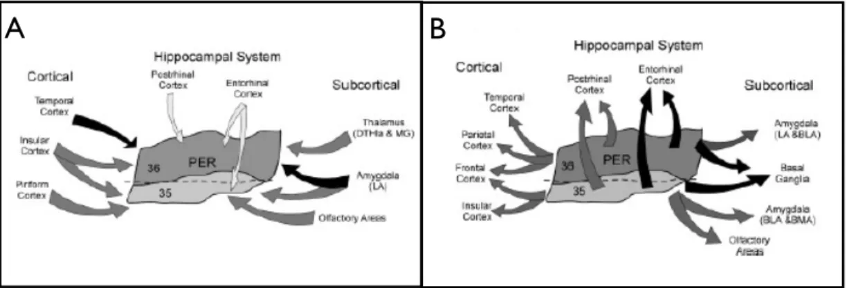
Perihinal cortex is part of the hippocampal system that comprises the hippocampal formation (dentate gyrus, CA fields, and subiculum) and the parahippocampal region (the perirhinal PRh, postrhinal POR, and entorhinal ENT cortices together with the presubiculum and the parasubiculum) (Furtak et al., 2007). The hippocampal system is involved in various memory functions. It is located dorsal to the ENT and rostral to the POR, it can be differentiated from the ENT by the presence of large, heart-shaped pyramidal cells in layer V. The rostral boundary of the PRh is marked by the subcortical posterior limit of the claustrum. At its dorsal limit, the PRh is bordered by the ventral temporal cortex (TEv)(Amaral, 1998; Shi and Cassell, 1999).
Using Broadman’s classification of the brain areas, we refer to rat perirhinal cortex as to area 35 and area 36, respectively located above and below the rhinal sulcus (see Fig. 3-1). They differentiate for a number of cytoarchitectural characteristics, for example, layer thickness and cell density (Burwell et al., 1995; Burwell, 2001). Area 35 is a narrow strip of cortex that primarily occupies the ventral bank of the fundus of the rhinal sulcus. It is an agranular cortex characterized by a broad layer I, a layer II populated by small round cells and layer V characterized by the small cells located superficially and progressively larger cells located more deeply in the layer. Area 36 is a broader, more dorsally situated strip of cortex that includes much of the dorsal bank of the rhinal sulcus as well as a portion of the dorsally adjacent cortex. It is characterised by a prominent layer II containing mostly round cells larger and often darker than those seen in area 35. The granular layer is very weak and contains granule cells and cells that constitute layers III and V. Layer IV becomes more prominent in the dorsal portion of the field. (See Fig. 3-1 B) (Burwell and Amaral, 1998b).
11
Fig. 3-1 A) The hippocampal system: perirhinal cortex (PER), entorhinal cortex (EC), postrhinal coretx
(POR) and the hippocampus (HC) B) Nissl-stained standard coronal sections through the perirhinal cortex of layers of area 35 and ventral area 36 (B) (adapted from Furtak et al., 2007 and Burwell et al., 1995).
3.2 Connections of the rat perirhinal cortex
Perirhinal cortex is an associative cortex that receives and project to different brain areas (Fig. 3-2). Based on the percentage of retrogradely-labeled cells of a study published in 2007 by Furtak and Burwell, following are the major connections of perirhinal cortex (Furtak et al., 2007).
3.2.1 Cortical connections
About half of all afferent connections to area 36 of the PRh originate from cortical structures. The temporal cortex (especially its ventral portion) provides the heaviest input to area 36 (about half of the cortical input) conveying auditory, olfactory, and visual sensory information (Burwell and Amaral, 1998a). Area 36 also receives input from primary and secondary auditory regions within the temporal cortex. Nearly one third of all cortical input to caudal area 36 are provided by the occipital regions, with a large portion of the projections originating in the visual association regions. Area 36 send projections to the parietal, temporal, and frontal areas. The strongest efferent projection from area the rostral part of area 36 arises terminates in somatosensory cortex. The
rostrocaudal and caudal levels of area 36 strongly project to temporal regions and the piriform cortex, respectively. Projections to the cingulate and occipital regions are weak. (Furtak et al., 2007).
About half of all afferent connections to area 35 of the PRh originate from cortical structures. Area 35 receives strong afferent projections from both the piriform cortex and insular regions, (Burwell and Amaral, 1998a). It receives fewer cortical afferents from the temporal cortex as compared to area 36 and they mainly terminate in the mid-rostrocaudal level of area 35. Projections from area 35 to cortical structures are weaker than those of area 36. The strongest projection terminates in frontal areas and in the insular areas. Projections arising in area 35 terminate also in the supplementary and primary motor regions. Area 35 also has a moderate efferent connection to the parietal cortex.
3.2.2 Subcortical connections
Subcortical structures contribute roughly one-third of the total afferent connections of area 36 and the principal subcortical afferent from the amygdala and contributes to roughly half of the subcortical inputs. In particular, the lateral nucleus of the amygdala contributes most heavily to this strong projection, which terminates largely in rostral area 36. Inputs originating from the thalamus (especially its dorsal part) strongly innervate area 36. The septal nuclei, the basal ganglia and the hypothalamus provide weak input. Area 36 of PRh cortex provides widespread inputs to subcortical structures. Most are modest but a very heavy projection targets the caudate putamen. In addition, there is a strong efferent projection that terminates throughout the amygdala nuclei. There are some strong projections from the mid-rostrocaudal division of area 36 to the olfactory area. Moderate projections are sent to the thalamus. Based on the percentage of retrogradely-labeled cells, subcortical area 35 of the PRh. The strongest projection to area 35 originates in the olfactory areas, followed closely by the amygdala and the claustrum. The amygdala, the lateral nucleus in particular, strongly projects to caudal area 35, while the claustrum contributes input to all rostrocaudal levels of area 35. Area 35 receives a moderate (and
13 little) projection respectively from the dorsal thalamus, the ventral thalamus along with the septal nuclei, the basal ganglia, and the hypothalamus.
Overall, the subcortical efferents of area 35 are weak. The exception is the very strong projection that terminates in the basal ganglia, The next largest projection is to olfactory areas. Moderate projections are sent to the basomedial nucleus of the amygdala, weak projections to the claustrum, thalamus and hypothalamus.
3.2.3 Hippocampal connections
Overall, the hippocampal system projects weakly to area 36, the POR and ENT provide the majority of the parahippocampal input to area 36. less than one fifth of area 36 input arises from the ventral hippocampus. This projection originates primarily in field CA1. Area 36 provides only modest input to the hippocampal formation (CA1 and the Subiculum) and provides very strong input to the ENT. Moderate input terminate in the POR. Input to area 35 originates in the parahippocampal region. In particular, ENT accounts for roughly three-quarters of the hippocampal system input. In addition, caudal area 35 receives a modest input from the POR. Area 35 receives modest input from the dorsal and ventral HPC. Heavy efferent projections from area 35 terminate almost exclusively in the ENT. The POR, in contrast, receives a moderate projection that originates in caudal area 35.
Fig. 3-2 Connections of the rat perirhinal cortex (PER), area 35 and 36. Projection to PER (A) and from
PER (B) with cortical and subcortical structures. (Adapted from Furtak et al., 2007).
3.3 The role of perirhinal cortex in learning and memory
One of the most important functions of perirhinal cortex regards a form of declarative memory known as recognition memory. Recognition memory consists in the ability of recognising as familiar something that has been previously encountered. This form of memory is very important in everyday life. Two tests can be easily used to study the formation of recognition memory in laboratory: the delay-non-matching-to-sample (DNMS) task and the novel object preference (NOP) test. The classic DNMS consists in training the animal to displace an object to obtain a food reward. A sample object is presented to the subject and after a delay, the sample is presented again, along with a new stimulus. The subject is rewarded for selecting the new stimulus. Unlike the DNMS, the NOP does not require long training sessions, it is easy to learn and quicker to perform. It exploits the natural tendency of the animal to explore novel objects over familiar objects. It consists of two phases. During the sample phase the animal is presented with two identical objects to be explored, after a delay, test starts and the animal is presented with a novel object and an object identical to the sample objects. If the animal recognise the object seen during the sample phase as familiar, he will spend more time exploring the new object. The use of these tasks in combination with drug infusions or in vivo recording studies has brought to a better understanding of the mechanisms underlying recognition memory. Normal glutamatergic transmission in rat perirhinal cortex appears to be essential to recognition memory performance. NMDA or metabotropic glutamate receptors produce impairments at long (24 h), though not shorter (20 min) delays (Barker et al., 2006a,b). Intriguingly, blockade of kainate glutamatergic receptors produces the opposite pattern of impairment: a deficit after a delay of 20 min but not after a delay of 24 h (Barker et al., 2006b). Infusion of the L-type voltage-dependent calcium channel blocker impairs perirhinal long -term recognition memory (Seoane et al., 2009). Again, long-term (24 h) recognition memory is impaired by interference with the actions of phosphorylated CAMKII (calcium calmodulin kinase, 2) (Tinsley et al., 2010), BDNF (brain-derived neurotrophic factor) (Seoane et al., 2010), or PKMzeta
15 (Outram et al., 2010). Infusions of the muscarinic receptor antagonist scopolamine into monkey PRh disrupts DNMS object recognition, a result that is consistent with the finding that PRh ACh release increases significantly in monkeys performing the DNMS task (Tang and Aigner, 1996; Tang et al., 1997). One recent study has demonstrated the importance of CREB protein phosphorylation in PRh long-term object recognition memory (Warburton et al., 2005). In this study, CREB inhibition within rat PRh impaired NOP performance with a long (24-h) but not a short (15-min) retention delay and also disrupted the normal decremental response of PRh neurons to familiar versus novel pictures. Recording studies in vivo support a role for the perirhinal cortex in visual recognition memory. The responses of a subset of neurones in primate perirhinal cortex have been shown to be repetition -sensitive in response to visual stimuli, their response is maximal to the first stimulus and significantly reduced to repeated presentations (Xiang and Brown, 1998). These neurones can be classified into novelty, recency or familiarity neurones according to the circumstances in which a decrement is seen. In the case of a novelty neurons the decrement is only seen the first time that the stimulus is repeated and not in subsequent repetitions (i.e. when the stimulus becomes familiar). Furthermore, when the stimulus becomes familiar, the response becomes much briefer upon first and repeat viewings. Recency neurons show a decrement in response to repeat stimuli whether or not it is already familiar to the animal. These neurones therefore only detect whether or not the stimulus has been seen in the recent past. Familiarity neurons show no decrement between the initial first and second presentations of novel stimuli but do show a decrement during first and repeat presentations of familiar stimuli. Approximately 25 % of visually responsive neurones in the perirhinal cortex change their response with stimulus repetition (Xiang and Brown, 1998; Brown and Aggleton, 2001). The remaining ~75 % of visually responsive neurones are thought to encode information pertaining to the physical characteristics of the stimulus.
Different studies have also reported an involvement of perirhinal cortex in forms of memory other than recognition memory. Evidences have shown that PRh lesions made before or after training produce deficits in contextual fear conditioning (Corodimas & LeDoux, 1995; Bucci et al., 2000; Burwell et al.,
2004)and fear conditioning to discontinuous but not continuous auditory cues (Kholodar-Smith et al., 2008; Lindquist et al., 2004).
4 The rat amygdala
4.1 Anatomy of the rat amygdala
The amygdala originates its name from the Greek term for almond because in the early 19th century, when it was first described as an independent brain region, it comprised the only almond -shaped basolateral nucleus and not the whole structure. Subsequently, a large number of structures that surround the basolateral complex have been identified in many species and constitute what is now known as the amygdaloid complex. The amygdala is located in the temporal lobe and there has been much debate about how the amygdala should be classified on the basis of histological criteria such as density, configuration, shape, size of the cells or trajectory of the fibers. The most traditional view is that amygdala consists of an evolutionary primitive division associated with the olfactory system (the corticomedial region: cortical, medial, central nuclei) and and evolutionary newer division associated with the neocortex (the basolateral region: lateral, basal and accessory basal nuclei). A more recent hypothesis suggests that the amygdala is neither a structural nor a functional unit, but a region that belongs to other regions or system of the brain. According to this scheme, for example, the lateral and basal nuclei are seen as extensions of the cortex, while the central and medial nucleus are viewed as ventral extensions of the striatum (LeDoux, 2007). Independently of which hypothesis one could support, the functions performed by the aforementioned nuclei still remain the same. We will review the connections and the functions of the amygdala following the classification used by the first hypothesis in order to make the understanding easier. Moreover, because this study is conduced in the rat and given the large number of studies conduced on the rat amygdala in the literature, we will review the rat amygdala according to the classification done by Price and colleagues (Price et al.1987). Many other studies are present in other species such as monkeys or cats (Amaral et al., 1992; Price et al., 1987).
basolateral group that includes the lateral nucleus, the basal nucleus, and
accessory basal nucleus; the cortical group which includes the cortical nucleus and the nucleus of the olfactory tract and the centromedial group consisting of the medial and central nuclei. Two more structures are considered part of the amygdaloid complex but are not included in any of the previous groups: the intercaleted cell masses and the amygdalohippocampal area.
The basolateral nuclei
The lateral nucleus (LA) is located dorsally in the amygdala and it is bordered laterally by the external capsule (ec) and medially by the central nucleus (Ce). It has three subdivisions: the smaller celled dorsolateral subdivision, the larger celled ventrolateral subdivision, and the medial subdivision. The basal nucleus (BA) is located ventral to the LA and is subdivided into the rostral magnocellular subdivision and the more caudal intermediate and parvicellular subdivisions. The accessory basal nucleus (AB) is found ventral to the basal nucleus and lies adjacent to the amygdalohippocampal area (AHA). It is comprised of the magnocellular subdivision, the intermediate subdivision, and the parvicellular subdivision (Sah et al., 2003).
Cortical nuclei
They comprise the nucleus of the lateral olfactory tract (lOT), the bed nucleus of the accessory olfactory tract (BAOT), the anterior and posterior cortical nucleus (CoA and CoP, respectively), and the periamygdaloid cortex (PAC). The BAOT is at the very rostral part of the amygdala where it is bordered laterally by the CoA. The CoA is a layered structure located lateral to the NLOT. The CoP is also three layered and is located in the most caudal parts of the amygdala where it borders the AHA dorsally and the PAC laterally. The PAC is found ventral to the basal nucleus and is subdivided into three subdivisions: the periamygdaloid cortex, the medial division, and the sulcal division (Sah et al., 2003).
19 Centromedial nuclei
The centromedial group is in the dorsomedial portion of the amygdaloid complex and consists of the central (CeA), medial (M), and the amygdaloid part of the bed nucleus of stria terminalis (BNST). The CeA is located dorsomedially in the rostral part of the amygdala, bordered laterally by the basolateral complex, dorsally by the globus pallidus, and medially by the stria terminalis. The CeA has four divisions: the capsular subdivision (CeC), lateral subdivision (CeL), intermediate subdivision (CeI), and medial subdivision (CeM) (McDonald et al., 1992). The medial nucleus is found near the surface bounded medially by the optic tract. It begins at the level of the NLOT and extends caudally. It has four subdivisions: rostral, central (dorsal and ventral), and caudal (Sah et al., 2003).
4.2 Connections of the rat amygdaloid complex
4.2.1 Afferents to the amygdaloid complex
Based on the information content of the afferents, inputs to the amygdala can be separated into those arising in cortical and thalamic structures and those arising in the hypothalamus or brain stem. Cortical and thalamic inputs supply information from sensory areas and structures related with memory systems. Hypothalamic and brain stem inputs arise from regions involved in behaviour and autonomic systems. The major source of sensory information to the amygdala is the cerebral cortex. These projections are glutamatergic, predominantly arising from layer V pyramidal neurons. The majority are ipsilateral and enter the amygdala via the external capsule. Most cortical projections originate in association areas and transmit processed information by a series of cortico -cortical connections originating in the primary sensory cortex. These inputs can be divided into those that relay modality-specific sensory information, those that are polymodal, and those arising in the medial temporal lobe memory system (Sah et al., 2003).
The amygdala receives inputs from all modalities: olfactory, somatosensory, gustatory and visceral, auditory, and visual.
All regions of the olfactory stream have projections to the amygdaloid complex. Olfactory projections arise from the main and accessory olfactory bulbs as well as the primary olfactory cortex. The main olfactory bulb projects mainly to the nucleus of the lateral olfactory tract, anterior cortical nucleus, whereas the accessory olfactory bulb projects to the bed nucleus of the accessory olfactory tract, the medial nucleus, and posterior cortical amygdala (Scalia et al., 1975). The piriform cortex and anterior olfactory nucleus have projections to the lateral amygdala, basal, and accessory basal nuclei.
For somatosensory inputs, few projections arise directly from primary somatosensory areas. Most afferents reach the amygdala via the insular cortex and target the lateral, basal, and central nucleus (Shi and Cassell, 1998a/b). Somatosensory information also reaches the amygdala by projections from the parabrachial nucleus and thalamic nuclei, the medial geniculate and the posterior internuclear nucleus (PIN), which have been suggested to be involved in the transmission of nociceptive information. Inputs arising in the PIN target all subdivisions of the LA, but also innervate the accessory basal nucleus and the medial subdivision of the central nucleus (Linke et al., 2000).
Auditory and visual information also reach the amygdala from association areas rather than primary cortex. These pathways are thought to be particularly relevant during fear conditioning. For auditory information, area Te1, the primary auditory cortex in rat, has no direct projections to the amygdala (Shi and Cassell, 1997). Injections of anterograde tracers in Te3 show fibers in the LA, with the dorsolateral subdivision being the most common target. Retrograde tracing studies have shown that these projections arise from cortical layers II and IV (LeDoux et al., 1991). Subcortical acoustic inputs arise from the thalamic medial geniculate nucleus and target the same areas of the LA (Ledoux et al., 1990). As with acoustic inputs, visual cortical projections to the amygdala also originate both from thalamic and high-order visual areas. Cortical projections from these areas follow a cascade to the amygdala in large part via Te2 (Shi and Cassell, 2001). These fibers terminate in the dorsal subdivision of the LA, the CeL, and some in the magnocellular basal nucleus.
21 originating in areas related to long -term declarative memories, including prefrontal cortex, perirhinal cortex, the entorhinal cortex and the hippocampus. Projections between the amygdala and these structures are reciprocal and strong (Pitkänen, 2000). The prefrontal cortex projects mostly to the basal nucleus but afferents to the LA as well as accessory basal, central, and medial nuclei have also been described (McDonald et al., 1996). The perirhinal cortex instead sends its projections mostly to the medial portion of the LA although projections to basal and cortical nuclei have also been described (Shi and Cassell, 1999). The entorhinal cortex in comparison appears to project to most amygdalar nuclei. Inputs from hippocampus targets mainly the basal nucleus.
4.2.2 Efferents from the amygdaloid complex
The amygdaloid nuclei have widespread projections to cortical, hypothalamic, and brain stem regions. In general, projections from the amygdala to cortical sensory areas are light and originate in cortical and basolateral areas of the amygdala. The perirhinal area, along with other areas in the frontal cortex that project to the amygdala, receive reciprocal connections from the LA, BA, AB, M, and periamygdaloid cortex (Pitkänen et al., 2000). The cortical nuclei that receive olfactory projections all send substantial reciprocal projections back to the olfactory cortex.
The basolateral complex (LA, B, AB) has a substantial projection to the medial temporal lobe memory system with afferents to hippocampus and perirhinal cortex (Petrovich et al., 2001). A large projection is also found to the nucleus accumbens (McDonald et al., 1991). Similar to the LA, the basal nucleus also has substantial projections to hippocampus, but in addition has a major projection to prefrontal cortex, nucleus accumbens, and the thalamus. Efferents from the basolateral complex arise from pyramidal-like neurons and are thought to be glutamatergic (Parè et al., 1995). Activation of the central nucleus induces this autonomic response by stimulating groups of neurons in the brain stem that control the autonomic system, or alternatively by stimulating hypothalamic nuclei that modulate these centres (LeDoux et al., 1988). In agreement with these behavioural responses, the medial subdivision of the central nucleus has
substantial projections to the hypothalamus, bed nucleus of the stria terminalis, and several nuclei in the midbrain, pons, and medulla both CeA and BNST have strong projections to ascending monoaminergic and cholinergic neuron groups. These include the noradrenergic locus coeruleus, the dopaminergic substantia nigra and ventral tegmental area, the serotonergic raphae, and the cholinergic nucleus basalis (Amaral et al., 1992; Davis et al., 2001). These systems innervate large regions of the forebrain and temporal lobe memory systems. Large numbers of neurons in the medial subdivision of the central nucleus and medial nucleus are GABAergic, and these projections from the central nucleus have been suggested to be inhibitory (Pitkänen et al., 1994; Saha et al., 2000). Functionally, activation of CeA neurons in the rat results in rises in blood pressure and heart rate. A GABAergic projection from the CeA suggests these fibers are likely to innervate local inhibitory cells in brain stem nuclei.
4.2.3 Intra-amygdaloid connections
The lateral nucleus gives rise to the most extensive set of intra-amygdaloid connections. It sends projections to the central nucleus, the medial nucleus, the basal nucleus and the accessory basal nucleus. The AB projects to the LA, the central nucleus and to the medial nucleus. The basal nucleus is reciprocally connected with LA and sends projections to the Ce. The medial nucleus instead project to the LA, the CeA and the AB (Fig. 4-1)
23
Fig. 4-1 Intra-amygdaloid connections (from Aggleton, 1992).
The hierarchical organization of the intra-amygdaloid connections explains what happens after the information enters the amygdaloid complex. When the information enters, local filtering mechanisms within the amygdala circuitries might determine whether incoming neuronal activity will evoke a response. If a response is evoked, neuronal activity spreads within the division or becomes distributed to the other divisions or to the other amygdaloid nuclei in point-to-point manner. As a consequence, representations of the input information are established in parallel in different locations of the amygdaloid complex, with each location receiving input from other selective areas of the brain. After information becomes associated with or modulated by information from the other functional systems processed in parallel in different locations of the amygdala, it enters the output regions of the amygdala, particularly the central nucleus and the amygdalo–hippocampal area. The convergence of inputs in these areas might serve to gather the modulated stimulus representations and to bring them together finally to elicit appropriate behavioural responses. Studies of internal circuitries show that the amygdala has a clear and precise organization that is tailored to the computational functions it performs (for a review Pitkänen et al., 1997).
4.3 The role of the amygdala in learning and memory
The amygdala is involved in a wide range of behavioural functions and a malfunction or changes in its structures have been linked with different psychiatric disorders in humans, including anxiety disorders (the post -traumatic stress disorder, phobia and panic state), schizophrenia, depression and autism. (Kucharska-Pietura et al., 2003; Munson et al., 2006; Koenings et al., 2009; Townsand et al., 2010). The first study that suggests a role of the amygdala in processing emotions was conduced in the 1930s from Klüver and Bucy. The authors reported a change in the emotional behaviour of monkeys after bilateral ablation of a portion of the medial temporal lobe, that later on was recognized as a bilateral amygdala ablation. These changes resulted in: 1) “psychic blindness,” or the inability to recognize the emotional significance of objects; 2) hypersexuality, often directed indiscriminately; 3) altered emotional behaviour, particularly placidity; 4) hyperorality and the ingestion of inappropriate objects (pica); 5) “hypermetamorphosis,” or the tendency to react to every visual stimulus; and 6) memory deficits. This finding suggests for the first time that the amygdala could play an important role in emotional behaviour and in modulating memory. Since the first studies, a wide range of findings have reported a role of the amygdala in processing emotions, in particular fear, and its fundamental role in conditioning learning (LeDoux, 1992) and in modulating memory (McGaugh, 2002) . The test that has been widely used to study fear in rodents and in humans with some adaptations (Orr et al., 2000) is the fear conditioning test. This test is a type of Pavlovian learning task in which animals are presented with a neutral conditioning stimulus (CS) that is paired with an aversive unconditioned stimulus (US). The animals learn that the CS predicts the US and will exhibit specific behavioural responses such as freezing when the CS is presented alone. Additionally subjects also learn to associate the environment in which the CS-US pairings take place with the US, and will exhibit specific behavioural responses when in the environment in the absence of the CS. It is a simple and quick way to examine associative learning that is long lasting. This task can be designed to assess many types of conditioning sensitive to either the hippocampal system, the amygdalar system, or both. When a single or discrete CS, such as a tone, is associated with the US, this
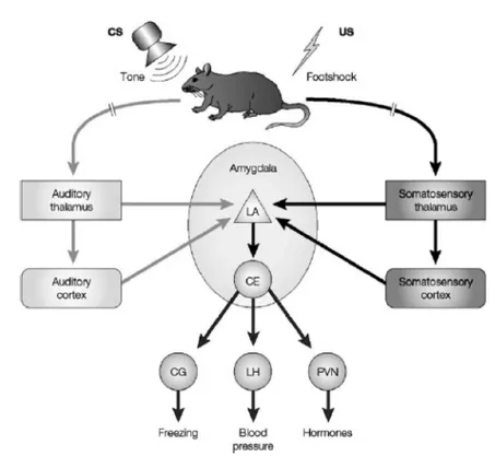
25 type of conditioning is dependent upon the amygdala (Fanselow and LeDoux, 1999; LeDoux, 2000; Maren, 2001). In a recent study it has been shown that specific activation of LA pyramidal cells as an US, in the absence of a peripheral shock US, produced fear conditioning, confirming that this nucleus is fundamental for associative conditioning learning (Johansen et al., 2010). A large number of studies have provided convincing evidences that associative plasticity in the LA contributes to fear memory formation (Sah et al., 2008; Maren et al., 2004; Blair et al., 2001). It is widely believed that LA plasticity underlying fear learning occurs as a result of a Hebbian mechanism whereby the shock US directly depolarizes LA pyramidal cells that are concurrently activated by weaker CS inputs, resulting in potentiation of the CS input synapses (Paré, 2002; Rogan et al., 2000). It has been proposed and now widely accepted that auditory information representing the CS and somatosensory information representing the US reach the lateral nucleus of the amygdala (LA) from both thalamic and cortical sources. Within the LA, individual neurons respond to both auditory and somatosensory stimuli suggesting convergence of CS and US inputs at the cellular level. Sensory information from the LA is then relayed to the central nucleus, both directly and indirectly via the basal, accessory basal, and intercalated nuclei. The central nucleus, in turn, projects to areas of the brainstem and hypothalamus that control the expression of defensive behaviours, hormonal secretions and autonomic responses (Sigurdsson et al., 2007) (Fig 4-2).
Fig. 4-2 Neural circuits underlying auditory fear conditioning (adapted from Sigurdsson et al., 2007)
The amygdala also plays a critical role in modulating memory consolidation, especially mediating the effects of acute stress on learning and memory (for a review Roozendaal et al., 2009). Acute and chronic stress exposure can induce functional and morphological changes as well as neuronal remodelling in the amygdala (Vyas et al., 2002 and 2004; for a review Roozendaal et al., 2009). Studies have reported for example that noradrenergic activation of the basolateral amygdala modulates consolidation of object recognition memory in rats (Roozendaal et al., 2008) and also inhibitory avoidance task (Ferry et al., 1999), contextual fear conditioning (Huff et al., 2005) or water -maze spatial training (Hatfield et al., 1999).
5 Excitatory synaptic transmission in the central
nervous system
Communication between neurons happen when an action potential reaches the terminal of a presynaptic neuron and voltage-dependent calcium (Ca2+) channels located in the presynaptic membrane open allowing Ca2+ to enter the cell. This influx of calcium ions triggers a series of events, which ultimately results in the release of the neurotransmitter from a synaptic vesicles into the synaptic cleft. Neurotransmitters drift across the synaptic space and bind special proteins located on the postsynaptic membrane called receptors. Neurotransmitters can initiate a process of excitatory synaptic transmission or inhibitory synaptic transmission, depending on which neurotransmitter is released and whether it causes a depolarization or a hyperpolarisation of the postsynaptic neurons.
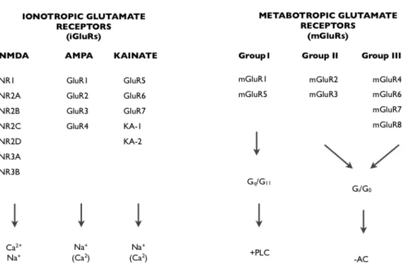
The amino acid glutamate is the major excitatory neurotransmitter in the mammalian central nervous system (CNS) and it exerts its effects by binding to glutamate receptors. It binds to two categories of receptors: ionotropic glutamate receptors (iGlurs) and metabotropic glutamate receptors (mGluRs)
(Fig. 5-1). The iGluRs are the
α-amino-3-hydroxy-5-methyl-4-isoxazolepropionate (AMPA) receptors, N-methyl-D-aspartate (NMDA) receptors and kainate (KA) receptors. (AMPA, Kainate, NMDA). For the purpose of this manuscript I will give only a brief description of mGluRs and a more detailed description of iGluRs.
Fig. 5-1 Glutamete Receptors and their constitute subunits. iGluRs can be subdivided based on
sequence homology and pharmacology, and are tetrameric complexes that allow the conductance of cations such as Ca2+ and Na+. mGluRs are G-protein coupled receptors and can be subdivided based on their intracellular signalling mechanisms. Group I mGluRs are positively coupled to phospholipase C (PLC), and group II and group III mGluRs are negatively coupled to adenylyl cyclise (AC). (Image adapted from Kew and Kemp, 2005).
5.1 Ionotropic glutamate receptors (iGluRs)
The ionotropic receptors are ligand gated ion channels and they are activated in response to the binding of a ligand molecule such as glutamate. Once the ligand is bound to the receptor, the receptor opens and positive charged ions such as Na+ and Ca2+ pass through the channel located in the centre of the
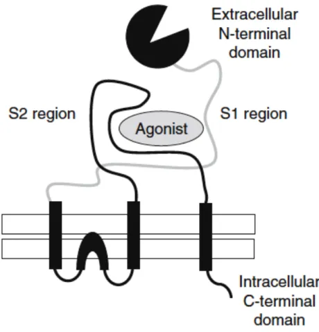
receptor complex. This flow of ions results in a depolarisation of the plasma membrane and the generation of an electrical current that is propagated down the processes (dendrites and axons) of the neuron to the next in line. The structure of ionotropic glutamate receptors is shown in Fig. 5-2. They are formed by a large extracellular N-terminal that cointains the ligand binding domain, three transmembrane regions and the intracellular C-terminal domain.
29
Fig. 5-2 Schematic representation of a ionotropic glutamate receptors (adapted from Kew and Kemp,
2005).
5.1.1 AMPA Receptors
AMPA receptors are responsible for the fast synaptic transmission in the CNS. They are permeable to Na+ and when glutamate binds to this receptor, the influx of Na+ ions results in neuronal depolarisation and the generation of an excitatory postsynaptic potential (EPSP) with the resultant generation of an action potential if threshold is reached creating a depolarization of the postsynaptic neuron.
AMPAR receptors are hetero -oligomeric proteins, four different genes (GluR1-4) encode AMPAR subunits (Hollmann and Heinemann, 199(GluR1-4). The extracellular and transmembrane regions of AMPAR subunits are very similar but vary in their intracellular cytoplasmic tails: GluR1, GluR4 and the long splice form of GluR2 have long cytoplasmatic carboxy -terminal tail (c-tail), while
GluR2, GluR3 and a short splice form of GluR4 have short and similar c -tails. Alternative splicing of the C -terminal domains determines the binding of the subunits to specific interacting proteins as well as the models of regulation of the receptors by protein phosphorylation. All four AMPAR subunits also occur in two alternatively spliced versions, Flip and Flop (Sommer et al., 1990) and form part of the extracellular ligand-binding domain. Flip variants are predominant prenatally, whereas Flop variants become expressed postnatally and reach levels equivalent to those of Flip in the adult. The Flip and Flop splice variants have effects on the rate and extent of desensitisation of heteromeric AMPA receptors and also influence their sensitivity to allosteric modulators (Kew and Kemp, 2005) (Fig. 5-3).
Fig. 5-3 Structure of the AMPAR subunits and the tetrameric channel. The individual subunits are
composed of four transmembrane domains, and the channel consists of four subunits, which are usually two dimers. The dimers are usually two different subunits, such as GluR1 and -2 or GluR2 and -3 (Shepherdand Huganir, 2007)
Native AMPA receptor channels are impermeable to calcium, a function controlled by the GluR2 subunit. The calcium permeability of the GluR2 subunit is determined by the post-transcriptional editing of the GluR2 mRNA, which changes a single amino-acid in the TMII region from glutamine (Q) to arginine (R). This is the so called Q/R editing site -GluR2(Q) is calcium permeable whilst GluR2(R) is not. Almost all the GluR2 protein expressed in the CNS is in the GluR2(R) form, giving rise to calcium impermeable AMPA receptors.
31 The C-terminus of the GluR2 subunit contains binding sites for a large number of interacting proteins such as NSF, AP2, as well as a terminal PDZ domain that binds PICK1 and GRIP, while the GluR1 subunit interacts with SAP97. These interactions are crucial for understanding the role of these receptors in synaptic plasticity (see next chapter).
5.1.2 NMDA Receptors
NMDA receptors mediate postsynaptic current that has a slow rise time and decay time. NMDAR has some basic properties: it is an ion channel sensitive to Na+, K+ and Ca2+, it is voltage-dependent because it needs a strong depolarization of the postsynaptic cell for its activation and it requires the binding of both glutamate and glycine to open. At resting membrane potentials, NMDARs are closed and ions cannot flow through the receptor channel due to a block by the Mg2+ ion. This is because at resting membrane potential, the
driving force for Mg2+, which is concentrated extracellularly, to enter the cell is
high. The Mg2+ therefore competes with Na+ and Ca2+ for access to the cell. However, as Mg2+ ions are too large to pass through the pore, the channel becomes effectively blocked. If the cell is depolarised then the Mg2+ block is removed and the current can flow (Dingledine et al., 1999). For these properties, NMDARs are known as “coincidence detectors” for postsynaptic depolarization and presynaptic release of glutamate.
Fig. 5-4 Schematic representation of the structure of NMDA receptor complex (adapted from:
http://www.frca.co.uk/article.aspx?articleid=100515).
NMDARs are heteromeric assemblies of NR1 (GluN1, with 8 different splice variants), NR2 (GluN2A, GluN2B, GluN2C and GluN2D) and NR3 (GluN3A and GluN3B) subunits that form ligand-gated channels with various cellular, biophysical and pharmacological properties depending on the composition of subunits and splice variants. Structurally they have the same membrane topology of ionotropic glutamate receptors. The c -terminal of both NR1 and NR2 interacts with several intracellular scaffolding proteins, it contains many serine/thereonine phosphorylation sites for proteins such as the cAMP -dependent protein kinase A (PKA), protein kinase C (PKC) and CaMKII (Chen and Roche, 2007), it is also involved in the regulation of receptor trafficking and function (Groc et al., 2009). Glutamate binds to NR2 subunits while the co-agonist glycine binds to the NR1 subunit. The NMDAR -dependent rise in
33 postsynaptic Ca2+ is important because it activates kinases (CAMKII, PKA, PKC and mitogen-activated protein kinase MAPK), and protein phosphatases, which ultimately results in an increase or decrease of AMPAR density and/or conductance relevant for long -term synaptic plasticity processes (Kerchner & Nicoll, 2008; Newpher & Ehlers, 2009) (Fig. 5-4).
5.1.3 Kainate Receptors
The last class of ionotropic glutamate receptors, the Kainate Receptors (KARs), have not been studied extensively so far, due to the lack of specific pharmacological antagonists. KARs are tetrameric receptor complexes composed of combinations of GluR5-7, KA1 and KA2, and have similar topology to AMPAR and NMDAR complexes. For the purpose of this manuscript I will not describe them in details.
5.2 Metabotropic glutamate receptors (mGluRs)
Metabotropic glutamate (mGlu) receptors are G-protein coupled receptors (GPCRs) that have been subdivided into three groups: mGluR1, mGluRII and mGluRIII, each group cointains different subunits for a total of eight sunbunits: mGlur1-8. The mGluRs bind glutamate within a large extracellular domain and transmit signals through the receptor protein to intracellular signalling partners. Group I mGlu receptors (mGlu1 and mGlu5) are positively coupled to PLC and intracellular calcium signalling, while group II (mGlu2 and mGlu3) and group III receptors (mGlu4, mGlu6, mGlu7 and mGlu8) are negatively coupled to adenylyl cyclase. The widespread expression of these receptors makes their study particularly interesting for drug targeting in neurological and psychiatric disorders such as Alzheimer’s disease, parkinson’s disease, anxiety, depression and schizophrenia.
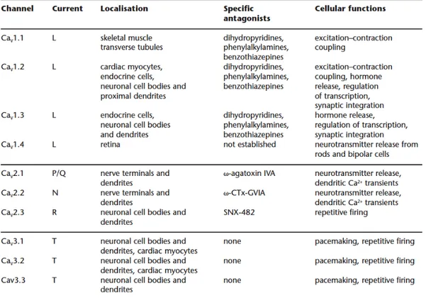
5.3 Voltage -dependent calcium channels (VDCCs)
Voltage -dependent calcium channels are the best source of Ca2+ influx into
neurons, muscles cells, endocrine cells and sensory cells. They activate in response to membrane depolarization. Electrophysiological studies have identified different forms of VDCCs, depending on their threshold of activation in response to depolarization. The high-voltage activated (HVA) channels require a strong depolarization and the low-voltage activated (LVA) channels that activate in response to a weak depolarization. The HVA are L -type, N -type, P/Q -type and R -type. The LVA comprises the T -type VDCC (see Fig. 5-5)
Fig. 5-5 Classification of the voltage dependent calcium -channels (adapted from International Union
of Pharmacology)
All VDCCs contain a pore -forming subunit, the α1 subunit that determines the
main biophysical and pharmacological properties of the different forms of VDCCs. There are three families of the α1 subunit: the Cav1 (that encodes the
35 and the Cav3 (that encodes the LVA T -type VDCC). The LVA channels are
thought to be formed by the only α1 subunit (Catterall W., 2000).
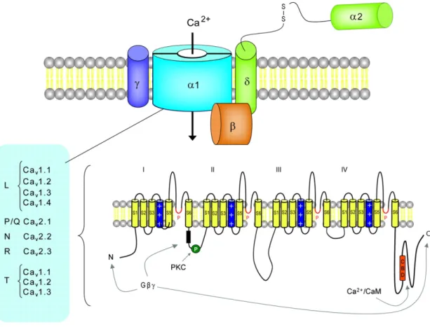
The HVA are heteromultimeres formed by the α1 subunit associated to three
auxiliary subunits: Cavβ, α2Δ and γ2. The α1 subunit of all channels (Cavα1) is
composed by four transmembrane domains, each consisting of six transmembrane helices (S1-S6). The segment four, S4, functions as the voltage sensor while S5 and S6 constitute the pore of the channels. The cytoplasmatic region of the α1 subunit is important for phosphorylation and interaction with
regulatory proteins, the intracellular c -terminal contains a binding site for calmodoulin CaM which mediates Ca2+ -triggered inactivation of the channels
upon prolonged membrane depolarization (Catterall W., 2000). The intracellular subunit Cavβ has α helices but no transmembrane segments; the γ2 is a
glycoprotein with four transmembrane segments and α2 is an extracellular,
extrinsic membrane protein (Fig. 5-6).
Calcium currents recorded in different cell types have shown various physiological and pharmacological properties. For example, the L-type channels require a strong depolarization for activation, they activate and inactivate slowly and are blocked by the organic calcium channels antagonists (i.e. dihydropyridines, phenylalkylamines and benzothiazepines). The other HVA channels (P/Q-,N-,R -type) also require a strong depolarization and are blocked respectively by agatoxin, conotoxin and SNX -482.
Voltage-gated Ca2+ channels are critical for signalling, plasticity, and injury in the nervous system. Studies in knockout or natural mutant mice indicate that many of these channels provide a target for pharmacologic treatment of absence epilepsy, cerebellar ataxia, and neuropathic pain (for a review see Bennaroch, 2007).
Fig. 5-6 General structure of voltage -gated calcium channels (VDCCs) (Bennaroch, 2007).
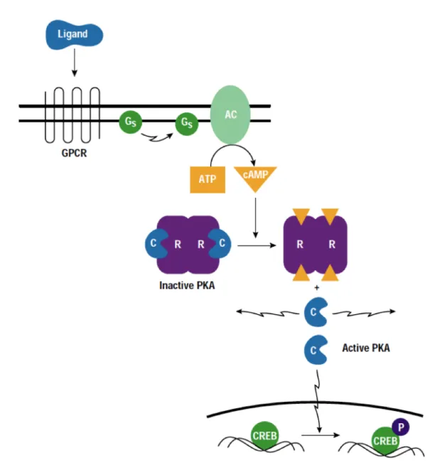
5.4 Cyclic Adenosine 3',5'-Monophosphate -dependent Protein Kinase A (cAMP -PKA)
The cAMP-dependent protein kinase, PKA is a second messenger -dependent enzyme (Smith et al., 1993) that has been linked to a wide range of cellular processes, including transcription, metabolism and apoptosis (Huggenvik et al., 1991, Hubbard et al., 1993, Matten et al., 1994).
PKA is composed of two regulatory (R) and two catalytic (C) subunits. The R subunits exist in two forms (RI and RII). Four genes encode the R subunits (RI-Alpha, RI-Beta, RII-Alpha and RII-Beta), and three encode the C subunits (C-Alpha, C-Beta and C-Gamma). Depending upon the associated RI and RII, PKA can be classified as Type I (predominantly located in the cytoplasm) and Type II (anchored to specific locations within the cell by A Kinase-Anchoring Protein, AKAP). Anchored PKA modulates the activity of various cellular proteins, including AMPA/Kainate channels, Glutamate receptor-gated ion channels,
L-37 type Ca2+ channels in skeletal muscle, hormone-mediated Insulin secretion in clonal beta cells, Vasopressin-mediated translocation of Aquaporin-2 into the cell membrane of renal principal cells, motility of mammalian sperm and the sperm Acrosome reaction (Dodge -Kafka and Kapiloff, 2006; Trewhella, 2006). Regulation of PKA in the cell is related primarily to modulation of its phosphotransferase activity. The holoenzyme contains two C subunits bound to homo -or heterodimers of either RI or RII subunits. The C subunits do not interact with one another. The R subunits each have an N-terminal dimerization domain and two cAMP binding sites.
Activation proceeds by the cooperative binding of two molecules of cAMP to each R subunit, which causes the dissociation and subsequent activation of each C subunit from the R subunit dimer (Fig. 5-7). cAMP is a cyclic nucleotide that serves as an intracellular and extracellular “second messenger” mediating the action of many peptide or amine hormones. The level of intracellular cAMP is regulated by the balance between the activity of two types of enzyme: AC (Adenylyl Cyclase) and the cyclic nucleotide PDE (Phosphodiesterase). Several receptors are responsible for the activation of cAMP-PKA pathway, GPCRs (G-Protein Coupled Receptors) being the most common receptors. When cyclic AMP levels are low, catalytic subunits are bound to a regulatory subunit dimer and are inactive. As the concentration of cyclic AMP increases, it binds to the regulatory subunits, leading to an allosteric change conformation which causes unleashing of the catalytic subunits. Free catalytic subunits are active and begin to phosphorylate their targets.
It has been shown that at the mossy fibers synapses, presynaptic LTP induction involves the cAMP/PKA pathway (Weisskopf et al., 1994). A similar sequence of events has been demonstrated to underlie presynaptic LTP induction at cerebellar parallel fiber synapse (Salin et al.,1996) corticothalamic synapses (Castro -Alamancos et al., 1999) and at cortico-lateral amygdala synapses (Fourcaudot et al., 2008).
6 Synaptic Plasticity
Synaptic plasticity is the ability of our neurons to change the efficacy or the strength of synaptic communication. These changes consist in either an enhancement or a depression of synaptic transmission at these synapses and they can be short- or long-lasting.
6.1 Short -term plasticity
Transient forms of synaptic plasticity have been associated with short-term adaptations to sensory inputs, transient changes in behavioural states and short-lasting forms of memory. Two forms of short-term plasticity are post-tetanic potentiation and post-post-tetanic depression (see Zucker and Regehr, 2002; Shepherd, 1998 for review).
Post-tetanic potentiation is a transient increase in the amplitude of a synaptic response that is seen after a brief train of stimuli. If a pair of stimuli is delivered and a potentiation of the second EPSP is observed, the phenomenon is defined ‘paired-pulse facilitation’ (PPF). This type of plasticity is largely believed to be pre-synaptic in origin (Bear et al., 1994). The first pulse leads to depolarisation of the presynaptic terminal and to an increase in intracellular Ca2+ that results in neurotransmitter release. If an optimal interval occurs between the first and second pulse, residual Ca2+ from the first pulse, plus the influx of Ca2+ due to the second pulse results in a greater increase in presynaptic Ca2+. This increases the probability of glutamate release from a given synapse, which results in a global increase in the amount of transmitter released and therefore a subsequent greater postsynaptic response to the second pulse (Sheperd, 1998; Zucker et al., 2002).
Post-tetanic depression is also thought to rely primarily on presynaptic mechanisms (Zucker et al., 2002). Depression of a synaptic response can occur
if there is a repetitive activation of a synapse that leads to a transient depletion of the presynaptic pool of neurotransmitter, or by the action of an inhibitory neurotransmitter such as GABA. Depression may also result from desensitisation of postsynaptic receptors after repeated binding of neurotransmitter (Zucker et al., 2002). If a pair of stimuli is delivered and a depression of the second EPSP is observed, the phenomenon is defined ‘paired-pulse depression’ (PPD).
6.2 Long-term plasticity
Lasting changes of synaptic transmission are thought to play an important role in the construction of neural circuits during development, and in the formation of long-term memories in the mature nervous system. Long-term plasticity consists in an enhancement (LTP) or depression (LTD) of synaptic strength, that may persist for many hours, weeks or more.
6.2.1 Long-term potentiation
In the early 1970s Bliss and Lomo found that repetitive stimulation of the excitatory synapses in the hippocampus causes an increase in synaptic strength that could last for hours or even days (Bliss and Lomo, 1973); this mechanisms is now known as long-term potentiation. Since then this phenomenon has been widely studied in different brain regions and it became the best candidate for understanding the cellular and molecular mechanism by which memories are formed and stored.
The classical model of long-term potentiation is the one studied at excitatory synapses between Schaffer collaterals and commissural axons and the apical dendrites of CA1 pyramidal cells of the hippocampus. The basic properties of this form of LTP that occurs in the CA1 region of the hippocampus are: it is
input-specific, it is elicited at the synapses that are activated by afferent activity
41 strong activation of a group of synapses can enhance the synaptic strength at adjacent synapses on the same cell if they are both activated within a small temporal window. Moreover it is triggered rapidly but can last for long periods of time. Three phases characterise the development of LTP: induction, expression and mantenance.
LTP induction
When a weak stimulation is applied to the presynaptic cell, its depolarization allows the release of neurotransmitter from the presynaptic vesicles. In excitatory synapses this neurotransmitter is usually glutamate and it binds to the postsynaptic AMPA receptors. The AMPA receptor is one of the main excitatory receptors in the brain, and is responsible for most of its rapid, moment-to-moment excitatory activity.Glutamate binding to the AMPA receptor triggers the influx of positively charged sodium ions (Na2+) into the postsynaptic
cell, causing a short depolarization called the excitatory postsynaptic potential (EPSP). NMDARs are present at postsynaptic membranes but at resting potential they are blocked by magnesium that does not allow calcium to enter the cell.
Figure 6-1 Model of the induction of LTP. During normal synaptic transmission, Glu is released from the
presynaptic bouton and acts on both AMPARs and NMDARs. However, Na+ flows only through AMPAR because the NMDAR is blocked by Mg+ (left). Depolarization of the postsynaptic cell relieves the Mg+ block of the NMDAR channel, allowing Na+ and Ca2+ to flow into the dendritic spine by means of the NMDAR (right). The increase of calcium influx is the crucial trigger for LTP (Malenka et al., 1999).
When a set of repeated stimuli is given at high frequency (usually at 100 Hz), the postsynaptic cell is progressively depolarized. In synapses that exhibit NMDA receptor-dependent LTP, sufficient depolarization unblocks NMDARs from the magnesium ion block allowing Ca2+ flow into the cell. The rapid rise in intracellular calcium concentration triggers a series of processes that mediate the early phase of LTP (see Fig. 6-1). For example, the transient rise of calcium activates the calcium/ calmodulin-dependent protein kinase (CaMKII), present at high concentration in the postsynaptic density (PSD), that in turn activates different downstream signalling cascade that are important for the later expression of LTP (Fukunaga et al., 1993; Lee et al., 2009). Moreover, protein kinase A (PKA) can be activated during this early phase of long -term potentiation, especially during early postnatal development when CaMKII expression is low (Malinow et al., 1993; Yasuda et al., 2003). Protein kinase C (PKC) has also been found to be important for LTP induction (Hu et al., 1987; Bliss and Collingridge, 1993). Lately also the mitogen-activated protein kinase (MAPK) cascade that activates extracellular signal-regulated kinases (ERKs) (Sweatt, 2004), the phosphatidylinositol 3-kinase (PI3 kinase) (Man et al., 2003) and the tyrosine kinase Src (Salter and Kalia, 2004) have been shown to play a role in triggering LTP.
LTP expression
The expression of LTP is a phenomenon that can depend either on presynaptic or postsynaptic mechanisms, or both. When LTP is expressed postsynaptically (for example, in most of NMDAR -dependent LTP) two major postsynaptic mechanisms are involved: the increase in the number of AMPARs at the synapse via trafficking, and the modification of AMPARs via the phosphorylation of the GluR1 subunit (Malenka et al., 1999; Malinow et al., 2002; Song et al., 2002; Bredt et al., 2003; Lee et al., 2003; Malenka et al., 2004). In other cases, although LTP is triggered postsynaptically, it is expressed presynaptically through the activation of a retrograde messenger (e.g. nitric oxide) to communicate from the postsynaptic cell back to the presynaptic terminal (Malenka et al., 1999 and 2004). The identification of this last form of LTP still remains elusive. However, the coexistence of both forms of long -term potentiation has been shown at glutamatergic synapses in the lateral nucleus of the amygdala (LA). In fact, conventional pairing-induced LTP and spike
timing-43 dependent LTP in thalamic projections to the LA are expressed postsynaptically and may implicate trafficking of AMPA receptors at stimulated synapses (Humeau et al., 2005; Rumpel et al., 2005), whereas LTP in cortical input to the LA is expressed presynaptically, resulting from an increase in the probability of neurotransmitter release (Tsvetkov et al., 2002).
LTP maintainance
One of the most exciting feature of LTP is its long-lasting property. It has been shown both in vitro and in vivo that LTP can last for hours or even days and years (Abraham et al., 2002). The processes that permit long-term potentiation to be long-lasting depend on proteins synthesis and gene expressions. Signalling molecules that are thought to link LTP induction to changes in gene transcription include calmodulin-dependent protein kinase IV (CaMKIV), mitogen activated protein kinase (MAPK) and PKA, which act downstream to phosphorylate the transcription factor CREB and zif268 (Lynch, 2004b; Warburton et al., 2005; Miyamoto 2006; Reymann et al., 2007).
A key role is played by PKMζ, an isoform of PKC believed to be critical for the maintenance of LTP. PKMζ becomes upregulated and activated approximately ten minutes following tetanic stimulation via an unknown mechanism. Unlike the kinases involved in LTP induction (PKA, PKC, MAPK, CaMKII), PKMζz lacks a regulatory domain and therefore may remain ‘persistently’ active, sustaining AMPA receptor phosphorylation (Osten et al., 1996). The role of PKMζ has been extensively studied in vitro in the hippocampus (Sacktor et al., 1996) and
in vivo in the neocortex (Shema et al., 2006) and in relation with specific
behaviours such as fear conditioning (Serrano et al., 2008).
Recent lines of research show that LTP can be maintained in the absence of new protein synthesis, albeit under defined experimental conditions. For example, it has been shown that application of mature brain-derived neurotrophic factor (BDNF) stabilizes LTP for at least several hours, even during application of the protein synthesis inhibitor anisomycin (Pang et al., 2994; Santi et al., 2006). Thus, in the presence of sufficient BDNF, LTP can become protein synthesis-independent.