Modulation of the Catalytic Activity of Neutrophil Collagenase
MMP-8 on Bovine Collagen I
ROLE OF THE ACTIVATION CLEAVAGE AND OF THE HEMOPEXIN-LIKE DOMAIN*
Received for publication, November 13, 2001, and in revised form, April 5, 2002 Published, JBC Papers in Press, April 12, 2002, DOI 10.1074/jbc.M110873200
Magda Gioia‡§, Giovanni Francesco Fasciglione‡§, Stefano Marini‡, Silvana D’Alessio¶, Giampiero De Sanctis储, Oliver Diekmann**, Michael Pieper**, Vincenzo Politi¶,
Harald Tschesche**, and Massimo Coletta‡ ‡‡
From the ‡Department of Experimental Medicine and Biochemical Sciences, University of Roma Tor Vergata, Via di Tor Vergata 135, I-00133 Roma, Italy,¶PoliFarma, Via di Tor Sapienza 138, I-00155 Roma, Italy,
储Department of Molecular, Cellular and Animal Biology, University of Camerino, I-62032 Camerino (MC), Italy, and
**Department of Biochemistry I, University of Bielefeld, D-33051 Bielefeld, Germany
The cleavage of bovine collagen I by neutrophil colla-genase MMP-8 has been followed at pH 7.4, 37 °C. The behavior of the whole enzyme molecule (whMMP-8), dis-playing both the catalytic domain and the hemopexin-like domain, has been compared under the same exper-imental conditions with that of the catalytic domain only. The main observation is that whMMP-8 cleaves bovine collagen I only at a single specific site, as already reported by many others (Mallya, S. K., Mookhtiar, K. A., Gao, Y., Brew, K., Dioszegi, M., Birkedal-Hansen, H., and van Wart, H. E. (1990) Biochemistry 29, 10628 –10634; Kna¨ uper, V., Osthues, A., DeClerk, Y. A., Langley, K. A., Bla¨ ser, J., and Tschesche, H. (1993) Biochem. J. 291, 847– 854; Marini, S., Fasciglione, G. F., De Sanctis, G., D’Alessio, S., Politi, V., and Coletta, M. (2000) J. Biol. Chem. 275, 18657–18663), whereas the catalytic domain lacks this specificity and cleaves the collagen molecule at multiple sites. Furthermore, a meaningful difference is observed for the cleavage features displayed by two forms of the catalytic domain, which differ for the N terminus resulting from the activation process (i.e. the former Met80of the proenzyme (MetMMP-8) and the for-mer Phe79 of the proenzyme (PheMMP-8)). Thus, the PheMMP-8 species is characterized by a much faster kcat/Km, fully attributable to a lower Km, suggesting that
the conformation of the catalytic domain, induced by the insertion of this N-terminal residue in a specific pocket (Reinemer, P., Grams, F., Huber, R., Kleine, T., Schnierer, S., Piper, M., Tschesche, H., and Bode, W. (1994) FEBS Lett. 338, 227–233), brings about a better, although less discriminatory, recognition process of cleavage site(s) on bovine collagen I.
Neutrophil collagenase MMP-81is a Zn2⫹
metallo-endopep-tidase, which is able to cleave native triple-helical collagen I (1–3) at a specific peptide bond between Gly775and the residue
at position 776 (which can be either Leu or Ile), leading to the formation of one-quarter and three-quarter fragments (4). MMP-8 is predominantly expressed by neutrophil precursors (5, 6), but very recently it has been demonstrated that it is also expressed in human articular chondrocytes (7) and that it might also be responsible for aggrecanase activity (8, 9). MMP-8 is expressed initially as a proenzyme and stored in the specific granules of neutrophils (10). The activation of MMP-8 can be accomplished either by mercurials, oxidative processes, and proteinases (11–14) thus removing a cysteinyl residue, which coordinates the Zn2⫹atom, rendering the enzyme inac-tive. In particular, it was observed that cathepsin G, a neutro-phil serine proteinase, and stromelysin MMP-3 activate proMMP-8 through two different processes, the first one cleav-ing the Phe79–Met80peptide bond (11), whereas MMP-3
acti-vates through the cleavage of the Gly78–Phe79 peptide bond
(15). Therefore, the resulting active MMP-8 displays two dif-ferent N termini (i.e. Met80 if activated by cathepsin G and
Phe79if activated by MMP-3), and it has been shown that these
two forms display meaningful differences in the catalytic activ-ity toward synthetic substrates or inhibitors (16, 17). The final active enzyme is made of a catalytic domain, where the Zn2⫹
atom coordinated to 3 histidyl residues is located, and a he-mopexin-like domain, which is connected to the catalytic do-main through a linker region (18, 19).
Collagen I is one of the major components of the extracellular matrix for most tissues, such as skin, tendon, blood vessels, cartilage, bones, and basal laminae (20). It displays a triple-helical structural arrangement (21), which is kept by most of the molecule during the first cleavage event by MMP-8 (22), even though three-dimensional structures of collagenases indi-cate that the substrate binding cleft appears too narrow to accommodate a triple-helical collagen molecule. The two sets of data are not necessarily in contradiction, because a partial unwinding of collagen may be envisaged during the enzymatic action (23–25) while keeping the overall three-dimensional structure of the whole collagen molecule.
Several studies have shown that, although the actual enzy-matic action of collagenases occurs in the catalytic domain, the hemopexin-like domain plays an important role, because its
* This work was supported in part by funds from the Italian Minis-tero dell’Universita` e della Ricerca Scientifica e Tecnologica (MURST COFIN MM03185591 to M. C.) and the Deutsche Forschungsgemein-schaft, Bonn, Grants SFB 549 and SFB A5 (to H.T.) and Project Ts 8/35 (to H. T.). The costs of publication of this article were defrayed in part by the payment of page charges. This article must therefore be hereby marked “advertisement” in accordance with 18 U.S.C. Section 1734 solely to indicate this fact.
§ These authors contributed equally to the work.
‡‡ To whom correspondence should be addressed: Dept. of Experi-mental Medicine and Biochemical Science, University of Roma Tor Vergata, Via di Tor Vergata 135, I-00133 Roma, Italy. Tel.: 39-06-72596365; Fax: 39-06-72596353; E-mail: [email protected].
1The abbreviations used are: MMP-8, Zn2⫹metallo-endopeptidase-8
neutrophil collagenase; whMMP-8, whole MMP-8 molecule; Met-MMP-8, catalytic domain of Met-MMP-8, where the N terminus is Met80of
the former proenzyme; PheMMP-8, catalytic domain of MMP-8, where the N terminus is Phe79of the former proenzyme.
© 2002 by The American Society for Biochemistry and Molecular Biology, Inc. Printed in U.S.A.
Ile(Leu)776even in the absence of the C-terminal domain (27).
Therefore, the question of the role played by the hemopexin-like domain remains partially open, because it is not evident whether it is absolutely necessary for the enzymatic action or else it has a modulatory function, regulating substrate recog-nition and processing (28). Furthermore, very recent data on MMP-2 indicate a role as well for the hemopexin-like domain in the collagenolytic activity of this MMP (29).
To have a better insight into the role of the hemopexin-like domain, we carried out a kinetic investigation on the fragmen-tation of bovine collagen I by the whole MMP-8 (whMMP-8, i.e. the molecule with both the catalytic and the hemopexin-like domains) and by the catalytic domain only. Furthermore, in the attempt to better characterize the potential in vivo effects of the different activation cleavages (11, 15), we compared the enzymatic activities on bovine collagen I of the catalytic do-main displaying Phe79as the N terminus (PheMMP-8) and the
catalytic domain displaying Met80 as the N terminus
(Met-MMP-8). The results allow us to envisage a more complex role played by the hemopexin-like domain, which appears very im-portant in the recognition process of the first cleavage site on the whole collagen molecule. In addition, the superactivation of the MMP-8 displaying Phe79 as the N terminus (16, 17) is
confirmed also for natural substrates such as bovine collagen I.
MATERIALS AND METHODS
Isolation of whMMP-8 and expression, purification, and refolding of recombinant MetMMP-8 and PheMMP-8 were carried out as previously reported (6).
Isolated purified whMMP-8 proenzyme was activated by incubating 0.1 ml of a 0.1g/ml procollagenase solution with p-aminophenyl mer-curic acetate (Sigma) at 37 °C for 2 h. This treatment shifts the equi-librium of the conformation toward the open, autocatalytically acti-vated form involving the cleavage of the region between residues 71 and 81. Because BB-94 (known also as Batimastat, a peptidomimetic MMP inhibitor, kindly provided by British Biotech Pharmaceuticals, Cowley, Oxford, UK) fully inhibits MMPs stoichiometrically, we employed it to titrate the active amount of the enzyme.
Catalytic domains displaying either Phe79 (PheMMP-8) or Met80
(MetMMP-8) were kept activated in small aliquots at – 80 °C until used for the experiments.
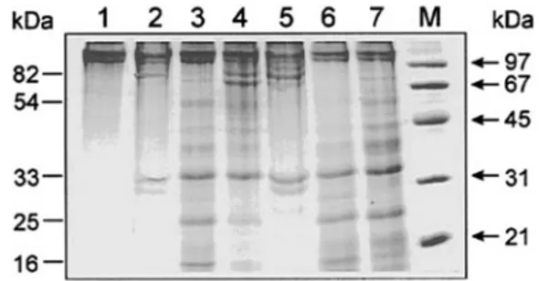
The purity of whMMP-8, MetMMP-8 and PheMMP-8 was checked by SDS-polyacrylamide gel electrophoresis according to Laemmli (30). Af-ter gels were run, they were stained using a silver staining kit (Bio-Rad). Zymography was performed as follows: 2l of purified MMP-8 was mixed with a 5-fold excess of sample buffer (0.25MTris, 0.8% SDS, 10% glycerol, and 0.05% bromphenol blue) and run on 12% SDS-poly-acrylamide gels containing either 1 mg/ml gelatin or bovine collagen I as described previously (30). After electrophoresis, SDS was removed from the gels by washing twice for 15 min in 2% Triton X-100. Gels were then incubated at 37 °C for 18 h in incubation buffer (50 mMTris/HCl,
pH 7.6, 0.15MNaCl, 10 mMCaCl2, 2% Triton X-100), stained with 0.5%
Coomassie Blue, and destained in 10% acetic acid and 40% methanol until pale proteinase bands were clearly visible. The proteinase band was further characterized by adding 20 mMEDTA or 0.3 mM
1,10-phenantroline (MMP inhibitors) or 1 mMphenylmethylsulfonyl fluoride (serine proteinase inhibitor) in the incubation buffer. Protein markers (Sigma) were used as the molecular weight standard (22, 31).
Bovine collagen I was dissolved in acidified water at room tempera-ture for 2 days. Afterward, the suspension was centrifuged for 1 h at 10,000⫻ g, and the supernatant containing the dissolved collagen was used. The amount of collagen was quantified as described by Bradford (32). The triple-helical structural arrangement was checked by circular dichroism in agarose gel-entrapped bovine collagen I (22), and only samples displaying a correct ellipticity in the 230 –220 nm range (33, 34) were employed for experimental assays.
Fragmentation kinetics were carried out as previously reported (22), employing different concentrations of bovine collagen (spanning 0.3 to
1.2M) at pH 7.4. The enzyme(s) concentration ranged between 0.1 and 1 nM.
Electrophoretic spots, corresponding to different fragmentation in-termediates of bovine collagen I, were analyzed as a function of time by laser densitometry (LKB 2202 UltraScan), and their intensity was calibrated (in order to obtain concentration values) using standard collagen solutions.
RESULTS AND DISCUSSION
Fig. 1 shows the electrophoretic pattern of bovine collagen fragmentation at 37 °C by the whole neutrophil collagenase MMP-8 (whMMP-8), by the catalytic domain of MMP-8 display-ing Met80as the N terminal (MetMMP-8), and by the catalytic
domain of MMP-8 displaying Phe79 as the N terminal
(PheMMP-8). It appears immediately obvious that whereas whMMP-8 cleaves the bovine collagen only at one site, showing only two fragments after 3 and 6 h (see Fig. 1, lanes 2 and 5), as already reported by us (22), both MetMMP-8 and PheMMP-8 show multiple cleavage sites, with several bands of differing molecular weight (see Fig. 1, lanes 3, 4, 6, and 7). This obser-vation clearly indicates that the removal of the hemopexin-like domain in MMP-8 brings about a loss of specificity in recogniz-ing the cleavage site on bovine collagen between Gly775and the
residue in position 776, which is instead peculiar to the enzy-matic action of whMMP-8 (4). This result is significantly dif-ferent from what has been reported for MMP-1, where the removal of the C-terminal domain was observed to abolish the capability of cleaving collagen (26). However, an additional difference is represented by the temperature, which was 25 °C in the case of MMP-1 (26) and 37 °C in our case (MMP-8). To test whether this fact might account for the difference in be-havior, we carried out the kinetics of collagen processing at 25 °C (Fig. 2, A and B). The outcome of this experiment was very interesting. It showed that whereas at 37 °C after 6 h the catalytic domains had processed more collagen I than whMMP-8 had, at 25 °C after 18 h (the longer time interval is because the kinetics are much slower at 25 °C than at 37 °C) whMMP-8 had processed some collagen I, and essentially no collagen has been processed by the catalytic domains (see Fig. 2A, lanes 5 and 6). Only after 48 h did we observe a significant amount of collagen I processed also by the catalytic domains (Fig. 2B, lanes 2 and 3). Therefore, this result, which confirms for MMP-8 the behavior reported for MMP-1 (26), clearly indi-cates that catalytic domains are able to process collagen I but the temperature dependence of their enzymatic activity is dra-matically increased, such that their processing rate is much slower than whMMP-8 at 25 °C and somewhat faster at 37 °C. Such a feature underlies the possibility of a larger activation energy of the catalytic activity for MetMMP-8 and PheMMP-8 with respect to whMMP-8, likely related to a larger structural flexibility for the catalytic domains alone, which indeed should be less structured than whMMP-8. However, this striking
tem-FIG. 1. Comparison of the fragmentation of bovine collagen I
by whMMP-8 (lanes 2 and 5), MetMMP-8 (lanes 3 and 6), and PheMMP-8 (lanes 4 and 7). Lane 1, intact bovine collagen I; lane M,
molecular weight markers; lanes 2–7, SDS-polyacrylamide gel of sam-ples after 1.5 (lanes 2– 4) and 6 h (lanes 5–7) of digestion at 37 °C. For further details, see “Materials and Methods.”
perature dependence of the catalytic activity may also (but not only) be correlated to the partial decrease of the compactness of the triple-helical arrangement of collagen I as the temperature
is raised (Fig. 2C); this effect might render collagen more open to attack even in a less specific fashion (as undertaken by catalytic domains, see above). In this respect, it may be
out-FIG. 2. A, SDS-polyacrylamide gel elec-trophoresis of bovine collagen I fragmen-tation at 25 °C by whMMP-8 (after 3 h,
lane 2 and after 18 h, lane 4), Met-MMP-8
(after 3 h, lane 3 and after 18 h, lane 5), and PheMMP-8 (after 18 h, lane 6). Lane
1, bovine collagen I incubated for 18 h
without enzyme. B, SDS-polyacrylamide gel electrophoresis of bovine collagen I fragmentation after 48 h at 25 °C by whMMP-8 (lane 1), MetMMP-8 (lane 2), and PheMMP-8 (lane 3). C, circular di-chroism spectra of agarose gel-entrapped bovine collagen I at 10 °C (a), 25 °C (b), and 37 °C (c). For further details, see “Ma-terials and Methods.”
lined that the decrease of ellipticity at 37 °C (see Fig. 2C) may also be related to some physical effect on the agarose gel, because at 37 °C we are approaching the melting point for agarose, and the extent of gelification could have some influ-ence on the compactness of the triple-helical arrangement of the collagen molecule.
The catalysis of bovine collagen can then be followed with the three enzymatic forms for longer time intervals and analyzed through the temporal evolution of different intermediate spe-cies with varying molecular weight (22). Fig. 3 shows an exam-ple of this type of experiments, with each lane corresponding to the species present at a given time interval after the incubation of bovine collagen I with PheMMP-8. To obtain a quantitative estimate of the concentration of intermediates, the different spots have been calibrated with known amounts of bovine collagen. Independent experiments have demonstrated that under these experimental conditions (i.e. pH 7.4 and 37 °C) no autodegradation of the bovine collagen occurs over the period time of our observations (data not shown). It may be important to underline at this point that the conformation of bovine collagen I has been checked by circular dichroism in the 250 – 200 nm region (32, 33), and only samples characterized by the typical positive ellipticity (Fig. 4) have been employed for ex-periments. The catalytic action of whMMP-8, as well as of MetMMP-8 and PheMMP-8, leads to the disappearance of the positive ellipticity at 224 nm (Fig. 4, A and B), due to the unwinding of the triple-helical arrangement (33, 34). The ki-netics of this process, however, appears very different in the case of whMMP-8 with respect to the catalytic domains Met-MMP-8 and PheMet-MMP-8 (Fig. 4C). Therefore, the lack of the hemopexin-like domain appears also to bring about a much faster unwinding and degradation of bovine collagen I (with enhancement of the helicase activity of MMP-8), even though circular dichroism observations alone do not allow us to detail the differences in collagen degradation among the three species (Fig. 4C).
However, this information can be obtained following the kinetics of collagen degradation by SDS electrophoresis. Fig. 5 shows the time evolution of different intermediate species of bovine collagen, which have been detected during the incuba-tion with whMMP-8 (panel A), MetMMP-8 (panel B), and PheMMP-8 (panel C). In the case of whMMP-8 only three species can be observed, as already reported (21). The overall behavior can be described according to reaction Scheme I, where A is the whole bovine collagen molecule, B and D corre-spond to the species at about 85 and 30 kDa, respectively, formed at the beginning of the reaction, and C, which is ob-served only at some later stage, corresponds to the species of about 50 kDa (see Fig. 5A). For MetMMP-8 and PheMMP-8 we
observed four major populations, the amount of which has changed with time, but which were all present from the begin-ning of enzymatic processing. They appear to correspond to fragments of about 85, 55, 33, and 25 kDa (see Fig. 5, B and C),
FIG. 3. SDS-polyacrylamide gel of fragmentation kinetics at
37 °C. Different lanes correspond to the following intervals after
incu-bation of bovine collagen I with 0.7 nMPheMMP-8: 0 (lane 1), 0.25 (lane
2), 0.5 (lane 3), 1 (lane 4), 2 (lane 5), 4 (lane 6), 6 (lane 7), and 24 h (lane 8). Lane M, molecular mass markers. For further details, see “Materials
and Methods.”
FIG. 4. A and B, circular dichroism spectra of agarose gel-entrapped bovine collagen I as a function of time after exposure to proteolytic fragmentation by MetMMP-8 (A) and PheMMP-8 (B). C, time evolution of the decrease of ellipticity at 224 nm of bovine collagen I after expo-sure at 37 °C to whMMP-8 (*), MetMMP-8 (E), and PheMMP-8 (⫻). For further details, see text.
even though it must be emphasized that species of larger mo-lecular mass are less defined, and therefore we cannot rule out the possibility that more than one species is formed with a molecular mass ranging between 80 and 100 kDa. In any event, the occurrence of four (or five) species since the beginning of the cleavage process suggests that the catalytic domain alone (ei-ther MetMMP-8 and/or PheMMP-8) is active on (at least) three different cleavage sites on the bovine collagen I molecule, lead-ing to a variety of intermediate species. Scheme II describes the simplest mechanism that can be sketched on the basis of the observed species, where A is the whole bovine collagen mole-cule, B and C are two large molecular weight fragments, which might range between 80 and 100 kDa, D is the⬃55-kDa frag-ment, E is the⬃33-kDa fragment, F is the ⬃25-kDa fragment, and G represents smaller undefined fragments (not reported as populations in Fig. 5, B and C). The continuous curves repre-sent the best simulation of observed data employing the reac-tion mechanism reported in Scheme I (for Fig. 5A) and in Scheme II (for Fig. 5, B and C); they appear to describe the observed behavior satisfactorily, indicating that the mecha-nism reported in Scheme II is a realistic representation of the actual event(s). The kinetic parameters employed to simulate data reported in Fig. 5 are listed in Table I. However, the complexity of Scheme II impedes the possibility of assigning a physical significance to all these parameters, because a cross-correlation exists among different parameters of steps subse-quent to the first one(s). In a previous analysis of the cleavage process of bovine collagen I by MMP-8, we have shown that the first cleavage step can be analyzed in greater detail according to the Michaelis-Menten assumption (22), as also suggested by the linearity of the corresponding Lineweaver-Burk plot. We have thus attempted this analysis in our system, measuring the amount of product during the first 3 h (corresponding to the formation of species B and D of Scheme I and of species B, C, D, E, and F of Scheme II) as a function of collagen concentra-tion; the slope of the concentration rising for each species, Vi,
has been utilized to obtain Lineweaver-Burk plots (Fig. 6). Their excellent linearity (see Fig. 6) indeed suggests that the Michaelis-Menten assumption might also be employed for the analysis of kinetics for the initial steps in Scheme II, even though we have to be cautious in giving these parameters an absolute quantitative validation. However, although only to a semiquantitative extent, the resulting catalytic parameters for
SCHEMEI
SCHEMEII
FIG. 5. A, time evolution of the concentration of different fragments of bovine collagen I (starting concentration⫽ 1. 2M) at 37 °C by 0.55 nM
whMMP-8 (with symbols corresponding to the following species (see Scheme I): E, B; *, C; and⫻, D. B, 0.3 nMMetMMP-8 (with symbols
corresponding to the following species (see Scheme II): E, (B⫹ C); ⫻, D; ⫹, E; and *, F. C, 0.7 nMPheMMP-8 (with symbols corresponding to the following species (see Scheme II): E, (B⫹ C); ⫻, D; ⫹, E; and *, F. Data are plotted on a logarithmic time scale. Continuous lines were obtained according to kinetic Schemes I and II, employing parameters reported in Tables I and II. For further details, see text.
the investigated cleavage processes (i.e. A3 B, A3 C, A 3 D; see Scheme II and Table II) may be usefully compared with those obtained previously at this pH for whMMP-8 (22). Ex-ploiting these parameters, we can then calculate for these steps the observed rate constants,
ki⫽ kcat关E0兴/共Km⫹ 关collagen]) (Eq. 1) where i⫽ 1 for Scheme I and i ⫽ 1, 2, 3 for Scheme II, and E0
is the total enzyme concentration. Such values of k have been then imposed on the respective step(s) to draw continuous lines in the description of the temporal evolution of each species (see Fig. 5). This has been described as a derivative of each species
concentration as a function of time, being iterated according the k values and influencing that species for finite time inter-vals. As an example, if we pick species B in Scheme II,
关B兴x⫽ 关B兴x⫺1⫹ 共k1⬘关A兴x⫺1⫺ k4⬘关B兴x⫺1兲 䡠 dt (Eq. 2) where [B]xis the concentration of species B at time⫽ x, [B]x⫺ 1
is the concentration of B at (time ⫺dt) (the same is true for [A]x⫺ 1), and k1⬘ and k4⬘ are the rate constants for the process reported in Scheme II. The same procedure has been applied to all species appearing in Schemes I and II.
This procedure allows us to give a semiquantitative descrip-tion of the mechanism reported in Fig. 5 employing parameters that might have some physical significance, at least for the first step(s). Other parameters referring to further steps (and re-ported in Table I) have only a phenomenological meaning.
A closer look at the parameters reported in Table II allows a comparison of the catalytic mechanism of collagen cleavage between the three forms of MMP-8 with reference to the first enzymatic attack on the intact collagen molecule. If we focus our attention on the formation of B from whMMP-8 (see Scheme I), which can be compared with the formation of either B or C from MetMMP-8 and PheMMP-8 (see Scheme II), we can immediately outline that, as compared with the value observed for whMMP-8, kcat/Km is somewhat faster in
Met-MMP-8 and much faster in the case of PheMet-MMP-8 (see Table II). This overall observation is perfectly in line with what was observed previously in synthetic substrates, where PheMMP-8 turned out to be much more active than MetMMP-8 (17). This feature has been attributed to a different conformation of the catalytic domain of the enzyme according to whether the N terminus is Met80, likely being freely rotating in the bulk, or
Phe79, which appears inserted in a hydrophobic pocket near the
active site (16). However, if we extend our observation to other catalytic parameters, such as kcatand Km, the formation of
species B in whMMP-8 in Scheme I (resulting in a fragmenta-tion of the collagen molecule in one 85-kDa and in one 30-kDa portion) can be better compared with the process leading in Scheme II to the formation of species B (of⬃80/85 kDa) and species E (of⬃33 kDa). Therefore, looking at the parameters for this process we can say that the removal of the hemopexin-like domain (as observed from the comparison between whMMP-8 and MetMMP-8, in both cases the N terminus being Met80) brings about a significant decrease in the rate constant
for the rate-limiting step, which is somewhat overcompensated by the decrease of Km; this suggests a marked increase of affinity for MetMMP-8 toward the similar site in the collagen (see Table II). Therefore, the removal of the hemopexin-like domain seems to induce a conformational change in MMP-8, which leads to an increase of the number of sites on the bovine collagen I being recognized by the enzyme. It also enhances significantly the affinity of MMP-8 not only for sites in common with whMMP-8 but also for new sites. On the other hand, such a conformational change, related to the removal of the he-mopexin-like domain, reduces to a relevant extent the rate constant for the catalytic rate-limiting step (this being espe-cially relevant for the newly recognized sites, i.e. A3 C and A 3 D). On the whole, after the removal of the hemopexin-like
FIG. 6. Lineweaver-Burk plots of bovine collagen I
concentra-tion dependence on the first three steps from Scheme II, namely k1ⴕ (E), k2ⴕ (ⴛ), and k3ⴕ (*) for MetMMP-8 (A) and PheMMP-8 (B); Tⴝ 37 °C. Continuous lines were obtained employing parameters re-ported in Table II, which were obtained by nonlinear least-squares fitting of data. For further details, see text.
whMMP-8 0.05⫾ 0.02 0.04⫾ 0.01 0.12⫾ 0.03
MetMMP-8 0.15⫾ 0.04 0.15⫾ 0.03 0.15⫾ 0.04 0.035⫾ 0.009 0.20⫾ 0.03
domain, MMP-8 cleaves individual sites on collagen with a reduced efficiency with respect to the action of whMMP-8. The overall effect, however, is an indiscriminate fragmentation of the collagen molecule, which leads to a much faster and more extended unwinding of the collagen.
A comparison of the behavior observed for the two catalytic domains, MetMMP-8 and PheMMP-8 (both lacking the he-mopexin-like domain), is very interesting because in this case the presence or not of an additional residue (i.e. Phe79) is the
result of two varying activation processes of the proenzyme (11, 15). In this case we observed for PheMMP-8 an overall increase of the kcat/Kmvalues for all three cleavage sites, even though
the functional bases for this increase are different for the three sites (see Table II). Therefore, focusing on the cleavage process that is likely common to that observed in whMMP-8 (i.e. the A 3 B process, as seen above) we can see that the presence of Phe79 as an N terminus of the catalytic domain further
de-creases the value of kcatwith respect to MetMMP-8, keeping a significantly higher affinity for the substrate as compared with whMMP-8 and MetMMP-8. Therefore, the conformational change detected in PheMMP-8 with respect to MetMMP-8 (16) dramatically decreases the speed of the rate-limiting step, which is largely overcompensated by an enhanced affinity to the substrate binding for the formation of the ES complex, thus resulting in a much higher value for kcat/Km(see Table II).
Further, in comparing whMMP-8 on one side and the two catalytic domains (i.e. MetMMP-8 and PheMMP-8) on the other side, we turn our attention also toward subsequent steps, al-though in these cases we must bear in mind that kinetic con-stants have only a phenomenological significance (see above). In such a case, we observe that already the removal of the hemopexin-like domain (as from the comparison between k2for
whMMP-8 and k4⬘ for MetMMP-8; see Table I) induces an
⬃3-fold rate enhancement, whereas the presence of Phe79as
the N terminus in the activated enzyme (as in the comparison of k4⬘ between MetMMP-8 and PheMMP-8; see Table I) brings
about an additional 10-fold rate enhancement.
In conclusion, it seems that the enzymatic processing of bovine collagen I by MMP-8 displays several modulatory mech-anisms, which appear to involve different domains of the mol-ecule. As already suggested previously (22), domains located far from the active site are heavily involved in the substrate recognition mechanism. This has been also confirmed by a recent investigation on the x-ray structure of MMP-8 bound to an inhibitor (35), where it is proposed that the bound substrate interacts with an exosite (formed by residues Asn188 and
Tyr189), leading to a major contact with blade 2 of the
C-terminal domain. Such an observation then leads to the possi-bility also that the catalytic domain alone of MMP-8 should be able to display a collagenolytic activity (35). Such a prediction is further substantiated by this investigation, because the re-moval of the hemopexin-like domain indeed has a gross
influ-ence on the value of Km, which becomes lower (thus reflecting
an increased affinity for substrate). Therefore, it appears that the hemopexin-like domain in some way restricts the accessi-bility of the recognition site of MMP-8 for collagen, rendering the formation of the ES complex with collagen more difficult. As a matter of fact, after the removal of the hemopexin-like domain the recognition mechanism becomes much less selec-tive; this clearly suggests that the hemopexin-like domain plays a more crucial role in rendering specific the action of MMP-8 on collagen than on rendering possible the collageno-lytic activity itself. Furthermore, from the circular dichroism data the unwinding of the collagen appears to occur much more rapidly, suggesting that the removal of the hemopexin-like domain uncovers a helicase-like site on MMP-8. However, it must also be stressed that the removal of the hemopexin-like domain seems to bring about a conformational change in MMP-8, which alters the structural arrangement of the active site such that in the catalytic domain alone the rate-limiting step shows a much slower kcat. In this respect, the activation
process of the proenzyme appears very important, because the newly formed N terminus and its structural arrangement (16) play an important role in modulating the rate of the catalysis-limiting step, kcat, which becomes dramatically reduced also for
a natural substrate by the presence of Phe79as the N terminus.
It should be emphasized that the larger activation energy for collagen cleavage displayed by catalytic domains alone (as sug-gested by the crossing-over of rates at different temperatures; see Figs. 1 and 2) might be in keeping with a less structured active site in the catalytic domain(s) alone, which require(s) a larger kinetic energy to become fully active, resulting in a less efficient kcat.
This observation, extended and demonstrated also for a nat-ural substrate, gives strength to the possible role of the acti-vation process in giving rise to a “superactive” enzyme form, which may be relevant in triggering pathological events. Fur-thermore, this study enforces the idea that extending the sit-uation in vivo from observations made on synthetic substrates and on incomplete enzyme forms must always be done very cautiously, because different domains, even those far away from the active site, may play important roles in large macro-molecular natural substrates.
REFERENCES
1. Hasty, K. A., Jeffrey, J. J., Hibbs, M. S., and Welgus, H. G. (1987) J. Biol. Chem. 262, 10048 –10052
2. Kna¨uper, V., Osthues, A., DeClerk, Y. A., Langley, K. A., Bla¨ser, J., and Tschesche, H. (1993) Biochem. J. 291, 847– 854
3. Sanchez-Lopez, R., Alexander, C. M., Behrendtsen, G., Breathnach, R., and Werb, Z. (1993) J. Biol. Chem. 268, 7238 –7247
4. Mallya, S. K., Mookhtiar, K. A., Gao, Y., Brew, K., Dioszegi, M., Birkedal-Hansen, H., and van Wart, H. E. (1990) Biochemistry 29, 10628 –10634 5. Mainardi, C. L., Pourmotabbed, T. F., and Hasty, K. A. (1991) Am. J. Med. Sci.
302, 171–175
6. Tschesche, H. (1995) Methods Enzymol. 248, 431– 449
7. Cole, A. A., Chubinskaya, S., Schumacher, B., Huch, K., Cs-Szabo, G., Yao, J., Mikecz, K., Hasty, K. A., and Kuettner, K. E. (1996) J. Biol. Chem. 271,
TABLE II
Catalytic parameters obtained at pH 7.4 and 37 °C in the enzymatic processing of bovine collagen I from whMMP-8 and from the catalytic domain displaying Met80as N terminus (MetMMP-8) and the catalytic domain displaying Phe79as N terminus (PheMMP-8)
kcat/Km kcat Km M⫺1s⫺1 s⫺1 M whMMP-8 A3 B 8.91 (⫾1.07) ⫻ 104 4.75 (⫾0.57) 5.33 (⫾0.67) ⫻ 10⫺5 MetMMP-8 A3 B 1.31 (⫾0.23) ⫻ 105 1.17 (⫾0.16) 8.91 (⫾1.05) ⫻ 10⫺6 A3 C 2.57 (⫾0.36) ⫻ 105 0.21 (⫾0.03) 8.03 (⫾0.97) ⫻ 10⫺7 A3 D 4.44 (⫾0.53) ⫻ 105 0.23 (⫾0.03) 5.10 (⫾0.62) ⫻ 10⫺7 PheMMP-8 A3 B 7.16 (⫾0.89) ⫻ 105 0.28 (⫾0.03) 3.58 (⫾0.44) ⫻ 10⫺7 A3 C 6.20 (⫾0.76) ⫻ 105 3.51 (⫾0.45) 5.66 (⫾0.62) ⫻ 10⫺6 A3 D 8.96 (⫾1.09) ⫻ 105 2.46 (⫾0.33) 2.75 (⫾0.36) ⫻ 10⫺6
11. Kna¨uper, V., Kra¨mer, S., Reinke, H., and Tschesche, H. (1990) Eur. J. Bio-chem. 189, 295–300
12. Mookhtiar, K. A., and van Wart, H. E. (1990) Biochemistry 29, 10620 –10627 13. Bla¨ser, J., Kna¨uper, V., Osthues, A., Reinke, H., and Tschesche, H. (1991) Eur.
J. Biochem. 202, 1223–1230
14. Michaelis, J., Vissers, M. C., and Winterbourn, C. C. (1992) Arch. Biochem. Biophys. 292, 555–562
15. Kna¨uper, V., Wilhelm, S. M., Seperack, P. K., DeClerk, Y. A., Langley, K. E., Osthues, A., and Tschesche, H. (1993) Biochem. J. 295, 581–586 16. Reinemer, P., Grams, F., Huber, R., Kleine, T., Schnierer, S., Piper, M.,
Tschesche, H., and Bode, W. (1994) FEBS Lett. 338, 227–233
17. Farr, M., Pieper, M., Calvete, J., and Tschesche, H. (1999) Biochemistry 38, 7332–7338
18. Bode, W. (1995) Structure 3, 527–530
19. Massova, I., Kotra, L. P., Fridman, R., and Mobashery, S. (1998) FASEB J. 12, 1075–1095
20. Prockop, D. J., and Kivirikko, K. I. (1995) Annu. Rev. Biochem. 64, 403– 434 21. Wess, T. J., Hammersley, A. P., Wess, L., and Miller, A. (1998) J. Mol. Biol.
275, 255–267
25. Bode, W., Fernandez-Catalan, C., Tschesche, H., Grams, F., Nagase, H., and Maskos, K. (1999) Cell. Mol. Life Sci. 55, 639 – 652
26. Murphy, G., Allan, J. A., Willenbrock, F., Cockett, M. I., O’Connell, J. P., and Docherty, A. J. (1992) J. Biol. Chem. 267, 9612–9618
27. Lauer-Fields, J. L., Tuzinski, K. A., Shimokawa, K., Nagase, H., and Fields, G. B. (2000) J. Biol. Chem. 275, 13282–13290
28. Murphy, G., and Kna¨uper, V. (1997) Matrix Biol. 15, 511–518
29. Patterson, M. L., Atkinson, S. J., Kna¨uper, V., and Murphy, G. (2001) FEBS Lett. 503, 158 –162
30. Laemmli, U. K. (1970) Nature 227, 680 – 683
31. Fisher, C., Gilbertson-Beadling, S., Powers, A., Petzold, G., and Poorman, R. (1994) Dev. Biol. 162, 499 –510
32. Bradford, M. M. (1975) Anal. Biochem. 72, 241–245
33. Ackerman, M. S., Bhate, M., Shenoy, N., Beck, K., Ramshaw, J. A. M., and Brodsky, N. (1999) J. Biol. Chem. 274, 7668 –7673
34. Persikov, A. V., Ramshaw, J. A. M., Kirkpatrick, A., and Brodsky, B. (2000) Biochemistry 39, 14960 –14967
35. Brandstetter, H., Grams, F., Glitz, D., Lang, A., Huber, R., Bode, W., Krell, H.-W., and Engh, R. A. (2001) J. Biol. Chem. 276, 17405–17412