U
NIVERSITÀD
EGLIS
TUDID
IM
ESSINAT
ESI DID
OTTORATO DIR
ICERCA INB
IOLOGIAA
PPLICATA EM
EDICINAS
PERIMENTALEC
URRICULUM INM
EDICINAS
PERIMENTALEXXXI CICLO
SSD BIO/14
Suppression of NLRP3 inflammasome
pathway improves acute lung injury
Candidata:
D
OTT.
SSAR
OBERTAF
USCOCorrelatore: Relatore:
Ch.mo Prof. Ch.ma Prof.ssa
S
ALVATOREC
UZZOCREAR
OSANNAD
IP
AOLACoordinatore:
Ch.ma Prof.ssa MARIA ASSUNTA LO GULLO ANNO ACCADEMICO 2016-2018
ii ABSTRACT
NLRP3 inflammasome is a pathway stimulated by a different cellular insults to activate innate immune defenses via the activation of Caspase-1 and several of pro-inflammatory cytokines, such as IL-1β and IL-18. NLRP3 expression is increased in numerous human inflammatory diseases, including pulmonary diseases. Pleural injection of carrageenan caused an acute inflammatory response, characterized by tissue damage, leukocyte infiltration, inflammatory exudates and increased myeloperoxidase activity. The aim of this study was to investigate the effect of the administration of two different inflammasome blocking agents BAY 11-7082 (30 mg/kg, i.p.) and Brilliant Blue G (BBG) (45.5 mg/kg, i.p.) in a mouse model of carrageenan-induced pleurisy. BAY 11-7082 or BBG administration 1 h after carrageenan injection reduced pulmonary membrane thickening and polymorphonuclear leukocyte infiltration, decreased NF-kB translocation in the nucleus the assembly of the NLRP3 inflammasome complex. Moreover ,they down-regulated iNOS, nitrotyrosine, and poly-ADP-ribosyl polymerase expression and repressed carrageenan-induced apoptosis. The results demonstrate that inflammasome-blocking agents administration can reduce the development of acute carrageenan-induced lung injury
iii TABLE OF CONTENTS
LIST OF FIGURES ...vi
LIST OF ABBREVIATIONS...viii CHAPTER 1. INTRODUCTION...1 1.1 Lung Diseases...1 1.2 Pleurisy...2 1.3 Inflammasomes...4 1.4 The NLRP3 inflammasome...8 1.5 BAY 11-7082...11 1.6 Brilliant Blue G...12
2. AIM OF THE STUDY...14
3. MATERIALS AND METHODS…...15
3.1 Animals...15
3.2 Carrageenan-induced pleurisy...15
3.3 Experimental group...15
iv 3.5 Immunohistochemical localization of nitrotyrosine, PAR, NLRP3, ASC, Caspase-1, IL-18, IL-1β, TNF-α, Bax, Bcl-2, myeloperoxidase, iNOS, manganese
superoxide dismutase and NRF2...17
3.6 MPO...18
3.7 Western blot analysis for IkB-α, NF-kB, NLRP3, ASC, Caspase-1, 18, IL-1β, iNOS, manganese superoxide dismutase and NRF2...19
3.8 Materials...20
3.9 Statistical evaluation...20
4. RESULTS...22
4.1 Effects of BAY 11-7082 or BBG treatment on carrageenan-induced pleurisy and histologic examination...22
4.2 Effects of BAY 11-7082 or BBG treatment on MPO expression and release of proinflammatory cytokines induced by carrageenan...24
4.3 Effects of BAY 11-7082 or BBG treatment on IkB-α and NF-kB expression induced by carrageenan...26
4.4 Effects of BAY 11-7082 or BBG treatment on NLRP3 expression induced by carrageenan...28
4.5 Effects of BAY 11-7082 or BBG treatment on ASC and Caspase-1 expression induced by carrageenan...30
4.6 Effects of BAY 11-7082 or BBG treatment on IL-1β and IL-18 expression induced by carrageenan...32
v 4.7 Effects of BAY 11-7082 or BBG treatment on MnSOD and NRF2 expression induced by carrageenan...34 4.8 Effects of BAY 11-7082 or BBG treatment on iNOS expression induced by carrageenan...36 4.9 Effects of BAY 11-7082 or BBG treatment on nitrotyrosine and PAR expression induced by carrageenan...38
4.10 Effects of BAY 11-7082 or BBG treatment on Bax and Bcl-2 expression induced by carrageenan...40
5. DISCUSSION...42 REFERENCES...45
vi LIST OF FIGURES
Figure
1. Mechanisms of the Inflammasomes...4
2. Composition of NLRP1, NLRP6, NLRP12, NLRC4, AIM2 and NLRP3...6
3. NLRP3 inflammasome activation...9
4. BAY 11-7082...12
5. Brilliant Blue G...13
6. Effect of BAY 1-7082 or BBG administration on histologic alterations, exudate volume, and PMN infiltration in the lung...23
7. Effect of BAY 1-7082 or BBG administration on the immunohistochemical localization of MPO and TNF-α...25
8. Effect of BAY 1-7082 or BBG administration on IkB-α degradation and nuclear NF-kB quantification...27
9. Effect of BAY 1-7082 or BBG administration on NLRP3 expression...29
10. Effect of BAY 1-7082 or BBG administration on ASC and Caspase-1 expression…...31
11. Effect of BAY 1-7082 or BBG administration on IL-1β and IL-18 expression……...33
12. Effect of BAY 1-7082 or BBG administration on MnSOD and NRF2 expression...35
vii 14. Effect of BAY 1-7082 or BBG administration on nitrotyrosine and PAR expression...39 15. Effect of BAY 1-7082 or BBG administration on Bax and Bcl-2 expression…...41
viii LIST OF ABBREVIATIONS
ASC Apoptosis-associated speck-like protein containing a caspase recruitment domain
ATP Adenosine triphosphate BBG Brilliant Blue G CAR Carrageenan
CARD Caspase activation and recruitment domain DAMP Danger Associated Molecular Patterns
FIIND Function-to-find domain IL- Interleukin-
LRRs Leucine-rich repeat
iNOS Inducible nitric oxide synthase MPO Myeloperoxidase
NACHT Nucleotide-binding oligomerization
NF-κB Nuclear factor kappa-light-chain-enhancer of activated B cells
NRF2 NF-E2–related factor 2
NLR Nucleotide-binding domain leucine rich repeat containing receptor (NLR) P2X7 Purinergic receptor
ix PKC Protein kinase C
PYD Pyrin
PLC Phospholipase C
PMN Polymorphonuclear leukocyte PRR Pattern Recognition Receptors ROS Reactive oxygen species
SOD Superoxide dismutase TNF Tumor necrosis factor
1 1. INTRODUCTION
1.1 Lung Diseases
Lung diseases are responsible for one-sixth of deaths worldwide and represent one of the world's major health problems. The incidence of lung diseases has remained unchanged since the beginning of the century and probably the situation will not change for several decades (Talley et al., 2004). They cause disability and premature death, resulting in a significant cost related to treatment, hospital care and loss of productivity of patients who are unable to work (Fragoso, 2017; McCallum et al., 2017). Infections, genetics and smoking are responsible for most of this diseases (Schwartz, 2012). The lungs are part of a composite apparatus, expanding and relaxing each day to bring in oxygen and eject carbon dioxide. Problems in any part of this system cause lung diseases. They can affect: airways, alveoli, interstitium, blood vessels, chest wall or pleura. The trachea branches into two tubes called bronchi, which in turn divide to become progressively smaller throughout the lungs. Diseases affecting the airways are: asthma, chronic obstructive pulmonary diseases, chronic bronchitis, emphysema, acute bronchitis and cystic fibrosis (Willen et al., 2018). The airways divide into tiny tubes, called bronchioles, which dead-end into air sacs (alveoli). Alveoli constitute the most of the lung tissue. Diseases that affect the alveoli are: pneumonia, tuberculosis, pulmonary edema, lung cancer, acute respiratory distress syndrome and pneumoconiosis (Adjemian et al., 2017). The interstitium is the thin lining between the alveoli. Blood vessels go through the interstitium and let gas exchange between the blood and alveoli. Lung diseases affecting the interstitium include: interstitial lung disease, pneumonias and pulmonary edemas (Cheng et al., 2018). The right side of the heart gets blood from the veins and pumps it into pulmonary arteries that takes to it the lungs. Diseases that affects this blood vessels are pulmonary embolism and pulmonary hypertention (Klinger, 2016). The chest wall also has a key role in breathing. Muscles take
2 the ribs together and help the chest to expand. Obesity hypoventilation syndrome and neuromuscular disorders are classified as lung diseases that can affect the chest wall (Robinson, 2016). The pleura is a lining that surrounds the lung. Diseases that affect the pleura are: mesothelioma, pneumothorax and pleurisy (Boutin et al., 1999).
1.2 Pleurisy
Pleurisy is a disabling and severe disease characterized by a progressive decline in lung function. It affects men more often than women. Pleurisy is on the rise worldwide: every year there are 30-35,000 new diagnoses in Europe, while in the United States the disease affects about 200 thousand people. Pleurisy (PLOOR-ih-see) is an acute or chronic inflammation of the pleura. It is an anatomical structure that consisting of two very thin layers of tissue that cover the outer surface of the lungs (visceral pleura) and which continue without interruption to cover the inner surface of the thoracic cavity (parietal pleura). In normal condition, this two layers of tissue are very close and slip on each other during normal breathing (English et al., 2006). They are wet by a very small amount of liquid (pleural fluid) that acts as a lubricant to allow the movement of the lungs into the thoracic cavity. Pleurisy occurs when the pleura is inflamed and irritated. In this case, the two layers of the membrane rub against each other as two pieces of sandpaper, causing pain when you exhale and inhale (English et al., 2006). Pleurisy is also characterized by a fluid build-up, called pleural effusion, that increases the space between this two layer putting pressure on the lungs and produces them to stop working properly. At the beginning, this fluid causes sharp chest pain (pleuritic pain), but after a while it may act like a cushion, leading to chest pain disappear. However, as soon as the fluid increases a patient with a pleural effusion will experience shortness of breath. Eventually also an
3 infection in the fluid can occur causing chills, fever and a dry cough. It is called empyema. Several underlying conditions can induce pleurisy:
Bacterial infection (pneumonia); Viral infection (influenza); Fungal infection;
Autoimmune disorders (rheumatoid arthritis); Medications;
Rib fracture;
Lung cancer close to the pleural surface.
Treatment of pleurisy includes treating the underlying condition and pain control. Instillation of carrageenan (CAR) into the pleural cavity caused local inflammation, lung injury, and recruitment of polymorphonuclear leukocytes (PMNs) (Oliveira et al., 2010). This experimental model has been usually employed to analyse the pathophysiology of acute lung inflammation and to investigate the mediators involved in cellular injury. The acute response stimulated by CAR is characterized by production of pleural exudate; accumulation of inflammatory mediators, such as cytokines and chemokines, proteases and complement activation products; and high expression of reactive oxygen species (ROS) (Ward, 2010). Several evidences suggest that one of the main intracellular pathway elicited during this disease is the NLRP3 inflammasome. Expression of inflammasome proteins can be detected in a lot of immune and non-immune cell types such as T cells (Lalor et al., 2011), monocytes/macrophages (Shalhoub et al., 2011), myofibroblast/fibroblast (Artlett et al., 2011), epithelial cells (Feldmeyer et al., 2010; Mortaz et al., 2011), keratinocytes (Feldmeyer et al., 2010; Dai et al., 2011) and hepatic
4 stellate cells (Watanabe et al., 2009) underlying the importance of this pathway in the immune response.
1.3 Inflammasomes
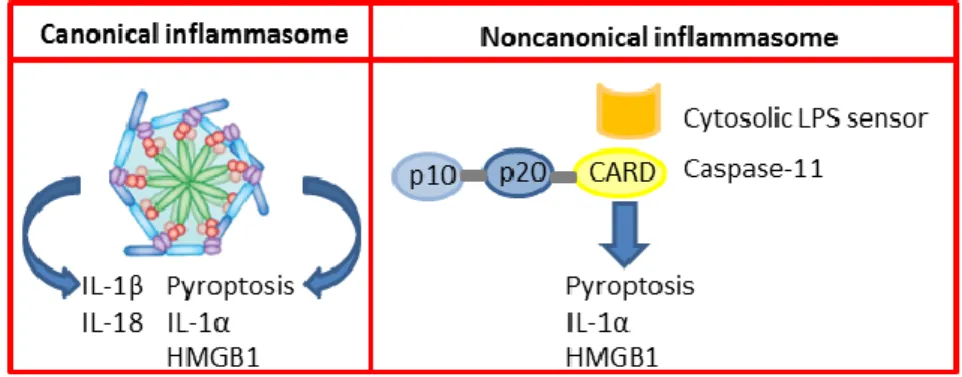
Inflammasomes are the main inflammatory pathway of the innate immune system. They are pattern recognition receptors able to recognize a wide range of conserved molecular motifs unique to microbes. Additionally, they can identify chemical alarm signals created by activated cells that are involved in tissue damage and host infection (Martinon et al., 2005). They are constituted by multiprotein oligomers that answer to inflammatory stimuli by starting an intracellular inflammatory cascade (Schroder et al., 2010). At the end of this cascade there is the activation and up-regulation of the innate immune system. We can distinguish two different kind of inflammasomes: canonical inflammasomes and non-canonical inflammasomes. The first one converts pro-Caspase-1 into the catalytically active enzyme, while the second promotes the activation of pro-11. Both Caspase-1 and Caspase-1Caspase-1 induce pyroptosis but only Caspase-Caspase-1 induces the secretion of the interleukins IL-1β and IL-18 (Martinon et al., 2005) (Figure 1).
Figure 1. Mechanisms of the Inflammasomes
Canonical inflammasome complexes induces autoactivation of Caspase-1. It subsequently converts proIL-1β and proIL-18 into the bioactive cytokines and triggers pyroptosis.
Non-5 canonical inflammasome can be activated in by the detection of cyotosolic LPS triggers activation of Caspase-11 in macrophages infected with Citrobacter rodentium, Escherichia coli and other Gram-negative bacteria the cytosol. Caspase-11 induces pyroptosis and release of IL-1α and HMGB1 directly and mediates secretion of IL-1β and IL-18 indirectly through canonical Nlrp3 inflammasome.
There are several classes of pattern recognition receptors, e.g. leucine rich repeats, toll like receptors, NOD-like receptors (NLRs), and all of them are evolutionary conserved. It has been recently shown that leucine rich repeats included within NLRs are a new class of pattern recognition receptor that are responsible for the three dimensional curved structures of the proteins (Bella et al., 2008; Hindle et al., 2009). These structures can bind specific ligands present on pathogens (Bell et al., 2005). NLRs are constituted by an N terminal effector domain, a central NACHT or NOD (a domain showed on CIITA, NAIP, TP-1 and HET-E proteins) and a C terminal leucine rich repeat domain (Inohara et al., 2001). There have been identified twenty-three NLRs holding NOD and leucine rich repeat domain, all classified by their N terminal region; NAIPs contain a BIR protein; NLRP 1-14 contain pyrin and NLRC 1-5 contain a caspase recruitment domain (CARD) protein (Kanneganti et
al., 2007). The NLR receptor is the sensor of the inflammasome, it detects the alarm signal.
The mechanism by which it happens still need to be clarified, but once triggered, the sensor starts the formation of the inflammasome complex. This sensor enables the recruitment of Caspase-1 either directly, through a CARD, or indirectly, through a PYRIN domain, which can link the PYRIN-CARD containing adaptor ASC. The resulting interaction of these proteins in turn leads to the cleavage and activation of Caspase-1. Additionally, recent studies have shown other families of genes that can assembly the inflammasomes: the RIG-I-like receptor (RLR) family (Ireton et al., 2011) and the AIM2-like receptor (ALR) family, which has a HIN200 DNA binding domain instead of an LRR
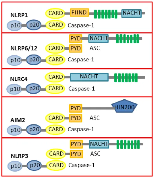
6 (Schattgen et al., 2011). Among all this different inflammasomes families the most characterized are NLRP1, NLRP6, NLRP12, NLRC4, AIM2 and NLRP3 (Figure 2).
Figure 2. Composition of NLRP1, NLRP6, NLRP12, NLRC4, AIM2 and NLRP3 NLRs (Nod-like receptor) are composed by CARD (caspase recruitment domain) or PYD (pyrin) motif, NACHT (nucleotide binding and oligomerization domain) and a number of LRRs (leucine-rich repeat). NLRP1 lacks the amino-terminal PYD motif and is autocatalytically cleaved FIIND (domain with function to find) domain. NLRP1 and NLRC4 recruit Caspase-1 through their CARD motifs. AIM2 is characterized by an amino-terminal PYD and a carboxy-terminal DNA-binding HIN200 domain. The PYD-CARD adaptor protein ASC stabilizes is required for assembly of the AIM2 and NLRP3 inflammasomes.
7 The first inflammasome discovered was the NLRP1 (Martinon et al., 2002). It has two natural ligands: the Bacillus anthracis lethal toxin and the muramyl dipeptide (MDP), a peptidoglycan fragment from Gram positive and negative bacteria (Boyden et al., 2006; Faustin et al., 2007). They can only activate human NLRP1(Faustin et al., 2007; Kovarova
et al., 2012), but not mouse because it displays species-specific differences at the genetic
level. Mouse NLRP1 can be activated by anthrax toxin. Humans have a single NLRP1 gene with both a CARD and a PYRIN domain, while mice possess three homologous genes Nlrp1 a, b and c containing CARD domain.
NLRP6 is extremely expressed in goblet and epithelial cells in the intestine (Elinav et al., 2011; Wlodarska et al., 2014), and in hematopoietic cells (Chen et al., 2011). The most important role of this inflammasome pathway is maintaining the intestinal homeostasis (Chen et al., 2011; Elinav et al., 2011; Normand et al., 2011; Hu et al., 2013).
NLRP6 has several characteristics that resemble NLRP12. As NLRP6, NLRP12 has a key role in protecting against AOM/DSS-induced colon cancer and DSS-induced colitis (Zaki
et al., 2011; Allen et al., 2012). Moreover, it maintains intestinal homeostasis by
negatively modulating several inflammatory signalling pathways such as MAPK and NF-kB (Zaki et al., 2011; Allen et al., 2012).
NLRC4 can be activated by many different bacterial pathogens such as Pseudomonas aeruginosa, Legionella pneumophila, Shigella flexneri and Salmonella typhimurium (Mariathasan et al., 2004; Amer et al., 2006; Franchi et al., 2006; Miao et al., 2006; Lamkanfi et al., 2007; Sutterwala et al., 2007; Suzuki et al., 2007; Miao et al., 2008). NLRC4 interacts with bacterial flagellin or structural components of the bacterial type III secretion system, that are leaked or injected into the host cell (Miao et al., 2010). These bacterial proteins directly bind by NLR-family apoptosis-inhibiting proteins (NAIPs) in the
8 cytosol. NAIPs in turn interact with NLRC4 and starts the assembly of the NLRC4 inflammasome complex, leading to the activation of Caspase-1, release of IL-1β, IL-18 and pyroptosis.
AIM2 is a member of the PYHIN family, proteins containing a PYRIN domain and the conserved DNA-binding domain hematopoietic IFN-inducible nuclear protein (Schattgen
et al., 2011). Theoretically, these proteins can bind nucleic acids and recruit ASC to start
the formation of an inflammasome. AIM2 can assembly an inflammasome by recognition of cytosolic DNA of viral or bacterial origin (Fernandes-Alnemri et al., 2010; Jones et al., 2010; Rathinam et al., 2010; Sauer et al., 2010), or self-DNA from apoptotic cells (Choubey, 2012; Zhang et al., 2013).
The most important and studied cytosolic inflammasome is the NLRP3 inflammasome. It contains the NOD-like receptor NLRP3, the adapter protein apoptosis-associated speck-like protein containing a caspase recruitment domain (ASC), and pro-Caspase-1, who cleaves into its active form. Caspase-1 in turn activates IL-18 and IL-1β, which contributes to the inflammation.
1.4 The NLRP3 inflammasome
Activation and prime of the NLRP3 inflammasome pathway needs different exogenous (bacterial hemolysins, pneumolysin, etc.) and endogenous (ATP, uric acid crystals, etc.) stimuli (Franchi et al., 2012). The precise mechanism still needs to be clarified, but new evidences indicate that a 2-step mechanism is required to activate NLRP3 (Juliana et al., 2010) (Figure 3).
9 Figure 3. NLRP3 inflammasome activation
Upon exposure to DAMPs or PAMPs, TRLs are phosphorylated and activate kB. NF-kB promotes the priming event. Extracellular ATP can induce K+/potassium efflux through a purinergic P2X7-dependent pore. These stimuli activate the NLRP3 inflammasome by facilitating the oligomerization of inactive NLRP3, ASC and pro-Caspase-1. The formation of this complex led to the activation of Caspase-1, which contributes to the production of active IL-1β and IL-18.
At the beginning, NLRP3 transcription is activated by NF-kB–inducing factors (Bauernfeind et al., 2009). Protein expression is not enough for activation of the full pathway: another transcription-independent step is required. It can be triggered by stimuli such as ATP, phagocytosis of monosodium urate crystals, or pore-forming toxins. The induction of this pathway leads to mitochondrial and lysosome damage, production of ROS and dysregulated ionic balance (K+ efflux, elevated intracellular Ca2+). Multiple publications indicates that the purinergic receptor takes part in lung inflammatory events
10 (Dostert et al., 2008). In particular, the purinergic receptor P2X7 participates to the release of IL-1β and has been proposed to be upstream of the NLRP3 inflammasome oligomerization (Ferrari et al., 2006). It is an extracellular ATP-gated plasma membrane ion channel receptor exposed on a variety of immune cells and has many biologic functions (Ferrari et al., 2006). P2X7 can be activated by extracellular ATP at the site of inflammation and tissue injury (Dostert et al., 2008), leading to cation flow across the plasma membrane (Burnstock et al., 2011). This receptor is involved in immune responses induced by extracellular ATP, including lung injuries (Riteau et al., 2010; Belete et al., 2011), through its implication in immune processes such as apoptosis (Woods et al., 2012) and inflammasome activation. P2X7 joins in lung disease, such as pulmonary fibrosis and pleurisy (Moncao-Ribeiro et al., 2011), asthma and chronic obstructive disease (Denlinger
et al., 2009; Eltom et al., 2011). Upon activation, NLRP3 recruit the PYD- and
CARD-containing bipartite adapter ASC (apoptosis-associated speck-like protein CARD-containing a CARD) via PYD/PYD interactions (Masumoto et al., 1999). ASC in turn recruits Caspase-1 via CARD/CARD interactions. The penultimate step for the assembly of the inflammasome is the cleavage and activation of Caspase-1. Caspase-1 (also defined as IL-1β converting enzyme, ICE) is first present as a 45 kDa inactive precursor (Walker et al., 1994), which is constituted by a N-terminal subunit of 15 kDa, a central subunit of 20 kDa, ASC proteins together resulting in the cleavage of Caspase-1 (Yamin et al., 1996). The active Caspase-1 is composed by a tetramer consisting of two fragments of 20 kDa and two fragments of 10 kDa (Wilson et al., 1994). Once activated, Caspase-1 can cleave other protein precursors (Carta et al., 2006; Ogura et al., 2006). Many of these proteins have a role in glycolysis (Shao et al., 2007), in the cytoskeleton of the cell (Shao et al., 2007; Keller et al., 2008), and inflammation and mitochondria function (Keller et al., 2008). Moreover, once activated, Caspase-1 causes its own secretion (Keller et al., 2008).
11 The two most important proteins cleaved by Caspase-1 are IL-1β and IL-18. They are secreted from the cells containing an activated inflammasome. The inactive IL-1β is a protein of 30.7 kDa, while inactive IL-18 is 22.3 kDa. They are respectively cleaved by Caspase-1 to the active 17.5 kDa and 17.3 kDa active forms. Once activated and secreted by the cells, IL-1β and IL-18 can be involved in paracrine and autocrine signalling. They are structurally similar and are in particular for the β-pleated sheets (Dinarello, 1999). IL-1β is a pleiotropic cytokine that can be found in localised inflammation targeting bacterial, parasitic or viral infections; or in systemic inflammation. It is produced by different cell types such as fibroblast, ephitelial cells and T cells; in response to damaged tissues or pathogens. IL-1β can also modulate its own mRNA expression through a signalling pathway involved p38 MAPK phosphorylation and NF-kB activity (Dinarello, 1998). IL-1β secretion requires two signals: the first one is commonly obtained through toll-like receptor signalling or IL-1β receptor self up-regulation of IL-1β gene transcription, the second one is through the inflammasome activation. The release of IL-1β into the peripheral blood leads to increase expression of other pro-inflammatory mediators, up-regulate cortisol levels and induce fever acting on the hypothalamus. IL-18 secretion, previously known as interferon-γ inducing factor, is also induced by inflammasome activation by the effect of this cytokine still needs to be completely elucidated. It is constitutively expressed in its inactive form in several cell types and only necessitates Caspase-1 cleavage for its activation and secretion. Like IL-1β, IL-18 can increase its own mRNA expression and its signalling pathway involves NF-kB activity (Dinarello, 1999). It activates T cells leading to increase expression of IL-2, TNF-α and GM-CSF (Dai
et al., 2011).
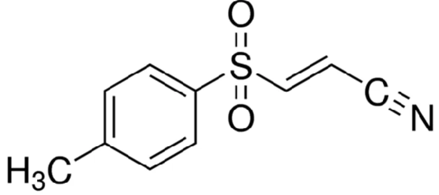
12 Small molecules are able to inhibit the pro-apoptotic and pro-inflammatory effects of NLRP3 inflammasome. Among these, one of the most studied is Bay 11-7082 (Figure 4). It is an NF-kB inhibitor, which is able to selectively block the Ikb kinase activity and to inhibit the activation of the NLRP3 inflammasome by blocking its ATPase activity required to recruit ASC. Its efficacy has been shown in endothelin-1 induced lung edema and oxidative stress (Piechota et al., 2011), burn-induced remote acute lung injury (Sio et
al., 2010; Han et al., 2015) and postnatal lung inflammation (Hou et al., 2015).
Figure 4. BAY 11-7082
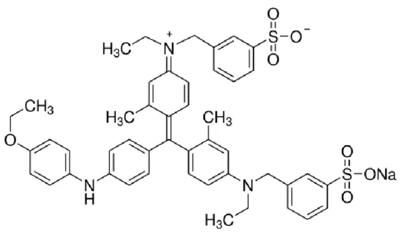
1.6 Brilliant Blue G
Another way to avoid the activation of the NLRP3 inflammasome is targeting its ATPase activity (Uberti et al., 2004). Brilliant Blue G (BBG) blocks the membrane-bound purinergic P2X7 receptor (Díaz-Hernández et al., 2009) (Figure 5). This receptor is expressed on multiple lung cell types such as pulmonary endothelia, type I alveolar epithelial cells and resident cells of the immune system. BBG can down-regulate the expression of ASC and avoid NLRP3 inflammasome activation (Zhao et al., 2013). It has been tested in silica-induced lung changes (Monçao-Ribeiro et al., 2014), acute lung injury
13 (Grailer et al., 2014) and pulmonary edema after subarachnoid hemorrhage (Chen et al., 2014).
14 2. AIM OF THE STUDY
The aim of this project was to clarify the role of the NLRP3 inflammasome in the pathogenesis of respiratory diseases, in particular pleurisy, testing two inflammasome-blocking agents, BAY 11-7082 and BBG, in a murine model of carrageenan-induced pleurisy. The main focuses were: pulmonary membrane thickening, polymorphonuclear leukocyte infiltration, NF-kB translocation in the nucleus, assembly of the NLRP3/ASC/caspase-1 complex, iNOS, nitrotyrosine, poly-ADP-ribosyl polymerase expression and CAR-induced apoptosis.
15 3. MATERIALS AND METHODS
3.1 Animals
CD1 male mice weighing 25–30 g (Harlan Nossan,Milan, Italy) were kept in a controlled location with standard rodent water and chow. Mice were kept in stainless cages at 22±1°Cunder a 12h light/dark cycle. The study was approved by the University of Messina Review Board for the care of animals. All animal experiments obeyed regulations in the United States (Animal Welfare Assurance A5594-01, Department of Health and Human Services), Europe (O.J. of E.C. L 358/1, 12/18/1986), and Italy (DM 116192).
3.2 Carrageenan-induced pleurisy
Pleurisy was induced by Carrageenan (CAR) as previously described (Cuzzocrea et al., 2000). Animals were anesthetized with isoflurane and cut at the sixth intercostal space. Saline containing 2% l-CAR (0.1 ml) or saline alone (0.1ml) was injected into the pleural cavity. The incision was sutured, and animals were allowed to recover. Mice were sacrificed by inhalation of CO2 at 4 h after CAR injection. The pleural cavity was injected
with 2ml of saline solution containing heparin (5 U/ml) and indomethacin (10 mg/ml). This solution and exudate were aspirated, and the total volume was measured. Exudate having blood was eliminated from the study. The volume of exudate was determined from the total volume recovered by subtracting the injected one (2ml). The leukocytes enclosed in the exudate were suspended in PBS and counted with a Burker’s chamber after vital Trypan Blue staining.
16 Mice were randomly separated into the following groups (n = 10 for each):
CAR + saline group: mice were subjected to CAR induced pleurisy.
CAR + BBG group: same as the CAR + saline group, but BBG (45.5 mg/kg, i.p.) was administered 1 h after CAR injection.
CAR + BAY 11-7082 group: same as the CAR + saline group, but BAY 11-7082 (30 mg/kg, i.p.) was administered 1 h after CAR injection.
Sham + saline group: sham-surgery group in which the same surgical procedures as in the CAR group were performed, except that saline was injected instead of CAR.
Sham + BBG group: same as the sham + saline group, but BBG (45.5 mg/kg, i.p.) was administered 1 h after saline injection.
Sham + BAY 11-7082 group: same as the sham + saline group, but BAY 11-7082 (30 mg/kg, i.p.) was administered 1 h after saline injection.
The used dose for BBG and for BAY 11-7082 was chosen in agreement with Minutoli et al. (Minutoli et al., 2015). The minimum number of animals for each group was found using the statistical test a priori power analysis of G-power software. This statistical test provides an efficient method for choosing the sample size necessary to perform the experiment.
3.4 Histological examination
Lung tissues were collected 4 h after CAR injection. Samples were fixed for 24 h in PBS-buffered formaldehyde solution (10% w/v) at room temperature, dehydrated using graded ethanol, and embedded in Paraplast (Sherwood Medical, Mahwah, NJ, USA). Seven μm
17 sections were deparaffinized with xylene and stained with hematoxylin and eosin. All sections were analysed using an Axiovision microscope (Zeiss, Milan, Italy). The following morphologic criteria were employed for scoring: 0, normal lung; grade 1, minimal edema or infiltration of bronchiolar or alveolar walls; grade 3, moderate edema and inflammatory cell infiltration without obvious damage to lung architecture; grade 4, severe inflammatory cell infiltration with obvious damage to lung architecture. All histologic studies were performed in a blinded fashion.
3.5 Immunohistochemical localization of nitrotyrosine, PAR, NLRP3, ASC, Caspase-1, IL-18, IL-1β, TNF-α, Bax, Bcl-2, myeloperoxidase, iNOS, manganese superoxide dismutase and NRF2
Four hours after CAR injection, lung samples were fixed in PBS-buffered formaldehyde (10% w/v) and embedded in paraffin, and 7-μm sections were used from samples. After deparaffinization, endogenous peroxidase was quenched with hydrogen peroxide (0.3% v/v) in methanol (60% v/v) for 30 min. Samples were permeabilized with Triton X-100 in PBS (0.1% w/v) for 20 min. Nonspecific adsorption was reduced by incubating the sections in normal goat serum in PBS (2% v/v) for 20 min. Endogenous avidin or biotin binding sites were stopped by incubation for 15 min with avidin and biotin (Vector Laboratories, Burlingame, CA, USA), respectively. Sections were probed overnight with purified goat polyclonal antibody directed toward anti-nitrotyrosine antibody (06-284; Millipore, Billerica, MA, USA), anti-PAR antibody (H-250: sc-7150, 1:500 in PBS, v/v; Santa Cruz Biotechnology), anti-NLRP3 (sc-66846, 1:400 in PBS, v/v; Santa Cruz Biotechnology), anti-ASC antibody (N-15: sc-22514-R, 1:500 in PBS, v/v; Santa Cruz Biotechnology), anti–Caspase-1 p20 (G-19: sc-1597, 1:500 in PBS, v/v; Santa Cruz
18 Biotechnology), anti–IL-18 antibody (H-173: sc-7954, 1:500 in PBS, v/v; Santa Cruz Biotechnology), anti–IL-1β antibody (H-153: sc-7884, 1:500 in PBS, v/v; Santa Cruz Biotechnology), anti–TNF-α antibody (N-19: sc-1350, 1:500 in PBS, v/v; Santa Cruz Biotechnology), anti-Bax (P-19: sc-526, 1:555 in PBS, v/v; Santa Cruz Biotechnology), anti–Bcl-2 (N-19: sc-492, 1:500 in PBS, v/v; Santa Cruz Biotechnology), anti-myeloperoxidase (MPO) antibody (2C7: sc-59600, 1:325 in PBS, v/v; Santa Cruz Biotechnology), anti-iNOS antibody (610432, 1:500 in PBS, v/v; Transduction Laboratories, Lexington, KY, USA), anti-MnSOD antibody (06-984, 1:500 in PBS, v/v; Millipore), or anti-NRF2 antibody (C-20: sc-722, 1:550 in PBS, v/v; Santa Cruz Biotechnology). Samples were washed with PBS and incubated with secondary antibody. Specific labelling was detected with a biotin-conjugated goat anti-rabbit IgG and avidin– biotin peroxidase complex (Vector Laboratories, Burlingame, CA, USA). Immunohistochemistry photographs were evaluated by densitometry by using Optilab Graftek software (Ljubljana, Slovenia).
3.6 MPO
MPO activity, an indicator of polymorphonuclear leukocyte accumulation, was quantified as previously described (Mullane et al., 1985). Lung tissue from each animal was removed and weighed. Each sample was homogenized in a solution containing 0.5% hexa-decyl-trimethylammonium bromide dissolved in 10 mM potassium phosphate buffer (pH 7) and centrifuged at 20,000 g at 4°C for 30 min. A solution of tetra-methylbenzidine (1.6 mM) and 0.1 mMH2O2 was then allowed to react with an aliquot of the supernatant. The rate of change in absorbance was measured spectrophotometrically at 650 nm. MPO activity was
19 quantified in units per gram weight of wet tissue and was calculated as the quantity of enzyme degrading 1 mmol of peroxide per minute at 37°C.
3.7 Western blot analysis for IkB-α, NF-kB, NLRP3, ASC, Caspase-1, IL-18, IL-1β, iNOS, manganese superoxide dismutase and NRF2
Lung tissues from each mouse were suspended in extraction Buffer A containing 0.15 mM pepstatin A, 0.2 mM PMSF, 1 mM sodium orthovanadate, and 20 mM leupeptin; homogenized at the highest setting for 2min; and centrifuged at 1000 g for 10min at 4°C. Cytosolic fraction was represented by the supernatants. The pellets, containing enriched nuclei, were resuspended in Buffer B containing 150mM NaCl, 1% TritonX-100, 1mM EGTA, 10 mM Tris-HCl (pH 7.4), 0.2 mM PMSF, 1 mM EDTA, 0.2 mM sodium orthovanadate, and 20 mm leupeptin. After centrifugation for 30min at 15,000 g at 4°C, the nuclear protein contained in the supernatants was stored at 280°C for further analysis. The levels of IkB-α, NLRP3, ASC, Caspase-1, IL-18, IL-1β, iNOS, and manganese superoxide dismutase (MnSOD) were quantified in the cytosolic fraction, whereas NF-kB p65 and NRF2 levels were quantified in the nuclear fraction. The filters were blocked with 1X PBS and 5% (w/v) nonfat dried milk for 40 min at room temperature and probed with specific IkB-α antibody (C-21: sc-371, 1:550 in PBS, v/v; Santa Cruz Biotechnology), anti–NF-kB p65 (F-6: sc-8008, 1:400 in PBS, v/v; Santa Cruz Biotechnology), anti-NLRP3 (sc-66846, 1:400 in PBS, v/v; Santa Cruz Biotechnology), anti-ASC antibody (N-15: sc- 22514-R, 1:500 in PBS, v/v; Santa Cruz Biotechnology), anti–Caspase-1 p20 (G-19: 1597, 1:500 in PBS, v/v; Santa Cruz Biotechnology), anti–IL-18 antibody (H-173: 7954, 1:500 in PBS, v/v; Santa Cruz Biotechnology), anti–IL-1β antibody (H-153: sc-7884, 1:500 in PBS, v/v; Santa Cruz Biotechnology), anti-iNOS antibody (610432, 1:500
20 in PBS, v/v; Transduction Laboratories), anti-MnSOD antibody (06-984, 1:500 in PBS, v/v; Millipore), or anti-NRF2 antibody (C-20: sc-722, 1:550 in PBS, v/v; Santa Cruz Biotechnology) in1X PBS (5%w/v) non fat dried milk, and 0.1% Tween-20 (PMT) at 4°C overnight. Membranes were incubated with peroxidase-conjugated bovine anti-mouse IgG secondary antibody or peroxidase-conjugated goat anti-rabbit IgG (1:2000; Jackson ImmunoResearch, West Grove, PA, USA) for 1 h at room temperature. To establish that blots were loaded with equal amounts of proteins, membranes were also incubated in the presence of the antibody against β-actin protein (1:10,000; Sigma-Aldrich, St. Louis, MO, USA). The relative expression of the protein bands of IkB-α (37 kDa), NF-kB p65 (65 kDa), NLRP3 (106 kDa), ASC (24 kDa), Caspase-1 (20 kDa), IL-18 (24 kDa), IL-1β (33 kDa), iNOS (130 kDa), MnSOD (24 kDa), and NFR2 (57kDa) was quantified by densitometric scanning of the X-ray films with a GS-700 Imaging Densitometer (Bio-Rad Laboratories, Milan, Italy) and a computer program (MolecularAnalyst; IBM,Armonk,NY, USA) and standardized for densitometric analysis to β-actin levels.
3.8 Materials
All materials were acquired from Sigma-Aldrich. BAY 11-7082 was obtained from Adipogen (Liestal, Switzerland), and Brilliant Blue G (BBG) was obtained from Sigma-Aldrich. All stock solutions were set in nonpyrogenic saline (0.9% NaCl) (Baxter, Thetford, United Kingdom).
21 All parameters in the text and figures are expressed as means ± SEM of n evaluations. For the in vivo studies, n symbolizes the number of animals used. In the experiments including histology or immunohistochemistry, the figures shown are demonstrative of at least 3 experiments (histologic or immunohistochemistry coloration) performed on different experimental days on the tissue collected from all the animals in each group. The results were analyzed by 1-way ANOVA followed by Bonferroni’s post hoc test for multiple comparisons. A value of P < 0.05 was considered statistically significant. Data are expressed as means ± SEM from 10 mice per group.
22 4. RESULTS
4.1 Effects of BAY 11-7082 or BBG treatment on carrageenan-induced pleurisy and histologic examination
When compared with lung tissues taken from sham animals (data not shown), histologic examination of lung samples from carrageenan-treated mice showed significant edema and tissue injury (Fig. 6A; see histologic score in Fig. 6D). Treatment with BAY 11-7082 reduced the degree of lung damage (Fig. 6B; see histologic score in Fig. 6D). Also, BBG administration decreased carrageenan-induced lung injury (Fig. 6C; see histologic score Fig. 6D). Moreover, when compared with the sham mice, intrapleural injection of carrageenan led to the development of acute pleurisy, generating turbid exudates with large amount of PMNs (Fig. 6E and F). Administration of BAY 11-7082 or BBG 1 h after carrageenan injection caused a reduction of pleural exudates and inflammatory cells recruitment in the pleural cavity (Fig. 6E and F). There was no significant difference between BAY 11-7082 and BBG treatment.
23 Figure 6. Effect of BAY 1-7082 or BBG administration on histologic alterations, exudate volume, and PMN infiltration in the lung
Lung tissues from CAR-treated mice displayed edema and tissue injury (A, D), and infiltration of the tissue with polymorphonuclear cell (E, F). BAY 11-7082 (B) or BBG (C) administration significantly decreased lung injury (D) and neutrophil infiltration (E, F). Values are means ± SEM (n = 10 mice per each group). *P <0.05 vs. sham-treatment group, °P <0.05 vs. CAR.
24 4.2 Effects of BAY 11-7082 or BBG treatment on MPO expression and release of
proinflammatory cytokines induced by carrageenan
Analysis of lung samples taken from carrageenan-treated mice showed an increased infiltration of neutrophils as assessed by MPO expression and activity (Fig. 7A, G and H), an indicator of polymorphonuclear cell infiltration, compared with sham-treated mice (data not shown). BAY 11-7082 or BBG administration reduced in the same way MPO expression in tissue obtained from treated mice (Fig. 7B, C and G). Carrageenan injection also led to an up-regulated release of pro-inflammatory cytokines such as TNF-α (Fig. 7D and G) compared with sham- treated animals (data not shown). Treatments equally reduced TNF-α production release (Fig. 7E and G).
25 Figure 7. Effect of BAY 1-7082 or BBG administration on the immunohistochemical localization of MPO and TNF-α
After four hours carrageenan injection, MPO activity was elevated in vehicle-treated animals (A, G, H), whereas BAY 11-7082 (B) or BBG (C) administration significantly down-regulated MPO activity (G, H). Lungs from carrageenan-treated mice displayed positive staining for TNF-α (D, G). There was a clear reduction in the immunostaining for TNF-α in tissues from mice administrated with BAY 11-7082 (E, G) or BBG (F, G). Values are means ± SEM (n = 10 mice per each group). *P <0.05 vs. sham-treatment group, °P <0.05 vs. CAR.
26 4.3 Effects of BAY 11-7082 or BBG treatment on IkB-α and NF-kB expression induced by carrageenan
Basal expression of α was detected in samples from sham-treated mice, whereas IkB-α levels were notably decreased in tissues from carrageenan-treated mice (Fig. 8A). BAY 11-7082 administration prevented carragenan-induced IkB-α degradation (Fig. 8A). Additionally, carrageenan-injection up-regulated NF-kB levels in the nuclear fractions compared with the sham-treated animals (Fig. 8B). Treatment with BAY 11-7082 significantly decreased the levels of NF-kB (Fig. 8B). Moreover, BBG administration reduced IkB-α degradation (Fig. 8C) and increased NF-kB expression (Fig. 8D). There was no significant difference between BAY 11-7082 and BBG.
27 Figure 8. Effect of BAY 1-7082 or BBG administration on IkB-α degradation and nuclear NF-kB quantification
Basal levels of IkB-α were found in tissues from sham animals (A) and were significantly down-regulated in samples from carrageenan-injected mice (C). BAY 11-7082 (A) or BBG (C) administration significantly reduced IkB-α degradation. Four hours after
carrageenan instillation, NF-kB levels were markedly increased in the nuclear fraction (B) compared with the sham-animals (D). BAY 11-7082 (B) or BBG (D) significantly reduced NF-kB in the nucleus. Values are means ± SEM (n = 10 mice per each group). *P <0.05 vs. sham-treatment group, °P <0.05 vs. CAR.
28 4.4 Effects of BAY 11-7082 or BBG treatment on NLRP3 expression induced by carrageenan
In order to evaluate the effect of BAY 11-7082 and BBG in the inflammasome activation, we investigated NLRP3 expression. Lung samples from carrageenan-treated mice displayed positive staining for NLRP3 (Fig. 9A and D) compared with sham-treated animals (data not shown). BAY 11-7082 or BBG both reduced this staining (Fig. 9B and D). Western blot analysis also confirmed these data. We found up-regulated NLRP3 expression in the carrageenan-treated group compared with the sham animals and a reduction of this expression in samples obtained from mice administrated with BAY 11-7082 or BBG (Fig. 9E and G).
29 Figure 9. Effect of BAY 1-7082 or BBG administration on NLRP3 expression
Lung tissues taken from carrageenan-treated animals showed positive immunostaining for NLRP3 (A, D). BAY 11-7082 (B, D) or BBG (C, D) reduced this staining. Western blot analysis confirmed up-regulated levels of NLRP3 expression in samples collected from carrageenan-injected animals compared with the sham mice (E–G). BAY 11-7082 (E, G) or BBG (F, G) administration decreased NLRP3 expression. Values are means ± SEM (n = 10 mice per each group). *P <0.05 vs. sham-treatment group, °P <0.05 vs. CAR.
30 4.5 Effects of BAY 11-7082 or BBG treatment on ASC and Caspase-1 expression induced by carrageenan
Lung samples obtained from carrageenan-treated animals showed positive staining for ASC (Fig. 10A and D). No positive staining was observed in samples from sham-treated mice (data not shown). BAY 11- 7082 or BBG administration reduced this staining (Fig. 10B and D). Lung sections from sham-treated animals did not indicate staining for Caspase-1 (data not shown), whereas samples from carrageenan-treated mice exhibited positive staining for Caspase-1 (Fig. 10D and H). Treatment with BAY 11-7082 or BBG reduced the staining for Caspase-1 (Fig. 10F and H). Western blot analysis exposed an increased expression of ASC and Caspase-1 in tissue collected from carrageenan-treated mice (Fig. 10I and L) compared with sham-treated animals. BAY 11-7082 or BBG administration reduced ASC and Caspase-1 expression (Fig. 10I and L). There was no significant difference between the inhibitors.
31 Figure 10. Effect of BAY 1-7082 or BBG administration on ASC and Caspase-1 expression
Four hours after carrageenan injection, positive ASC immunostaining in lungs collected from vehicle-treated animals was found (A, D). BAY 11-7082 (B, D) or BBG (C, D) administration reduced this staining. This treatment also decreased Caspase-1 positive staining (F, H) compared with vehicle-treated animals (E, H). Western blot analysis exposed increased expression of ASC and Caspase-1 in tissues collected from CAR-treated mice (I, J) compared with the sham-treated animals (K, L). BAY 11-7082 (I, J) or BBG (K, L) significantly inhibited this up-regulation. Values are means ± SEM (n = 10 mice per each group). *P <0.05 vs. sham-treatment group, °P <0.05 vs. CAR.
32 4.6 Effects of BAY 11-7082 or BBG treatment on IL-1β and IL-18 expression induced by carrageenan
Tissues obtained from carrageenan-treated animals showed positive staining for IL-1β (Fig. 11A and D). No positive staining was found in samples from sham-treated mice (data not shown). BAY 11-7082 or BBG treatment equally reduced IL-1β-positive staining (Fig. 11B and D). Tissues from sham-treated mice did not expose staining for IL-18 (data not shown), whereas lung samples from carrageenan-treated animals displayed positive staining for IL-18 (Fig. 11D and H). BAY 11-7082 or BBG decreased the degree of positive staining for IL-18 (Fig. 11F and H). Western blot analysis confirmed an improved expression of IL-1β and IL-18 in samples taken from carrageenan-treated mice (Fig. 11I and L) compared with sham-treated animals. BAY 11-7082 and BBG decreased IL-1β and IL-18 expression in the same way.
33 Figure 11. Effect of BAY 1-7082 or BBG administration on IL-1β and IL-18 expression
Lung sections obtained from CAR-treated mice exposed positive staining for IL-1β (A, D). Immunostaining for IL-1β showed a clear reduction in the lungs of mice administrated with BAY 11-7082 (B, D) or BBG (C, D). Positive IL-18 immunostaining in lung tissues from vehicle-treated animals was found (E, H). Administration with BAY 11-7082 (F, H) or BBG (G, H) reduced this staining. Western blot analysis of lung samples obtained from CAR-injected animals showed increased expression of IL-1β and IL-18 compared with the sham-treated animals (I, L). BAY 11-7082 (I, J) or BBG (K, L) reduced this expression. Values are means ± SEM (n = 10 mice per each group). *P <0.05 vs. sham-treatment group, °P <0.05 vs. CAR.
34 4.7 Effects of BAY 11-7082 or BBG treatment on MnSOD and NRF2 expression induced by carrageenan
Whether, to investigate carrageenan modulates the oxidative process, we tested the lung expression of the antioxidant enzyme MnSOD. Staining of samples from sham-treated mice displayed a constitutive expression of MnSOD (data not shown), while carrageenan significantly decreased lung MnSOD staining (Fig. 12A and D). BAY 11-7082 or BBG administration significantly increased MnSOD staining (Fig. 12B and D). Tissues from sham-treated mice showed basal staining for NRF2, carrageenan injection notably reduced this staining (Fig. 12E and H). Treatment with BAY 11-7082 or BBG improved NRF2 staining (Fig. 12F and H). Western blot analysis showed the up-regulation of MnSOD and NRF2 expression by BAY11-7082 or BBG administration compared with carrageenan-treated mice (Fig. 12I and N). No statistical difference was found between the inhibitors.
35 Figure 12. Effect of BAY 1-7082 or BBG administration on MnSOD and NRF2 expression
No positive staining for MnSOD was found in tissues from CAR-treated mice four hours after the injection (A, D). BAY 11-7082 (B, D) or BBG (C, D) administration increased this staining. Also no positive staining for NRF2 was detected in tissues from CAR-treated mice (E, H). BAY 11-7082 (F, H) or BBG (G, H) increased NRF2 staining. Western blot analysis confirmed a decrease of MnSOD expression in the cytosolic fraction of lung samples taken from CAR-treated animals compared with the tissues from sham-treated mice (I, K). Treatment with BAY 11-7082 (I, K) or BBG (J, K) restored MnSOD level. Western blot analysis displayed a reduction of NRF2 expression in the nuclear fraction of samples collected from CAR-treated mice compared with sham-treated animals (L, N). BAY 11-7082 (L, N) or BBG (M, N) administration increased NRF2 levels. Values are means ± SEM (n = 10 mice per each group). *P <0.05 vs. sham-treatment group, °P <0.05 vs. CAR.
36 4.8 Effects of BAY 11-7082 or BBG treatment on iNOS expression induced by carrageenan
Four hours after carrageenan injection, immunohistochemical analysis of tissues taken from the vehicle-treated group showed positive staining for iNOS (Fig. 13A and D) compared with samples from the sham-treated mice (data not shown). Treatment with BAY 11-7082 or BBG significantly attenuated this staining (Fig. 13B and D). iNOS expression in samples from carrageenan-treated mice was also found amplified in Western blot analysis. Western blot analysis also confirmed that treatment with BAY 11-7082 (Fig. 12E and G) or BBG (Fig. 12F and G) decreased iNOS expression. There was no significant difference between BAY 11-7082 or BBG administration.
37 Figure 13. Effect of BAY 1-7082 or BBG administration on iNOS expression
Lung tissues taken from CAR-treated animals displayed positive staining for iNOS (A, D). iNOS-positive staining was notably decreased in tissue sections from mice administrated with BAY 11-7082 (B, D) or BBG (C, D). An increase in iNOS quantification, measured by Western blot analysis, was detected in lungs obtained from mice subjected to CAR-injection compared with tissues from sham-treated animals (E, G). BAY 11-7082 (E, G) or BBG (F, G) significantly reduced this protein quantification. Values are means ± SEM (n = 10 mice per each group). *P <0.05 vs. sham-treatment group, °P <0.05 vs. CAR.
38 4.9 Effects of BAY 11-7082 or BBG treatment on nitrotyrosine and PAR expression induced by carrageenan
Immunohistochemical analysis of lungs from carrageenan-injected animals displayed positive staining for nitrotyrosine (Fig. 14A and G). No positive staining for nitrotyrosine was found in the samples of carrageenan-injected mice that received BAY 11-7082 or BBG (Fig. 14B, C and G). Four hours after carrageenan injection, lungs were collected, and immunohistological staining for poly-ADP–ribosylated proteins was performed. In carrageenan treated mice, positive staining for the PAR was detected (Fig. 14D and G). The two inhibitors equally reduced this staining (Fig. 14E, F and G).
39 Figure 14. Effect of BAY 1-7082 or BBG administration on nitrotyrosine and PAR expression
Lungs from CAR-treated mice exposed intense positive staining for nitrotyrosine (A, G). This staining was notably reduced in animals treated with BAY 11-7082 (B, G) or BBG (C, G) Also, positive PAR immunostaining was found in tissues collected from vehicle-treated animals (D, G). BAY 11-7082 (E, G) or BBG (F, G) administration decreased this staining. Values are means ± SEM (n = 10 mice per each group). *P<0.05 vs. sham-treatment group, °P <0.05 vs. CAR.
40 4.10 Effects of BAY 11-7082 or BBG treatment on Bax and Bcl-2 expression induced by carrageenan
Tissues obtained from sham-treated mice did not express any staining for Bax (data not shown), whereas lungs from carrageenan-treated animals displayed positive staining (Fig. 15A and G). BAY 11-7082 or BBG administration reduced Bax staining (Fig. 15B, C and G). Moreover, tissues from sham-treated animals revealed positive staining for Bcl-2 (data not shown), whereas in the carrageenan-treated group Bcl-2 staining was notably reduced (Fig. 15D and G). Treatment with BAY11-7082 and BBG significantly down-regulated the staining for Bcl-2 in mice subjected to carrageenan-induced pleurisy (Fig. 15E and G). There was no significant difference between them.
41 Figure 15. Effect of BAY 1-7082 or BBG administration on Bax and Bcl-2 expression Lungs from CAR-treated mice showed positive staining for Bax (A, G). There was a clear reduction of this staining in samples from mice administrated with BAY 11-7082 (B, G) or BBG (C, G). Four hours after CAR injection, no positive Bcl- 2 immunostaining was detected in lung tissues obtained from CAR-treated mice (D, G). BAY 11-7082 (E, G) or BBG (F, G) increased Bcl-2 staining. Values are means ± SEM (n = 10 mice per each group). *P <0.05 vs. sham-treatment group, °P <0.05 vs. CAR.
42 5. DISCUSSION
Carrageenan-induced pleurisy is a usually employed model of acute inflammation (Di Rosa et al., 1971; Velo et al., 1973). This injection into the pleural cavity causes leukocyte infiltration in the lungs and inflammatory exudates in the chest (Impellizzeri et al., 2011a). The animal model lends itself to analysis of the principal mediators involved in inflammation.. New data related NLRP3 inflammasome activation to acute injury induced by carrageenan administration (Wang et al., 2015). It has also been described that two blocking agents BAY 11-7082 and Brilliant Blue G (BBG) attenuated the inflammatory pathway by inhibiting the activation of the NLRP3 inflammasome (Minutoli et al., 2015). In particular, BAY 11-7082 inhibits the IkB kinase activity (Uberti et al., 2004) and in turn the translocation of NF-kB into the nucleus carrageenan-induced, whereas BBG blocks the membrane-bound purinergic P2X7 receptor (Díaz-Hernández et al., 2009), leading to the suppression of ASC expression and inhibiting the NLRP3 inflammasome activation. This study showed that both the administrations of the inflammasome blocking agents BAY 11-7082 and BBG significantly attenuated the histologic signs and biochemical markers of pleurisy. The two treatments reduced the exudate volume and the infiltration of the PMNs into the pulmonary parenchyma. Moreover, the reduction of the infiltrating cells well correlated with the reduced MPO expression found in tissues collected from BAY 11-7082 and BBG administrated animals. MPO release in the early phase of the inflammatory reaction to carrageenan injection may be related with the events that triggered the late mononuclear cell migration and numerous pro-inflammatory cytokines secretion, such as TNF-α (Kon et al., 2001). BAY 11-7082 and BBG were able to reduce TNF-α over-expression induced by carrageenan. As already mentioned, several studies underlined that these stimuli led to the activation of the NF-kB pathway (Impellizzeri et al., 2011b). In normal condition, NF-kB is located in the cytosol, in its inactive state, bound to an
43 inhibitor IkB protein (in this case, IkB-α). This inhibitor can be phosphorylated by a kinase enzyme (in this case IkB kinase) allowing it to release NF-kB. During inflammation a variety of extracellular signals, also through the intermediation of membrane integral receptors, can activate the IkB kinase enzyme. Once IkB-α is phosphorylated, it goes to ubiquitation and degradation in the proteasome, whereas NF-kB goes to the nucleus. It regulates the expression of genes influencing a broad range of biological processes including inflammation, innate and adaptive immunity and stress responses. Treatment with BAY 11-7082 or BBG significantly inhibited NF-kB translocation in the nucleus in two different way (Uberti et al., 2004; Díaz-Hernández et al., 2009). Among the transcription of the genes activated by NF-kB (Impellizzeri et al., 2011b) there is also the NLRP3 inflammasome (Stutz et al., 2013). NLRP3 is an ATPase and its activity is required to trigger the formation of the NLRP3 inflammasome complex, leading to ASC oligomerization an Caspase-1 activation (Fernandes-Alnemri et al., 2007; Proell et al., 2013). Once activated Caspase-1 proteolytically activates pro–IL-1β and pro–IL-18 (Thornberry et al., 1992; Gu et al., 1997) and induces their release (Dinarello, 2009). Treatment with BAY 11-7082 and BBG reduced NLRP3 expression, in turn ASC pyroptosome formation and pro-Caspase-1 self-cleavage in active Caspase-1. This inhibitors also decreased IL-1β and IL-18 expression in lungs. Several studies underlines that carrageenan administration led to a large increase of ATP release in lung epithelial cells (Saïd-Sadier et al., 2012). It is an endogenous DAMP and one of the first described NLRP3 inflammasome activators. High ATP extracellular concentrations led to P2X7R activation which in turn supports a rapid production of ROS (Cruz et al., 2007). Generation of ROS resulted in oxidative stress, which produced cell damage directly or by altering signalling pathways (Salvemini et al., 1996; Cuzzocrea et al., 2006). Several redox-sensitive proteins are up-regulated or down-regulated from the oxidative stress (Bowie et
44
al., 2000). This study demonstrated that treatment with BAY 11-7082 or BBG was able to
stabilize NF-E2–related factor 2 (NRF2) and up-regulate the levels of the antioxidant enzyme MnSOD. The experiments also confirmed the increase in iNOS expression in lung tissues from carrageenan-treated animals. Treatments with the tested inflammasome inhibitors attenuated iNOS expression. Among the stimuli induced by the generation of ROS, that characterized the pleurisy induced by carrageenan, there are the increased formation of nitrotyrosine expression (Impellizzeri et al., 2011b) and poly-ADP-ribosyl polymerase clivation after single DNA strand breakage (Cuzzocrea et al., 1998). Animals treated with BAY 11- 7082 or BBG showed reduced nitrotyrosine and PAR staining. It has been demonstrated that animals may respond to stress induced by carrageenan injection in a number of ways including pathways that induce apoptosis (programmed cell death) (Elmore, 2007). By inhibiting the inflammasome activation, BAY 11-7082 and BBG treatments were able to reduce the expression of the pro-apoptotic protein BAX and restore the expression of the anti-apoptotic protein Bcl-2 to the sham levels.
Taken together, the data showed in this study demonstrate the activation of the NLRP3 inflammasome pathway induced by carrageenan injection and its modulation by BAY 11-7082 and BBG. This two inflammasome blocking agents may be an useful strategy to attenuate the symptoms of pleurisy.
45 References
Adjemian J, Frankland TB, Daida YG, Honda JR, Olivier KN, Zelazny A, et al. (2017). Epidemiology of Nontuberculous Mycobacterial Lung Disease and Tuberculosis, Hawaii, USA. Emerging infectious diseases 23(3): 439-447.
Allen IC, Wilson JE, Schneider M, Lich JD, Roberts RA, Arthur JC, et al. (2012). NLRP12 suppresses colon inflammation and tumorigenesis through the negative regulation of noncanonical NF-kappaB signaling. Immunity 36(5): 742-754.
Amer A, Franchi L, Kanneganti TD, Body-Malapel M, Ozoren N, Brady G, et al. (2006). Regulation of Legionella phagosome maturation and infection through flagellin and host Ipaf. The Journal of biological chemistry 281(46): 35217-35223.
Artlett CM, Sassi-Gaha S, Rieger JL, Boesteanu AC, Feghali-Bostwick CA, Katsikis PD (2011). The inflammasome activating caspase 1 mediates fibrosis and myofibroblast differentiation in systemic sclerosis. Arthritis and rheumatism 63(11): 3563-3574.
Bauernfeind FG, Horvath G, Stutz A, Alnemri ES, MacDonald K, Speert D, et al. (2009). Cutting edge: NF-kappaB activating pattern recognition and cytokine receptors license NLRP3 inflammasome activation by regulating NLRP3 expression. Journal of
immunology 183(2): 787-791.
Belete HA, Hubmayr RD, Wang S, Singh RD (2011). The role of purinergic signaling on deformation induced injury and repair responses of alveolar epithelial cells. PLoS One 6(11): e27469.
Bell JK, Botos I, Hall PR, Askins J, Shiloach J, Segal DM, et al. (2005). The molecular structure of the Toll-like receptor 3 ligand-binding domain. Proceedings of the National
46 Bella J, Hindle KL, McEwan PA, Lovell SC (2008). The leucine-rich repeat structure.
Cellular and molecular life sciences : CMLS 65(15): 2307-2333.
Boutin C, Frenay C, Astoul P (1999). [Endoscopic diagnosis of mesothelioma]. Revue des
maladies respiratoires 16(6 Pt 2): 1257-1262.
Bowie A, O'Neill LA (2000). Oxidative stress and nuclear factor-kappaB activation: a reassessment of the evidence in the light of recent discoveries. Biochemical pharmacology 59(1): 13-23.
Boyden ED, Dietrich WF (2006). Nalp1b controls mouse macrophage susceptibility to anthrax lethal toxin. Nature genetics 38(2): 240-244.
Burnstock G, Kennedy C (2011). P2X receptors in health and disease. Advances in
pharmacology 61: 333-372.
Carta S, Tassi S, Semino C, Fossati G, Mascagni P, Dinarello CA, et al. (2006). Histone deacetylase inhibitors prevent exocytosis of interleukin-1beta-containing secretory lysosomes: role of microtubules. Blood 108(5): 1618-1626.
Chen GY, Liu M, Wang F, Bertin J, Nunez G (2011). A functional role for Nlrp6 in intestinal inflammation and tumorigenesis. Journal of immunology 186(12): 7187-7194.
Chen S, Zhu Z, Klebe D, Bian H, Krafft PR, Tang J, et al. (2014). Role of P2X purinoceptor 7 in neurogenic pulmonary edema after subarachnoid hemorrhage in rats.
PLoS One 9(2): e89042.
Cheng AJL, Sadler TJ (2018). Unexpected case of pneumomediastinum and subcutaneous emphysema: primary or secondary aetiology? BMJ case reports 2018.
Choubey D (2012). DNA-responsive inflammasomes and their regulators in autoimmunity.
47 Cruz CM, Rinna A, Forman HJ, Ventura AL, Persechini PM, Ojcius DM (2007). ATP activates a reactive oxygen species-dependent oxidative stress response and secretion of proinflammatory cytokines in macrophages. The Journal of biological chemistry 282(5): 2871-2879.
Cuzzocrea S, Mazzon E, Calabro G, Dugo L, De Sarro A, van De LF, et al. (2000). Inducible nitric oxide synthase-knockout mice exhibit resistance to pleurisy and lung injury caused by carrageenan. Am J Respir Crit Care Med 162(5): 1859-1866.
Cuzzocrea S, Mazzon E, Di Paola R, Peli A, Bonato A, Britti D, et al. (2006). The role of the peroxisome proliferator-activated receptor-alpha (PPAR-alpha) in the regulation of acute inflammation. J Leukoc Biol 79(5): 999-1010.
Cuzzocrea S, Zingarelli B, Gilad E, Hake P, Salzman AL, Szabo C (1998). Protective effects of 3-aminobenzamide, an inhibitor of poly (ADP-ribose) synthase in a carrageenan-induced model of local inflammation. Eur J Pharmacol 342(1): 67-76.
Dai X, Sayama K, Tohyama M, Shirakata Y, Hanakawa Y, Tokumaru S, et al. (2011). Mite allergen is a danger signal for the skin via activation of inflammasome in keratinocytes. The Journal of allergy and clinical immunology 127(3): 806-814 e801-804.
Denlinger LC, Shi L, Guadarrama A, Schell K, Green D, Morrin A, et al. (2009). Attenuated P2X7 pore function as a risk factor for virus-induced loss of asthma control.
Am J Respir Crit Care Med 179(4): 265-270.
Di Rosa M, Giroud J, Willoughby D (1971). Studies of the mediators of the acute inflammatory response induced in rats in different sites by carrageenan and turpentine. The
Journal of pathology 104(1): 15-29.
Díaz-Hernández M, Díez-Zaera M, Sánchez-Nogueiro J, Gómez-Villafuertes R, Canals JM, Alberch J, et al. (2009). Altered P2X7-receptor level and function in mouse models of
48 Huntington’s disease and therapeutic efficacy of antagonist administration. The FASEB
Journal 23(6): 1893-1906.
Dinarello CA (1998). Interleukin-1 beta, interleukin-18, and the interleukin-1 beta converting enzyme. Ann N Y Acad Sci 856: 1-11.
Dinarello CA (1999). Interleukin-18. Methods 19(1): 121-132.
Dinarello CA (2009). Immunological and inflammatory functions of the interleukin-1 family. Annual review of immunology 27: 519-550.
Dostert C, Petrilli V, Van Bruggen R, Steele C, Mossman BT, Tschopp J (2008). Innate immune activation through Nalp3 inflammasome sensing of asbestos and silica. Science 320(5876): 674-677.
Elinav E, Strowig T, Kau AL, Henao-Mejia J, Thaiss CA, Booth CJ, et al. (2011). NLRP6 inflammasome regulates colonic microbial ecology and risk for colitis. Cell 145(5): 745-757.
Elmore S (2007). Apoptosis: a review of programmed cell death. Toxicologic pathology 35(4): 495-516.
Eltom S, Stevenson CS, Rastrick J, Dale N, Raemdonck K, Wong S, et al. (2011). P2X7 receptor and caspase 1 activation are central to airway inflammation observed after exposure to tobacco smoke. PLoS One 6(9): e24097.
English JC, Leslie KO (2006). Pathology of the pleura. Clinics in chest medicine 27(2): 157-180.
Faustin B, Lartigue L, Bruey JM, Luciano F, Sergienko E, Bailly-Maitre B, et al. (2007). Reconstituted NALP1 inflammasome reveals two-step mechanism of caspase-1 activation.