Transvaginal ultrasound assessment of myometrial and cervical stroma
invasion in women with endometrial cancer -interobserver reproducibility
among ultrasound experts and gynaecologists.
Linda SE Eriksson1, Pelle G Lindqvist2, Angelique Flöter Rådestad1, Margit Dueholm3, Daniela Fischerova4, Dorella Franchi5, Ligita Jokubkiene6, Fransesco Paolo Leone7, Luca Savelli8, Povilas Sladkevicius6, Antonia Carla Testa9, Thierry Van den Bosch10, Lieveke Ameye11, Elisabeth Epstein1
This article has been accepted for publication and undergone full peer review but has not been through the copyediting, typesetting, pagination and proofreading process, which may lead to differences between this version and the Version of Record. Please cite this article as doi: 10.1002/uog.14645
1. Department of Women and Children’s Health, Karolinska Institutet, Stockholm, Sweden
2. Department of Obstetrics and Gynecology, CLINTEC Karolinska University Hospital, Stockholm, Sweden.
3. Department of Obstetrics and Gynecology, Aarhus University Hospital, Aarhus, Denmark.
4. Department of Obstetrics and Gynecology, First Faculty of Medicine and General University Hospital, Prague, Czech Republic.
5. Gynecologic preventive Unit, Division of Gynecology, IEO, Milano, Italy.
6. Department of Obstetrics and Gynecology, Skane University Hospital, Malmoe, Sweden.
7. Department of Obstetrics and Gynecology, Hospital L. Sacco, Milan, Italy. 8. Gynecology and Early Pregnancy Ultrasound Unit, S. Orsola-Malpighi Hospital, Bologna, Italy.
9. Department of Oncology, Catholic University of the Sacred Heart, Rome, Italy.
10. Department of Obstetrics and Gynecology, KU Leuven -University of Leuven, Leuven, Belgium.
11. Department of Development and Regeneration, KU Leuven, University of Leuven, Leuven, Belgium
Key words (MeSH): ultrasonography, diagnostic imaging, reproducibility of results, endometrial neoplasms, uterine neoplasms, neoplasm staging
Corresponding author: Linda SE Eriksson, Department of Obstetrics and Gynecology, Karolinska University Hospital, 171 76 Stockholm, Sweden, E-Mail:
[email protected], Telephone +46733968737
Abstract Objectives:
To assess interobserver reproducibility among ultrasound experts and gynaecologists in the prediction of deep myometrial- and cervical stroma invasion by transvaginal ultrasound in women with endometrial cancer.
Methods:
Video-clips of the corpus- and cervix uteri of 53 women with endometrial cancer, examined preoperatively by the same ultrasound expert, were integrated in a
digitalized survey. Nine ultrasound experts and 9 gynaecologists evaluated presence or absence of deep myometrial- and cervical stroma invasion. Histopathology from
hysterectomy specimen was used as gold standard.
Results:
As compared to gynaecologists, ultrasound experts showed higher sensitivity, specificity and agreement to pathology in the assessment of cervical stroma invasion (42%, 95% CI 31-53 % vs. 57%, 95% CI 45-68%, p<0.01, 83%, 95% CI 78-86% vs. 87%, 95% CI 83-90%, p=0.02, kappa 0.45, 95% CI 0.40-0.49 vs. 0.58, 95% CI 0.53-0.62, p<0.001) but not of deep myometrial invasion (73%, 95% CI 66-79% vs. 73%, 95% CI 66-79%, p=1.0, 70%, 95% CI 65-75% vs. 69%, 95% CI 63-74%, p=0.76 and kappa 0.48, 95% CI 0.44-0.53 vs. 0.52, 95% CI 0.48-0.57, p=0.11). Though interobserver
reproducibility in the context of test proportion “good” and “very good” regarding deep myometrial invasion did not differ between the groups (experts 34% vs. gynaecologists 22%, p=0.13), ultrasound experts assessed cervical stroma invasion with significantly higher interobserver reproducibility than gynaecologists (53% vs. 14%, p<0.001).
Conclusion:
Preoperative ultrasound assessment of deep myometrial- and cervical stroma invasion in endometrial cancer is best performed by ultrasound experts, as they show a higher degree of agreement to histopathology and higher interobserver reproducibility in the assessment of cervical stromal invasion.
Introduction
Endometrial cancer is the most common gynaecological malignancy in Western Europe and North America 1. Due to early symptoms with bleeding most cases are diagnosed in stage I, where prognosis is very good with 5-year survival of 90% 2. The prognosis is worse for women with high-risk endometrial cancer, as defined by presence of deep myometrial invasion, cervical stroma invasion, grade 3 tumour or non-endometroid histotype, and they are at increased risk of lymph node metastases. In contrast to women with high risk endometrial cancer women with low risk endometrial cancer do not benefit from pelvic or paraortic lymphadenectomy, as the procedure does not affect survival in this group and is associated with increased morbidity 1, 3, 4. Improved
preoperative identification of women with high-risk endometrial cancer helps in the selection of patients requiring lymphadenectomy and helps to avoid over treatment of patients at low-risk.
Whereas tumour type and grade can be obtained preoperatively through histopathology from endometrial biopsy, presence of deep myometrial invasion and cervical stroma invasion requires imaging. Identification of high-risk endometrial cancer is improved when biopsies are combined with transvaginal ultrasound (TVS) or Magnetic Resonance
Imaging (MRI) identifying up to 80% of women with high-risk endometrial cancer before surgery 5, 6.
TVS and MRI have been shown to perform equally well in preoperative staging of endometrial cancer when carried out by expert practitioners 7-10. Using TVS, subjective assessment of myometrial and cervical stroma invasion is better than, or as good as, objective measurement techniques 11. As opposed to MRI, TVS has the advantage of being easily accessible and confers no additional cost as it is implemented in the everyday work of gynaecologists. Whether gynaecologists can perform preoperative staging as well as ultrasound experts has not been studied. This could prove valuable in triage of women with high-risk endometrial cancer to tertiary cancer centres for
surgery.
In this study we assess interobserver reproducibility among ultrasound experts and gynaecologists in the prediction of deep myometrial- and cervical stroma invasion by transvaginal ultrasound in women with endometrial cancer.
Methods
Women with biopsy verified endometrial cancer scheduled for surgery at the Department of Obstetrics and Gynaecology at Karolinska University Hospital in Stockholm were examined by TVS by one ultrasound expert as part of their
preoperative work up and the video clips were saved in an image database. Ethical approval was obtained from the Ethical Committee in Sweden (LU-2012-400). Eighty consecutive women were examined between June 2011 and October 2012. Of these, 20 were not eligible for inclusion as they lacked video-clips of either the uterine cervix or
the uterine body. Sixty women with complete video-clips were retrospectively included in the study and made anonymous. Seven of these were excluded, because of diagnosis of sarcoma (n=1), that surgery was never undertaken (n=3), cancer of the cervix uteri according to final histopathology (n=1), no cancer found in hysterectomy specimen (n=1) and technical reasons (n=1). Video-clips from the remaining 53 women were saved in the best possible resolution directly from the ultrasound machine and integrated in a digitalized survey, where 9 ultrasound experts and 9 gynaecologists, blinded to clinical data, independently and subjectively evaluated myometrial invasion (<50%/≥50%), cervical stroma invasion (no/yes), video clip quality and certainty in the assessment (visual analogue scale:0-100, where “0” represented “very poor” or “totally unsure” and “100” represented “excellent” or “entirely sure”) on their own computer. Each case contained one or two longitudinal video-clips from side to side containing the entire uterine body and cervix and could be played multiple times. All video-clips had been recorded by an ultrasound expert with 17 years experience as a second opinion sonographer, using a Voluson E8 with a 5-9 mHz 3D transducer.
Surgery was performed in median 20 (3-53) days after ultrasound examination in all but one case, where surgery was delayed five months because of diagnosis of massive pulmonary embolism, during which time the patient received treatment with
chemotherapy. Histopathology from hysterectomy specimen was used as gold standard.
The ultrasound experts consisted of subspecialised gynaecologists working as second opinion sonographers in Europe since 6-10 years (n=2), 11-15 years (n=4) and 16-20 years (n=3). The gynaecologists were clinically active specialists at a private hospital (n=1) or at the Department of Obstetrics and Gynaecology at Karolinska University
Hospital in Stockholm (n=8), since 1-5 years (n=3), 6-10 years (n=3) and 11-15 years (n=2) and 20+ years (n=1), using ultrasound in their everyday work but having no experience in ultrasound staging of endometrial cancer. One month prior to the survey, the gynaecologists received a 1-hour introductory lecture about ultrasound assessment of myometrial- and cervical stroma invasion by endometrial cancer.
Statistical analysis:
The chi-square test was used for all categorical data, the t-test was used for normally distributed continuous data and the Mann-Whitney test was used in non-normally distributed data as appropriate. Mc Nemars test was used for paired categorical data. Cohen´s Kappa was used to express agreement for a single observer and Fleiss kappa for a group of multiple observers. Spearmans correlation coefficient was used to assess statistical dependence between two variables. A p-value <0.05 was considered
significant. Most calculations were done using SPSS (Statistical Package for Social
Sciences, IBM, New York, USA) software, version 22.0. The overall Kappa statistic among the 9 raters, based on the Fleiss methodology, was calculated using SAS (Statistical Analysis System) software, version 9.4, SAS Institute Inc., USA, with SAS macro magree.
Results
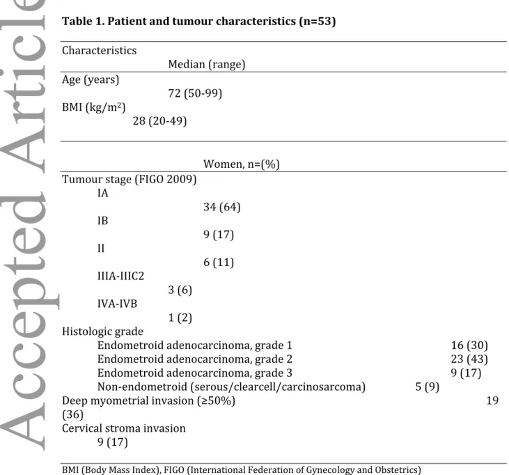
Demographic characteristics of the study population (n=53) show that the vast majority (81%) had stage I endometrial cancer at the time of diagnosis. Thirty-six percent had deep myometrial invasion and 17% had cervical stroma invasion according to
histopathology (Table 1).
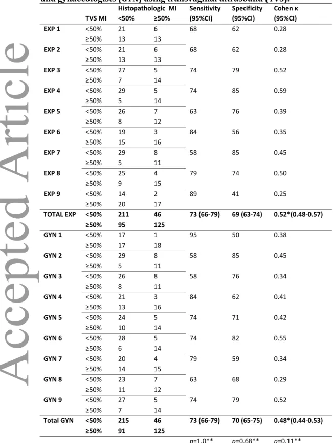
No difference was found between ultrasound experts and gynaecologists in predicting deep myometrial invasion (Table 2), but ultrasound experts assessed cervical stroma invasion with significantly higher sensitivity (p<0.01) and specificity (p=0.02)(Table 3).
A subanalysis of the agreement to histopathology among women with or without deep myometrial invasion and cervical stroma invasion showed that ultrasound experts had a higher agreement to histopathology than gynaecologists in the 34 cases without myometrial invasion (kappa 0.42, 95% CI 0.37-0.48 vs. kappa 0.36, 95% CI 0.31-0.42, p=0.04) but not in the 19 cases with myometrial invasion (kappa 0.44, 95% CI 0.36-0.51 vs. kappa 0.40, 95% CI 0.33-0.47, p=0.25). When all cases of myometrial invasion were combined, no significant difference could be found in degree of agreement to
histopathology between ultrasound experts and gynaecologists (kappa 0.52, 95% CI 0.48-0.57 vs. kappa 0.48, 95% CI 0.44-0.53, p=0.11, Table 2). Ultrasound experts had a higher agreement to histopathology than gynaecologists regarding the assessment of cervical stroma invasion. This finding holds for all 53 cases (kappa 0.58, 95% CI 0.53-0.62 vs. kappa 0.45, 95% CI 0.40-0.49, p<0.001, Table 3), as well as restricted to the 9 cases with cervical invasion (kappa 0.56, 95% CI 0.45-0.67 vs. kappa 0.44, 95% CI
0.33-0.55, p=0.04) and the 44 cases without cervical invasion (kappa 0.47, 95% CI 0.42-0.52 versus kappa 0.41, 95% CI 0.36-0.46, p=0.02).
Ultrasound experts rated a higher certainty in the assessment of both deep myometrial invasion and cervical stroma invasion (VAS 74, 95% CI 71-77 vs. 56, 95% CI 53-60, p=0.008, VAS 77, 95% CI 74-80 vs. 58, 95% CI 53-62, p=0.01). Ultrasound experts and gynaecologists assessed video-clip quality the same, regarding both deep myometrial invasion and cervical stroma invasion (VAS 64, 95% CI 60-68 vs. 56, 95% CI 52-61, p=0.23, VAS 71, 95% CI 68-74 vs. 59, 95% CI 54-63, p=0.07).
Correct assessment of deep myometrial invasion for ultrasound experts and
gynaecologists combined was correlated to certainty in the assessment (Correlation coefficient 0.49, p<0.001) but not to video-clip quality, stage or body mass index (BMI)(Correlation coefficient 0.24, p=0.08, 0.16, p=0.3 and 0.04, p=0.8). Correct assessment of cervical stroma invasion for experts and gynaecologists combined was correlated to video-clip quality, certainty in the assessment and stage (Correlation coefficient 0.36, p=0.01, 0.62, p<0.001 and -0.43, p<0.001) but not to BMI (Correlation coefficient -0.11, p=0.4).
When dividing the material into histopathological preoperative low-risk (endometroid cancer grade 1-2, n=39) and high-risk (endometroid cancer grade 3 and
non-endometroid cancer, n=14) no correlation was found between preoperative risk according to histopathology and correct assessment of deep endometrial invasion or cervical stroma invasion (0.74 vs. 0.63, p=0.4, 0.80 vs. 0.76, p=0.6). This finding holds
when subdividing ultrasound experts (0.73 vs. 0.64, p=0.5, 0.82 vs. 0.80, p=0.4) and gynaecologists (0.75 vs. 0.62, p=0.2, 0.77 vs. 0.71, p=0.5).
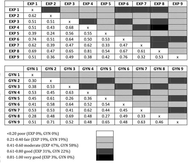
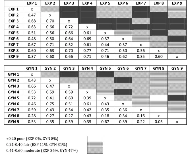
Interobserver reproducibility did not differ among ultrasound experts and among gynaecologists in the assessment of myometrial invasion (p=0.13) (Figure I) whereas ultrasound experts had a higher interobserver reproducibility than gynaecologists in the assessment of cervical stroma invasion (p<0.001) (Figure II).
Discussion
The findings of this study suggest that ultrasound experts, as compared to
gynaecologists, have a higher agreement to histopathology- and a higher interobserver reproducibility, in predicting cervical stroma invasion. However, both groups
performed equally well regarding deep myometrial invasion.
To the best of our knowledge this is the first study on interobserver reproducibility in ultrasound staging in endometrial cancer and the first study comparing interobserver reproducibility between ultrasound experts and gynaecologists. Previous interobserver reproducibility studies on endometrial cancer staging involve histopathological
assessment of deep myometrial invasion 12 and cervical stroma invasion 13 in
hysterectomy specimen and preoperative imaging through MRI, using more than two observers 14. In the latter, where four radiologists assessed 57 women with endometrial cancer, interobserver agreement according to Kappa was 0.39 for deep myometrial invasion and 0.50 for cervical stromal invasion. In the present study, interobserver agreement to Kappa was 0.52 and 0.48 for deep myometrial invasion and 0.58 and 0.45 for cervical stromal invasion by ultrasound experts and gynaecologists respectively. Though direct comparison should be made with caution as the Kappa coefficient is affected by the prevalence of the finding under observation 15, there is no indication that interobserver reproducibility differs markedly between TVS and MRI.
The present study use histopathology of hysterectomy as gold standard, but one must consider that also within the field of histopathology assessments differ. Interobserver studies have shown a good interobserver agreement for myometrial invasion (kappa
0.83) 12 but at most a fair to good agreement in the assessment of cervical stroma invasion (kappa 0.34-0.69) 13 in hysterectomy specimen. This should be kept in mind when viewing the results of the present study. Problematic factors mentioned in the histopathological assessment of cervical stromal invasion include determination of the junction between the lower uterine segment and upper endocervix, the distinction between cervical glandular involvement and stromal involvement and the distinction between cervical glandular involvement and reactive non-neoplastic lesions of the endocervical glands, factors also likely to complicate ultrasound assessment.
Previous studies on prediction of deep endometrial invasion and cervical stromal invasion in endometrial cancer using two dimension (2D) ultrasound found sensitivity and specificity of deep myometrial invasion to be 68-88% and 71-90%, respectively, and sensitivity and specificity of cervical stromal invasion to be 19-93% and 82-99%, respectively 5, 7-11, 16-21. Within the frame of the present study, sensitivity and specificity of deep myometrial invasion by ultrasound experts was 73% and 69%, respectively and sensitivity and specificity of cervical stromal invasion was 57% and 87%, respectively (Table 2 and 3) which is in the lower range of the studies described. That preoperative ultrasound assessment together with histopathology can improve preoperative
identification of women with high-risk endometrial cancer has already been shown in previous studies 5, 6 and the aim of the present study was not to assess that aspect further. The present study design with video-clips was chosen to assess interobserver reproducibility in multiple observers in an identical material under identical
circumstances and the fact that ultrasound experts could not perform live examination might explain why previous sensitivity- and specificity levels were not reached. The video-clips could, on the other hand, possibly have posed an advantage for the
gynaecologists, as the video-clips guide directly to the region of interest, which might be difficult to find for a less experienced examiner. Despite this possible advantage for the gynaecologists, ultrasound experts still showed a significantly higher interobserver reproducibility in the assessment of cervical stroma invasion.
Though it could be suspected that increased BMI would influence video-clip quality negatively, according to a subanalysis BMI did not influence the assessment of either myometrial invasion or cervical stroma invasion, which supports the findings of another recent study 22.
Advantages of the present study are the consecutive series of patients included, where poor image quality or difficult cases were not considered as exclusion criteria, the relatively large number of observers (n=18) and that all observers evaluated an identical material collected by the same ultrasound expert.
The main disadvantage of the present study is that ultrasound assessment could not be made through live examination. However, such a study with the same number of assessors from different parts of Europe would be difficult to perform and ethically questionable.
The fact that no significant difference could be seen between ultrasound experts and gynaecologists in predicting deep myometrial invasion with regard to sensitivity, specificity, degree of agreement to histopathology (Table 2) and interobserver reproducibility (Figure I) raise the question whether intensified training of
invasion to the same level as that of the ultrasound experts. The effect of dedicated training programs remains to be studied and could prove valuable in areas where ultrasound experts are not easily accessible, and aid in triage of women with high risk endometrial cancer to tertiary cancer centres for surgery.
The present study suggests that preoperative ultrasound staging in endometrial cancer is best performed by ultrasound experts, as they show a higher agreement to
histopathology- and a higher interobserver reproducibility in the assessment of cervical stroma invasion.
Acknowledgements
This study would not have been possible without the invaluable participation of M.D. Magnus Hagmar, M.D. Ulrika Joneborg M.D. Charlotte Klynning, M.D. Katja Lampinen, M.D. Cornelia Liebau, M.D. Kolbrun Pàlsdottir, M.D. Katarina Remeaus, M.D. Arne Rådestad and M.D. Ingela Svenberg.
References
1. May K, Bryant A, Dickinson HO, Kehoe S, Morrison J. Lymphadenectomy for the management of endometrial cancer. Cochrane Database Syst Rev. 2010. DOI 10.1002/14651858.CD007794.pub2.
2. Creasman WT, Odicino F, Maisonneuve P, Quinn MA, Beller U, Benedet JL, Heintz APM, Ngan Hys, Pecorelli S. Carcinoma of the corpus uteri. FIGO 26th Annual Report on the Results of Treatment in Gynecological Cancer. Int J Gynaecol Obstet. 2006;95:105-43.
3. Benedetti Panici P, Basile S, Maneschi F, Alberto Lissoni A, Signorelli M, Scambia G, Angioli R, Tateo S, Mangili G, Katsaros D, Garozzo G, Campagnutta E, Donadello N, Greggi S, Melpignano M, Raspagliesi F, Ragni N, Cormio G, Grassi R, Franchi M, Giannarelli D, Fossati R, Torri V, Amoroso M, Crocè C, Mangioni C. Systematic pelvic lymphadenectomy vs no lymphadenectomy in early-stage endometrial carcinoma: randomized clinical trial. J Natl Cancer Inst. 2008;100:1707-16.
4. Kitchener H, Swart AM, Qian Q, Amos C, Parmar MK. Efficacy of systematic pelvic lymphadenectomy in endometrial cancer (MRC ASTEC trial): a randomised study. Lancet. 2009;373:125-36.
5. Ortoft G, Dueholm M, Mathiesen O, Hansen ES, Lundorf E, Moller C, Marinovskij E, Petersen LK. Preoperative staging of endometrial cancer using TVS, MRI, and hysteroscopy. Acta Obstet Gynecol Scand. 2013;92:536-45.
6. Van Holsbeke C, Ameye L, Testa AC, Mascilini F, Lindqvist P, Fischerova D, Frühauf F, Fransis S, de Jonge E, Timmerman D, Epstein E. Development and external validation of (new) ultrasound based mathematical models for preoperative prediction of high-risk endometrial cancer. Ultrasound Obstet Gynecol. 2014;43:586-95.
7. Savelli L, Ceccarini M, Ludovisi M, Fruscella E, De Iaco PA, Salizzoni E, Mabrouk M, Manfredi R, Testa AC, Ferrandina G. Preoperative local staging of endometrial cancer: transvaginal sonography vs. magnetic resonance imaging. Ultrasound Obstet Gynecol. 2008;31:560-6.
8. Celik C, Ozdemir S, Kiresi D, Emlik D, Tazegul A, Esen H. Evaluation of cervical involvement in endometrial cancer by transvaginal sonography, magnetic resonance imaging and frozen section. J Obstet Gynaecol. 2010;30:302-7.
9. Ozdemir S, Celik C, Emlik D, Kiresi D, Esen H. Assessment of myometrial invasion in endometrial cancer by transvaginal sonography, Doppler ultrasonography,
magnetic resonance imaging and frozen section. Int J Gynecol Cancer. 2009;19:1085-90.
10. Antonsen SL, Jensen LN, Loft A, Berthelsen AK, Costa J, Tabor A, Qvist I, Hansen MR, Fisker R, Andersen ES, Sperling L, Nielsen AL, Asmussen J, Høgdall E, Fagö-Olsen CL, Christensen IJ, Nedergaard L, Jochumsen K, Høgdall C. MRI, PET/CT and ultrasound in the preoperative staging of endometrial cancer - a multicenter prospective comparative study. Gynecol Oncol. 2013;128:300-8.
11. Mascilini F, Testa AC, Van Holsbeke C, Ameye L, Timmerman D, Epstein E.
Evaluating myometrial and cervical invasion in women with endometrial cancer: comparing subjective assessment with objective measurement techniques. Ultrasound Obstet Gynecol. 2013;42:353-8.
12. Nedergaard L, Jacobsen M, Andersen JE. Interobserver agreement for tumour type, grade of differentiation and stage in endometrial carcinomas. Apmis. 1995;103:511-8.
13. McCluggage WG, Hirschowitz L, Wilson GE, Oliva E, Soslow RA, Zaino RJ. Significant variation in the assessment of cervical involvement in endometrial carcinoma: an interobserver variation study. Am J Surg Pathol. 2011;35:289-94.
14. Haldorsen IS, Husby JA, Werner HM, Magnussen IJ, Rorvik J, Helland H, Trovik J, Salvesen ØO, Espeland A, Salvesen HB. Standard 1.5-T MRI of endometrial
carcinomas: modest agreement between radiologists. Eur Radiol. 2012;22:1601-11.
15. Viera AJ, Garrett JM. Understanding interobserver agreement: the kappa statistic. Fam Med. 2005;37:360-3.
16. Sawicki W, Spiewankiewicz B, Stelmachow J, Cendrowski K. The value of ultrasonography in preoperative assessment of selected prognostic factors in endometrial cancer. Eur J Gynaecol Oncol. 2003;24:293-8.
17. Fishman A, Altaras M, Bernheim J, Cohen I, Beyth Y, Tepper R. The value of transvaginal sonography in the preoperative assessment of myometrial invasion in high and low grade endometrial cancer and in comparison to frozen section in grade 1 disease. Eur J Gynaecol Oncol. 2000;21:128-30.
18. Szantho A, Szabo I, Csapo ZS, Balega J, Demeter A, Papp Z. Assessment of myometrial and cervical invasion of endometrial cancer by transvaginal sonography. Eur J Gynaecol Oncol. 2001;22:209-12.
19. Akbayir O, Corbacioglu A, Numanoglu C, Guleroglu FY, Ulker V, Akyol A, Guraslan B, Odabasi E. Preoperative assessment of myometrial and cervical invasion in endometrial carcinoma by transvaginal ultrasound. Gynecol Oncol.
20. Cicinelli E, Marinaccio M, Barba B, Tinelli R, Colafiglio G, Pedote P, Rossi C, Pinto V. Reliability of diagnostic fluid hysteroscopy in the assessment of cervical invasion by endometrial carcinoma: a comparative study with transvaginal sonography and MRI. Gynecol Oncol. 2008;111:55-61.
21. Ruangvutilert P, Sutantawibul A, Sunsaneevithayakul P, Boriboonhirunsarn D, Chuenchom T. Accuracy of transvaginal ultrasound for the evaluation of
myometrial invasion in endometrial carcinoma. J Med Assoc Thai. 2004;87:47-52.
22. Fischerova D, Frühauf F, Zikan M, Pinkavova I, Kocián R, Dundr P, Nemejcova K, Dusek L, Cibula D. Factors affecting preoperative local staging of endometrial cancer. Ultrasound Obstet Gynecol. 2014;43:575-85.
Table 1. Patient and tumour characteristics (n=53) Characteristics Median (range) Age (years) 72 (50-99) BMI (kg/m2) 28 (20-49) Women, n=(%) Tumour stage (FIGO 2009)
IA 34 (64) IB 9 (17) II 6 (11) IIIA-IIIC2 3 (6) IVA-IVB 1 (2) Histologic grade
Endometroid adenocarcinoma, grade 1 16 (30)
Endometroid adenocarcinoma, grade 2 23 (43)
Endometroid adenocarcinoma, grade 3 9 (17)
Non-endometroid (serous/clearcell/carcinosarcoma) 5 (9)
Deep myometrial invasion (≥50%) 19
(36)
Cervical stroma invasion 9 (17)
Table 2. Prediction of deep myometrial invasion (MI) by ultrasound experts (EXP) and gynaecologists (GYN) using transvaginal ultrasound (TVS).
Histopathologic MI Sensitivity Specificity Cohen κ
TVS MI <50% ≥50% (95%CI) (95%CI) (95%CI)
EXP 1 <50% 21 6 68 62 0.28 ≥50% 13 13 EXP 2 <50% 21 6 68 62 0.28 ≥50% 13 13 EXP 3 <50% 27 5 74 79 0.52 ≥50% 7 14 EXP 4 <50% 29 5 74 85 0.59 ≥50% 5 14 EXP 5 <50% 26 7 63 76 0.39 ≥50% 8 12 EXP 6 <50% 19 3 84 56 0.35 ≥50% 15 16 EXP 7 <50% 29 8 58 85 0.45 ≥50% 5 11 EXP 8 <50% 25 4 79 74 0.50 ≥50% 9 15 EXP 9 <50% 14 2 89 41 0.25 ≥50% 20 17 TOTAL EXP <50% 211 46 73 (66-79) 69 (63-74) 0.52*(0.48-0.57) ≥50% 95 125 GYN 1 <50% 17 1 95 50 0.38 ≥50% 17 18 GYN 2 <50% 29 8 58 85 0.45 ≥50% 5 11 GYN 3 <50% 26 8 58 76 0.34 ≥50% 8 11 GYN 4 <50% 21 3 84 62 0.41 ≥50% 13 16 GYN 5 <50% 24 5 74 71 0.42 ≥50% 10 14 GYN 6 <50% 28 5 74 82 0.55 ≥50% 6 14 GYN 7 <50% 20 4 79 59 0.34 ≥50% 14 15 GYN 8 <50% 23 7 63 68 0.29 ≥50% 11 12 GYN 9 <50% 27 5 74 79 0.52 ≥50% 7 14 Total GYN <50% 215 46 73 (66-79) 70 (65-75) 0.48*(0.44-0.53) ≥50% 91 125 p=1.0** p=0.68** p=0.11**
* Fleiss Kappa for multiple observers. ** Mc Nemars test.
Table 3. Prediction of cervical stroma invasion (CI) by ultrasound experts (EXP) and gynaecologists (GYN) using transvaginal ultrasound (TVS).
Histopathologic CI Sensitivity Specificity Cohen κ
TVS CI No CI CI (95%CI) (95%CI) (95%CI)
EXP 1 no CI 40 5 44 91 0.37 CI 4 4 EXP 2 no CI 40 3 67 91 0.55 CI 4 6 EXP 3 no CI 39 3 67 89 0.51 CI 5 6 EXP 4 no CI 37 4 56 84 0.35 CI 7 5 EXP 5 no CI 42 5 44 95 0.46 CI 2 4 EXP 6 no CI 31 3 67 70 0.26 CI 13 6 EXP 7 no CI 41 6 33 93 0.31 CI 3 3 EXP 8 no CI 39 4 56 89 0.42 CI 5 5 EXP 9 no CI 35 2 78 80 0.44 CI 9 7 Total EXP no CI 344 35 57 (45-68) 87 (83-90) 0.58*(0.53-0.62) CI 52 46 GYN 1 no CI 36 4 56 82 0.32 CI 8 5 GYN 2 no CI 39 5 44 89 0.33 CI 5 4 GYN 3 no CI 38 6 33 86 0.20 CI 6 3 GYN 4 no CI 35 6 33 80 0.11 CI 9 3 GYN 5 no CI 33 3 67 75 0.31 CI 11 6 GYN 6 no CI 38 4 56 86 0.39 CI 6 5 GYN 7 no CI 35 5 44 80 0.20 CI 9 4 GYN 8 no CI 38 8 11 86 0.03 CI 6 1 GYN 9 no CI 35 6 33 80 0.11 CI 9 3 Total GYN no CI 327 47 42 (31-53) 83 (78-86) 0.45*(0.40-0.49) CI 69 34 p<0.01** P=0.02** P<0.001** * Fleiss Kappa for multiple observers.
Figure I. Interobserver agreement according to Kappa among ultrasound experts (EXP) and gynaecologists (GYN) in the assessment of deep myometrial invasion.
EXP 1 EXP 2 EXP 3 EXP 4 EXP 5 EXP 6 EXP 7 EXP 8 EXP 9 EXP 1 x EXP 2 0.62 x EXP 3 0.51 0.51 x EXP 4 0.51 0.43 0.68 x EXP 5 0.39 0.24 0.56 0.55 x EXP 6 0.74 0.51 0.64 0.50 0.53 x EXP 7 0.62 0.39 0.47 0.62 0.33 0.47 x EXP 8 0.69 0.47 0.65 0.81 0.54 0.67 0.61 x EXP 9 0.51 0.36 0.49 0.38 0.42 0.76 0.32 0.53 x
GYN 1 GYN 2 GYN 3 GYN 4 GYN 5 GYN 6 GYN 7 GYN 8 GYN 9
GYN 1 x GYN 2 0.30 x GYN 3 0.38 0.53 x GYN 4 0.53 0.45 0.63 x GYN 5 0.45 0.61 0.26 0.36 x GYN 6 0.41 0.58 0.64 0.52 0.54 x GYN 7 0.53 0.53 0.41 0.62 0.44 0.45 x GYN 8 0.28 0.48 0.69 0.48 0.27 0.49 0.33 x GYN 9 0.51 0.71 0.52 0.48 0.65 0.48 0.63 0.46 x
<0.20 poor (EXP 0%, GYN 0%) 0.21-0.40 fair (EXP 19%, GYN 19%) 0.41-0.60 moderate (EXP 47%, GYN 58%) 0.61-0.80 good (EXP 31%, GYN 22%) 0.81-1.00 very good (EXP 3%, GYN 0%)
Figure II. Interobserver agreement according to Kappa among ultrasound experts (EXP) and gynaecologists (GYN) in the assessment of cervical stroma invasion.
EXP 1 EXP 2 EXP 3 EXP 4 EXP 5 EXP 6 EXP 7 EXP 8 EXP 9 EXP 1 x EXP 2 0.47 x EXP 3 0.68 0.70 x EXP 4 0.63 0.66 0.72 x EXP 5 0.51 0.56 0.66 0.61 x EXP 6 0.48 0.50 0.64 0.69 0.37 x EXP 7 0.67 0.71 0.52 0.61 0.44 0.37 x EXP 8 0.60 0.63 0.70 0.77 0.71 0.50 0.56 x EXP 9 0.37 0.60 0.66 0.71 0.46 0.62 0.35 0.60 x
GYN 1 GYN 2 GYN 3 GYN 4 GYN 5 GYN 6 GYN 7 GYN 8 GYN 9
GYN 1 x GYN 2 0.43 x GYN 3 0.66 0.47 x GYN 4 0.53 0.59 0.59 x GYN 5 0.72 0.41 0.60 0.39 x GYN 6 0.46 0.75 0.51 0.61 0.43 x GYN 7 0.59 0.43 0.54 0.42 0.35 0.36 x GYN 8 0.28 0.27 0.27 0.43 0.18 0.34 0.16 x GYN 9 0.53 0.35 0.59 0.35 0.67 0.39 0.22 0.05 x
<0.20 poor (EXP 0%, GYN 8%) 0.21-0.40 fair (EXP 11%, GYN 31%) 0.41-0.60 moderate (EXP 36%, GYN 47%) 0.61-0.80 good (EXP 53%, GYN 14%) 0.81-1.00 very good (EXP 0%, GYN 0%)