Oxododecanoyl)-
L
-Homoserine-Lactone Induces HLA-G Expression in
Human Immune Cells
Daria Bortolotti,aJoel LeMaoult,bClaudio Trapella,cDario Di Luca,aEdgardo D. Carosella,bRoberta Rizzoa Department of Medical Sciences, Section of Microbiology and Medical Genetics, University of Ferrara, Ferrara, Italya
; CEA, Institute of Emerging Diseases and Innovative Therapies (iMETI), Research Division in Hematology and Immunology (SRHI), Saint-Louis Hospital, Paris, Franceb
; Department of Chemical and Pharmaceutical Sciences, University of Ferrara, Ferrara, Italyc
HLA-G is a nonclassical class I human leukocyte antigen (HLA) involved in mechanisms of immune tolerance. The objective
of this study was to determine whether N-(3-oxododecanoyl)-
L-homoserine lactone (3O-C
12-HSL), a quorum sensing molecule
produced by Pseudomonas aeruginosa, could modify HLA-G expression to control the host immune response. We evaluated the
ability of 3O-C
12-HSL to induce HLA-G expression in primary immune cells, monocytes (U937 and THP1), and T-cell lines
(Jur-kat) in vitro and analyzed the cellular pathway responsible for HLA-G expression. We studied the HLA-G promoter with a
lucif-erase assay and interleukin-10 (IL-10) and p38/CREB signaling with enzyme-linked immunosorbent assay and
immunofluores-cence, respectively. We observed that 3O-C
12-HSL is able to induce HLA-G expression in human monocytes and T cells. We
showed that the induction of HLA-G by 3O-C
12-HSL is p38/CREB and IL-10 dependent. 3O-C
12-HSL treatment is able to arrest
only the U937 cell cycle, possibly due to the peculiar expression of the ILT2 receptor in the U937 cell line. Our observations
sug-gest HLA-G as a mechanism to create a protected niche for the bacterial reservoir, similar to the role of HLA-G molecules during
viral infections.
B
acterial diseases can result in serious or life-threatening
com-plications, such as bacteremia, kidney failure, and toxic shock
syndrome. For this, the fight against bacterial infection represents
one of the high points of modern medicine. Lack of progress in
controlling mortality and morbidity associated with severe
bacte-rial infections in part reflects our limited understanding of the
complex biological pathways that bacteria use to regulate host
immune response. Many bacteria are capable of forming a
well-organized bacterial population during host infection, and
Pseu-domonas aeruginosa is one of the most commonly studied. As the
cell population increases, P. aeruginosa increases the expression of
quorum sensing (QS) molecules that bind to the transcriptional
activators, enabling the expression of target genes involved in P.
aeruginosa virulence (
1
). P. aeruginosa has two well-studied QS
systems, las and rhl (
2–4
). The las system consists of the LasR
transcriptional regulator and the LasI synthase protein, which is
essential for the production of the signal molecule
N-(3-oxodo-decanoyl)-
L-homoserine lactone (3O-C
12-HSL) that also is
re-quired for LasR activation (
4
). Recent studies have shown that
3O-C
12-HSL has the potential to modify the functions of host
immune cells (
5–9
). In particular, 3O-C
12-HSL inhibits
profes-sional immune cells, such as dendritic and T cells (
10
), promotes
immune cell apoptosis (
11–13
), and blocks the response of
mac-rophages and monocytes to toll-like receptor (TLR) signals (
14
).
However, the mechanism of this immune-regulatory activity has
yet to be fully characterized.
HLA-G is a nonclassical class I human leukocyte antigen
(HLA) characterized by the presence of membrane-bound
(G1-G4) and soluble (G5-G7) isoforms (
15
). HLA-G is involved in
mechanisms of immune tolerance under several conditions,
in-cluding pregnancy, organ transplantation, and autoimmune and
inflammatory diseases by inhibiting cytolytic functions of natural
killer cells, cytotoxic T lymphocytes, and T and dendritic cells, as
well as by inhibiting alloproliferative responses (
16
). Both soluble
and membrane-bound HLA-G isoforms have similar functions
and interact with specific inhibitory receptors (ILT-2 and ILT-4)
expressed by immune cells. During viral infections, HLA-G
mol-ecules are upregulated by the virus as a mechanism of immune
escape (
16
), inhibiting the host immune response. Few data are
available on the effect of bacterial infections on HLA-G
expres-sion.
The objective of the present study was to determine whether P.
aeruginosa 3O-C
12-HSL could modify HLA-G expression byim-mune cells, supporting the hypothesis of a direct involvement of
HLA-G molecules in P. aeruginosa’s ability to control host
im-mune response.
MATERIALS AND METHODS
Cell lines. U937 (ATCC CRL1593.2) and THP-1 (ATCC TIB202)
mono-cyte cell lines, a Jurkat (ATCC TIB152) T-cell line, and the 721.221 (ATCC CRL1885) B-cell line were grown in RPMI 1640 (Gibco) with 10% fetal bovine serum (FBS), 10% HEPES buffer, 5% penicillin-streptomycin at 37°C with 5% CO2.
Received 18 June 2015 Returned for modification 9 July 2015 Accepted 14 July 2015
Accepted manuscript posted online 20 July 2015
Citation Bortolotti D, LeMaoult J, Trapella C, Di Luca D, Carosella ED, Rizzo R. 2015.
Pseudomonas aeruginosa quorum sensing molecule N-(3-oxododecanoyl)-L-homoserine-lactone induces HLA-G expression in human immune cells. Infect Immun 83:3918 –3925.doi:10.1128/IAI.00803-15.
Editor: L. Pirofski
Address correspondence to Roberta Rizzo, [email protected].
Copyright © 2015, American Society for Microbiology. All Rights Reserved.
doi:10.1128/IAI.00803-15
on September 11, 2015 by guest
http://iai.asm.org/
PBMC purification. Peripheral blood mononuclear cells (PBMCs)
were isolated from whole blood of 10 control subjects by Ficoll gradient (Cederlane, Hornby, Ontario, Canada) and resuspended in RPMI me-dium (EuroClone, Milan, Italy) with 10% fetal calf serum (FCS), 100 U/ml penicillin, and 100 U/ml streptomycin (Sigma-Aldrich, St. Louis, MO). Ethical approval was obtained from the University of Ferrara Re-view Board.
Cell treatments. The cells were treated with 3O-C12-HSL (Sigma-Al-drich) at concentrations of 10, 25, and 50M, as suggested by the litera-ture (17), at different time points and then analyzed.
For the inhibitory experiments, Fc receptors first were blocked using human serum, and cells were incubated for 30 min at 37°C with a final concentration of 10g/ml isotype control monoclonal antibody (MAb) or blocking anti-interleukin-10 (IL-10; BioLegend) monoclonal antibody prior to use.
Cytometric analysis. For flow cytometric analysis, 106cells were washed and incubated for 30 min on ice in 100l of phosphate-buffered saline (PBS) containing 1% FBS, 10 mM sodium azide plus appropriately diluted fluorescent MAb. After two washes with cold washing buffer, cells were washed, fixed in 2% formaldehyde, and analyzed by flow cytometry with a FACSVantage flow cytometer (Becton Dickinson, San Jose, CA) using standard settings and CellQuest software (Becton Dickinson, San Jose, CA) for data analysis. The membrane-bound HLA-G antigens were detected by anti-HLA-G fluorescein isothiocyanate (FITC) MAb (87G; Exbio, Prague, Czech Republic), and ILT2 and ILT4 receptors were ana-lyzed by specific MAbs (Becton Dickinson). Isotype controls (Exbio, Prague, Czech Republic) were performed. PBMCs were analyzed using anti-CD3-peridinin chlorophyll protein, anti-CD14-phycoerythrin (PE), anti-CD45-PE, and anti-CD56-PE (BD) MAbs.
The cell cycle was analyzed by propidium iodide (PI) staining. Cells were resuspended cells in 500l PI–Triton X-100 staining solution (0. 1% [vol/vol] Triton X-100 [Sigma] in PBS, 2 mg DNase-free RNase A [Sigma], 0.40 ml of 500g/ml PI [Roche]), incubated for 30 min at RT, and analyzed by flow cytometry.
Immunofluorescence microscopy. For immunofluorescence
micros-copy, 106cells were washed and incubated for 30 min on ice in 100l of PBS containing 1% FBS, 10 mM sodium azide, and the appropriately diluted fluorescent MAb. After two washes with cold washing buffer, cells were washed again, fixed in 2% formaldehyde, and analyzed by fluores-cence microscopy. Cyclic AMP (cAMP) response element-binding pro-tein (CREB) and phosphorylated CREB (pCREB) (Immunological Sci-ences), as well as p38 and phosphorylated p38 (pp38) (Santa Cruz), were detected by indirect immunofluorescence with the secondary antibody goat anti-mouse FITC (Dako).
Western blot analysis. Cell pellets were treated with lysis buffer with
fresh protease inhibitors, biotinylated with 0.2 mg/ml EZ-Link sulfo-NHS-LC-biotin (Pierce, Rockford, IL) in 1⫻ PBS (pH 8.0) for 30 min at 4°C. Samples then were immunoprecipitated for 2 h at RT with CREB/ pCREB (Immunological Sciences) and p38/pp38 (SantaCruz) MAbs or actin MAb (Santa Cruz) as a positive loading control, washed twice in 1⫻ PBS, and incubated overnight with protein G-Sepharose beads (Santa Cruz, CA) at 4°C. The samples were washed twice and resuspended in 20 l of Laemmli buffer (Bio-Rad, Segrate, Italy). Immunoprecipitates were denatured at 100°C for 5 min. Proteins were loaded with native or reduc-ing (in the presence of SDS) runnreduc-ing buffers in 10% TGX-precast gel (Bio-Rad, Segrate, Italy), with subsequent electroblotting transfer onto a polyvinylidene difluoride (PVDF) membrane (Millipore, MA) (1). The membrane was incubated with a horseradish peroxidase (HRP)-conju-gated anti-mouse antibody (1:5,000; Amersham Biosciences, NJ) and de-veloped with the ECL kit (Amersham Biosciences, NJ). The images were acquired by GelDoc (Bio-Rad).
ELISA. Soluble HLA-G5 levels in cell culture supernatants were
mea-sured by enzyme-linked immunosorbent assay (ELISA) using as capture antibody the MAb 5A6G7 (Exbio, Prague, Czech Republic), which recog-nizes the HLA-G5 molecule. The intra-assay coefficient of variation (CV)
was 1.3%, and the interassay CV was 3.7%. The limit of sensitivity was 1.0 ng/ml (18).
IL-10 levels were analyzed using a human IL-10 ELISA detection kit (EBioscience).
MTT cell viability assay. One hundred microliters of cells at a density
of 1⫻ 106/ml were seeded into 96-well plates and treated with 3O-C 12 -HSL (10M and 25 M) for 6, 12, and 24 h. After incubation, 10 l of MTT [3-(4,5-dimethyl-2-thiazolyl)-2,5-diphenyl-2H-tetrazolium bro-mide; Sigma-Aldrich] was added for 4 h at 37°C. Cells then were lysed by adding 100l of MTT solvent. Plates then were read at 570 nm for viabil-ity evaluation using an ELISA reader (Victor; Perkin-Elmer).
mRNA preparation. Total cellular RNA was prepared from each cell
culture with TRIzol reagent (Life Technologies, NY). The RNA samples were digested with DNase. The quality and quantity of RNA samples were assessed by a 1% agarose gel electrophoresis, followed by ethidium bro-mide staining. These mRNA samples were immediately used for cDNA synthesis or stored frozen at⫺80°C until use.
Real-time PCR. To analyze the presence of HLA-G mRNA, 2g
mRNA was reverse transcribed for each sample using a SuperScript first-strand synthesis system (Invitrogen, San Giuliano Milanese, Italy) accord-ing to the manufacturer’s instructions. The primers and the detection probe for HLA-G gene expression analysis were the following: forward primer HLAG-F, 5=-CCCACCATCCCCATCATG-3=; reverse primer HLA-G-R, 5=-CCAGTGACTACAGCTGCAAGGA-3=; and the MGB probe, 5=-TATCGTTGCTGGCCTGG-6-carboxyflourescein-3= (Applied Biosystems) (19). Fold changes in expression were determined by the 2⫺⌬⌬CTmethod. Amplification was performed with 100 ng of RNA con-verted into cDNA with TaqMan 2⫻ universal PCR master mix in a final volume of 50l (Applied Biosystems) by using the following protocol: 2 min at 50°C for AmpErase UNG activation, 20 s at 95°C for initial dena-turation, and then 40 cycles of 20 s at 95°C and 60 s at 60°C for amplifi-cation. All reactions were performed in triplicate.
Reporter constructs and expression vectors. Luciferase reporter
plasmids were generated by cloning genomic promoter fragments into pGL3-Basic (Promega, Madison, WI). These constructs contain a 1,438-bp promoter fragment of HLA-G (pGL3-G1500) and a 269-bp AspI-AhaII-HLA-B7 promoter fragment (pGL3-HLA-B) (kind gift of Sam J. P. Gobin) (20). All inserts were verified by sequence analysis.
The Renilla luciferase control plasmid pRL-actin was used as a trans-fection efficiency control.
Transient transfection. 721.221 cells were transfected by Amaxa
nucleofector technology (Lonza) with a DNA precipitate of 1g of pGL3 reporter plasmid, 1 or 0.5g of expression vector, and 0.1 g of Renilla luciferase control plasmid (pRL-actin) per well. Luciferase activity was determined using a luminometer (Victor; PerkinElmer) and corrected for transfection efficiency with the Renilla luciferase activity values.
Statistical analysis. Since the values presented a normal distribution
(Kolmogorov-Smirnov test), the differences were evaluated by Student t test using Stat View software (SAS Institute Inc., Cary, NC). The P value was considered to be statistically significant when it was⬍0.05.
RESULTS
P. aeruginosa 3O-C12
-HSL induces HLA-G expression in
hu-man monocytes and T cells. We first analyzed the ability of P.
aeruginosa 3O-C
12-HSL to induce HLA-G transcription and
transduction in human primary immune cells. We exposed
pe-ripheral blood mononuclear cells (PBMCs) from 10 control
sub-jects to P. aeruginosa 3O-C
12-HSL (
17
). PBMCs were negative for
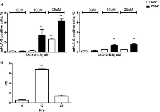
HLA-G staining before the treatment. Both CD3
⫹and CD14
⫹cells induced membrane-bound HLA-G expression, with the
highest levels occurring after 12 h of incubation with 25
M
3O-C12-HSL (2.7%
⫾ 0.3% CD3
⫹HLA-G
⫹and 6.4%
⫾ 0.6% CD14
⫹HLA-G
⫹cells) (
Fig. 1a
) that decreased after 24 h of incubation
(0.6%
⫾ 0.1% CD3
⫹HLA-G
⫹and 1.6%
⫾ 0.4% CD14
⫹HLA-Bacterial Quorum Sensing and HLA-G Molecules
on September 11, 2015 by guest
http://iai.asm.org/
G
⫹). As a confirmation, we performed a real-time PCR
quantifi-cation of HLA-G mRNA in PBMCs after 3O-C12-HSL treatment.
We observed a 6-fold increase in HLA-G mRNA transcription 12
h after the incubation with 3O-C12-HSL (
Fig. 1b
) (P
⬍ 0.0001)
that was lost after 24 h. The analysis of B and NK cells showed no
HLA-G induction (data not shown).
To confirm our data in a standardized in vitro model, we used
Jurkat T-cell and monocyte cell lines that differ in maturation
stages (less mature THP-1 and U937 cells are at a more advanced
stage of differentiation). We treated these cell lines with 10 and 25
M 3O-C12
-HSL (
17
) at the 12-h time point, selected on the basis
of the results obtained with PBMC treatment (
Fig. 1a
). We
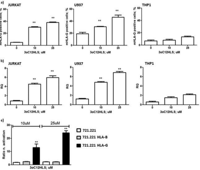
con-firmed the increased expression of HLA-G molecules on T and
monocyte cells (
Fig. 2a
,
b
, and
c
), as previously observed in
PBMCs (
Fig. 1a
), with a significantly lower upregulation in THP1
cells (
Fig. 2c
). These results also were confirmed by mRNA
anal-ysis. We observed a 6-fold increase in HLA-G mRNA
transcrip-tion 12 h after the incubatranscrip-tion of Jurkat and U937 cells with
3O-C
12-HSL (
Fig. 2b
) (P
⬍ 0.0001), while THP1 induced lower levels
of HLA-G mRNA after 3O-C12-HSL treatment (
Fig. 2b
). These
results also confirmed the effect of 3O-C
12-HSL on human
im-mune cells in a standardized in vitro model.
P. aeruginosa 3O-C
12-HSL induces HLA-G via p38/CREB
and IL-10 pathways. We wanted to investigate the mechanisms
used by 3O-C
12-HSL to induce HLA-G expression. To account for
the direct effect of 3O-C12-HSL on the HLA-G gene promoter, we
transfected the 721.221 cell line (classical and nonclassical
HLA-I-negative cells) with HLA-G or HLA-B promoters (
20
). These
cells were treated with 3O-C
12-HSL, and a luciferase assay was
performed. We observed an increased activation of HLA-G
pro-moter after 3O-C
12-HSL treatment. In particular, the promoter
activation was dose dependent, with an increased activation of
21-and 39-fold with 10
M and 25 M 3O-C12
-HSL, respectively
(
Fig. 2d
, black histograms). On the contrary, the HLA-B promoter
was not affected by 3O-C
12-HSL treatment (
Fig. 2d
, gray
histo-grams). These data suggest a specific effect of 3O-C12-HSL on the
HLA-G promoter, with the implication of specific transcription
factors. It is known that 3O-C12-HSL modifies transduction
path-ways (
21
), in particular inducing p38, a mitogen-activated protein
kinase (MAPK) that phosphorylates and activates CREB (
22
), a
transcription factor that regulates HLA-G gene expression (
21
).
We hypothesized that the p38/CREB pathway could be implicated
in the increased HLA-G expression via 3O-C
12-HSL. We
evalu-ated the expression of p38 and CREB and their phosphorylevalu-ated
isoforms after 3O-C
12-HSL treatment in U937 and THP1 cells.
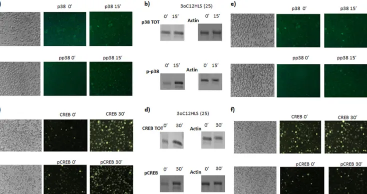
We observed a clear increase in the amount of p38 and
phosphor-ylated p38 after 15 min (
Fig. 3a
) and a consequent increase in
CREB and phosphorylated CREB after 30 min of 10
M
3O-C12-HSL treatment of U937 cells (
Fig. 3b
). Western blot analysis
con-firmed an increased expression of p38 (
Fig. 3b
) and CREB (
Fig.
3d
) protein with a consequent increase in the phosphorylated
iso-forms. Jurkat cells showed a similar pattern of activation (data not
shown). THP1 cells reached a lower rate of CREB
phosphoryla-tion (
Fig. 3e
and
f
), accounting for the lower HLA-G expression
observed in this cell line (
Fig. 2c
). These data suggest that the
induction of the p38/CREB pathway is one of the mechanisms
used by 3O-C
12-HSL to induce HLA-G expression.
Since IL-10 is one of the major inducers of soluble HLA-G5
expression (
23
), and since it is known that 3O-C
12-HSL is able to
increase IL-10 expression in macrophages (
17
), we evaluated if
this cytokine could be implicated in the induction of HLA-G5
secretion via 3O-C12-HSL. We analyzed the levels of IL-10 and
HLA-G5 expression in Jurkat, U937, and THP1 cell culture
super-natants after 3O-C12-HSL treatment. We observed an increased
FIG 1 (a) Membrane HLA-G expression in PBMCs from 10 healthy subjects. Cells were treated with 10 and 25M 3O-C12-HSL for 12 h (left) and 24 h (right). CD3⫹and CD14⫹cell results are reported. (b) HLA-G mRNA expression analysis in PBMCs after 12 h of treatment with 25M 3O-C12-HSL. RQ, relative quantitation. Means⫾ standard deviations (SD) are reported. **, P ⬍ 0.05 by Student t test.
on September 11, 2015 by guest
http://iai.asm.org/
secretion of IL-10 already 6 h after 3O-C
12-HSL addition in both
Jurkat and U937 cells (
Fig. 4a
, white histograms), while HLA-G5
levels increased after 12 h of treatment (
Fig. 4
, gray histograms).
On the contrary, THP1 presented no IL-10 induction after
3O-C
12-HSL treatment (
Fig. 4a
), coinciding with the absence of
HLA-G5 upregulation in THP1 cells (
Fig. 4b
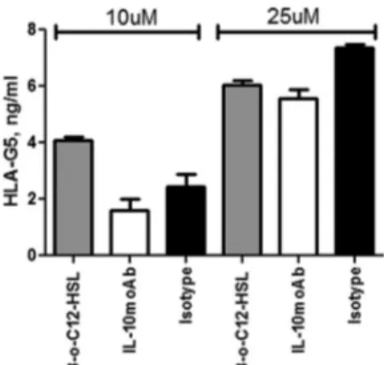
). To evaluate the role
of IL-10, we pretreated U937 cells with anti-IL-10 MAb and
ob-served the reduction of HLA-G5 secretion in 3O-C12-HSL-treated
U937 cell culture supernatants (
Fig. 5
, white histograms). This
blocking treatment was not able to completely inhibit HLA-G5
expression, even at increased anti-IL-10 MAb concentrations
(data non shown), demonstrating that IL-10 is only one of the
mechanisms used by 3O-C
12-HSL to induce HLA-G5 secretion.
P. aeruginosa 3O-C12
-HSL effect on monocyte and T-cell
vi-ability. Since 3O-C
12-HSL treatment was reported to induce cell
apoptosis (
13
), we verified if 3O-C12-HSL treatment could affect
monocyte and T cell viability. We treated Jurkat, U937, and THP1
cells with 10, 25, or 50
M 3O-C12-HSL for 6, 12, and 24 h. After
the treatments, cell viability was evaluated by MTT assay. We
ob-served no significant decrease in cell viability in both Jurkat and
THP1 cells (
Fig. 6a
and
c
). On the contrary, we found a 40%
decrease in U937 cell viability after 12 h of incubation with all
concentrations of 3O-C
12-HSL (
Fig. 6b
). Interestingly, U937 cells
reconstituted their viability after 24 h of incubation. The analysis
of the U937 cell cycle showed that the treatment with 3O-C
12-HSL
for 12 h blocked 62% of cells in the G0/G1
stage (
Fig. 6d
).
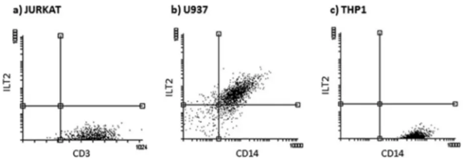
Since HLA-G interaction with its ligands (ILT2 and ILT4) is
able to modify immune cell proliferation (
24
,
25
), we tested U937,
THP1, and Jurkat cells for ILT2 and ILT4 expression. We observed
no ILT4 expression on any of the three cell lines, while ILT2 was
expressed only on the surface of U937 cells (
Fig. 7
).
DISCUSSION
The inability of the host to contain an invading pathogen is
asso-ciated with a systemic inflammatory response that, when
deregu-lated, can lead to organ injury. Recently, it has been established
that QS molecules not only are important in the regulation of
bacterial virulence genes but also interact with eukaryotic cells and
modulate immune responses (
5–14
). The investigations reported
here demonstrate that 3O-C12-HSL, a P. aeruginosa QS molecule,
is able to induce HLA-G expression in human monocytes and T
cells. Moreover, we showed that the induction of HLA-G by
3O-C
12-HSL is p38/CREB and IL-10 dependent. The implication of
IL-10 and p38/CREB pathways has been previously reported in
FIG 2 HLA-G membrane (87G-FITC MAb) (a) and mRNA HLA-G (b) expression in Jurkat, U937, and THP1 cell lines. Cells were treated with 10 and 25M 3O-C12-HSL for 12 h. (c) Luciferase assay of HLA-B and HLA-G promoter activation in 721.221-transfected cell lines after treatment with 10 and 25M 3O-C12-HSL for 12 h. Means⫾ SD are reported. **, P ⬍ 0.05 by Student t test.
Bacterial Quorum Sensing and HLA-G Molecules
on September 11, 2015 by guest
http://iai.asm.org/
bacterial infections. In particular, IL-10 expression previously has
been related to the role of 3O-C
12-HSL in P. aeruginosa infection
by simultaneously upregulating the anti-inflammatory cytokine
IL-10 and downregulating the proinflammatory cytokine tumor
necrosis factor alpha (
17
). IL-10 expression is prompted during
bacterial infections (
26
) as a mechanism of immune suppression,
and CREB phosphorylation is induced to modify cellular patterns
of protein expression (
27
). As a proof of concept of the
implica-FIG 3 Intracellular pathway activation analysis in U937 and THP1 cell lines. U937 and THP1 cells were treated with 25M 3O-C12-HSL. p38 and CREB analysis was performed by immunofluorescence for total (p38, CREB) and phosphorylated (pp38, pCREB) status in U937 (a and c) and THP1 (e and f) cells. Western blot analysis for total (p38; 38 kDa), CREB (37 kDa), phosphorylated (pp38, pCREB), and actin (loading control; 42 kDa) status was performed in U937 cells (b and d). Shown are the most representative results at 0 and 15 s for p38 and at 0 and 30 s for CREB after 25M 3O-C12-HSL treatment.
FIG 4 IL-10 (a) and HLA-G5 (b) levels in Jurkat, U937, and THP1 cell culture supernatants. Cells were treated with 10 and 25M 3O-C12-HSL for 6, 12, and 24 h. Means⫾ SD are reported. **, P ⬍ 0.05 obtained by Student t test.
on September 11, 2015 by guest
http://iai.asm.org/
tion of both IL-10 and p38/CREB pathways, the THP1 cell line
showed a lower increase of HLA-G expression than U937 after
3O-C
12-HSL treatment with corresponding lower p38/CREB
ex-pression and IL-10 induction. It is worth noting that U937 and
THP1 cell lines already have been found to behave in a different
way after exposure to compounds (
28
,
29
) and microbial
infec-tions (
30
), sustaining the data obtained in our study. In particular,
another bacterium, Chlamydia pneumoniae, already has been
shown to downregulate genes related to cell division only in U937
cells (
31
), supporting the peculiar effect of 3O-C12-HSL treatment
in arresting the U937 cell cycle. Moreover, only U937 cells express
the ILT2 receptor, and interacting with HLA-G molecules in the
cell culture supernatant might modify cell activation status (
24
).
However, we are aware that the effects of 3O-C
12-HSL on cell
viability are still to be understood.
The results obtained suggest HLA-G as a mechanism of
im-mune privilege that creates a protected niche for a bacterial
reser-voir, similar to the role of HLA-G molecules during viral
infec-tions (
16
). This hypothesis is in agreement with the previous
observations on the implications of HLA-G expression in bacterial
infections. First, HLA-G expression at the extravillous
cytotro-phoblast surface seems to increase the risk of Listeria
monocyto-genes infection (
32
). We suggest that this bacteria exploits
cytotro-phoblast constitutive HLA-G expression to evade host immune
cells. Moreover, soluble HLA-G molecules are expressed during
septic shock and are predictive of better survival (
33
). The patients
FIG 5 HLA-G5 levels in cell culture supernatants of U937 cells after 10 or 25
M 3O-C12-HSL treatment for 12 h (gray histogram) with anti-IL-10 (white histogram) or anti-isotype (black histogram) MAb addition. Means⫾ SD are reported. **, P⬍ 0.05 obtained by Student t test.
FIG 6 MTT cell viability assay. Cells were treated with 10, 25, and 50M 3O-C12-HSL for 6, 12, or 24 h. Jurkat (a), U937 (b), and THP1 (c) cell lines are shown. Means⫾ SD are reported. **, P ⬍ 0.05 obtained by Student t test. (d) Propidium iodide staining for cell cycle analysis in U937 cells treated with 25 M 3O-C12-HSL for 12 h. The percentage of cells in each cell cycle stage is reported. The most representative results are shown.
Bacterial Quorum Sensing and HLA-G Molecules
on September 11, 2015 by guest
http://iai.asm.org/
with sepsis are characterized by a deregulated immune response
(
10
), impaired chemotactic responses, and alterations in
neutro-phil functions (
34
) that could benefit from HLA-G–ILT2
interac-tion, allowing the restoration of a controlled immune activation
(
35
).
In conclusion, we present for the first time the effect of a
bac-terial QS compound, 3O-C12-HSL, on HLA-G induction in
hu-man immune cells. These results are important to understanding
bacterial virulence mechanisms and in evaluating the role of
HLA-G expression during bacterial infections. In fact, HLA-G
could be considered on the one hand as a natural way to create an
immune tolerance condition that could allow bacterial persistence
in the host and, on the other hand, as an immune factor that could
control impaired immune activation during bacterial infection
that is frequently harmful to patient health. As a future
perspec-tive, it will be of importance to recognize the cellular
characteris-tics that make a cell responsive to 3O-C12-HSL treatment for
HLA-G expression and the specific pathological contests that
could benefit or be damaged by the modulation of HLA-G
mole-cules.
ACKNOWLEDGMENTS
Daria Bortolotti, Roberta Rizzo, Claudio Trapella and Dario Di Luca are inventors of the patent FE2014A000005 (submitted November 2014), which is relevant to this work.
We thank Iva Pivanti for technical support, Marina Daouya for labo-ratory support, and Linda M. Sartor for language revision of the manu-script.
REFERENCES
1. Williams P. 2007. Quorum sensing, communication and cross-kingdom signalling in the bacterial world. Microbiology 153:3923–3938.http://dx .doi.org/10.1099/mic.0.2007/012856-0.
2. Davenport P, Griffin JL, Welch M. 2015. Quorum sensing is accompa-nied by global metabolic changes in the opportunistic human pathogen, Pseudomonas aeruginosa. J Bacteriol 197:2072–2082. http://dx.doi.org /10.1128/JB.02557-14.
3. Lee J, Zhang L. 2015. The hierarchy quorum sensing network in Pseu-domonas aeruginosa. Protein Cell 6:26 – 41. http://dx.doi.org/10.1007 /s13238-014-0100-x.
4. Williams P, Camara M. 2009. Quorum sensing and environmental ad-aptation in Pseudomonas aeruginosa: a tale of regulatory networks and multifunctional signal molecules. Curr Opin Microbiol 12:182–191.http: //dx.doi.org/10.1016/j.mib.2009.01.005.
5. Smith RS, Kelly R, Iglewski BH, Phipps RP. 2002. The Pseudomonas autoinducer N-(3-oxododecanoyl) homoserine lactone induces cycloox-ygenase-2 and prostaglandin E2 production in human lung fibroblasts: implications for inflammation. J Immunol 169:2636 –2642.http://dx.doi .org/10.4049/jimmunol.169.5.2636.
6. Ritchie AJ, Yam AOW, Tanabe KM, Rice SA, Cooley MA. 2003. Mod-ification of in vivo and in vitro T and B cell mediated immune responses by
the Pseudomonas aeruginosa quorum sensing molecule N-(3-oxododecanoyl)-L-homoserine lactone (OdDHL). Infect Immun 71: 4421– 4431.http://dx.doi.org/10.1128/IAI.71.8.4421-4431.2003. 7. Telford G, Wheeler D, Williams P, Tomkins PT, Appleby P, Sewell H,
Stewart GS, Bycroft BW, Pritchard DI. 1998. The Pseudomonas
aerugi-nosa quorum-sensing signal molecule N-(3-oxododecanoyl)-L-homoserine lactone has immunomodulatory activity. Infect Immun 66: 36 – 42.
8. Hooi DS, Bycroft BW, Chhabra SR, Williams P, Pritchard DI. 2004. Differential Immune modulatory activity of Pseudomonas aeruginosa quorum-sensing signal molecules. Infect Immun 72:6463– 6470.http://dx .doi.org/10.1128/IAI.72.11.6463-6470.2004.
9. Smith RS, Harris SG, Phipps R, Iglewski B. 2002. The Pseudomonas aerugi-nosa quorum sensing molecule N-(3-oxododecanoyl) homoserine lactone contributes to virulence and induces inflammation in vivo. J Bacteriol 184: 1132–1139.http://dx.doi.org/10.1128/jb.184.4.1132-1139.2002.
10. Boontham P, Robins A, Chandran P, Pritchard D, Cámara M, Williams
P, Chuthapisith S, McKechnie A, Rowlands BJ, Eremin O. 2008.
Sig-nificant immunomodulatory effects of Pseudomonas aeruginosa quo-rum-sensing signal molecules: possible link in human sepsis. Clin Sci 115: 343–351.http://dx.doi.org/10.1042/CS20080018.
11. Li H, Wang L, Zhang C, Gong F, Li H, Xie X, Xia C, Chen J, Song Y,
Shen A, Song J. 2009. Influence of Pseudomonas aeruginosa quorum
sensing signal molecule N-(3-oxododecanoyl) homoserine lactone on mast cells. Med Microbiol Immunol 198:113–121.http://dx.doi.org/10 .1007/s00430-009-0111-z.
12. Jacobi C, Schiffner F, Henkel M, Waibel M, Stork B, Daubrawa M,
Eberl L, Gregor M, Wesselborg S. 2009. Effects of bacterial N-acyl
ho-moserine lactones on human Jurkat T lymphocytes-OdDHL induces apoptosis via the mitochondrial pathway. Int J Med Microbiol 299:509 – 519.http://dx.doi.org/10.1016/j.ijmm.2009.03.005.
13. Tateda K, Ishii Y, Horikawa M, Matsumoto T, Miyairi S, Pechere JC,
Standiford TJ, Ishiguro M, Yamaguchi K. 2003. The Pseudomonas
aeruginosa autoinducer N-3-oxododecanoyl homoserine lactone acceler-ates apoptosis in macrophages and neutrophils. Infect Immun 71:5785– 5793.http://dx.doi.org/10.1128/IAI.71.10.5785-5793.2003.
14. Kravchenko VV, Kaufmann GF, Mathison JC, Scott DA, Katz AZ,
Grauer DC, Lehmann M, Meijler MM, Janda KD, Ulevitch RJ. 2008.
Modulation of gene expression via disruption of NF-kappaB signaling by a bacterial small molecule. Science 321:259 –263. http://dx.doi.org/10 .1126/science.1156499.
15. Rizzo R, Bortolotti D, Baricordi OR, Fainardi E. 2012. New insights into HLA-G and inflammatory diseases. Inflamm Allergy Drug Targets 11: 448 – 463.http://dx.doi.org/10.2174/187152812803590037.
16. Rizzo R, Bortolotti D, Bolzani S, Fainardi E. 2014. HLA-G molecules in autoimmune diseases and infections. Front Immunol 5:592.
17. Glucksam-Galnoy Y, Sananes R, Silberstein N, Krief P, Kravchenko VV,
Meijler MM, Zor T. 2013. The bacterial quorum-sensing signal molecule
N-3-oxo-dodecanoyl-L-homoserine lactone reciprocally modulates pro-and anti-inflammatory cytokines in activated macrophages. J Immunol
191:337–344.http://dx.doi.org/10.4049/jimmunol.1300368.
18. Rizzo R, Malagutti N, Bortolotti D, Gentili V, Rotola A, Fainardi E,
Pezzolo T, Aimoni C, Pelucchi S, Di Luca D, Pastore A. 2014. Infection
and HLA-G molecules in nasal polyposis. J Immunol Res 2014:407– 430. 19. Yao YQ, Barlow DH, Sargent IL. 2005. Differential expression of alter-natively spliced transcripts of HLA-G in human preimplantation embryos
FIG 7 ILT2 membrane expression, evaluated by flow cytometry (anti-ILT2 MAb) in Jurkat (a), U937 (b), and THP1 (c) cell lines. The most representative results
are shown.
on September 11, 2015 by guest
http://iai.asm.org/
and inner cell masses. J Immunol 175:8379 – 8385.http://dx.doi.org/10 .4049/jimmunol.175.12.8379.
20. Gobin SJP, Biesta P, de Steenwinkel JEM, Datema G, Van den Elsen PJ. 2002. HLA-G transactivation by cAMP-response element-binding protein (CREB). An alternative transactivation pathway to the conserved major histocompatibility complex (MHC) class I regulatory routes. J Biol Chem
277:39525–39531.
21. Kravchenko VV, Kaufmann GF, Mathison JC, Scott DA, Katz AZ,
Wood MR, Brogan AP, Lehmann M, Mee JM, Iwata K, Pan Q, Fearns C, Knaus UG, Meijler MM, Janda KD, Ulevitch RJ. 2006.
N-(3-oxo-acyl)homoserine lactones signal cell activation through a mechanism dis-tinct from the canonical pathogen-associated molecular pattern recogni-tion receptor pathways. J Biol Chem 281:28822–28830.http://dx.doi.org /10.1074/jbc.M606613200.
22. Deak M, Clifton AD, Lucocq LM, Alessi DR. 1998. Mitogen and stress-activated protein kinase-1 (MSK1) is directly stress-activated by MAPK and SAPK2/p38, and may mediate activation of CREB. EMBO J 17:4426 – 4441.http://dx.doi.org/10.1093/emboj/17.15.4426.
23. Rizzo R, Hviid TV, Stignani M, Balboni A, Grappa MT, Melchiorri L,
Baricordi OR. 2005. The HLA-G genotype is associated with IL-10 levels
in activated PBMCs. Immunogenetics 57:172–181.http://dx.doi.org/10 .1007/s00251-005-0788-0.
24. Shiroishi M, Tsumoto K, Amano K, Shirakihara Y, Colonna M, Braud
VM, Allan DS, Makadzange A, Rowland-Jones S, Willcox B, Jones EY, van der Merwe PA, Kumagai I, Maenaka K. 2003. Human inhibitory
receptors Ig-like transcript 2 (ILT2) and ILT4 compete with CD8 for MHC class I binding and bind preferentially to HLA-G. Proc Natl Acad Sci U S A 100:8856 – 8861.http://dx.doi.org/10.1073/pnas.1431057100. 25. Carosella ED, Gregori S, LeMaoult J. 2011. The tolerogenic interplay (s)
among HLA-G, myeloid APCs, and regulatory cells. Blood 118:6499 – 6505.http://dx.doi.org/10.1182/blood-2011-07-370742.
26. McNab FW, Ewbank J, Howes A, Moreira-Teixeira L, Martirosyan A,
Ghilardi N, Saraiva M, O’Garra A. 2014. Type I IFN induces IL-10
production in an IL-27-independent manner and blocks responsiveness to IFN-␥ for production of IL-12 and bacterial killing in Mycobacterium tuberculosis-infected macrophages. J Immunol 193:3600 –3612.http://dx .doi.org/10.4049/jimmunol.1401088.
27. Kim YO, Jung MJ, Choi JK, Ahn do W, Song KS. 2011. Peptidoglycan from Staphylococcus aureus increases MUC5AC gene expression via RSK1-CREB pathway in human airway epithelial cells. Mol Cells 32:359 – 365.http://dx.doi.org/10.1007/s10059-011-0118-3.
28. Panzarini E, Ramires PA, Miccoli MA, Tenuzzo B, Scordari A, Dini L. 2006. Differentiation of THP-1 and U937 cells in presence of synthetic hydrogels. Caryologia 59:395– 402.
29. Legdeur MC, Bontje PM, Ossenkoppele GJ, Beelen RH, van de
Loos-drecht AA, Broekhoven MG, Langenhuijsen MM, Thijsen SF, Hofstee H, Schuurhuis GJ. 1996. The role of BCL-2 and bax protein in
monocyte-mediated apoptosis in human leukemic cell lines. Exp Hematol 24:1530 – 1539.
30. Nonaka T, Kuwabara T, Mimuro H, Kuwae A, Imajoh-Ohmi S. 2003. Shigella-induced necrosis and apoptosis of U937 cells and J774 macro-phages. Microbiology 149:2513–2527. http://dx.doi.org/10.1099/mic.0 .26341-0.
31. Virok D, Loboda A, Kari L, Nebozhyn M, Chang C, Nichols C, Endresz
V, Gonczol E, Berencsi K, Showe MK, Showe LC. 2003. Infection of
U937 monocytic cells with Chlamydia pneumoniae induces extensive changes in host cell gene expression. J Infect Dis 188:1310 –1321.http://dx .doi.org/10.1086/379047.
32. Cao B, Mysorekar IU. 2014. Intracellular bacteria in placental basal plate localize to extravillous trophoblasts. Placenta 35:139 –142.http://dx.doi .org/10.1016/j.placenta.2013.12.007.
33. Monneret G, Voirin N, Krawice-Radanne I, Bohé J, Lepape A,
Rouas-Freiss N, Carosella ED. 2007. Soluble human leukocyte
anti-gen-G5 in septic shock: marked and persisting elevation as a predictor of survival. Crit Care Med 35:1942–1947.http://dx.doi.org/10.1097/01 .CCM.0000277039.84372.1C.
34. Kovach MA, Standiford TJ. 2012. The function of neutrophils in sepsis. Curr Opin Infect Dis 25:321–327. http://dx.doi.org/10.1097 /QCO.0b013e3283528c9b.
35. Bankey PE, Banerjee S, Zucchiatti A, De M, Sleem RW, Lin CF,
Miller-Graziano CL, De AK. 2010. Cytokine induced expression of
pro-grammed death ligands in human neutrophils. Immunol Lett 129:100 – 107.http://dx.doi.org/10.1016/j.imlet.2010.01.006.
Bacterial Quorum Sensing and HLA-G Molecules