UNIVERSITÀ DEGLI STUDI DI
ROMA
"TOR VERGATA"
FACOLTA' DI SCIENZE MATEMATICHE, FISICHE E
NATURALI
DOTTORATO DI RICERCA IN IMMUNOLOGIA
XXI CICLO
Immunomodulatory properties of cholera toxin B subunit
Manuela Colucci
A.A. 2008/2009
Tutor: Dott.ssa Francesca Quintieri
Coordinatore: Prof. Paolo Rossi
INDEX 1 ABSTRACT ... 4 1.1 English ... 4 1.2 Italian ... 6 2 INTRODUCTION ... 9 2.1 Cholera toxin ... 9
2.1.1 Transport of cholera toxin into target cells ... 10
2.1.2 Immunity or tolerance? ... 15
2.1.3 Adjuvants’ role: from CT to CTB ... 16
2.2 Dendritic cells ... 19
2.2.1 DC subsets ... 20
2.2.2 Different maturative stages of DC ... 23
2.2.3 Tolerogenic DC ... 28
2.3 Regulatory T cells ... 33
3 MATERIALS AND METHODS ... 39
3.1 Chemical and reagents ... 39
3.2 Human PBMC isolation ... 40
3.3 Generation and culture of MDDC ... 40
3.4 MLR ... 41
3.5 T cell culture ... 43
3.6 Cytokine assays ... 44
3.7 Analysis of T cell function (suppression assay) ... 44
3.8 Flow cytometry analysis ... 46
3.9 Statistical analysis ... 48
4 RESULTS ... 49
4.1 rCTB treatment affects maturation of human MDDC ... 49
4.2 rCTB-treated MDDC show a reduced ability to stimulate MLR
response. ... 52
4.3 rCTB-treated MDDC induce the generation of CD4+ T cells with reduced proliferative capacity and increased IL-10 production 53 4.4 rCTB-treated MDDC induce functional CD4+ Tr1 cells ... 57
5 CONCLUSIONS ... 63
6 DISCUSSION ... 64
7 REFERENCES ... 71
1
ABSTRACT
1.1English
Antigen-non-specific innate immunity and antigen-specific adaptive immunity synergize to eradicate invading pathogens through the actions of immune cells and their effector proteins, including complement, antibodies, cytokines and cytolytic factors. Adaptive immune responses are induced, coordinated and regulated by dendritic cells (DC). DC initiate immunity by the activation of naïve B and T cells - the effector cells of the adaptive immune system - and by the stimulation of natural killer cells - the crucial cellular instigators of innate resistance. Besides linking innate and adaptive immunity, DC limit excessive, tissue-damaging immune responses in order to prevent autoimmunity and non-essential reactions to innocuous agents through their ability to induce antigen-specific unresponsiveness of lymphocytes in primary and secondary lymphoid tissues by mechanisms that include deletion and induction of regulatory T cells (Tregs). Given the central role of these antigen presenting cells in immunity and tolerance, they are ideal therapeutic targets for pharmacological modulation of immune responses.
In the present study, we examined the possibility that recombinant CTB (rCTB) may affect human DC functions in response to toll-like receptor (TLR) stimulation and may induce the generation of DC with the capacity to generate Tregs. CTB - cholera toxin B subunit - is an efficient mucosal carrier molecule for the generation of immune responses to linked antigens, since it facilitates entry into the cell of the CTB-antigen complex by ligation of its surface receptor GM1. There is also good evidence that CTB acts as an immunosuppressant, as it is able to down-modulate human monocyte/macrophage cell line activation and to suppress Th1-type responses. Our findings show that rCTB partially prevents the lipopolysaccharide (LPS)-induced maturation process of monocyte-derived DC (MDDC) and decreases their interleukin-12p70 (IL-12p70) production with no relevant effect on IL-10 production. LPS-stimulated MDDC pre-treated with rCTB are able to promote the induction of low proliferating T cells, which show an enhanced IL-10 production associated with a reduced interferon-γ (IFN-γ) production and the same high levels of trasforming growth factor-β(TGF-β) as the control. These T cells suppress proliferation of activated autologous T cells. Transwell experiments and blockade of IL-10R and TGF-β showed that the immunomodulatory effect is mediated by soluble factors. Thus, T cells induced by rCTB-conditioned MDDC acquire a
regulatory phenotype and activity similar to those described for IL-10 producing type 1 Tregs (Tr1).
1.2 Italian
L'immunità innata antigene-non-specifica e l'immunità adattativa antigene-specifica collaborano per debellare i patogeni che invadono l'organismo tramite l'intervento delle cellule del sistema immunitario e delle loro molecole effettrici, che includono le proteine del complemento, gli anticorpi, le citochine e i fattori deputati alla lisi cellulare. Le risposte immunitarie adattative vengono indotte, coordinate e regolate dalle cellule dendritiche (DC). Le DC avviano le risposte immunitarie attivando i linfociti B e T naïve – che sono le cellule effettrici dell'immunità adattativa – e stimolando le cellule natural killer, che sono le principali cellule effettrici dell'immunità innata. Accanto al loro ruolo di collegamento tra immunità innata e adattativa, le DC hanno il delicato compito di limitare le risposte infiammatorie eccessive, che potrebbero causare danni tissutali, con lo scopo di prevenire l'insorgenza di reazioni indesiderate contro antigeni autologhi o innocui. A questo scopo, le DC inducono nei linfociti uno stato di “non-responsività” contro gli antigeni sia negli organi linfoidi primari che in quelli secondari, attraverso meccanismi che includono la delezione e
l'induzione di cellule T regolatorie (Tregs). Dal momento che presentano questo ruolo centrale sia nell'immunità che nella tolleranza, le DC dimostrano di essere un candidato ideale come bersaglio di farmaci immunomodulanti.
Nel presente studio abbiamo esaminato la possibilità che la CTB ricombinante (rCTB) possa influenzare le funzioni delle DC in risposta alla stimolazione tramite toll-like receptors (TLR), rendendo le DC capaci di indurre il differenziamento di Tregs.
La CTB – la subunità B della tossina colerica – è un'efficace proteina trasportatrice a livello mucosale, in quanto facilita l'internalizzazione degli antigeni ad essa legati tramite l'interazione con il proprio recettore di membrana GM1, e favorisce l'attivazione delle risposte immunitarie dirette contro tali antigeni.
Inoltre è stato dimostrato che la CTB agisce come un immunosoppressore, dal momento che è in grado di inibire l'attivazione delle linee cellulari monocito-macrofagiche e di sopprimere le risposte di tipo Th1.
Le nostre ricerche dimostrano che la rCTB previene parzialmente la maturazione di DC derivanti da monociti (MDDC) indotta da lipopolisaccaride (LPS) e ne diminuisce la produzione di IL-12p70 senza influenzarne la produzione di IL-10. Le DC pretrattate con CTB e
stimolate con LPS sono così in grado di promuovere l'induzione di cellule T scarsamente proliferanti che mostrano un'aumentata produzione di IL-10 associata ad una ridotta produzione di interferon-γ (IFN-γ) e ad analoghi elevati livelli di trasforming growth factor-β
(TGF-β), quando paragonati a quelli delle cellule di controllo.
Le cellule T così indotte sono capaci di sopprimere la proliferazione di cellule T autologhe attivate, tramite la produzione di fattori solubili, come dimostrano gli esperimenti effettuati mediante Transwell o con anticorpi bloccanti il recettore dell'IL-10 (IL-10R) ed il TGF-β. Tutte queste caratteristiche rendono le cellule T indotte da DC condizionate con rCTB simili alle Tregs di tipo 1 (Tr1) producenti IL-10.
2
INTRODUCTION
2.1 Cholera toxinCholera toxin (CT) is a toxin secreted by Vibrio Cholerae, a bacterium that colonizes the mucosal surface of the human small intestine and secretes the toxin. CT stimulates secretion of water and electrolytes by the cells of the small intestine, leading to the severe watery diarrhoea that is characteristic of cholera (Waldor & RayChaudhuri, 2000). CT consists of noncovalently associated A (active)- and B (binding)-subunits (Fig. 1A).
The A-subunit is cleaved into the A1 and A2 chains, which are linked by a disulfide bond and extensive noncovalent interactions. Five identical polypeptides (11.6 kDa each) assemble into a pentameric ring to form the B-subunit that binds to a membrane lipid ganglioside GM1 at the cell surface and allows cell entry of the holotoxin. The A2 chain protrudes with its C terminus through the central pore in the B-ring and has a KDEL (Lys-Asp-Glu-Leu) sequence, which is known to be an endoplasmic reticulum (ER) retrieval motif. The A1 chain enters the cytosol and increases the level of intracellular cyclic AMP (cAMP) inducing intestinal chloride secretion that causes the massive secretory diarrhoea seen in cholera (Fujinaga et al., 2003; Fujinaga, 2006; Pina &
Johannes, 2005; Smith, Lord, Roberts, & Johannes, 2004; Wolf, Fujinaga, & Lencer, 2002).
2.1.1 Transport of cholera toxin into target cells
The process by which CT enters the cells and is translocated to the cytosol, where it exerts its toxicity, remains obscure. Recently, the pathway followed by CT from the plasma membrane to the ER lumen has been clarified (Fujinaga, 2006).
The A-subunit is unable to enter into the cells alone. It needs to be non-covalently associated to its B-subunit, although the B-subunit trafficking is independent of the A-subunit. The structure of cholera toxin B subunit (CTB) alone, or in complex with its receptor, the pentasaccharide ganglioside GM1, reveals the existence of one receptor binding site per monomer with single hydrogen interactions between the sugar on GM1 and the protein residues on the toxin subunit (Merritt et al., 1994). The binding of GM1 to the five sites of CTB is known to be cooperative and the affinity of this interaction depends not only on its structure but also on the incubation time, the amount of GM1 molecules, and the cholesterol in cell membrane, although it is independent of temperature or pH variations (Dawson, 2005). GM1 expression on the surface of the plasma membrane is particularly relevant, since there are strong
variations depending on the type and state of the cells. Indeed, the presence of GM1 is associated with formation of characteristic membrane microdomains called “lipid rafts”, as they float into the membrane phospholipidic bilayer. These rafts are formed through segregation of composed glycosphingolipids (GM1 is a glycosphingolipid), cholesterol, and phospholipids, and their position and stability into the plasma membrane are variable (Fig. 2) (Pierce, 2002; Smith et al., 2004).
Fig. 1: (A) Schematic structure of cholera toxin. (B) The pathway followed by the cholera toxin from cell surface through the Golgi and the ER to the cytosol. Cholera
toxin bound to the cell surface GM1 in the lipid raft is endocytosed, and transported retrogradely to the Golgi and the ER as a holotoxin. At the ER, PDI (protein disulfide isomerase) unfolds and dissociates the A1 fragment from the B subunits. Then, at least the A1 subunit is translocated into the cytosol, probably through the Sec61 channel.
Fig. 2: Lipidic raft structure. Lipid rafts are sphingolipid- and cholesterol-rich
membrane microdomains in the outer leaflet of the plasma membrane. Sphingolipid microdomains float in a phospholipid bilayer, leading to the coining of the term ‘lipid rafts’. The membrane outer leaflet rafts are believed to be linked to an inner leaflet that is probably rich in phospholipids with saturated fatty acids and cholesterol. Rafts were shown selectively to include some proteins and to exclude others, so rafts provide a mechanism for the lateral sorting of proteins in the membrane. GPI, glycosylphosphatidyl inositol.
Because of their high lipid content, these microdomains are detergent-insoluble glycolipid-enriched complexes (DIG), and it is proposed that these rafts function as platforms for the attachment of proteins when membranes are moved around inside the cell and during signal transduction. Actually, lipid rafts can bind proteins: glycosylphosphatidylinositol (GPI)-anchored proteins, transmembrane proteins and doubly acylated tyrosine kinases of the Src family were demonstrated to be all associated with rafts and incorporated into DIG (Brown & London, 2000; Simons & Ikonen, 1997). Therefore, the localization of GM1 into DIG is important, since this molecule could have a relevant role in the activation of characteristic signalling mechanisms (Nashar, Betteridge, & Mitchell, 2001). This might explain why depletion of cholesterol, which disrupts rafts, inhibits the endocytosis and the intracellular transport of CT-GM1 complex - like a raft-mediated signalling - since DIG are cholesterol-rich domains (Fujinaga et al., 2003; Smith et al., 2004). Finally, the internalisation of CT-GM1 complexes depends on the mechanism of endocytosis: it has been observed that CTB-GM1 is internalised in a clathrin-independent manner (clathrin is a protein associated with intracellular transfer of membrane by coated vescicles) in cells that expressed high levels of
caveolin, a protein present in small invaginations of the plasma membrane named “caveolae”, and in a clathrin-dependent manner in the presence of low levels of caveolin (Simons & Ikonen, 1997; Smith et al., 2004).
To induce the pathology, both the A and B subunits have to enter the cell as a fully assembled holotoxin bound to GM1 molecule, following a retrograde route throughout the biosynthetic pathway from the plasma membrane to the ER lumen via the Golgi. At the ER, the protein disulfide isomerase (PDI) unfolds and dissociates the A1 fragment from the B subunit. Then, the A1 subunit is translocated into the cytosol, through the Sec61 channel, and ADP-ribosylates the trimeric G protein to activate adenylyl cyclase and increase the level of intracellular cyclic AMP (cAMP) (Fig. 1B) (Fujinaga, 2006).
So, the GM1 binding is necessary not only for the internalisation of the toxin, but also for the transfer to the ER, since it has been demonstrated that GM1 on the plasma membrane is able to go back to the ER in its physiological condition, and the CT-GM1 complex is stable during the entire route from the membrane to the Golgi, and to the ER (Fujinaga, 2006; Smith et al., 2004).
What is the final ending of CTB or the exact interaction of CTB-GM1 with the intracellular organelles has still to be investigated.
2.1.2 Immunity or tolerance?
The immune system has two basic functions. First, to protect from harmful pathogens by mounting appropriate immune responses against
non-self antingens (Ags), and second, to limit excessive,
tissue-damaging immune responses in order to prevent autoimmunity and non-essential reactions to innocuous agents. Tolerance to self Ags is a fundamental property of the normal immune system and the failure of self tolerance leads to autoimmune diseases. To prevent autoimmunity, self-reactive T cells are controlled through central and peripheral tolerance. Central tolerance takes place in the thymus, where self-reactive T lymphocytes are deleted by negative selection. Nevertheless, some self-reactive T clones escape negative selection and reach periphery, where they might recognize foreign molecules and cross-react with self Ags. This requires peripheral strategies to ensure tolerance, among which immunological ignorance of given Ags, apoptosis, induction of anergy, and generation of regulatory T cells (Tregs) with suppressor activity (Adler & Steinbrink, 2007). Furthermore, the mucosal immune system - present in the respiratory, gastro-intestinal, and genitourinary tracts - has the further difficulty to coexist with innumerable dietary Ags and intestinal flora. A key peculiarity of this
system is its capability to remain tolerant to these Ags, whereas it has to maintain the function to respond to those which are really pathogens. Moreover, tolerance induced on the mucosal surface can produce a generalized peripheral tolerance (Mayer & Shao, 2004). On these considerations, mucosal immunity can be used either to protect the mucosal surface against infections or to act as an immunological treatment for autoimmune disorders through the induction of peripheral Ag-specific tolerance, called “oral tolerance” (Holmgren et al., 2005).
2.1.3 Adjuvants’ role: from CT to CTB
Besides their action as mucosal immunogens, some toxins have an adjuvant effect since they strongly potentiate the immunogenicity of Ags conjugated with them or simply administered contemporary at the same mucosal site so to induce a robust immune response. (Mayer & Shao, 2004). A powerful adjuvant effect has been attributed to the entire CT. This toxin is able to induce a mucosal immune response through different mechanisms. CT can increase permeability of the intestinal epithelium leading to enhanced uptake of co-administered Ags (Lycke, Karlsson, Sjolander, & Magnusson, 1991), and can also directly affect functions and phenotype of immune cells. It can also enhance antigen presentation by dendritic cells (DC), (inducing their maturation through
enhancing MHC, CD80, and CD86 expression) (Gagliardi et al., 2000; Gagliardi et al., 2002; Gagliardi & De Magistris, 2003), macrophages (A. Bromander, Holmgren, & Lycke, 1991; A. K. Bromander, Kjerrulf, Holmgren, & Lycke, 1995; Cong, Weaver, & Elson, 1997), and B cells, (promoting B cell isotype differentiation and IgA switching) (Kim, Eckmann, Lee, Han, & Kagnoff, 1998; Lycke & Strober, 1989), and can affect proliferation and cytokine production of effector T cells (Lavelle et al., 2004). Furthermore, CT can induce mixed Th1 and Th2 polarization in naïve T cells (Gagliardi et al., 2000). However, since CT is normally considered to be toxic for human use, great efforts have recently been made to define whether it would be possible to separate the adjuvant and toxic activities as a basis for development of mucosal adjuvants for human use. To avoid toxicity, that has been associated to the CTA subunit, isolated CTB has been explored for its ability to augment immune responses also against coupled Ags. For example, CTB-coupled influenza virus hemagglutinin (HA) or ovalbumin (OVA) enhance antigen-presenting capacity of macrophages by up-regulating CD40, CD80 and CD86, and inducing interleukin-1 (IL-1) and interferon-γ (IFN-γ) production (George-Chandy et al., 2001). Beside its adjuvant effect, CTB conjugation with some Ags, i. e. auto-Ags, can give rise to Ag-specific tolerance, useful for cure/prevent autoimmune
disease. For example, studies in non-obese diabetic (NOD) mice have shown that oral administration of a CTB-insulin fusion protein induces IL-4 and transforming growth factor-β (TGF-β)-secreting CD4+ Tregs, with resultant prevention of type 1 diabetes (Ploix et al., 1999; Sobel, Yankelevich, Goyal, Nelson, & Mazumder, 1998). Also, pig, mouse and human insulin, that normally differ in their tolerogenic potency when used in a mouse model of type 1 diabetes, show increased and equal effectiveness at inducing tolerance when conjugated with CTB after oral administration (Mayer & Shao, 2004; Petersen et al., 2003). Furthermore, auto-Ags-CTB conjugates have been used in animal models to suppress the autoimmune disease development in experimental allergic encephalomyelitis, experimental autoimmune arthritis, and chondritis (Sun, Rask, Olsson, Holmgren, & Czerkinsky, 1996; Sun et al., 2000; Tarkowski, Sun, Holmdahl, Holmgren, & Czerkinsky, 1999).
More recently, immunomodulatory function of unconjugated CTB, in purified and recombinant form, has been explored in in vivo and in vitro studies. Parenteral administration of uncoupled CTB suppresses diabetes development in the NOD mice (Sobel et al., 1998). Moreover, oral administration of recombinant CTB (rCTB) inhibits the IL-12-mediated
murine experimental colitis induced by intrarectal administration of trinitrobenzene sulfonic acid (TNBS) (Boirivant, Fuss, Ferroni, De Pascale, & Strober, 2001). Furthermore, CTB is able to inhibit established Th1-mediated inflammation, as it inhibits IL-12 and IFN-γ production in ex vivo human Crohn’s disease mucosal explants (Coccia et al., 2005). CTB-treatment is also able to induce apoptotosis in CD8+ T cells, while CD4+ T cells have shown partial resistence to this apoptotic effect (Nashar, Williams, & Hirst, 1996). In vitro studies about purified immune cell populations showed that CTB decreases the expression of CD80 and CD86 on antigen-pulsed murine DC (Eriksson, Fredriksson, Nordstrom, & Holmgren, 2003). CTB was also reported to suppress in vitro human macrophage and monocyte reactivity, inhibiting the proinflammatory response to lipopolysaccharide (LPS) (reduced IL-6, tumor necrosis factor-α - TNF-α, IL-12, and nitric oxide - NO) without affecting the IL-10 production (Burkart et al., 2002).
2.2 Dendritic cells
The immune system has evolved into two distinct and sophisticate defense mechanisms against environmental pathogens: the Ag-nonspecific innate immunity, designed to rapidly respond to the
pathogen invasion, and the Ag-specific adaptive immunity, characterized by the activation of pathogen specific B and T cells. It is now clear that the adaptive phase is initiated by the innate recognition of infection. The key cellular players in translating innate information into adaptive immunity are DC, a family of antigen presenting cells (APC) with unique characteristics (Banchereau et al., 2000). DC are designed to capture, internalize, process, and present different Ags to Ag-specific T cells trough MHC-peptide complexes (Hart, 1997). This task has been made possible by the large repertoire of pattern recognition receptors (PRRs) expressed on DC that respond to evolutionarily conserved molecular signatures of microbes, parasites, and viruses, known as pathogen-associated molecular patterns (PAMPs). In response to signals induced from PRR, DC undergo a profound phenotypic and functional transformation to become immunogenic competent APC able to sustain the expansion and differentiation of Ag-specific T cells into appropriate effector cells (Joffre, Nolte, Sporri, & Reis e Sousa, 2009).
2.2.1 DC subsets
Because of their role in environment patrolling, DC are located in many tissues of the body. DC can be found in lymphoid tissue, peripheral
blood, epidermis and epithelia of gastrointestinal and respiratory systems, the sites through which most Ags can enter. They are a heterogenous population of cells that have arisen from distinct, bone-marrow derived (CD34+ stem cells) hematopoietic lineages. Under steady-state conditions, they exist as conventional DC (cDC) and precursor DC (pre-DC); the latter are able to rapidly adapt their morphology and function in response to stimuli from the extracellular environment. In mice, three subsets of cDC have been identified in the spleen and lymph nodes: CD8α+ DC, CD4+CD8– DC and CD4–CD8– DC. On the contrary, human DC do not express CD8, whereas CD4 expression results heterogeneic (Shortman & Liu, 2002; Thomson & Robbins, 2008). To date, two main pathways of differentiation into DC from pre-DC are hypothesized to exist. The myeloid pathway generates two subsets: Langerhans cells, which are found in stratified epithelia such as the skin; and interstitial DC, which are found in all other tissues. The lymphoid pathway generates plasmacytoid DC (pDC), which secrete large amounts of type I interferons (IFN-α and IFN-β) after viral infection (Banchereau & Palucka, 2005; Narbutt et al., 2004). DC and their precursors show remarkable functional plasticity. For example, monocytes can differentiate into either macrophages, which function as
scavengers, or DC that induce specific immune responses, after exposure to granulocyte/macrophage colony-stimulating factor (GM-CSF) and IL-4 (Banchereau & Palucka, 2005; Conti et al., 2008; Sallusto & Lanzavecchia, 1994). In contrast, pDC differentiate from precursors as a result of stimulation with IL-3 and CD40 ligand (CD40L) (Colonna, Trinchieri, & Liu, 2004). There is evidence that either polarized DC or distinct DC subsets might provide T cells with different signals that determine the class of immune response. Beside their pathways of differentiation, myeloid and plasmacytoid DC can be distinguished by specific markers expression. While myeloid DC express myeloid Ags such as CD11c, CD13, CD33, CD45RO, ILT1, and mannose receptors, pDC are characterized by IL-3 receptor α-chain (IL-3Rα or CD123), lymphoid markers such as CD2, CD5, CD7, CD45RA, CXC-chemokine receptor 3 (CXCR3), and produce mRNA for germ-line IgK and for pre-T-cell receptor-α (pTα). Finally, DC subsets differ for the response to microbial components through the expression of specific Toll-like receptor (TLR). For example, TLR9 (a receptor for de-methylated DNA) is expressed only by pDC, whereas myeloid DC preferentially express TLR2 and TLR4 (receptors for the bacterial products peptidoglycan - PGN - and LPS, respectively). Such differential expression might confer distinct maturation signals, yielding
distinct types of immune response (Banchereau & Palucka, 2005; Shortman & Liu, 2002).
2.2.2 Different maturative stages of DC
DC migrate as immature or precursor cells from the bone marrow into peripheral tissues where they encounter Ags as tissue-resident cells, or they circulate in the blood. A major functional characteristic of immature DC (iDC) is their capacity for endocytosis (receptor-dependent or -in(receptor-dependent internalization), phagocytosis (uptake process through cytoskeletal movement), and micropinocytosis (fluid endocytosis through internalization of small particles within small vescicles) (Banchereau et al., 2000; Lutz & Schuler, 2002). Indeed, iDC are characterized by the expression of different receptors on their surface designed for the internalization of the Ag, as Fcγ and ε receptor (Banchereau & Steinman, 1998), while they express only small quantities of MHC class II and costimulatory molecules, essential for T cell activation.
If DC stimulation does not occur in the periphery, DC remain immature and tissue-resident, since iDC express CC-chemokine receptor 1 (CCR1), CCR2, CCR5 and CXCR1 and are attracted to non-lymphoid tissues by their respective ligands, which are expressed constitutively in
peripheral tissues or are induced at inflammatory sites (Gunn et al., 1998; McColl, 2002).
After maturation, DC loose their Ag-capture function and become professional APC. DC maturation is triggered by numerous stimuli, including: a) endogenous factors that are released by necrotic cells (for example, heat-shock proteins), b) pro-inflammatory cytokines that are secreted by bystander cells (for example, TNF), c) exogenous PAMPs, and d) activated T cells that express ligands for co-stimulatory molecules such as CD40L. Moreover, extensive up-regulation of MHC II-peptide complexes and T cell co-stimulatory molecules, such as B7-1 (CD80), B7-2 (CD86), and CD40, is observed on the surface of mature DC (mDC). Furthermore, DC maturation results in the down-regulation of expression of CCR1, 2, and 5 receptors and the up-regulation of CCR7 expression that switches DC responsiveness to its ligands, CC-chemokine ligand 19 (CCL19) and CCL21, that guide migration from peripheral tissues to secondary lymphoid organs, where DC can present Ags to naïve T cells (Banchereau & Palucka, 2005; Hackstein & Thomson, 2004; Mellman & Steinman, 2001; Winzler et al., 1997). However, recent studies have outlined that spontaneous migration of DC into lymph nodes occurs also under healthy steady-state conditions (Lutz & Schuler, 2002). Finally, upon activation, DC produce
pro-inflammatory cytokines, such as IL-12p70, TNF-α, IL-1β, IL-6 and NO, and become fully mature immunogenic APC (Hackstein & Thomson, 2004; Joffre et al., 2009; Lutz & Schuler, 2002).
Different Ags are captured, processed and presented to T cells in different manner by DC. Soluble and particulate Ags are internalized by specific mechanisms, degraded in endosomes, and the generated polypeptides are targeted to MHC II compartments to form MHC II-peptide complexes. Ags originated from self or intracellular pathogens, instead, are processed in proteasome, and derived peptides are loaded onto the nascent MHC class I (MHC I) molecules in the endoplasmic reticulum (ER). Finally, lipidic and glycolipidic Ags are loaded on CD1 family proteins (CD1a-d) (Banchereau et al., 2000).
DC need to provide different “signals” to efficient prime naïve T cells (Fig. 3). DC activated by pathogen contact will normally present high levels of MHC-peptide complexes, which can engage T-cell receptors on naïve T cells. This event provides the first activating signal to the T cell and is therefore referred as “signal 1”. DC activated by pathogen encounter also express a variety of co-stimulatory molecules (in particular CD80 and CD86), which engage counter-receptors on T cells (CD28) and transmit signals that are important for T-cell proliferation and survival (signal 2). Finally, activated DC also produce mediators
that act on the T cell to promote its differentiation into an effector cell (signal 3). For example, IL-12 is a typical mediator released by many activated DC that can instruct the development of Th1 cells or cytotoxic T-lymphocytes (CTL) development. The integration of these three classes of signal by the T cell determines its subsequent fate. All signals appear to be required for full effector T-cell generation, whereas signal 1 without signals 2 and 3 (for example in the case of resident iDC) may be used to drive a naïve T cell into an inactive state, either through anergy, deletion, or induction of Treg (Fu et al., 1996; Joffre et al., 2009; Jonuleit, Schmitt, Schuler, Knop, & Enk, 2000; Lutz et al., 2000; Muller et al., 2002; Peche, Trinite, Martinet, & Cuturi, 2005; Sakaguchi, 2000). Nevertheless, in the last few years it has highlighted the existence of DC that express maturation marker without being able to prime T-cell responses and, in some instances, these cells induce tolerance (Albert, Jegathesan, & Darnell, 2001; Fujii, Liu, Smith, Bonito, & Steinman, 2004; Menges et al., 2002). This means that DC maturation in the commonly used phenotypic sense is not equivalent to DC immunogenicity and that “mature” DC may include cells with very distinct properties.
Hence, the fate of newly activated T cells is largely determined by the activation status of the presenting DC, mature but not immunogenic DC
fail to produce pro-inflammatory cytokines and are not able to evoke effector T cells (Joffre et al., 2009).
Fig. 3: Priming of naïve T cells by resting, mature, and activated DC. The fate of
newly activated T cells is determined largely by the activation status of the presenting DC. Stimulation of naïve T cells by resting DC results in cell death or anergy. Mature DC induce the clonal expansion of T cells but not their differentiation (no effector function).
Only (directly) activated DC provide all signals required for the priming of effector T-cell responses.
Moreover, there are several evidences that different intermediate stages of DC maturation exist. For example, under healthy steady-state conditions iDC are able to internalize tissue self-peptides and load them to MHC II and I molecules, up-regulating their expression at the cell surface. Co-stimulatory molecules expression is also partially increased. Finally, these DC down-regulate anchor receptors, and express relevant levels of CCR7, that guide them through the afferent lymph vessels into the lymph nodes. Thus, steady-state migrating DC have little in common with tissue-resident iDC on the functional point of view. However, in the absence of microbial or inflammatory stimulation these DC are not presumed to produce IL-12 or other pro-inflammatory cytokines and their maturation process seems to halt at a “semi-mature” stage (Lutz & Schuler, 2002).
2.2.3 Tolerogenic DC
In recent years, because of the phenotypical and functional plasticity of DC, it has progressively emerged that, besides their role to initiate immune reactivity, DC show a key ability to induce and maintain tolerance. A role for DC in tolerance induction was first demonstrated in
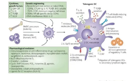
the context of central tolerance in the thymus, where thymic DC induce the apoptotic death of potentially reactive T cells presenting self-Ags to them (Adler & Steinbrink, 2007; Morelli & Thomson, 2007; Shortman & Heath, 2001). Nevertheless, DC tolerogenicity is not specific to a DC subset or restricted to the immature state of the APC. Although tolerogenic DC are able to present Ag to Ag-specific T cells, they fail to deliver adequate co-stimulatory signals with the result to inhibit effector T cell activation and proliferation. This may be manifested by T cell death, T cell anergy or Treg expansion or generation. Such regulatory capacity of DC has inspired numerous groups to explore and exploit the most important checkpoints of DC biology, which could be taken as pharmacological targets to induce tolerance (Hackstein & Thomson, 2004; Morelli & Thomson, 2007). There are several attributes that may characterize “tolerogenic” DC. They are immature, maturation-resistant or alternatively activated DC that express surface MHC molecules, have a low ratio of co-stimulatory to inhibitory signals, and an impaired ability to synthesize Th1-cell-driving cytokines (such as IL-12p70). In an effort to generate tolerogenic DC, in vitro-propagated DC have been manipulated through exposure to various anti-inflammatory and immunosuppressive agents, which target towards DC differentiation and function by various
mechanisms (Fig. 4). These agents include: a) IL-10 and TGF-β1; b) inducers of cAMP such as prostaglandin E2, histamine, β2 agonists, neuropeptides, the vitamin D3 metabolite 1α,25-dihydroxyvitamin D3 (1α,25(OH)2D3) and its analogues, glucosamine, the antioxidant N-acetyl-l-cysteine, ligands for inhibitory immunoglobulin-like transcript receptors (such as the MHC class Ib molecule HLA-G), and cobalt protoporphyrin (to induce haemoxygenase-1 expression). In addition, numerous experimental or clinically approved immunosuppressive drugs such as corticosteroids, cyclosporine, tacrolimus, rapamycin, aspirin, deoxyspergualin (DSG), mycophenolate mofetil (MMF) and sanglifehrin A have been also used. All these molecules prevent DC maturation and/or activation, or impair the capacity of DC to produce IL-12p70. In addition, some of these agents prevent the nuclear translocation of nuclear factor-κB (NF-κB) family members, which are required for the differentiation of DC (Adler & Steinbrink, 2007; Hackstein & Thomson, 2004; Morelli & Thomson, 2007).
Fig. 4: Generation of tolerogenic DC in vitro. DC that are generated in vitro from bone
marrow precursors (BMDC precursors) in rodents or blood monocytes in humans and non-human primates (NHP) have been rendered tolerogenic by controlling their culture conditions through exposure to cytokines, growth factors or pharmacological mediators, or by genetic engineering. DC generated under such conditions can down-regulate the outcome of the T-cell response by a single predominant function or, more frequently, by a combination of complementary mechanisms. CCR7, CC-chemokine receptor 7; CTLA4-Ig, cytotoxic T-lymphocyte antigen 4-immunoglobulin fusion protein; GM-CSF, granulocyte/macrophage colony-stimulating factor; HO1, haem oxygenase-1; IDO, indoleamine 2,3-dioxygenase; IκB, inhibitor of NF-κB; IL, interleukin; ILT, immunoglobulin-like transcript; NF-κB, nuclear factor-κB; ODN, oligodeoxynucleotide; PDL1, programmed cell death ligand 1; PGE2, prostaglandin E2; TGFβ1, transforming growth factor-β1; TNFR, tumour-necrosis factor receptor; VEGF, vascular endothelial growth factor.
Tolerogenic, partially mature DC can be also generated in vitro by combining agents that promote phenotypic maturation (TNF and LPS)
with agents that suppress the functional maturation of DC (such as IL-10 and TGF-β) (Hackstein & Thomson, 2004). Partially mature tolerogenic DC are characterized by high levels of MHC class II expression, variable levels of T-cell co-stimulatory molecule expression and an inability to produce bioactive IL-12. Also, granulocyte (G)-CSF indirectly favours the in vitro differentiation of peripheral blood monocytes into tolerogenic DC (post-G) through the release of IL-10, and IFN-α. Post-G DC possess a mature, MHC IIhighCD80/86high phenotype and release trace amounts of IL-12p70. Furthermore, post-G DC activate the in vitro differentiation of IL-10/TGF-β-producing Treg cells, capable of suppressing bystander T cells in cytokine-dependent manner (Rutella, Danese, & Leone, 2006).
Recently, advances in gene-transfer technology resulted in the enhancement of the tolerogenic potential of DC. These DC are genetically manipulated to: a) express “immuno-suppressive” molecules, which can inhibit or block the expression of cell-surface co-stimulatory molecules by inducing genetically the expression of IL-10, TGF-β1 or the CTL antigen 4 (CTLA4)-immunoglobulin fusion protein; b) prevent NF-κB activation by expressing a dominant negative form of IκB-kinase 2 (IKK2); c) prevent the proliferation of allogeneic T cells through the
expression of indoleamine 2,3-dioxygenase (IDO); d) induce and maintain T-cell anergy (by expressing programmed cell death 1 ligand (PDL1)); or e) promote the deletion of Ag-specific T cells through the expression of CD95 ligand (CD95L) or TNF-related apoptosis-inducing ligand (TRAIL) (Morelli & Thomson, 2007).
2.3 Regulatory T cells
Specialized T cells that inhibit the proliferation and activation of effector T cells are known as suppressor or regulatory T cells and were initially described in the 1970s when Ag-primed T cells were shown to induce tolerance after transfer to naïve mice (Rutella & Lemoli, 2004). A large body of evidence attributes an important role to these cells in the immunological dysregulation underlying autoimmune diseases, chronic inflammatory diseases, and cancer, as well as in the immunobiology of transplantation (Roncarolo et al., 2006). The best characterized Tregs are inscribed among the CD4+ subset and have been categorized into two major subgroups based on their ontogeny: the naturally occurring forkhead box P3 (FOXP3)+CD4+CD25+ Tregs, which develop in the thymus and are present in normal naïve mice and healthy individuals from birth, and the inducible Tregs, which are generated in the periphery
under various tolerogenic conditions. Many subsets of inducible Tregs have been reported, and their definition has largely been based on their different biological features, including specific cytokine secretion profile, cellular markers, ability to differentiate in response to Ag-specific stimuli, and dependency on cytokines vs. cell-cell contact mechanisms for mediating suppressive activity. Some of these Tregs include inducible FOXP3+CD4+CD25+ Tregs, which can be generated
in vitro from CD4+CD25- T cells in the presence of TGF-β, regulatory
type 1 (Tr1) cells, which produce high levels of IL-10, and TGF-β-producing Th3 cells (Carrier, Yuan, Kuchroo, & Weiner, 2007; Groux et al., 1997; Roncarolo et al., 2006; Roncarolo & Battaglia, 2007).
In particular, naturally occurring CD4+CD25+ T cells have been characterized in the peripheral blood of human subjects for their unique ability to suppress bystander T cells in an Ag-nonspecific and cell contact-dependent manner. They constitutively express a variety of cell surface molecules commonly associated with activated/memory cell phenotype. These include the α chain of the IL-2 receptor (CD25), CD45RBlow CD62L, CTLA4, and glucocorticoid-induced tumor necrosis factor receptor (GITR) family-related gene. Neverheless, none
of these surface markers is uniquely expressed by natural Tregs; in the last few years only FOXP3 has been demonstrated critically important for the development and function of Tregs. Genetic mutations in the gene encoding FOXP3 have been identified in both humans and mice, resulting in fatal autoimmune diseases. Humans with mutations in FOXP3 suffer from a severe and rapidly fatal autoimmune disorder known as the immune dysregulation- polyendocrinopathy-enteropathy X-linked (IPEX) syndrome. An analogous disease also occurs in mice, known as “scurfy” desease, owing to a spontaneous mutation in FOXP3. Furthermore, ectopic expression of FOXP3 can phenotypically and functionally convert effector T cells to Tregs (Le & Chao, 2007).It appears that, in mice, FOXP3 expression is both necessary and sufficient for Treg development. However, there is accumulating evidence demonstrating that, in contrast to mice, human FOXP3 is transiently expressed in activated dividing T cells and does not interfere with proliferation and cytokine production. At the same time, FOXP3 does not inhibit expression of the membrane-bound Treg markers CD25, CTLA4, and GITR. It is therefore possible that, depending on the cell subset and/or the stage of T cell differentiation, FOXP3 expression can exert different functions and act as a negative or positive regulator. Furthermore, it has been recently reported that FOXP3 is expressed in
mammary epithelial cells and acts as transcriptional repressor of oncogenes. Future studies will be necessary to better understand the functional consequences of FOXP3 expression in T and non-T cells (Sakaguchi, 2008). On the other hand, several studies have elucidated suppressive mechanisms of CD4+CD25+ Tregs. These cells are able to mediate suppression by cell-cell contact dependent mechanisms, to reduce CD4+ and CD8+ proliferation by inhibiting IL-2 transcription through combined action of CTLA4 and membrane-bound TGF-β, to induce apoptosis of effector T cells, and to produce regulatory cytokine as IL-10 and TGF-β (Roncarolo & Battaglia, 2007).
As opposite as CD4+CD25+ Tregs, inducible Tr1 and Th3 are Ag-specific Tregs, and mediate their suppression activity expecially through producing high levels of IL-10 and TGF-β, respectively.
Although TGF-β is a well-recognized immunoregulatory cytokine, the mechanism by which TGF-β-secreting T cells regulate immune responses is not well-defined. Th3 cells were initially described in mice after oral tolerance induction to myelin basic protein (MBP) for their ability to suppress MBP-specific experimental autoimmune encephalomyelitis (EAE) in vivo in a TGF-β-dependent manner. Th3 regulatory cells form a unique T-cell subset which primarily secretes
high amounts of TGF-β, provides help for IgA synthesis, and displays suppressive properties both for Th1 and for Th2 cells (Rutella & Lemoli, 2004). Furthermore, Th3 secrete low amounts of IL-4 and IL-10 and no IFN-γ or IL-2 upon TCR ligation. In the absence of inflammation, the secretion of TGF-β up-regulates the expression of the Foxp3 gene in activated T cells during T cell expansion which induces the differentiation of Tregs in the peripheral repertoire in the absence of thymic CD25+ Tregs. Thus, Th3 cells appear to be central mediators of peripheral immune tolerance both directly and indirectly by the induction of FOXP3+ Tregs (Carrier et al., 2007).
The most studied inducible Treg subset is constituted by Tr1 cells. Adaptive Tr1 cells arise in vitro and in vivo upon encounter with Ag in the presence of IL-10 and are characterized by a unique cytokine profile (IL-10+ IL-4– TGF-β+ IL-5+ IL-2low/– IFN-γlow/–). To date, no specific cell-surface marker for Tr1 cells has been identified (Roncarolo et al., 2006). Tr1 cells have low proliferative capacity following activation in vitro through the T-cell receptor (TCR), in part due to autocrine production of IL-10. However, their proliferative activity in
vivo is unknown. These cells regulate immune responses through the
they suppress both naïve and memory T-cell responses against tumors,
self and non-self-Ags, and pathogens (Rutella & Lemoli, 2004), and
downregulate the expression of co-stimulatory molecules and pro-inflammatory cytokines by APCs such as DC, Langerhan’s cells, and macrophages (Roncarolo et al., 2006). Furthermore, Tr1 cells favour the production of IgD, IgA and IgG by B cells. Importantly, Tr1 cells are inducible, Ag-specific, and need to be activated through their TCR to exert their suppressive functions. However, once activated, they mediate suppression in an Ag nonspecific manner. IL-10 is absolutely required for the differentiation and function of mouse Tr1 cells, whereas for human Tr1-cell differentiation, IL-10 is necessary but probably not sufficient. Other necessary factors include the presence of APCs, which provide, in addition to IL-10, other soluble and surface molecules that are crucial for Tr1-cell generation. Many different approaches have been explored for the induction of Tr1 cells both ex vivo and in vivo, such as treatment with immunosuppressive drugs, soluble proteins, and peptide antigens (Roncarolo & Battaglia, 2007).
3
MATERIALS AND METHODS
3.1 Chemical and reagentsThe V. cholerae strain 0395-tacCTB, lacking the CTA gene (kindly supplied by Dr. Rino Rappuoli, Istituto Ricerche Immunobiologiche, Chiron, Siena, Italy), was used as a source for rCTB production. rCTB was produced and purified as previously reported (Boirivant et al., 2001), according to the protocol described by Lebens et al. (Lebens, Johansson, Osek, Lindblad, & Holmgren, 1993) with minor modifications. All rCTB preparations contained < 1 EU/ml endotoxin, as determined by a quantitative, chromogenic Limulus amebocyte lysate test (QLC1000, BioWhittaker, Walkersville, MD, USA). rCTB was dissolved in culture medium and used at 10 µg/ml. A possible cytotoxic effect of the drugs was excluded using evaluation of cell viability with trypan blue (more than 95% viable cells). Any possible contamination by enterotoxins was excluded by performing control experiments with boiled rCTB.
3.2 Human PBMC isolation
Human PBMC were isolated by the Ficoll-Hypaque method from buffy coats from healthy, voluntary blood donors (courtesy of the Transfusional Medicine Department, Policlinico Umberto I, University “La Sapienza,” Rome).
3.3 Generation and culture of MDDC
Human MDDC were generated according to the protocol described by Sallusto and Lanzavecchia (Sallusto & Lanzavecchia, 1994). Briefly, monocytes were isolated from PBMC by MACS (Miltenyi Biotec, Germany) using anti-CD14 microbeads. The recovered monocytes - which were > 95% pure, as shown by flow cytometry with an anti-CD14 fluorescent antibody - were cultured at 5 x 10⁵/ml in RPMI 1640 (BioWhittaker), supplemented with 15% FCS (Hyclone, Logan, UT, USA), 1% Lglutamine, and 1% penicillin/streptomycin (Sigma-Aldrich, St. Louis, MO, USA) containing 50 ng/ml GM-CSF (Peprotech Inc., Rocky Hill, NJ, USA) and 1000 U/ml recombinant human (rh)IL-4 (Endogen, Pierce Biotechnology Inc., Rockford, IL, USA, distributed by Tema Ricerca, Italy) at 37°C and 5% CO2 for 5 days. At Day 5, after
monocyte differentiation in immature MDDC (> 90% CD1a+CD14- cells, analyzed by flow cytometry), separate cultures were treated or not with 10 µg/ml rCTB for 24 h. Subsequently, cells were harvested, washed extensively, and stimulated with 10 ng/ml LPS from
Escherichia coli (Sigma-Aldrich) (LPS-MDDC and CTB-LPS-MDDC)
or left untreated (MDDC and CTB-MDDC) for an additional 24 h or 48 h. At the end of the culture period, MDDC were washed extensively before further use. The phenotype of different MDDC populations was analyzed for CD40, HLA-DR, CD80, CD86, and CD83 expression by flow cytometry. Cell supernatants were collected and analyzed for IL-10 and IL-12p70 production. A portion of CTB-MDDC and LPS-MDDC was irradiated (3000 rads) and used to stimulate allogeneic PBMC in a mixed leukocyte reaction (MLR).
3.4 MLR
PBMC used as responder cells (1x10⁵) were cultured in 96-well plates (Costar Corp., Germany) with graded numbers of irradiated (3000 rads), allogeneic LPS-MDDC or CTB-LPS-MDDC. At day 4, cells were pulsed with methyl 3H-thymidine (Amersham Life Science, UK) for the
last 18 h of culture. 3H-Thymidine incorporation was measured in a LKB Betaplate liquid scintillation counter (Wallac, Inc., UK). Assays were performed in triplicate, and the results were expressed as mean cpm ± standard errors of the mean (SEM).
3.5 T cell culture
CD4+CD45RA+ lymphocytes were positively selected from human peripheral blood of healthy donors using the CD4+ Isolation Kit II followed by CD45RA magnetic beads (MACS, Miltenyi Biotec), according to the manufacturer’s instructions. Recovered CD4+CD45RA+ cells were more than 95% pure, as indicated by flow cytometry. Purified T cells were resuspended at the concentration of 1x10⁶/ml in RPMI 1640, supplemented with 10% of FCS, 1% L-glutamine, and 1% penicillin/streptomycin (Sigma-Aldrich; complete medium), and cultured with fresh, allogeneic LPS-MDDC or CTB-LPS-MDDC at the ratio of 10:1 for 1 week. After this period of culture, a portion of T cells stimulated with LPS-MDDC (TLPS-MDDC) and T cells stimulated with CTB-LPS-MDDC (TCTB-LPS-MDDC) was harvested, washed, and stimulated with platebound, anti-human CD3ε (10 µg/ml, R&D Systems Inc., Minneapolis, MN, USA) and soluble anti-human CD28 (2 µg/ml) mAb (R&D Systems Inc.) to analyze their cytokine production (after 24-48 h of culture) and proliferative capacity
(after 72 h of culture). In some experiments, TLPS-MDDC and TCTB-LPS-MDDC were harvested, washed, and stimulated with TCTB-LPS-MDDC. Supernatants were collected after 24 h for IFN-γ and 48 h for IL-10, TGF-β1, and IL-4.
3.6 Cytokine assays
Cytokine content in the culture supernatants was measured by sandwich ELISA using commercially available kits: IL-10, IL-12p70, and TGF-β1 ELISA kits (R&D Systems Inc.) and IL-10, IFN-γ, and IL-4 ELISA kits (Pierce Biotechnology Inc., distributed by Tema Ricerca). ODs were measured on a Bio-Rad Novapath ELISA reader, and the results were expressed as pg/ml ± SEM.
3.7 Analysis of T cell function (suppression assay)
Next, we investigated the capacity of TCTB-LPS-MDDC to suppress autologous TLPS-MDDC proliferation. To this purpose, parallel cultures of TLPS-MDDC and TCTB-LPS-MDDC were cultured in the presence of rhIL-2 (40 U/ml, Roche Diagnostics GmbH, Germany). After 6 days, cells were harvested and washed. TLPS-MDDC were then labeled with
5 µM final concentration of the intracellular fluorescent dye CFSE (VybrantTM carboxyfluorescein diacetate, succinimidyl ester cell tracer kit, Molecular Probes, Eugene, OR, USA). To distinguish the two populations, we stained TCTB-LPS-MDDC with PKH-26 dye (2x10⁻⁶ M; PKH26 red fluorescent cell linker kit, Sigma-Aldrich). After the cell-labeling, according to the manufacturer’s instructions of each product, cells were resuspended in complete medium and cultured together at different ratios. Results of proliferation and IL-10 production analyses indicated 1:1 as the optimal ratio. To induce activation, T cells were stimulated with LPS-MDDC from the same donor used for the CD4+CD45RA+ T cell priming at a 10:1 ratio for 4 days. In some experiments, 30 µg/ml neutralizing anti-IL-10R mAb (BD PharMingen, San Diego, CA, USA) and 50 µg/ml anti-TGF-β1, -β2, and -β3 mAb (R&D System Inc.) were added at the beginning and at Day 2 of the culture period. The proliferation of the CFSE-labeled TLPS-MDDC was assessed at Day 4.
To assess if cell-cell contact was necessary for the suppressive capacity of TCTB-LPS-MDDC, Transwell experiments were performed using CFSE-labeled TLPS-MDDC cells and PKH-26-labeled
TCTB-LPS-MDDC. We placed 4x10⁵ CFSE-labeled TLPS-MDDC cells with allogeneic LPS-MDDC (10:1 ratio) in the bottom well of a Transwell system (Costar, Corning Inc., Corning, NY, USA) and 4x10⁵ PKH-26-labeled TCTB-LPS-MDDC with allogeneic LPS-MDDC (10:1 ratio) in the upper Transwell chamber. Transwell cultures with 4x10⁵ CFSE-labeled TLPS-MDDC cells with allogeneic LPS-MDDC (10:1 ratio) in the upper chamber were performed as control. After 4 days, we measured the proliferative response of CFSE-labeled TLPS-MDDC cells in the bottom well. Proliferation of CFSE-labeled cells was assessed by a BD FACSCanto flow cytometer and FACSDiva software (BD Bioscience, San Jose, CA, USA). Results were analyzed with the proliferation model of ModFit LT software (Verity Software House, Inc., Topsham, ME, USA) and expressed as proliferation index (P.I.).
3.8 Flow cytometry analysis
Cells were washed with PBS and then stained with various fluorochromes using standard methods provided by the manufacturers. Antibodies used for surface staining of human monocytes and DC - PE-labeled anti-CD14 and FITC-PE-labeled anti-CD1a, -CD40, -CD80, -CD83,
-CD86, and -HLA-DR mAbs - were purchased from BD PharMingen. To analyze the CD4+ T cell population, FITC-labeled anti-CD4, PE-labeled anti-CD45RA, and PE-PE-labeled anti-CD25 mAbs (BD PharMingen) were used. For detection of forkhead box p3 (Foxp3)+ cells, T cells were fixed and permeabilized according to the manufacturer’s instructions and incubated with anti-human Foxp3-allophycocyanin mAb (e-Bioscience, San Diego, CA, USA). Isotype-matched Abs were used as controls, and cells were preincubated with human IgG to avoid nonspecific binding to FcRs. All samples were analyzed using a BD FACSCanto flow cytometer and FACSDiva software (BD Bioscience).
3.9 Statistical analysis
Results were expressed as mean ± SEM or as P.I. Statistical analysis was performed using Student’s t-test and Wilcoxon test for paired data as appropriate. P values < 0.05 were considered significant.
4
RESULTS
4.1 rCTB treatment affects maturation of human MDDC
We first assessed the influence of rCTB on the pattern of expression of costimulatory (CD40, CD80, CD86), maturation (CD83), and class II MHC (HLA-DR) surface molecules in MDDC, generated from monocytes cultured with GM-CSF and IL-4. To this purpose, separate cultures of MDDC were treated or not with 10 µg/ml rCTB for 24 h. Subsequently, cells were washed and stimulated with LPS (10 ng/ml; LPS-MDDC and LPS-MDDC) or left untreated (MDDC and CTB-MDDC). In preliminary experiments, the 24 h and 48 h treatment with LPS on CTB-MDDC and untreated MDDC has been tested. Indeed, we observed that the effect of rCTB on MDDC maturation was more evident during the first 24 h of LPS stimulation. Therefore, in subsequent experiments, we used the 24 h LPS stimulation. As shown in Table 1, pretreatment with rCTB induces a statistically significant reduction of the LPS-induced upregulation of CD40 (p = 0.037), and it does not affect the expression of the other molecules.
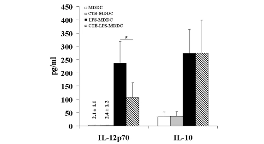
rCTB does not induce significant modification in the surface marker expression of MDDC not treated with LPS. Cytokine production was also investigated. CTB-LPS-MDDC produce a significantly decreased amount of IL-12p70 (p = 0.014) when compared with LPS-MDDC, with no variation of IL-10 production (Fig. 5). rCTB has no effect on cytokine production by MDDC not treated with LPS.
Fig. 5: rCTB inhibits IL-12p70 production by LPS-MDDC, whereas it does not affect IL-10 production. IL-12p70 and IL-10 concentration in the supernatants of human
MDDC, treated as described in Table 1, was measured by a specific sandwich ELISA. Data represent mean ± SEM of six independent experiments performed. *, p = 0.014; CTB-LPS-MDDC versus LPS-MDDC.
These results suggest that rCTB is able to prevent the LPS-induced maturation of MDDC, which acquire characteristics that resemble those of tolerogenic DC (Lutz & Schuler, 2002).
4.2 rCTB-treated MDDC show a reduced ability to stimulate MLR response.
The results obtained with the analyses of the surface molecule expression and cytokine production induced us to test whether rCTB has any effect on MDDC immunostimulatory capacity. Therefore, their ability to stimulate an allogeneic T cell response was assessed. To this purpose, graded numbers of CTB-LPS-MDDC and LPS-MDDC were cultured with a fixed number of allogeneic PBMC for 5 days. As expected, LPS-MDDC were quite efficient in inducing proliferation of alloreactive cells, while CTB-LPS-MDDC induced a significant (p < 0.05 by Wilcoxon test, n = 8), lower proliferative response (Fig. 6).
Fig. 6: rCTB pretreatment down-regulates the immunostimulatory capacity of LPS-MDDC in MLR. PBMC were cultured with graded numbers of allogeneic LPS-LPS-MDDC
(●) or CTB-LPS-MDDC (○). At Day 4, methyl 3H-thymidine (3H-TdR) was added for the
last 18 h of culture. One representative experiment out of eight is shown.
4.3 rCTB-treated MDDC induce the generation of CD4+ T cells
with reduced proliferative capacity and increased IL-10 production
To investigate the effect of CTB-LPS-MDDC on T cells, allogeneic CD4+CD45RA+ T cells were cultured with CTB-LPS-MDDC (TCTB-LPS-MDDC) or LPS-MDDC (T(TCTB-LPS-MDDC) for 1 week.
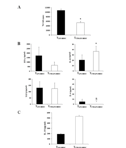
Cells were collected, washed, stimulated with anti-CD3/anti-CD28 mAbs, and analyzed for proliferative capacity and cytokine production. TCTB-LPS-MDDC showed a significantly reduced proliferative capacity when compared with TLPS-MDDC (p < 0.01; Fig. 7A).
Fig. 7: CD4+CD45RA+ T cells stimulated with CTB-LPS-MDDC show reduced
cultured with allogeneic LPS-MDDC (TLPS-MDDC) or CTB-LPS-MDDC (TCTB-LPS-MDDC) at a
10:1 ratio. After 1 week, cells were collected, washed, stimulated with CD3/ anti-CD28 mAb or LPS-MDDC, and analyzed for proliferative capacity and cytokine production. (A) Proliferation of TLPS-MDDC (solid bar) and TCTB-LPS-MDDC (open bar). Columns
represent mean ± SEM of three independent experiments. *, p = 0.01, TCTB-LPSMDDC versus
TLPS-MDDC. (B) Cytokine production by TLPS-MDDC (solid bars) and TCTB-LPS-MDDC (open bars)
measured in supernatants of CD3/CD28-stimulated cells (24–48 h, see Materials and Methods). Columns represent the mean ± SEM of three independent experiments. *, p < 0.05, TCTB-LPS-MDDC versus TLPS-MDDC. (C) IL-10 production by TLPS-MDDC (solid bars) and T CTB-LPS-MDDC (open bars) measured in supernatants of LPS-MDDC-stimulated cells (48 h).
Columns represent mean ± SEM of duplicate of two independent experiments.
The analysis of cytokine production showed that when compared with TLPS-MDDC, TCTB-LPS-MDDC produce significantly higher amounts of IL-10 (p < 0.05), lower amounts of IFN-γ (inhibition: 57%; p < 0.05), and the same levels of TGF-β and IL-4 (Fig. 7B). In some experiments, TLPS-MDDC and TCTB-LPS-MDDC were stimulated with LPS-MDDC for 48 h, and IL-10 production was analyzed. Also in this case, TCTB-LPS-MDDC produce consistently higher levels of IL-10 respect to TLPS-MDDC (Fig. 7C). Taken together, these results suggest that CTB-LPS-MDDC might induce the differentiation of Tregs.
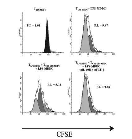
4.4 rCTB-treated MDDC induce functional CD4+ Tr1 cells To evaluate the ability of TCTB-LPS-MDDC to inhibit autologous TLPS-MDDC proliferation, the latter cell population was restimulated with LPS-MDDC in the presence or absence of TCTB-LPS-MDDC (see
Materials and Methods). TLPS-MDDC and TCTB-LPS-MDDC were
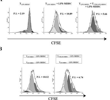
labeled with CFSE and PKH-26, respectively. In preliminary experiments, different numbers of TCTB-LPS-MDDC were added to TLPS-MDDC cultures to identify the optimal ratio, which at 1:1, was chosen for the suppressive activity and for the IL-10 production evaluation (data not shown). As shown in Figure 8A, TCTB-LPS-MDDC inhibited the proliferation of CFSE-labeled TLPS-TCTB-LPS-MDDC consistently (p < 0.05). Next, we investigated whether the inhibitory effect of TCTB-LPS-MDDC on TLPS-MDDC proliferation might be mediated by soluble factors. To this aim, LPS-MDDC-stimulated TCTB-LPS-MDDC and TLPS-MDDC were separated in a Transwell system that allows the free exchange of soluble factors but excludes
direct cell– cell contact. As shown in Figure 8B, the proliferation of TLPS-MDDC was still inhibited, indicating that soluble factors mediated the inhibitory effect.
Fig. 8. TCTB-LPS-MDDC exert regulatory activity on TLPS-MDDC, which is cell– cell
contact-independent.
TLPS-MDDC and TCTB-LPS-MDDC, after 6 days of culture with rhIL-2, were labeled with CFSE or
PKH-26, respectively. CFSE-labeled TLPS-MDDC were restimulated with LPS-MDDC at a
10:1 ratio in the presence or absence of TCTB-LPS-MDDC for 4 days. (A) Proliferation of
CFSE-labeled TLPS-MDDC was assayed at Day 4 by flow cytometry and expressed as P.I., calculated
by ModFit LT software. One representative experiment out of three is shown (p < 0.05; TLPS-MDDC+LPS-MDDC vs. TLPSMDDC +TCTB-LPS-MDDC+LPS-MDDC). (B) Parallel experiments
cultures with TLPS-MDDC in the upper and the lower chamber were used as control (see
Materials and Methods). Proliferation of CFSE-labeled TLPS-MDDC was assayed at Day 4.
One representative experiment out of three is shown (p < 0.05; TLPS-MDDC+LPS-MDDC vs.
TLPS-MDDC+TCTB-LPS-MDDC+LPS-MDDC).
As T cells stimulated with CTB-LPS-MDDC in the presence of anti-CD3/anti-CD28 mAbs produced TGF-β and IL-10, we performed additional experiments in the presence of blocking mAbs. As shown in Figure 9, the addition of anti-IL-10R and anti-TGF-β mAbs to the cultures completely reversed the inhibitory effect.
Fig. 9: TCTB-LPS-MDDC exert regulatory activity on TLPS-MDDC via IL-10 and TGF-β
production. CFSE-labeled TLPS-MDDC were stimulated in the presence of PKH-26-labeled
TCTB-LPS-MDDC as described in Figure 8. TCTB-LPS-MDDC were tested for their ability to suppress
the proliferation of CFSE-labeled TLPS-MDDC in the presence or absence of blocking IL-10R
cytometry and expressed as P.I. calculated by ModFit LT software. One representative experiment out of three is shown.
These results indicate that the TCTB-LPS-MDDC phenotype is associated with regulatory activity. Finally, we analyzed the intracellular expression of Foxp3, and we found that TCTB-LPS-MDDC and TLPS-MDDC did not differ in the expression of this marker (data not shown). Taken together, data indicate that TCTB-LPS-MDDC display the properties described for Tr1 cells.
5
CONCLUSIONS
In the present study, we examined the possibility that rCTB may affect human MDDC maturation/activation in response to TLR stimulation. Our findings showed that rCTB partially prevented the LPS-induced maturation process of MDDC and decreased their IL-12p70 production with no relevant effect on IL-10 production. Furthermore, LPS-stimulated MDDC pretreated with rCTB were able to promote the induction of low proliferating T cells, which showed an enhanced IL-10 production associated with a reduced IFN-γ production and the same high levels of TGF-β as the control. These T cells suppressed proliferation of activated autologous T cells. Transwell experiments and blockade of IL-10R and TGF-β showed that the immunomodulatory effect is mediated by soluble factors. Thus, T cells induced by rCTB-conditioned MDDC acquired a regulatory phenotype and activity similar to those described for Tr1 cells.
6
DISCUSSION
Many studies demonstrate that DC play an important role in the induction and maintenance of immune tolerance (Steinman, Turley, Mellman, & Inaba, 2000; Wakkach et al., 2003). In this study, we found that pretreatment of human MDDC with rCTB reduces their ability to produce IL-12 upon challenge with LPS and affects their maturation with an impaired capability of alloantigen presentation. Furthermore, we analyzed the influence of CTB-LPS-MDDC on T cells. Our data demonstrate that CTB-LPS-MDDC induce the in vitro differentiation of IL-10-producing T cells with low proliferative capacity, which behave as Tr1 cells, described by different authors (Battaglia, Gregori, Bacchetta, & Roncarolo, 2006; Groux et al., 1997; Levings et al., 2005; Roncarolo et al., 2006).
The CTB subunit, when conjugated with an antigen, is a highly efficient carrier molecule for the induction of mucosal antibody responses (Czerkinsky, Russell, Lycke, Lindblad, & Holmgren, 1989; McKenzie & Halsey, 1984) and oral tolerance (George-Chandy et al., 2001). The mechanism underlying this effect is probably a result of the ability of the CTB to promote the presentation of the coupled antigen with a concomitant increase of CD40 and CD86 expression on APC