UNIVERSITÀ DEGLI STUDI DI VERONA
SCUOLA DI DOTTORATO IN SCIENZE
DELLA VITA E DELLA SALUTE
DOTTORATO DI RICERCA IN BIOTECNOLOGIE MOLECOLARI,
INDUSTRIALI ED AMBIENTALI
CICLO XXII
Characterization of human TSE strains after passage in humanized
transgenic mice
Dottoranda: Roberta Galeno
Coordinatore: Prof. Roberto Bassi
Tutor: Prof. Salvatore Monaco
Tutor esterni: Dott. Franco Cardone
Prof. Maurizio Pocchiari
Index
ABBREVIATIONS ... 3
1. INTRODUCTION ... 5
1. 1 TRANSMISSIBLE SPONGIFORM ENCEPHALOPATHIES ... 5
1. 2 THE GENE PRNP AND THE PROTEIN PrP ... 8
1. 3 THE ETIOLOGICAL AGENT OF TSEs ... 11
1. 4 TSE STRAINS ... 12
1. 5 EXPERIMENTAL TRANSMISSION STUDIES AND THE SPECIES BARRIER ... 16
1. 6 SPORADIC CJD ... 17
1. 7 sCJD TRANSMISSION AND TRANSGENIC HUMANIZED MICE ... 20
2. AIM OF THE STUDY ... 23
3. METHODS ... 24
3. 1 ANIMALS ... 24
3. 1.1 Transgenic mice ... 24
3. 1.2 Confirmation of the genotype of the transgenic mice ... 24
3. 2 INOCULATION OF THE TRANSGENIC MICE ... 27
3. 2.1 Sporadic CJD patients ... 27
3. 2.2 Preparation of inocula from human brains ... 28
3. 2.3 Inoculation of mice ... 29
3. 3 EXAMINATION OF INOCULATED MICE ... 29
3. 3.1 Clinical signs ... 29
3. 3.2 Survival time ... 30
3. 3.3 Histological and immunohistochemical examination ... 30
3. 3.4 Preparation of the brain tissue for the Western blot analysis ... 32
3. 3.5 PrPTSE detection by Immunoblot ... 33
4. RESULTS ... 35
4. 1 ATTACK RATE ... 35
4. 2 SURVIVAL TIME ... 36
4. 3 TOPOGRAPHY OF SPONGIOSIS AND PrPTSE DEPOSITION ... 40
4. 4 PrPTSE GLYCOTYPE ... 50
5. DISCUSSION ... 52
6. CONCLUSIONS ... 58
7. BIBLIOGRAPHY ... 60
ABBREVIATIONS
aa: Amino acids
Ala: Alanine
BSE: Bovine spongiform encephalopathy CJD: Creutzfeldt-Jakob disease
CNS: Central nervous system CWD: Chronic wasting disease DTT: Dithiothreitol
DW: Drowsy
EEG: Electroencephalogram FFI: Fatal Familial Insomnia
FTIR: Fourier transformed infrared spectroscopy
G: Gauge
GPI: Glycosylphosphatidylinositol
GSS: Gerstmann-Sträussler-Scheinker disease GT: Gene targeting
H&E: Haematoxylin-eosin staining
HuTg: Transgenic mice expressing human PrP
HuMM: Transgenic mice expressing human PrP with methionine/methionine genotype at codon 129
HuMV: Transgenic mice expressing human PrP with methionine/valine genotype at codon 129 HuVV: Transgenic mice expressing human PrP with valine/valine genotype at codon 129
HY: Hyper
Iso: Isoleucine kDa: kiloDalton
LDS: Lithium dodecylsulphate mAb: Monoclonal antibody MM: Methionine/methionine MV: Methionine/valine NGS: Normal goat serum
NMR: Nuclear magnetic resonance PBS: Phosphate buffered saline PCR: Polymerase chain reaction
PK: Proteinase K
PRNP: Human gene encoding the prion protein
prnp: Mouse gene encoding the prion protein PrP: Prion protein
PrPC: Cellular prion protein
PrPTSE: Pathological isoform of prion protein RGI: Random genomic insertion
sCJD: Sporadic Creutzfeldt-Jakob disease vCJD: Variant Creutzfeldt-Jakob disease SDS: Sodium dodecylsulphate
SD: Standard deviation TBS: Tris buffered saline TBST: TBS/Tween 20 Thr: Threonine
TME: Transmissible mink encephalopathy TSE: Transmissible spongiform encephalopathy Val: Valine
VV: Valine/valine WB: Western blot
1. INTRODUCTION
1. 1 TRANSMISSIBLE SPONGIFORM ENCEPHALOPATHIES
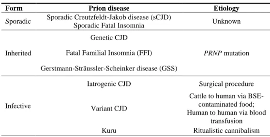
Transmissible Spongiform Encephalopathies (TSEs) are fatal neurodegenerative diseases affecting human and animals, caused by an infectious agent whose nature is still under debate. The main event characterizing TSEs is the accumulation in the Central Nervous System (CNS) of insoluble fibrils mainly composed of the disease-associated isoform of the host prion protein (PrPTSE), which is produced following a conformational change from the endogenous cellular prion protein (PrPC) (Prusiner, 1998). PrPTSE fibrils can sometimes aggregate into amyloid plaques that are easily detectable by conventional neurohistological examinations, together with other TSE-specific lesions such as spongiosis (i.e. a sponge-like vacuolation of cerebral grey matter), neuron loss, and gliosis (figure 1. 1). Pathological misfolding of host proteins links TSEs to other human neurodegenerative diseases like Alzheimer‟s disease (Amyloid ), Parkinson‟s disease (-synuclein), or Huntington‟ s disease (huntingtin). Among these however, only TSEs have been proved to be transmissible either naturally (human to human through medical procedures, or animal to animal via natural secretions), or experimentally (to laboratory animals) via different routes of infection.
According to their origin, human TSEs are classified as sporadic, inherited, or infectious forms (Ironside, 1998) (table 1. 1). Sporadic forms (including the vast majority of human TSE cases), occur without any apparent episode of infection and are mainly represented by sporadic Creutzfeldt-Jakob disease (CJD), a rapidly progressive dementia commonly associated with myoclonic jerks, pyramidal, extrapyramidal, and cerebellar signs.
Inherited forms, accounting for 10-15% of human cases, are associated to mutations (more than 50 have been described until now) of the prion protein gene (PRNP) and, besides a typical Creutzfeldt-Jakob disease picture, they may manifest as peculiar clinical and pathological syndromes (e.g. Fatal Familial Insomnia, FFI, Gerstmann-Sträussler-Scheinker disease, GSS).
Infectious forms reported in the literature originated after accidental inter-human transmission of the infectious agent by medical procedures or have a zoonotic origin through contaminated food. Animal TSEs include scrapie of sheep and goats (Hunter et al., 2000), a disease already reported in the eighteenth century (Schneider et al., 2008) and the first TSE described (by McGowan in 1922). Other animal TSEs include: chronic wasting disease (CWD) of deer and elk (Williams, 2005), the transmissible mink encephalopathy (TME) (Libersky et al., 2009) and bovine spongiform encephalopathy (BSE) (Wells et al., 1991) which reached an epidemic diffusion during the end of the last century and transmitted to humans, as variant CJD (vCJD), by ingestion of contaminated food (Will et al., 1996).
Despite intensive studies conducted during the last 50 years, many aspects of TSE diseases are still obscure such as the nature of the etiological agent, the molecular mechanism of prion propagation and replication, the neurodegeneration caused by PrPTSE accumulation in the brain, and the wide phenotypic spectrum observed even among the same forms of TSEs disease.
Table 1. 1: Spectrum of human prion diseases (modified from Aguzzi et al., 2004).
Form Prion disease Etiology
Sporadic Sporadic Creutzfeldt-Jakob disease (sCJD)
Sporadic Fatal Insomnia Unknown
Inherited
Genetic CJD
PRNP mutation
Fatal Familial Insomnia (FFI)
Gerstmann-Sträussler-Scheinker disease (GSS)
Infective
Iatrogenic CJD Surgical procedure
Variant CJD
Cattle to human via BSE-contaminated food; Human to human via blood
Figure 1. 1.Histological alterations observed in a CJD patient‟s brain. (a) spongiform degeneration; (b) PrPTSE deposition; (c) gliosis.
A
B
1. 2 THE GENE PRNP AND THE PROTEIN PrP
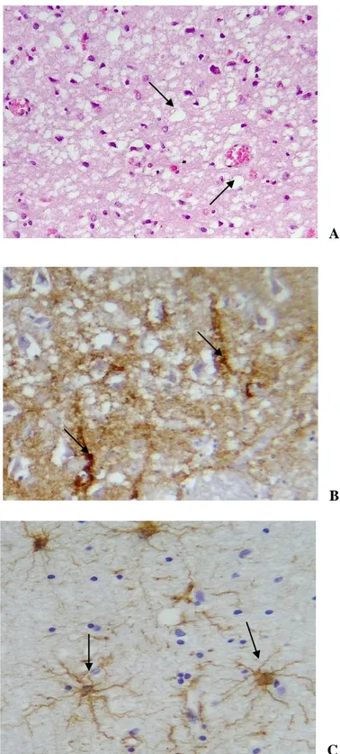
PrPTSE is the pathological isoform of the host-encoded cellular prion protein PrPC, a glycosyl-phosphatidylinositol (GPI) membrane-anchored glycoprotein of about 33-35 kDa. The protein is encoded by the PRNP gene, placed on the short arm of chromosome 20 in humans (Makrinou et al., 2002).
More than thirty point mutations and several insertions encoding additional copies of an octapeptide, regularly present in five copies on PRNP, are linked to the genetic forms of human TSEs (Kovács et al., 2005). Moreover, although different polymorphisms of PRNP have been discovered, only the methionine/valine (M/V) polymorphism at codon 129 has shown a critical influence on clinical, pathological and biochemical phenotype of human TSE diseases. It has also a role in the susceptibility to the disease (Pocchiari et al., 2004) (figure 1. 2): CJD mainly affects homozygous subjects, especially for methionine, while 129 heterozygosity seems to have a protective effect (Puopolo et al., 2003). Indeed 80% of patients with sCJD and 100% of patients with vCJD were homozygous for the methionine at codon 129 (Ironside et al., 2004; Alperovitch et
al., 1999).
Figure 1. 2. Mutations and polymorphisms of the prion protein associated with human TSEs (from Wadsworth et al.,
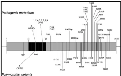
Human PrPC consists of 253 amino acids (aa) and its tridimensional structure, inferred by nuclear magnetic resonance (NMR) studies (Zahn et al., 2000) on recombinant, non glycosylated forms, consists of a long, unstructured, N-terminal domain of about 100 aa that starts with a signal peptide (aa 1-22) that targets the protein to the cell surface (Turk et al., 1988), followed by five repeats of a copper binding octapeptide (Viles et al., 1999). The C-terminal domain, highly conserved over many different species, consists of two short antiparallel beta-sheets and three long alpha-helices with a disulfide bridge (cysteine 179 and 214) between helix 2 and 3 (Haire et al., 2004) and a GPI anchor (aa 230) that binds the protein to the raft domains on the cell surface (Naslavsky et al., 1997). PrPC undergoes facultative N-glycosylation at two sites (aa 181 and 196), resulting in unglycosylated, monoglycosylated, and diglycosylated forms (figure 1. 3) (Endo et al., 1989; Bolton et al., 1985). This basic structure was confirmed by studies on a wide range of mammalian species (Cappai et al., 2004; Riesner, 2003).
Figure 1. 3.Human cellular prion protein.
1 22 51 91 253231 253 S-S 179 214 N-181 N-196 GPI Signal sequence Signal sequence Octapeptide region N-glycosylation sites
PrPC is expressed in a wide range of tissues including CNS (mainly in neurons and glia), blood cells, lymphatic tissue, intestine, muscles, lungs, spleen and heart (Shmakov et al., 2000). The physiological role of PrPC is still under debate. Several functions have been attributed to this protein ranging from signal transduction, antioxidant activity, cell membrane signaling, cell adhesion, synaptic transmission, to regulation of immune system and immune response as well as pro-apoptotic or anti-pro-apoptotic activity (Sauer et al., 2003; Gauczynski et al., 2001; White et al., 1999). The presence of PrPC is not essential for survival (Wopfner et al., 1999; Manson et al., 1994; Büeler
et al., 1992), but it is necessary for replication of the TSEs infectious agent and neurotoxicity to
occur (Collinge et al., 2007; Mallucci et al., 2005). The close relationship between infection and expression of PrPC in the host is clearly highlighted in knock-out mice for PrP gene (genotype
prnp0/0), which are resistant to the experimental infection with scrapie (Büeler et al., 1993).
PrPC and PrPTSE share the same amino acidic sequence and undergo to the same post-translational modifications (Harris et al., 2003), but differ profoundly in their conformation, that is accounted for their different biochemical and biophysical properties (Pan et al., 1993; Safar et al., 1993; Caughey
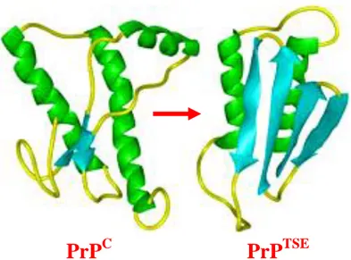
et al., 1991). PrPC is monomeric, proteinase-sensitive and soluble in non-ionic detergents, whereas PrPTSE is insoluble, hydrophobic, and partially resistant to proteinase and tends to aggregate. Indeed proteinase K (PK) digestion of PrPTSE originates a proteinase-resistant core with molecular weight of 27-30 kDa, which retains the full infectivity (Prusiner et al., 1984; Bolton et al., 1982). The insolubility of PrPTSE in physiological solutions and its aggregated state hamper the definition of its tridimensional structure by high-resolution techniques. Low-resolution optical spectroscopic measurements revealed that PrPTSE contains mostly beta-sheet structures (figure 1. 4) (Caughey et
al., 2001; Pan et al., 1993), suggesting that the differences between the two isoforms result from
Figure 1. 4. Tridimensional structure of mature PrPC obtained by NMR and presumable tridimensional structure of PrPTSE.
1. 3 THE ETIOLOGICAL AGENT OF TSEs
One of the unsolved issues of prion diseases is the definition of the nature of the etiological agent responsible for the pathology. Originally it was defined as a “slow unconventional virus” (Gajdusek, 1977) because of its viral size, peculiar biological properties (i.e. high resistance to inactivation and apparent absence of a nucleic acid), transmissibility, and the long incubation time that characterizes the disease. However, the failure to identify a TSE-specific nucleic acid despite uncountable efforts together with the identification of an insoluble and proteinase-resistant protein (PrPTSE) associated with infectious brain material and the highly unusual resistance to treatments that inactivate viruses or naked nucleic acids (e.g. heating, gamma radiations, nuclease) (Taylor, 2000; Alper, 1993), led to the genesis of the “protein only hypothesis” (Griffith, 1967). This atypical agent was defined by Prusiner as “prion” (proteinaceous infectious only particle) (Prusiner, 1982), to distinguish it from other infectious agent like virus or viroids. Prion particles are credited to be devoid of nucleic acid and to propagate in the host by directing the refolding of the
physiological PrPC protein into the PrPTSE conformation through an autocatalytic process which is still matter for speculation (Morris et al., 2009).
The “prion only” hypothesis is a reliable model to explain a number series of observations like the correlation between PRNP gene mutations and genetic TSEs (Gabizon et al., 1996), the strictly association between infectivity and PrPTSE (Gabizon et al., 1988), the finding that PrP knockout mice do not develop the disease (Büeler et al., 1993) and the absence of a specific immune response in the infected host (Berg et al., 1994; Casaccia et al., 1989).
On the other hand, a growing amount of data suggests that TSE infectivity can be separated from PrPTSE (Barron et al., 2007; Piccardo et al., 2007; Berardi et al., 2006; Shacked et al., 1999; Lasmèzas et al., 1997) and the recent finding of small RNAs possibly associated to the TSE infectious agent (Simoneau et al., 2009) is bringing to a renewed interest towards the viral hypothesis.
A third hypothesis lays in the middle: the agent may be a particle called “virino” that is composed by an exogenous nucleic acid enveloped in the host prion protein (Dickinson et al., 1988).
1. 4 TSE STRAINS
After the seminal observation that scrapie disease gave rise to different clinical syndromes in experimentally affected goats (“scratching” and “drowsy” phenotypes) (Pattison and Millson, 1961), it was only with the development of modern strain characterization techniques in the mouse model that the existence of multiple infectious prion “strains” or “isolates” was detailed and demonstrated beyond any doubt (Bruce et al., 1991).
Different strains are traditionally distinguished on the basis of the incubation period, the neuropathological profile and the clinical signs in the recipient host (Bruce, 2003).
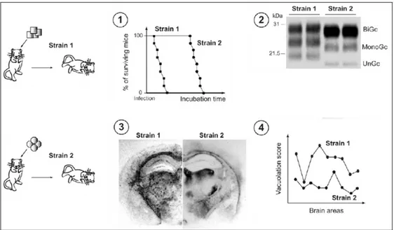
prion strains in the same host usually results in different and reproducible incubation times, this parameter should be cautiously regarded (Bruce, 1993). A second feature that allows to distinguish the strains is the neuropathological profile or “lesion profile”, represented by the distribution and the degree of neurological damages found in the brain of infected animals, according to a standardized procedure for grading the severity of spongiform degeneration in nine brain areas (Fraser and Dickinson, 1973). More recently, some biochemical properties of the pathological prion protein have been added to the list of the traits that characterize prion strains, such as the electrophoretic mobility after PK digestion (Collinge et al., 1996; Parchi et al., 1996), extent of PK resistance (Bessen et al., 1992a), stability towards denaturing agents (e.g. guanidine HCl), position and intensities of infrared bands associated with β-sheet structures, and metal binding capacities (Thomzig et al., 2004; Safar et al., 2000, 1998; Kuczius and Grouschup, 1999; Wadsworth et al., 1999). Moreover, an important factor contributing to the diversity among the strains is the PrPTSE glycosylation: the relative ratios of di-, mono-, and unglycosylated forms of the pathological protein differ in various prion strains (Khalilli-Shirazi et al., 2005) (figure 1. 5).
Figure 1. 5. Models of prion strains variation. Upon experimental inoculation of susceptible animals with identical
genetic background, prion strains exhibit specific traits („phenotype‟) such as: (1) incubation time and attack rate, (2) PrPTSE pattern in Western blot, (3) distribution of PrPTSE deposits in the brain, (4) distribution and intensity of vacuolation in standardized brain areas („lesion profile‟) (modified from Béringue et al., 2008a).
In some cases these differences are so evident that are used in prion strain typing, as for example to discriminate between sCJD (type 2A) from vCJD (type 2B). Fourier transformed infrared spectroscopy (FTIR) (Aucouturier et al., 1999; Caughey et al., 1998) and conformation dependent immunoassay (Bellon et al., 2003; Safar et al., 1998) confirm the hypothesis that different structures acquired by PrPTSE may account for prion strains differences. However, the molecular basis for the biochemical variety of PrPTSE and how this relates to disease features remain unidentified.
Among available experimental host species, the mouse model is widely used for TSE strain primary passage and characterization allowing to identify more than twenty phenotypically distinct strains from different sources: goat and sheep scrapie (Bruce et al., 1993), BSE from cattle (Bruce et al., 2002; Lasmezas et al., 1996), sCJD and GSS from human sources (Muramoto et al., 1992; Tateishi
et al., 1984; Manuelidis et al., 1978). Serial passages in the same species with constant biological
conditions permit to stabilize and define a prion isolate (see next paragraph).
On some occasions, the experimental passage into mice led to the isolation and separation of different infectious strains coexisting in a single natural host. A representative example is the isolation of “drowsy” (DY) and “hyper” (HY) strains after the injection of the agent responsible for transmissible mink encephalopathy (TME) in the Syrian hamsters (Bartz et al., 2000; Bessen et al., 1992b). After serial passages (hamster-to-hamster) incubation periods became stable and could be assigned to two groups of different clinical signs: 150 days to the group that presented lethargy (so this strain was called “drowsy”) and 60 days to the group characterized by hyperactivity (the strain was then called “hyper”). Differences among the two groups were also concerning the vacuolation distribution and PrPTSE deposition in different brain regions (Bessen et al., 1994), as well as electrophoretical mobility of the pathological isoform of the PrP (19 kDa for the unglycosylated band of DY, 21 kDa for that of HY) (Bessen et al., 1995). Furthermore, DY and HY had a different resistance to PK digestion, DY being more sensitive than HY (Bessen et al., 1992a).
1. 5 EXPERIMENTAL TRANSMISSION STUDIES AND THE SPECIES BARRIER
Transmission of TSE disease from a species to another is often characterized by a prolonged incubation period and an incomplete attack rate (the number of animals affected) as compared with the intra-species transmission. This phenomenon is known as “species barrier” and in some cases can be abrogated through serial passages into the same host, after which a reduction of incubation time and a higher attack rate is observed, reflecting the adaptation, or rather, the selection of the most virulent strain from the original inoculum into the new species (Béringue et al., 2008a). Once stabilized into the new host, each strain can be serially propagated in vivo with high reproducible incubation period, clinical signs, lesion profile, and biochemical characteristics of PrPTSE, together giving the “strain signature” in that host species.
Early studies suggested that the species barrier lies in the degree of homology of PrP amino acid sequence between the donor and the host species (Scott et al., 1989), as PrP sequence identity leads to an enhanced susceptibility to the disease and to a reduced incubation period. Consequently in the years a lot of different models of transgenic mice for sheep (Crozet et al., 2001; Vilotte et al., 2001; Westaway et al., 1994), bovine (Bèringue et al., 2006; Buschmann et al., 2005; Castilla et al., 2003; Scott et al., 1999) and human (Beringue et al., 2008b; Asano et al., 2006; Bishop et al., 2006; Korth
et al., 2003; Taguchi et al., 2003; Asante et al., 2002; Kitamoto et al., 2002; Telling et al., 1994)
were created with the purpose to break the barrier and facilitate transmission studies. Usually, transgenic mice with the same PrP primary structure as the donor species are more susceptible to the disease compared to the wild-type mice and can replicate some original strain properties, such as PrPTSE molecular profile.
On the other hand it is important to consider that PrP sequence diversity alone cannot explain the presence of species barrier. This aspect has been brought to the light by the capacity of the BSE
sporadic and genetic CJD isolates to bank voles (a newly discovered rodent model). Such human strains propagate in these rodents with a low or absent transmission barrier despite the wide divergence between human and vole PrP primary structure (Nonno et al., 2006). Moreover wild type mice are more susceptible to vCJD and BSE compared to human or bovine transgenic mice (Bishop et al., 2006). Actually a general opinion is that both PrP sequence and the strain properties contribute to the “species barrier” or, more correctly, to the “transmission barrier” (Scott et al., 2005).
1. 6 SPORADIC CJD
The sporadic form of CJD (sCJD) accounts for 85% of the total human cases of TSEs with an incidence of 1-2 cases per million people per year worldwide, with an equal incidence in men and women aged between 60-70 years (Ladogana et al, 2005). The aetiology of sCJD is still unidentified, since the disease occurs in individuals that do not present mutations on PRNP gene, nor there is any evidence of accidental exposure of the patients to an infectious source. It has been speculated that the disease is the consequence of spontaneous conversion of PrPC to PrPTSE as a rare stochastic event (Wadsworth and Collinge, 2007), possibly facilitated by a hypothetical presence of somatic PRNP mutations which would explain the late onset of the disease. Alternatively, a change in the biochemical properties of the microenvironment surrounding the protein could be the trigger for PrP pathological conversion (Zanusso et al., 2001).
Most cases of sCJD present at onset a rapidly progressive dementia with myoclonus. During the course of the disease the patient can develop extrapyramidal or pyramidal signs, visual or cerebellar disturbance and, at later stages, akinetic mutism. Other rarer forms of sCJD show different clinical presentations such as cerebellar ataxia rather than cognitive impairment (ataxic CJD, 10% of the total sCJD cases), persistent visual disturbances for several weeks before the appearance of other clinical signs (Heidenhain form of sCJD). In the majority of cases, the median duration of the
disease is around 3-6 months. A longer duration of 12 months is observed in 15% of cases, while in about 5% of patients the disease may last for more than 2 years.
Different clinical phenotypes may be accompanied by specific neuropathological lesion profiles built on the basis of the severity and distribution of spongiosis, neuronal loss, gliosis, and PrPTSE deposition (Appleby et al., 2009).
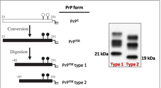
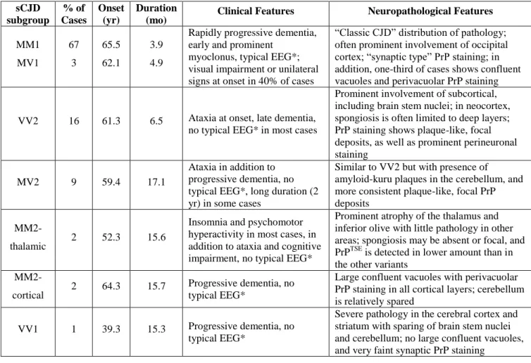
The vast majority of sporadic CJD patients can be categorized according to their genotype at the polymorphic codon 129 of PRNP gene and according to the electrophoretic mobility of the unglycosylated PK-resistant core of PrPTSE (PrPTSE type 1 and PrPTSE type 2) (Parchi et al., 1999, 1996). Unglycosylated PrPTSE type 1 has a molecular size of about 21 kDa with an N-terminus corresponding to the main PK cleavage site at amino acidic residue 82, while unglycosylated PrPTSE type 2 has a molecular size of 19 kDa with an N-terminus at amino acidic residue 97 (figure 1. 6). The combination of the two parameters mentioned above allows to classify the sCJD cases in six main subgroups (MM1, MV1, VV1, MM2, MV2, VV2) corresponding to different clinical-pathological phenotypes (table 1. 2).
Table 1. 2. Molecular and phenotypic features of the six subtypes of sCJD (modified from Parchi et al., 1999). sCJD subgroup % of Cases Onset (yr) Duration
(mo) Clinical Features Neuropathological Features
MM1 MV1 67 3 65.5 62.1 3.9 4.9
Rapidly progressive dementia, early and prominent
myoclonus, typical EEG*; visual impairment or unilateral signs at onset in 40% of cases
“Classic CJD” distribution of pathology; often prominent involvement of occipital cortex; “synaptic type” PrP staining; in addition, one-third of cases shows confluent vacuoles and perivacuolar PrP staining
VV2 16 61.3 6.5 Ataxia at onset, late dementia, no typical EEG* in most cases
Prominent involvement of subcortical, including brain stem nuclei; in neocortex, spongiosis is often limited to deep layers; PrP staining shows plaque-like, focal deposits, as well as prominent perineuronal staining
MV2 9 59.4 17.1
Ataxia in addition to progressive dementia, no typical EEG*, long duration (2 yr) in some cases
Similar to VV2 but with presence of amyloid-kuru plaques in the cerebellum, and more consistent plaque-like, focal PrP deposits
MM2-thalamic 2 52.3 15.6
Insomnia and psychomotor hyperactivity in most cases, in addition to ataxia and cognitive impairment, no typical EEG*
Prominent atrophy of the thalamus and inferior olive with little pathology in other areas; spongiosis may be absent or focal, and PrPTSE is detected in lower amount than in the other variants
MM2-cortical 2 64.3 15.7
Progressive dementia, no typical EEG*
Large confluent vacuoles with perivacuolar PrP staining in all cortical layers; cerebellum is relatively spared
VV1 1 39.3 15.3 Progressive dementia, no typical EEG*
Severe pathology in the cerebral cortex and striatum with sparing of brain stem nuclei and cerebellum; no large confluent vacuoles, and very faint synaptic PrP staining
* Typical EEG: electroencephalogram showing periodic sharp-waves complexes typical of sCJD
Polymorphism M/V at position 129 influences not only the susceptibility and the phenotypic features of the disease (Pocchiari et al., 2004), but also the production of PrPTSE types: MM homozygosity is more frequently associated with type 1 PrPTSE, while VV or MV favour the production of PrPTSE of type 2 (Parchi et al., 2000). From subsequent characterization studies of sCJD it was found that about 30% of cases accumulate both type 1 and type 2 in the brain, either in the same or in distinct anatomical areas (Notari et al., 2007; Polymenidou et al., 2005; Puoti et al., 1999).
As occurs with any other biological phenomenon, such a rigorous classification can not comprise the whole variability of clinical and pathological CJD phenotypes. However it remains a precious
tool to recognize atypical and rare forms of human TSEs that may represent emerging infectious strains with an unknown risk for humans.
1. 7 sCJD TRANSMISSION AND TRANSGENIC HUMANIZED MICE
Experimental transmission studies of human TSE diseases were first carried on non-human primates, such as chimpanzees, squirrel monkeys and cynomolgus macaques as they are evolutionary closer to humans than any other animal model and subsequently showed to have a similar PrP homology with the human counterpart (Williams et al., 2007; Herzog et al., 2005 Brown et al., 1994). In spite of their reliability, these studies are expensive, time consuming and burdened with ethical concerns, and are currently used only to produce data that are urgently needed to face public health problems.
Most types of sCJD did not transmit efficiently to wild-type laboratory mice (Nonno et al., 2006; Bruce et al., 1997), hence to overcome the human to mouse species barrier several lines of transgenic mice expressing the human prion protein have been developed (table1. 3).
Table 1. 3. Transgenic lines expressing human PrP. Tg line (genotype) Transgenic method (GT/RGIa)
Expression levelb Reference
Tg152 (MM) RGI 4-8 Telling et al., 1995
Tg110 (VV) RGI 1 Telling et al., 1994
Tg440 (MM) RGI 2 Telling et al., 1995
Tg650 (MM) RGI 6 Beringue et al., 2008b
HuMM (MM) GT 1 Bishop et al., 2006
HuVV (VV) GT 1 Bishop et al., 2006
HuMV (MV) GT 1 Bishop et al., 2006
Tg35 (MM) RGI 1-2 Asante et al., 2002
Tg45 (MM) RGI 4 Asante et al., 2002
Ki-Hu129M/M (MM) GT 1 Kitamoto et al., 2002 Ki-Hu129M/V (MV) GT 1 Kitamoto et al., 2002 Ki-Hu129V/V (VV) GT 1 Kitamoto et al., 2002
a) GT: gene targeting; RGI: random genomic insertion b) compared to PrP expression in the original host species
Most of such humanized transgenic lines were engineered on endogenous PrP null background in order to avoid any interfering effect of the resident murine PrP gene. Transgenic mice have contributed enormously to the abrogation of the species barrier creating new valuable models characterized by short incubation periods and high attack rates in transmission experiments, therefore allowing a better investigation of the molecular basis of human prion strains (Grouschup and Buschmann, 2008; Telling, 2008). The insertion of human PRNP gene in the murine genome
was performed by two different techniques. The random genomic insertion (RGI), is based on the microinjection of the DNA of interest into a fertilized mouse oocyte; this means that it is neither possible to control the insertion site nor to control the number of copies of the transgene integrated, thus leading to an overexpression of the human PrP in the murine background. Furthermore, the number of copies integrated is not directly proportional to the expression levels of PrP, thus highlighting the effects that the point of insertion may have on the expression of the newly integrated transgene. These models of transgenic mice may be useful to investigate the effect of PrP expression levels on the susceptibility to the disease and may represent a valuable resource as a rapid mouse bioassay mode (some of the lines developed clinical signs in less than 100 days post-inoculation) but they cannot model the natural pathogenesis of TSEs.
As a further drawback it is not possible a comparison between different mouse lines since any of it has different level of PrP expression.
The alternative gene targeting technique (GT) (Manson and Tuzi, 2001), is based on the homologous recombination and allows to replace the murine prnp gene with the corresponding gene derived from another species. In the case of humanized mice the human PRNP is present in a single copy in the same site where was located the prnp, therefore under the natural expression modifiers that determine physiological expression levels of the human PrP (Bishop et al., 2006). Moreover this system allows a direct comparison between the different mouse lines. The transgenic mice used to perform the studies described in this thesis, produced by means of GT technique, express the human PRNP with three alternative genotype at the polymorphic codon 129 (MM, MV, VV), the most recognised factor of susceptibility and pathological phenotype in sCJD.
2. AIM OF THE STUDY
Sporadic CJD, the most common form of human TSE diseases, presents distinctive phenotypes, that could be discriminated by clinical signs at onset, duration of disease, type and distribution of neuropathological lesions (spongiosis, gliosis, and neuronal loss), and by the type and the pattern of deposition of the pathological prion protein PrPTSE, an hallmark of TSE diseases. Parchi and collaborators (Parchi et al., 1996) have shown that the polymorphic codon 129 of the PRNP gene and the PrPTSE type (1 or 2) that accumulates in the patient‟s brain are the major determinants of disease phenotype. Thus, they classified all cases of sCJD into six subtypes (MM1, MM2, VV1, VV2, MV1, and MV2).
Transmission studies of the six sCJD subtypes into transgenic mice expressing the human prion protein suggested that partly the variety of the clinical-pathological phenotypes of sCJD might be related to the presence of distinct human strains of prion agent.
There are, however, rare cases of sCJD that do not fulfils any of the above criteria, suggesting that other minor infectious strains might be responsible of these rare variants. It is still controversial whether TSE strains are influenced by yet unknown endogenous or exogenous factors, but the understanding of the biological characteristics of these strains will improve our knowledge of the pathogenesis of the disease, and hopefully, to find an appropriate therapy for these otherwise fatal disorders.
Aim of my PhD work was to determine whether in 3 atypical Italian sCJD cases identified through the national surveillance of CJD and related disorders at the Istituto Superiore di Sanità, Rome, it was possible to identify novel human prion strains. This objective was pursued by inoculating brain extracts of each patient into gene-targeted transgenic mice carrying the human PRNP gene with the three possible genotypes (MM or VV or MV) at the polymorphic codon 129. Survival times, attack rates, lesion profiles, and molecular analysis of the PrPTSE type recovered from mice brains for each genotype/inoculum combination will be compared with data from the transmission of the “classical” sCJD subtypes to clarify the basis of these atypical cases.
3. METHODS
3. 1 ANIMALS3. 1.1 Transgenic mice
The transgenic model (HuTg) used in transmission experiments is represented by three transgenic lines expressing human PrP homozygous for methionine (HuMM), valine (HuVV) or heterozygous (HuMV) at the polymorphic position 129 in a 129/Ola background (Bishop et al., 2006). The murine prnp gene was entirely replaced by human PRNP gene by using gene targeting technique (Bishopet al., 2006; Manson and Tuzi, 2001)that allows the production of human PrP in the same tissues and at the same levels as that wild-type PrP. The three transgenic lines have identical genotype, except for the polymorphic codon 129. Thus the differences in the transmission properties can be directly attributable to the codon 129.
Initial breeding stocks (HuMM and HuVV mice), kindly supplied by Prof. Jean Manson (The Roslin Institute, Neuropathogenesis Division, University of Edinburgh), were used to establish successful colonies of human transgenic mice. The heterozygous HuMV line was generated by crossing the two homozygous lines. The experimental breeding procedures were carried in the animal facility of the Department of Cell Biology and Neuroscience - Istituto Superiore di Sanità, Roma in collaboration with qualified and authorized staff.
3. 1.2 Confirmation of the genotype of the transgenic mice
The following method was used to check both the genotype of the original breeding stocks and that of the mice used for the transmission experiments.
Genomic DNA was isolated from the mouse tail following standard laboratory procedures. Briefly, tailsnips were cut and incubated overnight at 53°C in 0.5 ml of tail solution (50mM Tris-HCl, pH 8,
DNA was amplified for mouse prnp orf with primers Mouse PrpF and R (figure 3.1) to verify the absence of endogenous gene. Positive and negative controls were included.
PCR was done using 200-500 ng of DNA in 50 l volume with standard buffers (Taq PCR Master Mix kit, Qiagen ). PCR was run through 40 cycles at 94°C for 30 seconds, at 58°C for 30 seconds, at 72°C for 30 seconds. PCR products were run on 1.5% agarose gel and visualized by ethidium bromide under UV light.
Transgene integration in mice was analyzed by PCR using human primers for PRNP (figure 3. 1) and the polymorphism at codon 129 was detected by direct sequencing of PCR products. PCR was done using 200-500 ng of DNA in 50 l volume with standard buffers (Taq PCR Master Mix kit, Qiagen). The first step was 95°C for 15 minutes, continued by 40 cycles of 94°C for 40 sec, 62°C for 40 sec, and 72°C for 40 sec and the final elongation step at 72°C for 10 minutes. The PCR positive products were run on 1.5% agarose gel, visualized by ethidium bromide under UV light and isolated using a QIAquick PCR Purification kit (QIAGEN) according to the standard protocol provided by QIAGEN. The purified PCR products were sequenced by the Sanger dideoxynucleotide chain termination method (Slatko et al., 2001) with the GenomeLab DTCS-Quick Start Kit (Beckman). Sequences were run for 25 cycles at 96°C for 30 minutes, 56°C for 15 minutes, and 60°C for 4 minutes. Purified sequences were then electrophoresed on the Beckmann Coulter CEQ 8000 capillary sequencer and analyzed using the sequence navigator software provided by the manufacturer. All PCR and sequencing cycles were performed using the Gene Amp PCR system 9700 (Perkin Elmer). All tests were performed in duplicate.
Figure 3. 1
Human sequence of the construct inserted in the mouse gene:
ATG GCG AAC CTT GGC TAC TGG CTG CTG GCC CTC TTT GTG ACT ATG TGG
ACT GAT GTC GGC CTC TGC AAA AAG CGG CCA AAG CCT GGA GGG TGG AAC
ACC GGT GGA AGC CGG TAT CCC ggg cag ggc agc cct gga ggc aac cgc tac
cca cct cag ggc ggt ggt ggc tgg ggg cag cct cat ggt ggt ggc tgg ggg cag cct cat ggt ggt ggc tgg ggg cag ccc cat ggt ggt ggc tgg gga cag cct cat ggt ggt ggc tgg ggt caa gga ggt ggc acc cac agt cag tgg aac aag ccg agt aag cca aaa acc aac atg aag cac atg gct ggt gct gca gca gct ggg gca gtg gtg ggg ggc ctt ggc ggc tac (G/A)tg ctg gga agt gcc atg agc agg ccc atc ata cat ttc ggc agt gac tat gag gac cgt tac tat cgt gaa aac atg cac cgt tac ccc aac caa gtg tac tac agg ccc atg gat gag tac agc aac cag aac aac ttt gtg cac gac tgc gtc aat atc aca atc aag cag cac acg gtc acc aca acc acc aag ggg gag aac ttc acc gag acc gac gtt aag atg atg gag cgc gtg gtt gag cag atg tgt atc acc cag tac gag agg gaa tct cag gcc tat tac cag aga gga tcg agc atg gtc ctc ttc tcc tct cca cct gtg atc ctc ctg atc tct ttc ctc atc ttc ctg ata gtg gga tga ggaaggCCT
Bold: primers for human PRNP screening by PCR
CAPITALS: mouse sequence Lower case: human sequence
Blue case: M129V polymorphism
Red case: primers used for PCR products sequencing
Primers used for PCR and sequencing: Hum F1 5’-gca gcc ctg gag gca acc gc-3’ Hum R1 5’-aac cac gcg ctc cat cat ctt-3’
Hum F 5’-cta ccc acc tca ggg cgg tgg tgg c-3’ Hum R 5’-tgg ttg ctg tac tca tcc at-3’
MousePrpF 5’ tgt ggc agg ggc tgc g-3’ MousePrpR 5’-gct gga tct tct ccc g-3’
Bold: primers for human PRNP screening by PCR
3. 2 INOCULATION OF THE TRANSGENIC MICE 3. 2.1 Sporadic CJD patients
Patients were recruited through the National CJD surveillance unit of the Istituto Superiore di Sanità, that received the permission from the ethical committee to collect tissue samples from patients referred to the unit and use them for research purposes. Patient CJD diagnoses were confirmed by histopathology, immunohistochemistry and Western blot (Cardone et al., 1999). Each of the three transgenic mice lines were inoculated with the sCJD cases described below:
A typical CJD case (referred in the thesis as MM1), characterized by a onset at 71 years old and a duration of 4 months. The patient had a pseudoperiodic EEG, typical for sporadic CJD, and manifested dementia, myoclonus and pyramidal, extrapyramidal, cerebellar and visual signs. After the death the brain was removed and half hemisphere was fixed in formaline for the histological investigations and the other half was frozen to perform biochemical, genetic and transmission studies. The brain presented intense spongiosis. Western blot analysis revealed presence of type 1 PrPTSE. Genetic analysis on PRNP displayed methionine homozygosity at polymorphic codon 129 and no presence of disease-associated mutations. The present case was used as control, because of it is the most common form of sporadic CJD.
An atypical case (referred in the thesis as MVx) (Zanusso et al., 2007), represented by a patient who exhibited behavioural and personality changes followed by rapidly evolving dementia. The duration was of 14 months. After the death the brain was removed and half hemisphere was fixed in formaline for the histological investigations and the other half was frozen to perform biochemical, genetic and transmission studies. Post-mortem neuropathological examination of the brain showed an atypical CJD phenotype characterized by intracellular prion protein deposition and the presence of axonal swellings filled with amyloid fibrils. Biochemical analysis of the pathological prion protein disclosed
a previously unrecognized PrPTSE tertiary structure lacking diglycosylated species. PRNP analysis revealed Met/Val heterozigosity at codon 129 and no pathological mutation.
A case (referred in the thesis as MV1/2) defined atypical because characterized by an early onset (38 years old), a long duration (36 months), psychiatric symptoms and sleep disturbances. After the death the brain was removed and half hemisphere was fixed in formaline for the histological investigations and the other half was frozen to perform biochemical, genetic and transmission studies. Neuropathological studies showed diffuse spongiosis and kuru-like plaques. Biochemical analysis of the pathological prion protein in the brain revealed the presence of both PrPTSE types (type 1 and type 2). The part of the brain that was inoculated for transmission studies in the transgenic mice contains only type 2 PrPTSE, in order to avoid having a confounding factor in the inoculum (co-presence of type 1 and type 2). Genetic investigation on PRNP showed Met/Val heterozigosity at codon 129 and no disease-linked mutations.
An atypical case (referred in the thesis as MV2At) characterized by an early onset (44 years old), a very long duration (62 months) and psychiatric symptoms. After death, the brain was removed and half hemisphere was fixed in formaline for the histological investigations and the other half was frozen to perform biochemical, genetic and transmission studies. Neuropathological analysis showed a diffuse spongiosis and deposition of kuru-plaques like in the cerebellum and Western blot displayed type 2 PrPTSE. Sequencing of PRNP gene revealed Met/Val heterozygosity at codon 129 and no pathological mutations.
3. 2.2 Preparation of inocula from human brains
sealed, and stored at -80ºC (Pocchiari et al., 1989). On the day of inoculation the homogenate was thawed, vortexed to homogeneity and then diluted to 1% (w/v) in sterile PBS. Preparation and dilution of inocula were carried out in a Class 2 microbiological Safety Cabinet, located in a Category 3 hazard risk room.
3. 2.3 Inoculation of mice
The animals were housed in compliance with Directive N. 86/609/EEC on the protection of animals used for experimental and other scientific purposes and the Legislative Decree of 27 January, N. 116/12 (Gazzetta Ufficiale-Suppl. Ord.- n. 40, 18 febbraio 1992). Before the inoculation procedure, mice were anesthetized by intraperitoneal administration of 0.1 ml of a solution prepared by mixing 1 ml Ketamine-50, 0.15 ml Xylazine, 9.75 ml of saline (NaCl 0.9%). The inoculum (20 µl of 1% human brain homogenate) was injected into the left cerebral hemisphere by using an insulin syringe with a 26G needle. The animals were sacrificed by CO2 asphyxia. Experimental inoculation
procedures were carried out in the animal facility of the Department of Cell Biology and Neuroscience - Istituto Superiore di Sanità, Roma in collaboration with qualified and authorized
staff.
3. 3 EXAMINATION OF INOCULATED MICE
3. 3.1 Clinical signs
The animals were observed two days per week from 100 days after the inoculation to record the onset of the clinical signs which manifest as motor dysfunctions (ataxia), rigidity of the tail, hunched back and weight loss (Carp et al., 1984) and were culled at the terminal stage of the disease.
3. 3.2 Survival time
For each combination inoculum/genotype we recorded the survival time, corresponding to the time between the inoculation and the culling, for animals clinically sick, or to the time intercurrent between inoculation and spontaneous death, for animals that did not show clinical signs.
Survival time of each group of animals is expressed as the average of the survival times of PrPTSE positive mice only. The survival curves were built according Kaplain-Meier method by considering PrPTSE positivity. All PrPTSE negative cases entered in the analyses as censored. Comparison between survival curves were carried out by the log-rank test.
3. 3.3 Histological and immunohistochemical examination
At postmortem, each brain was divided in two parts by a sagittal paramedian cut. One half was immediately frozen and stored at -80°C for Western blot analysis, while the other half was immersed and fixed in 4% buffered formaldehyde for neuropathological analyses.
Fixed brains were cut in five sections (2 mm thick) at standard coronal levels, transferred into embedding cassettes, immersed in 98% formic acid for 1 hour to reduce infectivity and washed for 2 hours in running water. The samples were then dehydrated in graded alcohols (70% o.n. - 95% o.n. - 100% for 2 h), clarified in xylene for 30 min and finally embedded in paraffin wax.
Five-µm-thick sections were cut on rotative microtome, collected on glass slides for histological analyses or on charged slides (Superfrost® Plus slides, Menzel-Glaser) for immunohistochemistry and let dry at 37°C overnight in an oven.
For histology, the slides were dewaxed in xylene (2 x 20 mins), rehydrated in decreasing graded alcohols (100% 2 x 20min, 95% - 70% - distilled water 5 min each), stained with haematoxylin and eosin (H&E), coded and examined blind with a Leica microscope for pathological assessment.
Dickinson (Fraser and Dickinson, 1968). Vacuolation scores are derived from at least four individual mice per group, and are reported as means ± standard error of the mean.
Table 3. 1. Definition of vacuolation scores.
Vacuolation score Description
0 No vacuoles
1 A few vacuoles widely and unevenly scattered 2 A few vacuoles evenly scattered 3 Moderate numbers of vacuoles, evenly scattered 4 Many vacuoles with some confluence
5 Dense vacuolation with most of microscopic field confluent, lace-like appearance
Table 3. 2. List of the brain regions of HuTg mice and corresponding numbers used to build up
the “lesion profile” curves.
Brain regions Corresponding numbers in the lesion
profile figures Dorsal medulla 1 Cerebellar cortex 2 Superior colliculus 3 Hypothalamus 4 Thalamus 5 Hippocampus 6 Septum 7
Posterior cerebral cortex 8 Anterior cerebral cortex 9
For immunohistochemistry, slides were dewaxed, rehydrated and immunostained for the presence of PrPTSE using the mouse monoclonal antibody (mAb) SAF84 (SPI-BIO, Massy, France). The sections were immersed for 10 min in methanol containing 3% H2O2 to block endogenous
peroxidase activity, washed in distilled water for 10 min and subjected to antigen retrieval by autoclaving at 121°C for 30 minutes in distilled water. The slides were cooled at room temperature, treated with 98% formic acid for 1 min to enhance staining, rinsed, and incubated with 4 M guanidine thiocyanate for 30 min at 4°C in a humid camber. After a one hour incubation in PBS containing 3% Normal Goat Serum (NGS), the slides were then incubated overnight at 4C with mouse mAb SAF84 (1.5 g/ml) in PBS/NGS 3%. Subsequent antibody detection involved incubation with a biotinylated goat anti mouse secondary antibody for one hour (1:200 dilution, Vector Laboratories) at room temperature, followed by incubation with the avidin-biotin-peroxidase complex (Vectastain ABC-Elite kit, Vector Laboratories) according to the manufacturer‟s instructions. The samples were stained with 3‟- 3‟diaminobenzidine (DAB, Sigma) or 3-amino-9-ethylcarbazole substrate (AEC Plus; Dako, Glostrup, Denmark) as chromogen to visualize the reaction product and then counterstained with hematoxylin. Positive (sections of previously immunostained brains) and negative (sections stained with the omission of the primary antibody from the incubation solution and sections from uninfected brains) control sections were included in each run.
3. 3.4 Preparation of the brain tissue for the Western blot analysis
The protocol described below was kindly provided by Doctor Matthew Bishop (National CJD Surveillance Unit, Western General Hospital, Edinburgh).
deoxycholate, 200 mM Tris-HCl pH 7.4) were added. After mixing by vortex, the sample was centrifuged at 2000 rpm for 5 minutes at 4 ºC in a microfuge.
An aliquot of 100 µl of supernatant was taken and treated with proteinase K (PK, Sigma) to a final concentration of 50 µg/ml. This step enables to detect only the pathological form and not the cellular form of PrP (completely digested after this treatment). The digestion was carried out at 37ºC for 1 hour with constant agitation (1000 rpm of Thermomixer), then was stopped by adding 2 µl of 25 X Complete Inhibitors Cocktail (Roche).
The samples were centrifuged at 14000 rpm for 60 minutes at 4 ºC in a microfuge. The resulting pellet was resuspended in 20 µl of LDS Sample buffer 2X prepared from NuPAGE LDS Sample buffer 4 X Invitrogen (106 mM Tris-HCl, 141 mM Tris base, 2% LDS, 10% glycerol, 0.51 mM EDTA, 0.22 mM Serva Blue G250, 0.175 mM Phenol red) plus DTT 0.5 M, boiled at 99 ºC for 10 minutes and stored at – 20 ºC.
Tissue manipulation was carried out in a Class 2 microbiological Safety Cabinet in a Category 3 hazard risk room.
3. 3.5 PrPTSE detection by Immunoblot
Denatured samples were loaded onto a 1 mm thick NuPAGE SDS Electrophoresis polyacrylamide gel (pH 7.0) with an acrylamide concentration of 12% (Invitrogen) and run for 2 h at 100 V constant voltage.
Once proteins were separated, they were electrotransferred onto a nitrocellulose membrane by a semi-dry cell (Semi-dry transfer unit, Hoefer Semi-phore) in transfer buffer (25 mM Tris-base, 192 mM Glycine, 20% methanol) for 1 hour at constant amperage of 125 mA. The membrane was then incubated for 1 hour at 37 °C in blocking buffer: 5% milk powder (Non-Fat Dry Milk, Bio Rad) diluted in TBS / 0.05% Tween-20 (TBST) with constant agitation. This step is necessary to cover non-specific binding sites on the membrane.
After two quick washes in TBST, the membrane was incubated with the anti-PrP 3F4 primary antibody diluted 1:1000 in 5% milk / TBST for overnight at 4°C with constant agitation. 3F4 monoclonal antibody binds residues 109-112 of PrP (Lund et al., 2007; Kanyo et al., 1999; Bolton
et al., 1991; Rogers et al., 1991) and was chosen for its high specificity and affinity for the human
PrP (Kascsak et al., 1987). After 5 washes of 5 minutes each in TBST, the membrane was incubated for 2 hours at 37°C with constant agitation with anti-mouse IgG peroxidase-linked secondary antibody (Amersham) diluted 1:5000 in 5% milk / TBST. PrP bands were finally revealed by incubation for 1 minute in the dark with a solution containing luminol (Western Blotting Luminol Reagent, Santa Cruz Biotechnology) and exposure of the membrane to light sensitive film (Amersham Hyperfilm).
4. RESULTS
4. 1 ATTACK RATEThe presence of spongiosis in H&E stained brain sections or the detection of the pathological form of PrP (PrPTSE) in the brain (by immunohistochemistry or Western blot) were the essential parameters to define the transmission of the disease. The attack rates (percentage of diseased brains) for each combination inoculum/genotype are summarized in table 4. 1.
MM1, MV1/2 and MV2At transmitted efficiently to all three lines with high percentages of attack rate. MV2At displayed the highest efficiency, with an attack rate of 100% in all three lines.
MVx case transmitted only to HuVV mice with a low attack rate value.
Table. 4. 1. Attack rate.
Inoculum from human
sporadic CJD Mouse genotype PrP
TSE
positive/tested Attack rate (% positive)
MM1 MM 18/19 95% VV 12/15 80% MV 18/18 100% MVx MM 0/14 0% VV 4/18 22% MV 0/14 0% MV1/2 MM 13/16 81% VV 20/20 100% MV 17/19 89% MV2At MM 16/16 100% VV 20/20 100% MV 17/17 100%
4. 2 SURVIVAL TIME
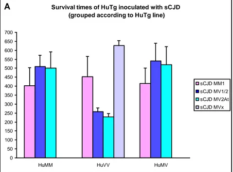
Transgenic mice inoculated with brain homogenates of sporadic CJD patients were difficult to evaluate for the onset of clinical signs because these were rather not specific, (i.e., hunching of the back, hypo-activity, lowering of the hind back, and ruffled fur), therefore, we regarded the time between inoculation and death as a more reliable end-point. The mean survival times for each inoculum/genotype combination are summarized in figure 4. 1 and in table 4. 2.
Figure 4. 1. Survival times of HuTg mice injected with different sporadic CJD cases.
Survival times of HuTg inoculated with sCJD (grouped according to HuTg line)
0 50 100 150 200 250 300 350 400 450 500 550 600 650 700
HuMM HuVV HuMV
sCJD MM1 sCJD MV1/2 sCJD MV2At sCJD MVx A
B Survival tim es of HuTg inoculated w ith sCJD (grouped by inoculum type)
350 400 450 500 550 600 650 700 HuMM B
Table 4. 2. Survival times in days (mean ± SD) for each combination inoculum/genotype.
TRANSGENIC LINES
sCJD INOCULUM HuMM HuVV HuMV
MM1 402.7 ± 99.33 453.0 ± 112.6 415.1 ± 86.2
MV1/2 510.2 ± 62.2 258.1 ± 21.1 540.8 ± 98.7
MV2At 501.9 ± 88.2 229.1 ± 17.6 519.0 ± 100.8
MVx 511.9 ± 106.9# 626.0 ± 28.6 453.6 ± 144.4#
#
Survival times of clinical positive but PrPTSE negative mice (no PrPTSE positive mice found)
The Kaplan-Meier survival curves drawn for each inoculum are in figure 4. 2, showing the survival differences between the 129 genotypes.
MM1 case successfully transmitted to the three lines with similar survival times and overlapping survival curves. Indeed no statistically significant differences among survival curves are present between different HuTg mice (p=0.13).
MVx case transmitted only to HuVV mice with very long survival times. No survival data in the other different lines are available for comparison.
MV1/2 successfully transmitted to all three HuTg lines, with significantly shorter survival times in HuVV as compared with HuMM and HuMV. In line with this observation, the survival curve for HuVV mice shows a very steep decay and is significantly different (p= 0.00) from those of HuMM and HuMV mice, which result statistically indistinguishable.
MV2At transmission gave the same results as the MV1/2 inoculum in the three HuTg mouse lines with shorter survival time in the HuVV line with respect to HuMM and HuMV lines. Statistical analyses show a significantly (p= 0.00) shift of HuVV survival curve with regard to those of HuMM and HuMV, which do not differ between them.
Figure 4. 2. Kaplan-Meier survival curves grouped by inoculum type.
The Kaplan-Meier survival curves of each HuTg line inoculated with different inocula are depicted in figure 4. 3. These curves allow to visualize the effect of the inoculum type in the same 129 genotype: in the HuMM line and HuMV line, the MM1 inoculum was faster than the MV1/2 and MV2At inocula whereas in the HuVV line, an opposite trend was observed with a relatively fast transmission in animals injected with MV1/2 and MV2At inocula, followed by MM1 and MVx
sCJD MM1 0 10 20 30 40 50 60 70 80 90 100 150 200 250 300 350 400 450 500 550 600 650 700
survival time (days)
c u m u la ti v e s u rv iv a l (% ) HuMM HuVV HuMV sCJD MVx 0 10 20 30 40 50 60 70 80 90 100 150 200 250 300 350 400 450 500 550 600 650 700
survival time (days)
c u m u la ti v e s u rv iv a l (% ) VV sCJD MV1/2 0 10 20 30 40 50 60 70 80 90 100 150 200 250 300 350 400 450 500 550 600 650 700
survival time (days)
c u m u la ti v e s u rv iv a l (% ) HuMM HuVV HuMV sCJD MV2At 0 10 20 30 40 50 60 70 80 90 100 150 200 250 300 350 400 450 500 550 600 650 700
survival time (days)
c u m u la ti v e s u rv iv a l (% ) HuMM Hu VV HuMV
Figure 4. 3. Kaplan-Meier survival curves grouped by HuTg mouse line. HuMM 0 10 20 30 40 50 60 70 80 90 100 150 200 250 300 350 400 450 500 550 600 650 700
survival time (days)
c u m u la ti v e s u rv iv a l (% ) sCJD MM1 sCJD MV1/2 sCJD MV2At HuVV 0 10 20 30 40 50 60 70 80 90 100 150 200 250 300 350 400 450 500 550 600 650 700
survival time (days)
c u m u la ti v e s u rv iv a l (% ) sCJD MM1 sCJD MVx sCJD MV1/2 sCJD MV2At HuMV 0 10 20 30 40 50 60 70 80 90 100 150 200 250 300 350 400 450 500 550 600 650 700
survival time (days)
c u m u la ti v e s u rv iv a l (% ) sCJD MM1 sCJD MV1/2 sCJD MV2At
4. 3 TOPOGRAPHY OF SPONGIOSIS AND PrPTSE DEPOSITION
To characterize the phenotype of affected mice, we analyzed the pattern of vacuolar degeneration in nine grey matter brain areas, which are represented by the lesion profile (figure 4. 4).
The MM1 case presented a pattern of spongiosis that resulted remarkably similar among the three human transgenic lines, with slightly more intense spongiform changes in HuMV mice in all the areas, except in the superior colliculus (area 3).
MV1/2 showed similar profiles of spongiform degeneration in HuMV and HuVV mice, with more severe changes in HuVV mice, confirming that this transgenic genotype is more susceptible to this inoculum. In HuMM mice the thalamus (area 5) was the area mainly affected similarly to HuMV mice, the other line with methionine (in heterozigosity) at codon 129.
MV2At case produced more intense spongiform changes in HuVV and HuMM involving all brain areas except the cerebellar cortex (area 2). A much lower degree of spongiosis could be appreciated in HuMV were the thalamus (area 5) was the most severely affected area similarly to what observed in HuMM mice, the other methionine 129 containing genotype.
While both the MV1/2 and the MV2At sCJD cases associated with vacuolation in the deep layer of the cerebral cortices, the MM1 determined vacuolation of superficial cortical layers in all infected animals (fig. 4. 5).
In most of the animals belonging to the three genotypes, large vacuoles can be observed in several white matter areas (i.e. striatum, internal capsule, cerebellar white matter and medulla). The same vacuolation is however present in age-matched control animals, suggesting that such phenomenon may not be related to the disease but rather to an aging process (fig. 4. 6).
All mice belonging to the three lines were negative at the neurophatological analysis both for the presence of spongiosis and for PrPTSE deposition once inoculated with the MVx case.
Figure 4. 4. Lesion profiles grouped by inoculum type. sCJD MM1 0 1 2 3 4 1 2 3 4 5 6 7 8 9
scoring area in brain
m e a n v a cuo la ti o n sc o re HuMM HuVV HuMV sCJD MV1/2 0 1 2 3 4 1 2 3 4 5 6 7 8 9
scoring area in brain
m e a n v a cuo la ti o n sc o re HuMM HuVV HuMV sCJD MV2At 0 1 2 3 4 1 2 3 4 5 6 7 8 9
scoring area in brain
m e a n v a cuo la ti o n sc o re HuMM HuVV HuMV
Figure 4. 5. Vacuolation in the cerebral cortex of HuMM mice infected with MM1 sCJD (a,b) and with MV2At sCJD
(c,d). Vacuoles are present in the superficial layers of the cortex in mice infected with MM1 sCJD (arrows in b) and in the deep layers of the cortex adjacent to the corpus callosum in mice infected with MV2At sCJD (arrows in d). b and d are higher magnifications of the boxed areas in a and c. CC: Corpus Callosum.
Figure 4. 6. White matter vacuolation in affected (a) and healthy (b) animals in cerebellar cortex (arrows) and in
vestibular nuclei (arrowheads).
d b
c a
When neuropathological data are analysed on the basis of the recipient genotype (figure 4. 7) it can be seen that in HuMM mice the three inocula determined different lesion profiles, highlighting the role of the strain on lesion distribution in the brain, despite the same 129 polymorphism in the host. Both MV cases (MV1/2 and MV2At) mainly targeted the thalamus (area 5), with the MV2At inoculum inducing a higher severity of the lesions in the hippocampus (area 6) and cerebral cortices (areas 8 and 9). HuMM mice infected with the MM1 sCJD case had a completely distinct lesions profile, characterized by a more severe vacuolation in the superior colliculus (area 3) and the septum (area 7), and a lower spongiform change in the thalamus (area 5).
In HuVV mice, the two MV cases (MV1/2 and MV2At) produced an identical pattern of vacuolar changes, with the higher spongiform change in the thalamus (area 5), hippocampus (area 6) and cerebral cortices (8 and 9 areas). This pattern was clearly different from that observed with the MM1 sCJD case, where the level of vacuolation was less severe in all the nine areas except for the superior colliculus (area 3) and the septum (area 7).
In HuMV mice, the MV1/2 and the MV2At sCJD cases had similar vacuolation scores in the posterior and central areas of the brain but lower severity of lesions in the hippocampus (area 6), septum (area 7) and cerebral cortices (areas 8 and 9) of animals injected with the MV2At sCJD inoculum. Again, mice injected with MM1 sCJD showed a completely different lesion profile characterized by the presence of lesions in all the nine areas examined.
Figure 4. 7. Lesion profiles grouped by HuTg line. HuMM 0 1 2 3 4 1 2 3 4 5 6 7 8 9
scoring area in brain
m e a n v a cuo la ti o n sc o re sCJD MM1 sCJD MV1/2 sCJD MV2At HuVV 0 1 2 3 4 1 2 3 4 5 6 7 8 9
scoring area in brain area
m e a n v a cuo la ti o n sc o re sCJD MM1 sCJD MV 1/2 sCJD MV2At HuMV 0 1 2 3 4 1 2 3 4 5 6 7 8 9
scoring area in brain
m e a n v a cuo la ti o n sc o re sCJD MM1 sCJD MV1/2 sCJD MV2At
Immunohistochemical analyses revealed a heterogeneous deposition of PrPTSE in different brain areas with all three CJD subtypes (table 4. 3). After inoculation of MM1 case in HuMM mice, granular deposition can be observed in the ventral thalamic nuclei and only rare and small plaque-like aggregates are present in the hippocampus. Scattered PrPTSE deposits were also found in the septum and in the mesencephalic nuclei in some of the animals analyzed.
In HuVV mice, the MM1 inoculum is characterized by the total absence of plaques and by the presence of little granular PrPTSE deposition in the hippocampus; the thalamic nuclei are involved with few scattered deposits.
In HuMV mice the thalamic nuclear group and the hippocampus are always sites of PrPTSE deposition, with the other brain areas involved only occasionally at the diencephalic (cortical) and mesencephalic level. Plaques are always absents. Animals with longer survival times are characterized by a more abundant PrPTSE deposition.
The MV1/2 case determined in HuMM mice granular PrPTSE deposition in the thalamic nuclei, invariably present in all the animals analyzed; a variable number of plaques are observed along the corpus callosum at the level of the hippocampus and in few mice in the retrosplenial cortex but are absent in all other levels. Some granular deposits can be observed in the mesencephalon and in the medulla. As in HuMM mice, the MV1/2 inoculum in HuVV mice is characterized by a granular deposition in the thalamic nuclei and plaques along the corpus callosum. In some animals the PrPTSE is also found in the motor cortex, in the mesencephalon and in the medulla.
The ventral thalamus is the only area of deposition in HuMV mice inoculated with MV1/2, but in such mice the deposition is mainly represented by plaques whereas the granular deposition is almost absent.
Regarding MV2At case in all the three mice lines, the pattern of PrP deposition is the same as for the MV1/2 inoculum, with granular deposits in the thalamus and plaques along the corpus callosum in HuMM and HuVV mice; and plaques, mainly present in the thalamus, in HuMV mice.