UNIVERSITY OF PISA
Department of Chemistry and Industrial Chemistry
PhD Thesis in Chemical Science
XXI, 2005-2008
Chemical characterization of
human breath
Sara Tabucchi
Supervisor: Prof. Roger Fuoco
Table of Content
Abstract
………. III1. State of the art
1.1 Introduction ………...1
1.2 State of the art . ……….……. 2
1.3 Tests used in the clinical diagnosis ……… 4
1.4 Exposure assessment ……….. 6
1.5 Physiology of respiration ………... 8
1.6 Breath sampling techniques ………... 14
1.7 Analytical techniques used for chemical breath characterization ……… 19
1.8 Breath pre-concentration techniques ……… 23
1.8.1 The Solid Phase Micro-Extraction (SPME) ………. 24
1.8.2 The Solid Phase Extraction (SPE) ………...… 25
1.9 Recovery of the analytes after pre-concentration ……… 30
2. Methods and Materials
2.1 Description of standard solutions preparation ……….… 332.2 Chromatographic, mass spectrometric and thermal desorption methods ….... 38
2.3 Adsorbent phases ……….… 41
2.4 Bags material ………... 42
3. Experimental part
3.1 Effect of instrument tuning ………... 433.2 Selection of the adsorbent phase and desorption tubes performance evaluation .. ………..…. 58
3.2.3 Blank evaluation adsorption tubes ………... 69
3.3 Determination of response factors for compounds quantification ……..….... 71
3.4 Instrumental calibration with liquid mixture ……….…... 73
3.5 Comparison between different sampling bags ………..….. 77
3.6 Extension of analytical procedure to samples with high water content …….. 84
3.7 Bags preparation reproducibility ………... 85
4. Clinical applications
4.1 Breath variability ………... 884.2 Human breath analysis medical applications 4.2.1 Diabetes mellitus ………... 94
4.2.2 Hepatic cirrhosis ……….…... 101
4.2.3 Dialysis ……….…… 103
5. Future developments
5.1 Breath sampling system ……….………...….... 1085.1.1 Description of the breath sampler prototype ……….….... 110
5.1.2 Preliminary measurements on the Breath Sampler performance ………... 115
5.1.3 Breath collection by the Breath Sampler ……….…... 118
5.2 Towards a different breath sample collection ………..……… 124
5.2.1 Method development ………..…... 125
5.2.2 Exhaled breath condensate collection device ………..…... 129
5.2.3 Human EBC sample collection and analysis ………... 132
Conclusions
………...….. 134Bibliography
………... 136Abstract
In the present work analysis of exhaled breath has been proposed as a novel way to detect disease, to monitor disease progression, and to monitor clinical intervention. An analytical method of analysis based upon two stages thermal desorption capillary gas chromatography and mass spectrometry was developed for the analysis of breath samples. All the steps of the analytical procedure were evaluated, trying to identify the critical aspects in order to optimize the entire procedure. A novel breath collection media has been developed that is cheap, disposable and readily available. This material allows breath samples to be collected in a novel manner. After validation the procedure was applied to real samples and preliminary experiments were performed aimed at estimating the variability of the composition of breath as a function of time of day and the inter-subject variability. In particular the trend in time of the two principal compounds in breath, acetone and isoprene was observed. The data showed a wide inter-variability between different people and also confirmed that a meal can influence breath composition. For this reason if possible, it was better to collect a breath sample in the morning before eating. Single substances or sets and patterns of exhaled markers were also investigated in order to establish correlations between the chemical composition of breath and patients’ clinical conditions.
Preliminary studies were performed on patients with end-stage renal disease. The results underlined the capacity of the analytical procedure to appreciate small variation in breath composition. In particularly in people under dialysis treatment two compounds were found to show significant differences in breath concentration between patients and healthy people before the dialysis and no important differences after. These two compounds should become an additional and important parameter to determine the end of the dialysis treatment.
In parallel a breath collection system prototype has been designed that enables samples of dead space air to be separated from end tidal breath and be collected independently. This device is also novel and has a great potential in breath analysis field.
CHAPTER 1
State of the Art
1.1 Introduction
How many times do we hear expressions such as: “To sigh with relief, to breathe deeply, to take a breath of fresh air, to feel choked, to take your breath away“. These are all phrases refer to our moods. We often feel tired, lazy, dulled or very commonly we feel stressed. When an emotion is exaggerated respect to an external stimulus, it means that there is not sufficient oxygen in the body and in the mind. Oxygen enables the mind to work and if there is a lack of oxygen, the blood has to flow faster. Meanwhile we lose clarity of thought and our mood changes for the worse. Everybody knows just how important breathing is, but in reality how much do we know about the respiration process? When we breathe many organs are involved, in fact the inspired air passes through the airways to reach the lungs, which represent the real respiration organ1. When we breathe, we bring oxygen into the body and the produced substances are eliminated. Besides containing principal gases (nitrogen, oxygen, carbon dioxide, inert gases) and 98% of water, human breath consists of a great variety of organic compounds present at very low concentrations. What is eliminated may come from products of our metabolism or from substances that we have inhaled and adsorbed from an external environment. Breathing is at the base of the life and enables our organism to carry out the principal vital function. Therefore, chemical composition of exhaled air reflects the general metabolic conditions and it may give useful and precious information on the state of health of a person. For these reasons breath has the potential to become a key for reading our organism.
1.2 State of the art
Breath scientific research started in 1784, when Antoine Laurent Lavoisier, father of modern chemistry, and Pierre Simon Laplace, analyzed the breath of a guinea pig and observed that the animal burned oxygen and produced carbon dioxide. This discovery was the first proof of food combustion in human body2, 3.
In the mid 19th century, colorimetric tests were developed that led to the detection of volatile organic compounds (VOCs) in breath at low concentrations. One of the first colorimetric analyses was developed by the German Nebelthau. He observed a rapid and intense colour variation in an alkaline iodine solution, bubbling in the breath of diabetics. The colour variation was induced by the presence of a high amount of acetone in breath3, 4.
In 1874 the British scientist Francis Anstie used a colorimetric method to highlight the presence of ethanol in breath. By bubbling breath through an acid chromic solution he observed a colour variation from red to green due to the presence of ethanol. With other experiments the scientist demonstrated that the amount of ethanol eliminated from breath was less than the ethanol ingested. He deduced that most of the ethanol was metabolized3.
It was only in 1971 that Linus Pauling carried out the first systematic breath characterization, condensing volatile compounds in a cold trap (made from a stainless steel tube cooled with dry ice). An analysis of the condensate using gaschromatography and mass spectrometry highlighted that human breath samples are very complicated as they contain thousand of different compounds3.
The most significant results on the studies of the correlation between metabolism and the presence of organic substances in human breath, and between pathologies and markers are reported below. However, in most cases the metabolic pathways that lead to their formation are not well known yet5,6, 7.
In the breath of patients with hepatic cirrhosis, sulfur compounds such as ethylmercaptan, dimethylsulphide and dimethyldisulphide were identified. These
compounds were produced in the human organism from an incomplete metabolism of methionine, intermediate of the transaminase process2, 6, 8, 9, 10.
In the breath of patients suffering from lung cancer, alkanes with low molecular weights, such as hexane, heptane, pentane, methylpentane and benzene derivatives were identified. They are produced from an alteration in the lipid peroxidation process (oxidative stress), probably due to an increase in oxygen free radical activities in the cancer cells. Active species of oxygen are continuously produced in the mitochondrion and exit in the cytoplasm where they cause peroxidative alterations in proteins, polyunsaturated fatty acids and in DNA. Oxygen free radicals degrade the cell membranes and convert polyunsaturated fatty acids into volatile alkanes, which are eliminated in breath. These alkanes could be converted into alkyl alcohol by cytochrome P450 (CYP) mixed with oxidase enzymes, which are active in lung cancer. The activation of CYP enzymes in patients affected by lung cancer can accelerate the degradation of alkanes and methylalkanes produced by oxidative stress4, 7, 11, 12, 13, 14.
Increases in 8-isoprostane, hydrogen peroxide and 3-nitrotyrosine concentration have been found in the breath condensate of people suffering from lung inflammation 15.
In patients undergoing dialysis a decrease of some compounds in blood was highlighted. Some of these include nitrogenous compounds such as urea, ammonia, methylamine, ethylamine, dimethylamine and trimethylamine16, other are aromatics compounds such as m–cresol and other uremic toxins such as malondialdehyde or oxalic acid16. In the literature there are few papers on breath variation after dialysis treatment and only some studies concerning pentane17 and ammonia2, 6, 18.
1.3 Tests used in the clinical diagnosis
Many tests on breath analysis have been approved by the U.S. Food and Drug Administration (FDA) and are now used for the diagnosis and monitoring of some diseases:
• A nitric oxide breath test is used for monitoring asthma therapy. During tidal respiration, the nitric oxide levels in asthmatic people can reach higher values than in healthy people. It has been demonstrated that the breath concentration of nitric oxide is proportional to the degree of eosinophilic inflammation of the airways and decreases if the subject starts a corticosteroid therapy19. It is known that nitric oxide forms almost exclusively in the superior and inferior tract of the airways and diffuses towards the lumen. The alveolar level of nitric oxide is very low due to the fact that it easily linked with hemoglobin in capillary blood. In the inflamed airways of asthmatic people the production of nitric oxide is higher than in healthy people20.
Other tests (listed below) determine the concentration of a volatile substance directly taken (ethanol test) or derived from the metabolism of a specific precursor, given to the patient some hours before the test.
• The ethanol screening test is used to determine alcohol levels in blood. The subject has to breathe inside a plastic tube; 1 cm3 of breath is collected through a piston. The sample is sent inside a combustible cell where an electron current is generated. The electric current measured is proportional to the alcohol concentration in blood21, 22.
• The hydrogen breath test is used to diagnose intestinal diseases, such as malabsorption syndrome3 or pancreas diseases. Usually this test is used with children with cystic fibrosis. A quantity of rice starch is given to the child; children with cystic fibrosis are not able to secrete a sufficient amount of amylase enzyme to digest the flour in the small intestine. The bacterial present in the intestine are the final part of the digestion process and produce high amounts of hydrogen. The hydrogen is eliminated by the breath, thus we can
• The 14CO2 breath test is used to diagnose other pancreas diseases. A quantity of 14C labeled triglycerides is given to the patient; the triglycerides are metabolized
from pancreas enzymes, producing a certain amount of labeled 14CO2. If the
amount of 14CO2 is lower than a specific value, it means that the pancreas
functionality is reduced3, 23.
• The infection caused by Helicobacter pylori is diagnosed by an urea breath test. Helicobacter have the urease enzyme, which is not present in human organism. It enables to urea to be metabolized thus producing CO2. If a quantity of labeled 14C urea is given to the infected subject, the bacteria metabolizes it in the
stomach, producing 14CO2. This gas is eliminated by breath, thus a breath
analysis is performed at a certain time after the administration, revealing the infection3, 23.
In addition to these tests, many studies on breath, analysis and the characterization of healthy people and subjects suffering from different pathologies have been reported in the literature. However, going from what is reported in the literature to real clinical practice is not straightforward, since the correlation between breath composition and a subject's state of the health is difficult to understand24, 25.
In terms of the medical application of breath analysis, the differences between the chemical breath composition of healthy people and people with diseases are not great. Only in a few cases were found compounds in the samples of people with a disease and not in healthy people. Usually small differences in compound concentrations are observed, thus very accurate analytical techniques are needed.
1.4 Exposure assessment
In human breath there are thousands of VOCs. These compounds can be generated inside the body (endogenous substances) or can be adsorbed from exposure to the environment (exogenous substances). In fact breath VOCs content can show whether a subject has been exposed to a polluting environment26, 27. The VOCs used in productive activities and industrial processes (laundry, the shoe industry, the production of paints or inks, etc.), can be adsorbed from the skin or through inhalation and are eliminated with breath28, 29, 30, 31. Most of these compounds are toxic and can cause several diseases, such as cancer (benzene and toluene) or pulmonary inflammation (acrolein and an α,β unsaturated aldehydes). In most cases it is very difficult to find a correlation between the chemical agents and the disease, due to the complexity of interactions between human organisms and the environment, particularly when a lot of time has passed between exposure and the onset of the disease32.
For some solvents biological monitoring involves sampling and the analysis of blood or urine. These are invasive methods, there are operative difficulties during sampling and it is possible to perform only a limited number of analyses. For this reason, breath analysis has been proposed as an alternative. Inhaled air during exposure stays inside the alveoli for sufficient time to permit to the VOCs to reach equilibrium with the blood of the pulmonary arteries. This equilibrium is regulated by partition coefficients, which determine the relative blood/breath concentration of each compound33. Thus the analysis of solvents in expired air may be an indication of their levels in blood28. Breath measurements principally reflect recent exposures. This is due to the fact that after a subject has stayed in a place for a few hours, equilibrium is reached with the ambient air; at this point breath principally reflects the actual exposure of the subject34.
Breath analysis is also used to determine occupational exposure to industrial solvents and many studies have demonstrated the correlation between expired VOCs and chemical substances in work places32. Guidotti et al.35 assessed the amount of chlorinated solvents in the breath of exposed subjects in the work place. Chloroform, trichloroethylene, tetrachloroethylene, tetrachloroethane and carbon tetrachloride were assessed. Detection limits of 0.5 and 5 ppb were found useful to analyze these compounds in the breath of subjects exposed to very low concentration levels.
Chen et al.36 evaluated the correlation between the exposure of normal people to toluene, xylenes and ethylbenzene and the concentrations in the breath of petrol station attendants. Attendants provided breath samples after their shift at the petrol station and ambient air samples were simultaneously collected. It was found that the levels of toluene and xylenes correlated with those of ambient air, whereas the ethylbenzene levels were too low to show a correlation with the exposure that a particular subject experienced. The concentrations of toluene, xylenes and ethylbenzene in breath were in the ranges 16 to 200, 5 to 66, 1 to 33 ng/l respectively.
Sweet et al.37 developed a method to determine perchloroethylene in breath. The measurements taken from exposed subjects gave values ranging from 7 to 44 μg/l, with variability coefficient of about 6 %.
Perbellini e al.38 demonstrated the correlation between the concentrations of 1,3-butadiene, 2,5-dimethylfurane and benzene in breath, together with urine and blood samples taken at the same time. The median concentrations of 1,3-butadiene, 2,5-dimetilfurano and benzene in breath were 1 ng/l, 0.5 ng/l, and 6 ng/l, respectively.
The American Conference of Governmental Industrial Hygienists (ACGIH)39 proposed indexes of biological exposure in breath for some compounds, which represent the probable concentration values for healthy workers exposed to these compounds in the air at a concentration equal to the TLV (Threshold Limit Value).
The biological exposure indexes for some compounds, as defined according to ACGIH, are reported as follows:
• Tetrachloroethylene is the solvent used in dry washing. Within the industry it is also used to skim the fat from metals and in the preparation of glues and adhesives. It is measured before the last shift of the working week and has a limit of 5 ppm.
• Ethylbenzene is an intermediate product in styrene production. It is used to obtain resins and synthetic rubbers, or as a solvent for paint or motor cleaning. It is measured at the end of the working week.
• Methylchloroform is used for degreasing and cleaning. It has fallen into disuse due to the damaged ozone. It is measured before the end of the daily shift; the TLV is 40 ppm.
• Trichloroethylene was banned in 1977 in the USA; it was used as an anesthetic and as a solvent for corn sterilization. Now it is used for metal degreasing, as a painting solvent and for dry washing.
Breath analysis is a potential biomarker for occupational and environmental exposure since its major advantage is that it involves a noninvasive procedure. However, more research is necessary for it to be used routinely. It is especially important to establish relationships between exposures (dose) and level of substances in the various breath samples32.
1.5 Physiology of respiration
The major function of the respiratory system is the gas exchange between the external environment and an organism's circulatory system. In humans, this exchange facilitates the oxygenation of the blood with a concomitant removal of carbon dioxide and other gaseous metabolic wastes from the circulation.40 The simultaneous activity of the respiratory apparatus and the cardio-circulatory system guarantees that oxygen is brought to the tissues and that carbon dioxide is removed. Oxygen has to reach every cell, to be used for metabolic oxidation and the production of energy, while carbon dioxide, which is an end-product of cell metabolism, has to be removed from venous blood and transferred to the lung to be eliminated41.
The respiratory system can be divided in two sections (Figure 1.1): dead space which mainly acts as a conducting airway (nose, pharynx, larynx, and other airways without alveoli), and a section whose main function is gas exchange (alveoli and alveolar sacs)42.
Figure 1.1 Respiratory system
Upon inhalation, a gas exchange occurs in the alveoli, the tiny sacs which are the basic functional component of the lungs. The alveolar walls are extremely thin (approx. 0.2 micrometers). These walls consist of single layers of epithelial cells in close proximity to the pulmonary capillaries which consist of single layers of endothelial cells. The close proximity of these two cell types allows permeability to gases and, hence, a gas exchange.
The blood supplying the alveoli is pumped to the lung from the right ventricle of the heart and then flows into the entire body. Thus it is poor in oxygen (used from the cells) and rich in carbon dioxide (produced from cells). (Figure 1.2)
Figure 1.2 Gas exchange in the alveoli
The chemical process of gas exchange is diffusion: a substance always diffuses from A to B if the concentration or pressure is higher in A rather in B. Thus in the alveoli the oxygen pressure (100-110 mmHg) is lower than in the inspired air and higher than in
blood capillaries (40 mmHg). Carbon dioxide pressure in the alveoli, on the other hand, is lower (40 mmHg) than in capillary blood (46 mmHg) and the CO2 moves in the
opposite direction of the O2. For the CO2 the difference in pressure is small, but it is
enough to eliminate the carbon dioxide produced by the organism as a result a good diffusivity of this gas. It is also important to consider that gas exchanges are also regulated by gas solubility in the liquid solution43.
A typical respiratory cycle in a time span of about 5 seconds involves the exchange of half a litre of air with the lungs (tidal volume), so that the total ventilation, i.e. the volume of air moved in and out of the lungs per unit of time, is about 6 l/min. Not all the air we breathe is useful for the renewal of respiratory gases.
Before inspiration, dead space is filled with end-tidal air remaining from the previous respiratory cycle. End-tidal air is the last fraction of expired air, whose composition resembles alveolar air. During inspiration, half a litre of fresh ambient air is then inhaled into the body, but only the first 350 ml reach the alveoli together with 150 ml of end-tidal air contained in the dead space, where they are diluted and mixed with alveolar air. During expiration, 150 ml of ambient air, which filled the dead space, and 350 ml of air coming from the alveolar region are exhaled through the nose and/or mouth in sequence. By analyzing a respiration cycle, it can be seen that dead space is alternately filled with ambient and end-tidal air, and that only 350 ml of ambient air actually ventilates the lungs. Since the volume of air contained in the lungs during normal breathing is approximately 3 l, it follows that the composition of alveolar air is pretty stable during respiration (the cyclic variations of oxygen and carbon dioxide are about 2% and 5%, respectively).
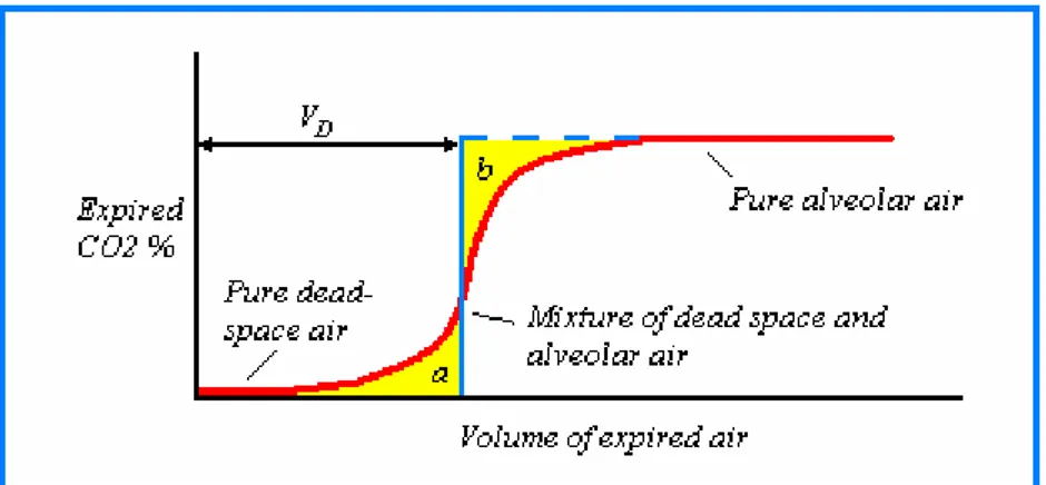
During expiration the breath composition changes in order to empty the airways and the lungs. Such a process can be followed by measuring the CO2 in breath, see
Figure 1.36,55.
Three phases can be identified: during Phase I the air in dead space is eliminated and the composition of this fraction is low in CO2, being similar to inspired air; during
Phase II the partial pressure of CO2 increases quickly until it reaches a maximum value
of 35 mmHg; only in Phase III is the alveolar air eliminated, containing the volatile compounds released from blood.
Figure 1.3 CO2 breath profile during the respiration process
Ambient air is the main source of xenobiotic contaminants and oxygen. Its content of oxygen and carbon dioxide is fairly constant, while the concentrations of xenobiotics may be highly variable.
Dead space air has a composition close to ambient air (but a higher water content). Differences may arise due to the chemicals originating and/or released in the conducting airways or to gas exchanges with the mucus layer (mainly for water soluble compounds).
The composition of alveolar air is due to the interaction of ambient air with blood through the alveolar membrane. There are considerable non-homogeneities in the composition of alveolar air in different lung regions even in healthy subjects, since posture and gravity alter both local ventilation (exchange of the air in the lungs) and perfusion (blood circulation). Pulmonary ventilation and perfusion are mainly regulated by respiratory and cardiac frequency44,55.
When the composition of ambient air changes, the time needed for each breath fraction to reach a new equilibrium ranges from a few seconds for dead space air to minutes or hours for alveolar air.
In most cases, blood is the main source of markers and for this reason alveolar air should be considered the most representative of an individual’s condition. If markers are released from conducting airways, dead space air should be sampled.
Dead space and end-tidal air can be distinguished on the basis of their composition. Unfortunately, a net separation cannot be made between the two due to the absence of a sharp anatomical boundary. A boundary region has been identified at the terminal bronchioles with a 2 mm diameter, where the convective flow gives way to diffusion43. The diffusion and mixing of gases in this region depend on their physical–chemical properties, on the rate of respiration and on the anatomy. The concentration of expiratory gases is also greatly influenced by the ratio of ventilation-to-volume, perfusion-to-volume and ventilation-to-perfusion inequalities45, as well as lung volume46, posture47 and flow rate48.
Thanks to the presence of different path lengths of dead space between the mouth and alveoli, the lack of a uniform velocity across the cross section of the tracheobronchial tree, and due to convective and diffusive mixing of gases at the boundary region, the transition in expired air is not so sharp, so that the concentration profile is rather S-shaped. (Figure 1.4)
In 1948 Fowler defined dead space as the volume of the conducting airway until the point where a large change in gas composition occurs, and he proposed a method for determining its value49.
Figure 1.4 CO2 concentration profile as a function of expired volume Fowler’s
graphic method is also shown: dead volume corresponds to the point where (a) and (b) regions have the same area.
This consisted of giving the subject a single breath of pure oxygen and monitoring the nitrogen concentration in the exhaled air. The nitrogen concentration profile in exhaled air resembles the carbon dioxide profile reported in Figure 1.4. The volumes i.e.,
shaded areas a and b are equivalent and represent the ideal transition point between dead space and alveolar air. Fowler used nitrogen as a tracer gas as he was able to use a fast sensor to measure its concentration levels in the expired air. In cases where the diffusivities were comparable, other gases, such as oxygen, carbon dioxide or helium, provided similar results50, 51.
Bohr using carbon dioxide as a tracer gas proposed a different method based on mass balance at the end of the 19th century52.His formula was the expression of a simplified model that considered the expired breath as the sum of two unmixed homogeneous fractions, the inspired air contained in the dead space and the alveolar air from the lungs:
VE × CE = (VE – VDS) CA + VDS × CDS (1)
Where VE and VDS are the volumes of expired and dead space air, CE is the average
concentration of carbon dioxide in expired air (the value that would be measured in a sampling bag), and CDS and CA are the concentrations of the same gas in dead space
and alveolar air respectively. The contribution VDS · CDS of dead space air to the carbon
dioxide balance can be neglected as the concentration of this gas in ambient air is close to zero. If the carbon dioxide concentration CA in the alveolar air is known (it is
reasonably close to the end-tidal CO2 concentration level), Bohr’s formula can be
rearranged and used to estimate either the dilution factor D of the alveolar air due to dead space air:
E E A A E DS E V V C D V V V C = = = − (2)
or the dead space volume:
(1 E) DS E A C V V C = − (3)
Even though they provide about the same estimate of dead space volume in healthy individuals, there is an important conceptual difference between Bohr’s and Fowler’s
approaches, originating from the use of tracer gases such as carbon dioxide and nitrogen which are involved or not involved in the respiration process. Fowler’s method estimates the volume of the conducting airways to the point where the pure oxygen delivered to the subject mixes with the nitrogen in alveolar air (anatomic dead space). Bohr’s method on the other hand, measures the volume of air not involved in the gas exchange with blood (physiological dead space).
The difference is due to the alveolar dead space, i.e. the volumes of the lung constituted by alveoli that are ventilated but insufficiently perfused (or perfused but poorly ventilated) for a gas exchange to be effective. Such volumes are normally very small (less than 5 ml) in healthy individuals, but can increase dramatically in patients affected by lung diseases characterized by ventilation or perfusion impairments such as bronchitis, emphysema or pulmonary embolism. Thus, if Bohr’s method is important to clinicians in order to assess the alveolar dead space, at the same time the estimate of the dilution factor may be used by researchers in breath analysis to normalize data and increase reproducibility42.
1.6 Breath sampling techniques
Despite its great potential, breath analysis is still far from being used in clinical practice. A list of argued reasons has been proposed by Risby et al53. From a technical point of view, one of the main problems is the lack of a standard sampling procedure and sufficiently accurate measurements for the trace compound analyses. To carry out a reliable quantitative analysis, breath samples need collecting in a standard and reproducible way.
In the absence of a standardization of the respiratory process and the breath collection procedure, the ratio between alveolar air and dead volume can vary a lot from breath to breath, leading to a large variability in the data.
Breath collection can be performed in two principal ways: mixed sampling and end-tidal sampling. In mixed breath sampling all the expired air is collected, including the air in the superior airways, which are not involved in a gas exchange (dead volume). In end-tidal sampling, only end-tidal air (principally alveolar air) is collected, which contains most of the information regarding compound composition in blood.
Mixed sampling is generally preferred for its simplicity, although dilution by dead volume air (15-20%), may depends on the general condition of a person, such as pulmonary capacity, pulmonary ventilation and blood flow6, 15, 55, 54. Pulmonary capacity varies according to age, gender, build. Thus in the case of mixed breath sampling it is important to normalize the data on CO2 concentrations. The amount of
CO2 in alveolar air during respiration, together with that present in mixed breath
sample, enables us to evaluate the dilution factor due to the dead anatomical volume6, 55, 56.
It would be useful to be able to collect dead volume and end-tidal during the expiratory phase separately, because it would enable us to identify the origin of the compounds in breath. The presence of exogenous (substances coming from ambient air) compounds, in fact, is one of the main sources of pollution. It would therefore be useful to distinguish them from endogenous compounds (substances produced from physiologic or pathologic metabolism).
The solution to this problem is dealt with in different ways: Phillips asserts that every compound needs to be quantified on the basis of the concentration gradient between breath and ambient air57; instead Schubert et al., assert that those compounds whose concentration in ambient air is comparable or higher than in breath, do not have to be considered in the characterization of the subject's breath58; and finally, to get round the problem, other scientists, give pure air59, 60 to the subject. In reality this latter solution is also questionable, in fact, after exogenous compounds have reached pulmonary alveoli dissolved in blood on the basis of their blood/air repartition coefficients, the compounds are then transferred through the body and released in the different tissues depend on the chemical affinity. The concentration of each compound in alveolar air is the result of a dynamic equilibrium that involves several compartments, each with its specific time constant. It has been demonstrated that different compounds have different elimination
times ranging from minutes to hours61, and this limits the use of pure air only to a few cases.
Further complications depend on the water solubility of some exogenous compounds, which dissolve in the mucus layer of the upper airways, and are thus present in breath in unpredictable amounts62. At the same time, it is known that some endogenous compounds of clinical interest, such as nitric oxide, are produced in the superior airways and do not come from alveolar air63.
All these considerations lead to the conclusion that separating the different breath fractions could be an effective for future research in this field.
There are many different sampling techniques for breath analysis. Several containers are used to sample exhaled air, such as glass tubes and plastic bags, from which a sub-sample is transferred directly to the analysis system by means of syringes, or solid adsorbents, from which the components are thermally desorbed32.
Several plastic materials were used in gaseous sampling field; among these the most used are Tedlar, Nalophan and the Cali-5-Bond, a multilayer material.
Tedlar is a polyvinylfluoride film with excellent chemical, electrical properties and a high mechanics resistance. Furthermore Tedlar material has the property to be a barrier at UV rays.
Nalophan (polyethylenterepthalate, PET) is a waterproof material and it is principally used in foods storage. It resists against most of the organic solvent, oils, weak acids and corrosive solutions; it also resists to temperature between -60°C and 220°C and it can not be welded.
The Cali-5-bond is composed by an internal layer of high density polyethylene, followed by polyamide, an aluminum sheet, which avoids diffusion processes, a polyvinylidenchloride layer and the last external layer a polyester film. This material was specifically projected for the sampling, the transport and the storage of ambient air or gaseous mixtures. This material guarantees a lower permeability and a high mechanics resistance, together with high chemical inertia.
Several types of sampling devices54, 64, 31, 35, 37 have been proposed to guarantee efficient sampling that will truly represent the content of exhaled air during the exhalation time, some of which are commercially available.
Pleil et al. collected breath in preconditioned stainless steel canisters56, 65. The subject controls the sampling, manually opening the valve through which the breath enters. The subject breathes ambient air and mixed samples are collected.
There are two commercially available samplers that permit end-tidal breath to be collected. The Quintron system which consists of a tee connector, connected to two one way valves. The heart of each valve is a silicon disc that seals the exit until the threshold pressure is reached. The valves are regulated to open at different pressures; a 250 ml bag is connected to the valve that opens at the lowest pressure, whereas a 750 ml bag is connected to the other valves. There is a mouthpiece at the entrance of the tee connector. When the subject breathes in the system, the pressure of the connector increases until the first valve opening and the bag are filled with dead volume air. The pressure increases again until the second valve opening and the second bag are filled with end-tidal breath.
The other collection system, BioVOC™, is produced by Markes International. In this case the subject is asked to breathe through a mouthpiece into an open cylinder. Only the last portion of end-tidal breath (150 ml) remains in the cylinder after expiration. The mouthpiece is then replaced by a piston, used to push the sample in an adsorption tube connected to the end part of the cylinder66.
Even if these systems are not the best solution, they are favourable in terms of their simplicity and cost. Their main limitations are the poor control of sampling conditions and the low volume collectable with a single breath.
A more sophisticated system, namely Breath Collecting Apparatus was developed by Menssana Research57. In this system the subject breathes through a mouthpiece in an instrument consisting of an inlet valve for ambient air inspiration and an exit valve connected to an open, stainless steel, cylindrical container held to 40°C to avoid water condensation. The sampling port, close to the mouthpiece, is connected to an adsorption tube, a flow meter and a pump. The system is controlled by a microprocessor, which, when necessary, starts the pump. The selective collection of end-tidal air in the
container is obtained by starting the pump at an appropriate time interval after expiration. The total volume collected in the adsorption tube during multiple breaths can be selected by the operator.
A breath sampler with CO2 control has been proposed by Schubert et al. for patients
who are mechanically ventilated67. When the percentage CO2 volume exceeds the
expected value an infrared sensor sends the data to an electronic processor, which activates a two way valve directing the flow towards an adsorbent trap55.
As regards the commercially available sampler, it is worth noting that BioVOC does not have a system to hold the temperature at a specific value to avoid water condensation, moreover the cylinder has a small hole into which the subject breathes, which causes high back pressure, making the sampler not suitable for subjects with pulmonary dysfunctions.
The same limitations apply to the Quintron system. Although the first bag volume can be modified on the basis of the weight and height of the subject, the different pulmonary capacities are not considered. The sampler offers a lower back pressure to the breathing respect to BioVOC sampler, but no remedy is taken for water condensation.
M. Phillips, on the other hand created a good prototype sampler. The device is built with inert materials, uses a disposable mouthpiece and is heated to avoid sample condensation, however only a portion of alveolar air is sampled (when the pump is sucking) and not all the alveolar air.
From this analysis on breath samplers it is evident that there are many difficulties to overcome: back pressure during the expiration phase, sample condensation and an accurate separation of the two breath fractions.
1.7 Analytical techniques used for chemical breath characterization
The methodologies described in the literature can be divided in two main groups:
• Analytical techniques providing a sample collecting phase and subsequent analysis using different instrumental techniques;
• Analytical techniques providing an analysis in real time directly on the patients using dedicated equipments.
Gaschromatography is certainly the best technique for the separation and the analysis of volatile organic compounds (VOCs) in liquid and gaseous samples and coupled with mass spectrometry (MS) it enables us to identify analytes in complex mixtures.
After collection the pre-concentration of the analytes is required, because of the very low concentration levels in the breath sample. The pre-concentration of the compounds can be performed with thermal adsorption tubes, through which a known volume of the sample is passed through (Solid Phase Extraction, SPE), or using the solid-phase micro extraction technique (SPME)92, 93, 94.
Information on breath composition can also be obtained from direct breath measurements performed by different mass spectrometric techniques: Selected Ion Flow Tube 68, 69, 70 (SIFT) or Proton Transfer Reaction Mass Spectrometry 71, 72, 73, 74 (PTR-MS). These techniques enable us to eliminate the collection and pre-concentration phases and give real time responses.
The SIFT technique was recently applied in the identification and quantification of gases present in air and breath samples. A primary ion current (i.e. H3O+) is generated
from an ionic source. A quadrupole mass filter directs the primary ions into a tube, where a flow of an inert carrier gas such as helium is passed through. Most of the organic substances have a greater proton affinity than water, thus the H3O+ ion is a
proton donor.
The gaseous sample, introduced in the tube, interacts with the primary ion stream; the collision between molecules causes the transfer of the proton from primary ion to the analyte, which receives a positive charge without any fragmentation. Before reaching the detector the produced ions are separated from each other and from the primary ion
stream, on the basis of the mass/charge ratio, using a quadrupole mass filter placed at the end of the tube.
All the produced ions are detected giving a single mass spectrum. When analyte concentrations in the sample are very low, the amounts of primary ions that react are lower than the total and the amount of ions produced by the analyte is proportional to the partial pressure in the sample73,74.
The PTR-MS technique is similar to SIFT and is shown in Figure 1.5.
Figure 1.5 Schema of PTR-MS
An ion source produces H3O+ from water vapour molecules. The H3O+ ions entering the
drift tube transfer the protons to volatile organic compounds, which have higher proton affinities than water.
The exothermic reaction is as follows:
H3O+ + VOC → VOCH+ + H2O (4)
The PTR-MS technique is different from SIFT in some ways that are used to increase the sensitivity of the method. The system works with an empty cathode ion source, which produces a high density of primary ions directing them into the tube without a
transport gas. For this reason, an increase of two orders of magnitude in sensitivity can be achieved compared to the SIFT technique74.
The two techniques can be used to take frequent and fast measurements without any pretreatment of the sample. However, the characterization of the substance only occurs on the basis of the mass/charge ratio and the chemical identification of the compounds is not easy73, 75, 76. Sometimes gas chromatography separation is needed to perform a quantitative, because ions produced from different analytes with the same mass/charge ratio can be present73, 74. Another problem is the undesirable fragmentation of the sample molecules, which complicate the situation considerably.
In recent years Ion Mobility Spectrometry (IMS) has been developed, which is a small and effective instrumentation for the detection of trace compounds in a gases76, 77, 78. IMS bases the characterization of compounds on their gas phase mobility, under a high electric field. Typically IMS is formed from an ionization chamber with β or a UV radiation source, an ion injector, a drift tube and an ion collector. The carrier gas (i.e. air or nitrogen) transfers the neutral analyte molecules in the vapour phase into the ionization chamber where a series of ion-molecule reactions occur. The reactant ions are injected into the drift tube, by periodically opening the shutter grid. The ions produced are formed in defined chemical reactions of neutral analyte molecules with reactant ions. The mobility of these produced ions may be used to identify the analyte molecules. The ions are selectively revealed on the basis of their characteristic drift time through the tube. The drift velocity (v) of the ion is related to the electric field strength (E) by the mobility (k), where78:
v = k x E (5)
Therefore the mobility is inversely proportional to the drift time, which is usually measured at a fixed drift length. In order to work the IMS does not need the vacuum and the ambient air may be used as carrier gas. Often the IMS is coupled with a gas-chromatographic system because, in the case of a complex mixture, the selectivity is low70, 71, 76, 77.
Recently spectroscopy techniques were developed to determine volatile compounds in gaseous samples. The laser spectroscopy technique for example measures the light attenuation of the laser after passing through the gaseous sample in the adsorption cell. The mid infrared region (λ=3-10 μm) is particularly interesting, because the vibrational transition of most of the organic compounds leads to adsorption lines in that spectral range. One of the main advantages of these techniques is the possibility of measuring breath components in real time, in pptv (parts per trillion in volume) concentrations. Moreover spectroscopy provides information on analyte concentrations in the sample during the different phases of respiration. This is different from other analysis techniques, which give information on the whole expiratory phase79,70.
Alongside traditional analytical techniques, over the last few years innovative instrumental techniques based on sensor have been on the increase4, 18, 74, 80, 81. Sensors for ethanol and acetaldehyde analysis in breath have been created, immobilizing alcohol oxidase (AOD) and aldehyde dehydrogenase (ALDH) enzymes on electrodes82. Furthermore MOS (metallic oxide semiconductor) gas sensors seem to provide a good methodological approach to breath analysis, because of the wide range of detectable gases, their high sensitivity, the fast response and the simplicity of the devices involved. The poor selectivity of the sensor is a limitation that may be overcome with the use of an array of non selective sensors. The simultaneous use of more than one sensor and the application of a multivariate analysis of the data enable us to obtain useful information on the chemical composition of the sample. This is despite the fact that the sensor array response is not univocally related to the concentration of a single compound, but rather to the combination of all the compounds in the breath sample83. Recently these sensor arrays have been used in the medical field, to diagnose some pathologies, such as diabetes, lung cancer, asthma or other respiratory inflammations74, 84, 85, 86, 87.
1.8 Breath pre-concentration techniques
Owing to the low concentrations of most of the compounds present in human breath (order of magnitude μg/l and ng/l), after sampling and before analysis, a concentration analyte step is required. The methods used in the sampling and the pre-concentration of breath substances include a chemical method, a cryogenic method2, 3, 5 and the most diffused adsorptive methods3.
In the chemical methods, breath is usually bubbled through a reagent solution, which only captures a specific compound. The method is simple and direct, and the trapped sample is easily analyzed by a spectrophotometric technique. The main disadvantages are the low sensitivity and the great effort required by the subject to breathe.
In the cryogenic method, breath is passed through a trap immersed in a cryogenic fluid. After sampling the trap is heated and the trapped compounds are released and transferred to the instrument by a carrier inert gas.
The main problems encountered during the collecting phase are linked to the high content of water, a relative humidity > 95%. In the cryogenic method, if the water is not removed, the formation of ice clogs the trap3. In the adsorptive method on the other hand, the dipolar interactions between the water and the polar analytes reduce the trap efficiency of the adsorbent used88.
To avoid interferences due to water, different resolutions were found which included the use of drying agents (Na2SO4 and CaCl2) through which the sample was passed, or
the use of membranes with adsorbent surfaces (MESI, Membrane Extraction with Sorbent Interface)89. These systems are limited due to the possible loss of the analytes and the contamination of the sample.
In the adsorptive method, breath comes in contact with a resin, which traps the organic compounds of interest. There are two different types of pre-concentration techniques that use adsorption in the solid phase:
• The Solid Phase Micro-Extraction (SPME), which uses fused silica micro-fiber coated with an appropriate stationary phase;
1.8.1 The Solid Phase Micro-Extraction (SPME)
The solid phase micro-extraction technique, developed by Pawliszyn in 1989, combines the collecting and analytes extraction phases in one step. The SPME does not use any solvents90, 91 and may be easily coupled with the most diffuse chromatographic technique, such as Gas-Chromatography (GC), High Performance Liquid Chromatography (HPLC) and Capillary Electrophoresis (CE)90.
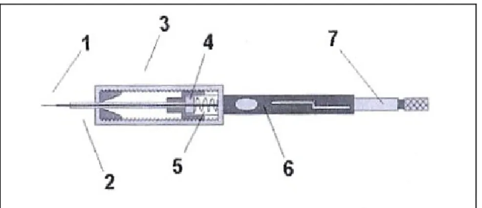
The complete system used for SPME is shown in Figure 1.6
Figure 1.6 SPME system: 1) withdrawn fused silica fiber, 2) stainless steel needle, 3) holder, 4) silicon septum, 5) spring, 6) tube support, 7) plunger.
The pre-concentration procedure exposes the fiber to the sampler for the time required by the analytes to reach equilibrium between the sample and the fiber. At the end of the equilibration time, the fiber is inserted in to the gas-chromatography injector and the analytes are thermally desorbed. Consequently the carrier gas then transfers the analytes into the column30, 91, 92, 93.
The fiber is coated with a thin stationary phase, made of polymers or a mixture of polymers with carbon materials. Different stationary phases, with different thicknesses and polarities are available and have affinities with different classes of compounds. Polydimethylsiloxane (PDMS), is suited to non polar compounds, carbowax-divinylbenzene (CW-DVB) and polyacrylate (PA) are suited to polar analytes and polydimethylsiloxane-divinylbenzene (PDMS-DVB) and carboxen-polydimethyl siloxane (CX-PDMS) do not have a specific application91, 93 , 94, 95.
This technique has several advantages in terms of its applicability to all kinds of samples (solid, liquid or gaseous) without needing any particular sample preparation, its easiness to use, the speed of the analysis and the possibility to perform a chemical qualitative screening of a gaseous sample. The principle disadvantages involve the low pre-concentration factor analytes are present at trace levels. It is also limited in terms of a quantitative analysis of a complex sample matrix, due to the competition of different compounds in the distribution process94, 95.
1.8.2 The Solid Phase Extraction (SPE)
In the solid phase extraction technique, the sample passes through an adsorption tube, filled with a suitable stationary phase and the analytes are retained on the basis of their chemical-physical properties. (Figure 1.7)
Figure 1.7 Adsorption tube used in SPE technique: the tube can be made of stainless steel or
glass
Carbon is the main constituent of the adsorbent in terms of its high chemical inertness and its thermal stability96.
Different kinds of carbon are available: • Active carbon
• Carbon molecular sieve • Graphite carbon
Each has different properties and retains different classes of compounds.
Active carbons have a complex superficial structure made up of many functional groups: phenolic, carboxylic, aldehyde, ketone , peroxy, quinone and lactone. The principle mechanism of interaction includes hydrophobic, hydrogen bond and cationic exchanges. Using active carbons the compounds were not able to be completely released due to their high affinity with the stationary phase96.
Carbon molecular sieves have a highly porous surface with almost uniform micro-porous diameters and a wide superficial area, suited for the retention of organic compounds. The adsorption mechanisms include dispersive interaction (London or dispersion forces, Van der Waal forces) and strong dipole-dipole interactions96. The thermal treatment of graphite carbon eliminates volatile compounds and produces a homogeneous surface without micro-pores, where the compounds are adsorbed on the basis of their size and shape. Most of the surface has non polar sites (carbon atoms) that do not react with molecules containing functional groups, thus dispersive interactions were predominant in the adsorption mechanism96.
Porous carbon is produced by impregnating a suitable silica gel with mixed resins of phenol-formaldehyde, phenol-hexamine, saccharose or other materials. After polymerization inside the silica porous gel, the polymer is transformed in to vitreous carbon by heating it to 1000°C. The silica portion is removed in order to form the pores. The material is then heated to 2000-2800°C in an inert atmosphere, to fit the surface, remove the micro-pores and depending on the temperature, produce a certain degree of graphitization. The porous carbon has a homogeneous hydrophobic surface and the interactions depend on the final heating and possibly on chemical treatments96.
The choice of which adsorbent phase to use has to be made on the basis of the following properties97:
• Functionality: expresses the affinity of the phase for the different organic compounds;
• Chemical inertness: the reaction capacity of the phase in relation to adsorbed molecules;
• Size and shape of the particles: influence the hydrodynamic conditions of the sample that passes into the tube and determines the surface area value;
• Pores dimension: a high porosity determines a larger total surface area;
• Surface area: influences the recovery and the reproducibility of the adsorption process.
If the sample has a high content of water it is convenient to choose a hydrophobic material to limit water interferences. The presence of water could interfere with the detector acquisition, modify retention time and cause the column to deteriorate (referred to the chromatographic technique)98, 99.
The use of a multi-bed tube is recommended if the sample contains compounds with different chemical-physical properties. (Figure 1.8)
Figure 1.8 Multi-bed tube filled with three different stationary phases with different adsorption
capacities.
The multi-bed tubes are filled with different phases in an increasing order of adsorption capacity. With these tubes the range of adsorbed compounds is wider than with the mono-phase tube provided the phases are properly chosen. Adsorption tubes are used both with gaseous and liquid samples by applying different collecting techniques.
In terms of gaseous samples, the collecting techniques are as follows: • Diffusive sampling
Diffusive sampling or passive sampling is the easiest procedure from an operative point of view. The tubes are exposed to the sample, i.e. air, for an established time; volatile compounds diffuse in the tubes and the analytes are retained by the adsorption material. This kind of sampling takes a long time. Since diffusive sampling can be performed with only mono-phase tubes, the simultaneous exposure of tubes filled with different stationary phases is required.
In the case of active sampling, a known volume of the sample passes through the adsorption tube using a pump, which maintains a constant flow. Active sampling, differently from passive sampling, enables the operator to know the amount of sample collected in the tube and for the same sampling time, a greater amount of analyte is collected.
Multi-bed tubes can be used in active sampling to retain a wide range of compounds with different boiling points and polarities, in just one collection.
The sampling flow may vary from a minimum of 10 ml/min to 200 ml/min, the optimal flow being 50 ml/min. With a flow under 10 ml/min, the analytes may diffuse along the stationary phase and with a flow higher than 200 ml/min, the recovery of the analytes may not be complete.
During the collection process, the operator must be certain that all of the analytes collected remain in the adsorbent phase. To assure that no analyte is lost during the sample collection, the Breakthrough Volume should be considered.
Breakthrough volume is defined as the calculated volume of carrier gas per gram of adsorbent resin that causes the analyte molecules to migrate from the front to the back of the adsorbent bed, at a specific temperature. This volume defines the adsorption capacity of a resin in relation to a specific analyte and depends on the affinity between the stationary phase and the considered compound, the amount of stationary phase in the tube and the temperature.
A breakthrough volume measurement can be performed flowing a gas with a known content of analyte through an adsorption tube connected directly to a detector, which
gives the signal in real time. The detector will give no response until the resin traps the analyte. When the breakthrough volume is exceeded, the detector will give a response proportional to the amount of analyte coming out. (Figure 1.9)
Figure 1.9 Percentage trend of the amount of analyte coming out of the tube with respect to the volume of gas passing through (Vb is the breakthrough volume)
For example, at a fixed temperature, if the breakthrough volume for an analyte is 1.0 litre per gram of resin, then the sample should not be purged with more than 500 ml of gas, for tubes filled with 1 g of the adsorbent phase.
The adsorbent capacity of resin is influenced by humidity, thus the breakthrough volume will also depend on the sample content of water and its evaluation should be carried out in the same humidity conditions as the sample. In fact it is appropriate to sample a maximum volume corresponding to 70% of the breakthrough volume (Safe Sampling Volume, SSV), because the amount of analyte in the sample and thus the breakthrough volume can vary significantly.
1.9 Recovery of the analytes after pre-concentration
The recovery of the sample from the stationary phase can be made by extracting a small amount of organic solvent, or alternatively by heating the adsorbent phase purging with an inert gas (thermal desorption). The absence of solvent enables us to identify the most volatile compounds, usually eluted with the solvent. In addition, the cost and the disposal of the solvent are eliminated.
The thermal desorption technique allows the volatile compounds linked to the stationary phase to be released using a rapid increase in temperature. The analytes are thus transferred to the chromatographic column, using a flow of an inert gas as a carrier. Although the sample is rapidly heated, the analyte desorption is not as rapid. Thus during the transfer to the chromatographic column, a broadening of the chromatographic signal can occur, with a consequent loss of resolution.
This problem is solved in the two stage desorption system; in fact the analytes desorbed from the tube are concentrated in a cryogenic packed trap, which has a reduced internal volume. The trap is maintained at a low temperature using a Peltier cell, in order to adsorb the analyte coming from the tube; the trap is then rapidly heated and in this case the desorption process is faster and the transfer to the column is performed with smaller carrier gas volumes. The tube and the trap are both desorbed in the opposite direction with respect to the flow during adsorption process. (Figure 1.10)
Figure 1.10 Direction of the flow during compound adsorption and desorption of both the trap and the tube
In order to completely release the analytes from the adsorption tube, to quantitatively adsorb the released compounds in the focusing trap and quantitatively transfer the analytes into the column using a smaller volume of carrier gas, following parameters need to be optimized:
• Packing material of the trap;
• Tube desorption, focusing and trap desorption temperatures; • Desorption flows and times from the trap and the tube.
Since the internal trap can operate at a lower temperature than ambient temperature and the volume of gas that passes through is smaller than the volume passing through the tube, a stationary phase can be used with a lower adsorption capacity compared to the resin filling the tube.
The selection of an appropriate temperature of the tube is required, in order to completely desorb the compounds of interest and avoiding their thermal decomposition. It is also best not to desorb near the maximum temperature indicated for a specific stationary phase. This is to avoid the degradation of adsorbent material and the subsequent release of interfering compounds.
During the focusing phase, the internal trap can be cooled up to -40°C, the temperature at which most of the compounds are retained. Where there is a high water content in the sample, a temperature of around 0°C is required to avoid ice formation with the consequent obstruction of the trap. The desorption temperature also has to allow for the quantitative recovery of the adsorbed analytes. The temperature limit is the maximum provided for the stationary phase used. Trap and tube desorption times may guarantee
the complete release of the compounds of interest and usually varies from 5 to 30 minutes depending on the carrier gas flow and desorption temperature.
The flow of auxiliary gas during tube desorption should be optimized. A low flow limits the extraction efficiency from the tube and the desorption time becomes too long. An extremely high flow could limit the retention capacity of the trap. Usually flows around 30-50 ml/min are suggested. During the transfer from the focusing trap to the chromatographic column, the carrier gas flow should guarantee a quantitative and fast transfer of the compounds. The lowest compatible flow is 7-8 ml/min. The carrier gas
flow through the trap is the sum of the flow into the column and the split injection flow. Capillary columns need a split flow of 5-10 ml/min with flow values in the column of 0.5-2.0 ml /min, to ensure a sufficiently rapid transfer.
One of the application fields of thermal desorption technique includes an environmental analysis for the determination of volatile and semi volatile organic pollutants in the atmosphere26, in the working environment30, 32, and in the emissions from building materials28, 29. It is also widely used for the extraction of VOCs from matrices such as solid samples, emulsions and saline solutions, which cannot be injected directly in the chromatography. Over the last few years, this technique has also been used in breath analysis, in diagnostic application or to evaluate human exposure to toxic substances100. A limitation of this technique obviously includes compounds that are not analyzable by gas chromatography, compounds that are easily thermally degradable and compounds with a high boiling point (C36 hydrocarbons, PAH, etc).
CHAPTER 2
Materials and Methods
2.1 Description of standard solution preparation
Stock A
The liquid mixture consisted of eighteen compounds and two labelled internal standards, Toluene-D8 and Isopropanol-D8 in methanol. Labelled isopropanol-D8 and toluene- D8 (purity 99.8 %) were purchased from ARMAR Chemicals and used as an internal standard, without any further purification.
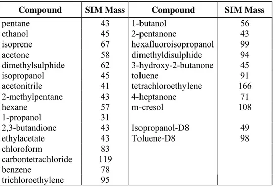
Isopropanol, 2-butanone, 2-pentanone, hexanal, 2-heptanone, 4-heptanone, heptanal, benzaldehyde were purchased from AccuStandard, Inc. Chemical Reference Standard (USA).
Pentane, 2-methylpentane, hexane, 1,1,1,3,3,3-hexafluoro-2-isopropanol, dimethylsulphide, acetone, carbon sulphide and toluene puriss p.a. standard for GC grade > 99% were purchased from Fluka. Dimethyldisulphide and isoprene puriss p.a. for GC grade > 99% were supplied by Sigma- Aldrich, all compounds were used without any further purification.
A stock solution consisting of 50 μl of each 18 neat compounds was prepared (SOL 1). Stock A was obtained by adding 20 μl of the stock solution and 50 μl of an intermediate labelled solution (Stock H) to 10 ml of methanol. Compound concentrations are reported in Table 2.1.
Table 2.1 Concentration of compounds in Stock A Stock A Concentration [ng/μl] pentane 70 isoprene 76 acetone 88 dimethylsulphide 94 carbon sulphide 141 isopropanol 87 2-methylpentane 73 hexane 73 2-butanone 89 2-pentanone 90 hexafluoroisopropanol 177 dimethyldisulphide 116 toluene 96 hexanal 91 4-heptanone 91 2-heptanone 91 heptanal 91 benzaldehyde 116 isopropanol-D8 111 toluene-D8 118
Stock B, Stock C and Stock D
Two solutions were prepared, consisting of 100 μl of twelve neat compounds (SOL 2) and 100 μl of thirteen neat compounds (SOL 3) respectively. A stock solution was prepared, consisting of 40 μl of SOL 2 and 40 μl of SOL 3 in 20 ml of methanol (Stock B) (Table 2.2).
Table 2.2 Concentration of compounds in Stock B Stock B Concentration of compounds in Stock B [ng/μl] tetrachloroethylene 267 3-hydroxy-2-butanone 164 1-butanol 135 trichloroethylene 245 benzene 146 carbontetrachloride 265 chloroform 247 ethylacetate 150 2,3-butandione 183 1-propanol 134 acetonitrile 131 ethanol 133 pentane 96 isoprene 105 acetone 122 dimethyl sulphide 130 isopropanol 121 2-methylpentane 100 hexane 101 2-pentanone 124 hexafluoroisopropanol 246 dimethyldisulphide 161 toluene 133 4-heptanone 126 m-cresol 158
Stock B solution was diluted to obtain Stock C and D (Table 2.3):
Table 2.3 Preparation of Stock C and D Stock C Stock D
Stock B (ml) 5 1
Methanol (ml) 5 9
Dilution factor 1:2 1:10
The concentrations of all compounds in the different stock solutions are reported in Table 2.4
Table 2.4 Standard solution concentrations
[ng/μl] Stock B Stock C Stock D
pentane 96 48 10 ethanol 133 67 13 isoprene 105 52 10 acetone 122 61 12 dimethylsulphide 130 65 13 isopropanol 121 60 12 acetonitrile 131 66 13 2-methylpentane 100 50 10 hexane 101 51 10 1-propanol 134 67 13 2,3-butandione 183 92 18 ethylacetate 150 75 15 chloroform 247 124 25 carbontetrachloride 265 133 27 benzene 146 73 15 trichloroethylene 245 123 25 1-butanol 135 68 14 2-pentanone 124 62 12 hexafluoroisopropanol 246 123 25 dimethyldisulphide 161 80 16 3-hydroxy-2-butanone 164 82 16 toluene 133 67 13 tetrachloroethylene 267 133 27 4-heptanone 126 63 13 m-cresol 158 79 16
50 μl of Stock H consisting of Toluene-D8 and Isopropanol-D8, were added to the three standard stock solutions. The concentrations of the internal standard compounds in each solution are reported in Table 2.5
Table 2.5 Concentrations of the internal standards in stock solutions B, C, and D [ng/μl] Stock B, C, D
Isopropanol-D8 111 Toluene-D8 118
Stock E
A gaseous Stock E sample was obtained by introducing 20 μl of liquid SOL 1 into a 2 L glass flask, held at 40 °C. The preparation of gaseous standards was carried out inside a oven held at 40°C, to avoid any condensation of the compounds. The corresponding concentrations are reported in Table 2.6.
Table 2.6 Concentration of compounds in Stock E Stock E Concentration [μg/l] pentane 348 isoprene 378 acetone 439 dimethylsulphide 470 carbon sulphide 703 isopropanol 436 2-methylpentane 363 hexane 366 2-butanone 447 2-pentanone 449 hexafluoroisopropanol 887 dimethyldisulphide 581 toluene 481 hexanal 453 4-heptanone 454 2-heptanone 456 heptanal 454 benzaldehyde 581
Stock F
5 μl of both isopropanol-D8 and toluene-D8 were evaporated in a 2 L glass flask, held at 40°C, to prepare a gaseous sample for use as an internal standard. The corresponding concentrations are reported in Table 2.7.
Table 2.7 Concentration of the internal standards in Stock F Stock F flask concentration [ppmv]
Isopropanol-D8 839 Toluene-D8 605
Stock G
20 μl of stock solution (Stock G) consisting of 100 μl of the neat compounds reported in Table 2.8 were vaporized into a 2 L glass flask, held at 40°C.
5 ml of this gaseous mixture were then introduced into a Nalophan bag filled with 15 L of pure air.
Table 2.8 Concentration of compounds in the flask and in Stock G.
Compounds Concentration of Stock G [ppmv] Concentration in bag [ppbv] Acetone 519 173 Isopropanol 628 209 Hexane 292 97 Methylethylketone 423 141 Dimethyldisulfide 429 143 Toluene 359 120
Stock H
An intermediate labelled solution was prepared consisting of 25 μl of isopropanol-D8, 25 μl of toluene-D8 and 950 μl of methanol.
2.2 Chromatographic, mass spectrometric and thermal desorption
methods
Samples were transferred into adsorption tubes using a membrane pump (NMP 50, SKC ITALY) at a constant flow rate measured by a soap bubble flow meter, (mod. Humonic Inc).
Thermal desorption (TD) and GC-MS analysis were performed by an automated two-stage thermal desorption unit (STD 1000, DANI Instrument, Italy) equipped with an internal focusing trap packed with 70 mg of Tenax GR (DANI Instrument, Italy), a GC device (Trace GC Ultra, Thermo Electron Corporation, USA) equipped with a DB-624 capillary column (6% cyanopropyl phenyl siloxane and 94% dimethylpolysiloxane), (60 m x 0.25 mm x 1.4 μm film thickness, from Agilent J&W) and a quadrupole mass spectrometer (Trace DSQ, Thermo Electron Corporation, USA)
A description of the conditions used for thermal desorption, chromatographic and spectrometric methods is given in Table 2.9, 2.10, 2.11