1) Clinica Medica “A. Murri”, Department of Biomedical Sciences and Human Oncology, University of Bari “Aldo Moro” Medical School, Bari, Italy
2) Gastrointestinal Endoscopy, “Perinei” Hospital, Altamura, Bari, Italy
3) Department of Gastroenterology, Henry Durant Hospital Center, Athens, Greece
Address for correspondence:
Prof. Piero Portincasa, MD, PhD
Clinica Medica “Augusto Murri”
Department of Biomedical Sciences and Human Oncology
University of Bari Medical School Piazza Giulio Cesare 11 70124 Bari - Italy [email protected]
Received: 30.04.2019 Accepted: 31.05.2019
Faster Detection of Helicobacter pylori Infection by
13C-Urea
Breath Test. Comparing Short versus Standard Sampling Time
Emilio Molina-Molina1, Leonilde Bonfrate1, Michele Lorusso1, Harshitha Shanmugam1, Giuseppe Scaccianoce2, Theodore Rokkas3, Piero Portincasa1INTRODUCTION
Helicobacter pylori (H. pylori) is a gram-negative spiral
microaerophilic bacterium that colonizes gastric mucosa. The worldwide prevalence of H.
pylori infections is approximately
50%, with a higher prevalence in low-income countries. H.
pylori infection is associated
with peptic ulcer disease, atopic disorders, gastric cancer and mucosa-associated lymphoid tissue (MALT) lymphoma [1]. Several guidelines recommend eradication of H. pylori infection in most of the diagnosed subjects [2, 3]. Current indications for diagnosis of H. pylori infection include non-invasive procedures
ABSTRACT
Background & Aims: 13C-Urea Breath Test (UBT) is a non-invasive, highly accurate and recommended test to
detect Helicobacter pylori (H. pylori) infection and to confirm post-therapy eradication. However, differences exist in terms of manufacturers, dose of labelled urea, addition of citric acid, solid vs. liquid formulation, and sampling times of breath samples. In this study, we compared the diagnostic accuracy of “short” (15 minutes) vs. “standard” (30 minutes) time for a single type of liquid UBT.
Methods: We compared the performance of a single UBT type (BREATHQUALITY, AB Analitica, Padua,
Italy, 10 mL of 75 mg 13C-Urea and 1.4 g citric acid) during a “short” vs. “standard” breath sampling time.
Enrolled were 151 subjects requiring UBT as naïve (N=92) or post-eradication (N=59) checks.
Results: UBT at 15 and 30 minutes were highly comparable, showing optimal correlation in all subsets of
patients (i.e. naïve vs. post eradication, negative vs. post eradication check). One discrepant result occurred at the borderline zone of the DOB 4‰, but proved to be true positive at a later confirmation by a second UBT and stool antigen test.
Conclusions: By shortening the testing time of BREATHQUALITY to 15 minutes (-50%) comparable
accuracy will be maintained and in addition, it will bring some benefits to patients’ waiting lists, compliance, and hospital staff.
Key words: breath test − chronic gastritis − infrared analysis − peptic ulcer − stable isotope. Abbreviations: DOB: delta over baseline; UBT: urea breath test.
such as urea breath test (UBT), fecal H. pylori antigen (active infection) and serology (active or prior infections). Invasive tests for H. pylori infection include upper gastrointestinal endoscopy, coupled to rapid urease test or histology. In this context, choosing the right diagnostic test depends on several aspects, namely patients’ choice, local availability of diagnostic tests, and clinical settings [4]. Urea breath test is the non-invasive, accurate and highly recommended test to detect H. pylori infection, employing the stable isotope 13C.
The urea breath test offers an important diagnostic role in younger subjects (generally aged less than 50) with upper uncomplicated gastrointestinal symptoms, who do not have “alarm” features (i.e., unintentional weight loss, unexplained iron deficiency anemia, persistent vomiting, progressive dysphagia or odynophagia, palpable mass or lymphadenopathy, and family history of upper gastrointestinal cancer). In these latter cases, upper gastrointestinal endoscopy or additional investigations are invariably performed [3, 4].
We incorporated the diagnosis of UBT in different settings looking at the impact of H. pylori infection in Italy, and outcome of different eradication therapies [5-8]. Patients
undergo UBT according to standard protocols recommended by manufacturers, taking breath samples of exhaled air generally 30 minutes after ingestion of the 13C-urea-containing
solution.
Because of the expanding role of UBT in the noninvasive diagnosis of H. pylori infection, there is a further and urgent need to rationalize the methodological aspects of the test. The time for breath sampling comprises a series of factors, which include time exposure of the gastric mucosa and H. pylori to the
13C-substrate, appearance of 13CO
2 in breath, sample collection,
and diagnostic outcome in terms of sensitivity and specificity. Additional factors also include the hospital stay of the patients, waiting lists and time devoted by doctors and nurses to the test, which generally requires at least 30 minutes between the substrate ingestion and the second breath sampling. In this study, therefore, we compared the diagnostic accuracy of “short” (15 minutes, T15) vs. “standard” (30 minutes, T30) time for a single type of liquid UBT currently adopted at our Hospital.
METHODS
Subjects
A total of 151 patients were enrolled (91 females, 60 males; age: 49.3±1.3 years) at the Academic Unit of Internal Medicine “Clinica Medica A. Murri”, in Bari, Italy. All subjects joining the study were outpatients from the public Hospital waiting list. Subjects were complaining of likely H. pylori-associated symptoms, including dyspepsia, gastroesophageal reflux (without alarm symptoms), a previous history of peptic ulcer, and use of chronic therapy with non-steroidal anti-inflammatory drugs (NSAIDs). Some patients had iron-deficiency-anaemia, vitamin B12 deficiency, and idiopathic thrombocytopenic purpura. A small subgroup of subjects was also referred by their own physicians because of a family history of H. pylori infection or personal concern about infection (risk of peptic ulcer, gastric cancer, atrophic gastritis), or with symptoms unrelated to H. pylori infection (urticarial, allergies, dermatological symptoms) [4]. All subjects underwent a visit including history, a general physical examination, UBT as well as the answer to a single question investigating the compliance of the short vs. standard UBT (see study protocol).
The study was totally noninvasive, and the protocol was approved by the local Ethics Committee of the University of Bari Medical School.
Urea Breath Test
The UBT test is the standard diagnostic tool currently available from the central pharmacy of the Policlinico Hospital. The kit is registered as a medical device (BREATHQUALITY UBT, AB Analitica, Padua, Italy) at the Italian Ministry of Health (A.I.C. n. 034510014), and consists of 75 mg 13C-labelled
urea in 10 mL of 1.4 g of citric acid solution. Subjects fasted for at least 8 hours and were free of medication which could influence the UBT results (i.e., of antibiotics in the prior month and no proton pump inhibitors in the previous two weeks) [4]. During the test, the patient swallowed the solution, added on-site to 200 mL tap water. UBT results depend on the gastric hydrolysis of urea by H. pylori. In case of infection, 13CO
2
appears in breath, while ammonia is released into the stomach. Breath samples were analyzed by the same 13C-infrared analyzer
(HeliFANplus®, FAN GmbH, Leipzig, Germany) kept at the Division of Internal Medicine. The differences between the values at T30 and baseline were expressed as delta over baseline (DOB, δ‰), with a normal cut-off up to 4‰. As recommended by the manufacturer, the DOB at T30 was the reference value, and this was compared with the results obtained from a sample taken at T15.
Study protocol
The protocol consisted of the following steps: 1) Full explanation of the purpose of the study and collection of the informed consent; 2) Collection of two baseline breath samples of expired air into glass vacutainers in the fasting subject at rest (8.00 AM). Smoking and physical exercise are forbidden for the whole duration of the test; 3) Ingestion of the 200 mL solution containing 13C-urea and citric acid; 4) Collection of
duplicate breath samples at two time points: T15 and at T30; 5) Recording the answer to one question: “Would you prefer a 15 min rather than a 30 min UBT?”; 6) Calculation of 13CO
2
concentrations in breath samples by infrared analyzer.
Statistical analysis
Data were expressed as mean±SEM. Results of UBT were compared between T15 and T30 by a paired t-test and visual inspection of single results. The comparisons included the whole group and two subgroups, i.e. according to UBT results (positive or negative) and according to the patient’s status (naïve or post-eradication). Proportions were compared by contingency tables and Chi-Square or Fisher’s exact tests. Correlations were assessed by calculating Spearman‘s test. Statistical analyses were performed with the NCSS statistical software (NCSS9 Statistical Software 2013. NCSS, LLC. Kaysville, Utah, USA, www.ncss.com/software/ncss) [9, 10]. The difference was considered statistically significant when the two-tailed probability (p-value) was less than 5%.
RESULTS
Among the 151 enrolled subjects, females (n=91) tended to be younger than males (n=60) (48.2±1.6 vs. 50.9±2.1 years respectively, p=0.30). Physicians referred 75% of the patients; other specialists, including us, referred 20% of the patients, while 5% asked to undergo UBT because of their own decision and personal concern. Females were more represented than males (60% vs. 40%, p=0.0005).
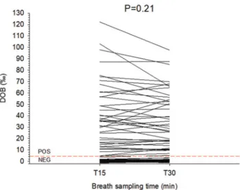
The results of the test comparing T15 and T30 appear in Fig. 1. With the given cut-off point set at DOB 4‰ at T30 (as recommended by the company), there were 41 UBT positive cases (27.2%) and 110 UBT negative patients (72.8%). Overall, mean DOBs were 11.1±1.9‰ (T15) and 10.5±1.7‰ (T30), with p=0.21. In addition, DOB was comparable in the case of a negative test (0.35±0.1‰ at T15 vs. 0.54±0.2‰ at T30, p=0.06) and positive test (40.8±4.6‰ at T15 vs. 38.1±3.9‰ at T30, p=0.13). The same occurred with respect to DOBs and patient’s status. For naïve patients there were 63 negative UBT (DOB 0.22±0.1‰ at T15 vs. 0.35±0.1‰ at T30, p=0.22) and 29 positive UBT (DOB 47.2±5.8‰ at T15 vs. 44.4±4.7‰
at T30, p=0.19). For post-eradication there were 47 negative UBT (DOB 0.23±0.12‰ at T15 vs. 0.42±0.12‰ at T30, p=0.16) and 12 positive UBT (DOB 23.2±4.0‰ at T15 vs. 21.3±3.9‰ at T30, p=0.09).
The overall correlations between DOB at T15 and T30 appear in Fig. 2. The highly significant correlation between the two time points was confirmed overall (n=151, r=0.97, p<0.001) (Fig. 2A). The correlation also persisted in the two
subgroups of patients with a negative UBT (Fig. 2B) and a positive UBT (Fig. 2C). Data dispersion, however, was much greater at lower DOB values, i.e. in the 110 UBT-negative cases (r=0.35, p=0.0006) (Fig. 2C).
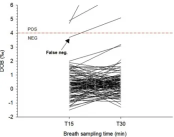
The visual comparison of DOBs at T15 and T30 disclosed a single discrepant result at T15 close to the borderline cut-off of 4‰ In this unique patient, DOB was 3.7‰ (negative) at T15, and increased to 5.1‰ (positive) at T30 (see Fig. 1A, arrow). This patient underwent a second UBT one week apart, and the results were positive (DOB 6.1‰ and 5.7‰ at T15 and T30, respectively), and further confirmed by a positive stool antigen test. We arbitrarily considered this case a “false negative” result. Based on the first-round results from the 151 cases, therefore, the overall sensitivity and specificity at T15 was 97%, compared to the given 100% of sensitivity and specificity at T30. The answer to the question about preferring the short- 15 min, rather than the standard 30 min UBT is summarized in Fig. 4. Over 90% of subjects were positive about the short version of UBT.
DISCUSSION
The UBT is one of the major non-invasive diagnostic tools for detection of H. pylori infection. The test is used worldwide, is totally non-invasive and can be used in children and pregnant women as well, with millions of subjects screened every year, both at a naïve level and after eradication therapies [4-7].
The results of the UBT provide important information which recapitulate further diagnostic and therapeutic decisions, in order to reduce the potential and deleterious Fig. 1. Comparison of exhaled breaths in subjects undergoing UBT
after 15 (T15) and 30 minutes (T30). The dotted horizontal line is set at 4‰ DOB and indicates the normal cutoff limit. Differences between T15 and T30 are not significant. Legend: DOB: delta over baseline; NEG: negative; POS: positive.
Fig. 2. A) Linear correlation between DOBs obtained at two time points (T15 and T30) in subjects
undergoing UBT. A very strong correlation is disclosed by the r=0.97. B) Linear correlation between DOBs obtained at two time points (T15 and T30) in subjects negative for UBT. The dotted horizontal and vertical lines indicate the normal cutoff limit of 4‰ DOB. Data dispersion is visible and disclosed by the r=0.35. C) Linear correlation between DOBs obtained at two time points (T15 and T30) in subjects positive for UBT. The single black triangle at the intersection of the dotted lines represents the “false-negative” result at T15. A very strong correlation is disclosed by the r=0.94. Legend: DOB: delta over baseline; the dotted horizontal and vertical lines indicate the normal cutoff limit of 4‰ DOB. .
consequences of the chronically infected gastric mucosa in humans [3, 11, 12].
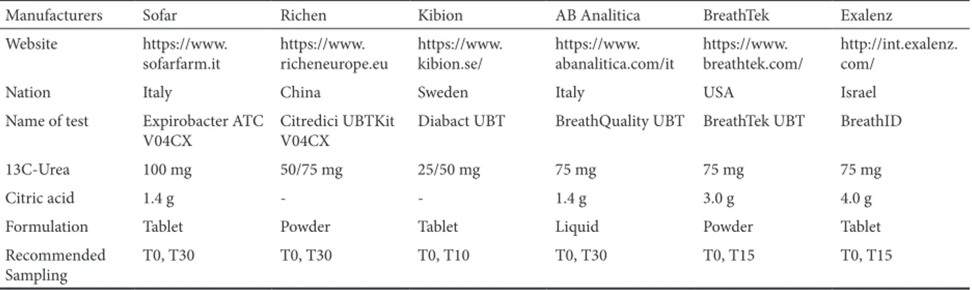
The sensitivity and specificity of UBT range from 88% to 95% and from 95% to 100%, respectively [13], with very uncommon false-positive results [14, 15]. In spite of such highly confident outcomes, differences still exist in terms of manufacturers, dose labelled urea (25, 50, 75 vs. 100 mg), addition of citric acid, solid vs. liquid formulation, and time required between each breath sampling (10, 15, 30 minutes) (Table I). In line with a previous study on 129 children and adolescents (age range: 2.1-19 years old), a dose of 25mg
13C-UBT was accurate for H. pylori diagnosis (prevalence:
41.1%), with an accuracy of 95.3% at T30 [16]. A need for evaluating different doses with bigger sample sizes across ages, populations and timing is therefore encouraged.
Thus, there is still room to rationalize further some methodological aspects of the test including testing time, i.e.,
time exposure of the gastric mucosa to the substrate without any significant diagnostic loss. In this study, therefore, we compared the sensitivity and specificity of “short” (T15) vs. “standard” (T30) time for a single type of liquid UBT provided by the local hospital pharmacy.
We show that this specific 13C-UBT (BREATHQUALITY)
brings consistent and reproducible results in patients evaluated at a 3rd referral hospital for suspected H. pylori infection or for confirmation of post-therapy eradication. In this setting, the kit for UBT provided highly reproducible results when performed at 15 min, as compared with 30 min (as recommended by the manufacturer). The test performed well in naïve patients, irrespective of the following positive or negative results, and in patients checking for eradication after different antibiotic treatments [4].
The correlation of DOB between T15 and T30, however, was somewhat weaker at lower DOB values, i.e. when the results of the test were negative. Likely, the finding depends on the absence of ammonia-producing bacteria but larger low-level “physiological” fluctuations of 13CO2 in expired samples of
healthy subjects.
Only in one case we found an apparent “false-negative” result at T15, as compared to T30, but it was very close to the borderline. This patient was enquired about potentially confounding factors, but all of them were later discarded. It is possible that the breath sample at T15 was insufficient, due to technical problems, especially at low DOB. That is why we performed a second UBT one week later, and found positive results at both T15 and T30, but still close to the borderline. In a real-life setting, this apparent discrepant result might have poor practical implications, since a crosscheck with a different test is advisable within the borderline range of 4±1‰ DOB.
In terms of management, reducing the test time from 30 to 15 minutes would align BREATHQUALITY with several others (see Table I) and shorten the necessary time to assess the patient, while decreasing waiting lists and increasing patient enrollment and compliance. This latter aspect also emerged from the one-question survey we conducted at the end of the test. In addition, other indirect costs could be affected, such as those involving public and private transportation or absenteeism, as well as a decrease in the working time of hospital personnel.
CONCLUSION
Shortening the testing time of BREATHQUALITY to 15 minutes (-50%) will keep comparable optimal accuracy and brings some benefits to patients’ waiting lists, compliance, and hospital staff.
Conflict of interests: None to declare.
Authors’ contributions: E.M.M. and M.L. designed the study
protocol, analyzed the data and wrote the draft. L.B. and H.S. collected the data and helped in data analysis. G.S. and T.R. interpreted the results and participated in writing the paper. P.P. analyzed the data and reviewed the manuscript.
Acknowledgements: The authors are indebted to Rosa De Venuto
and Paola De Benedictis for their skillful technical assistance. E.M.M.
Fig. 3. Comparison of exhaled breaths at different times in subjects
undergoing UBT. Compared to Fig. 1, the left Y-axis is set to a different scale (from -2 to 6 DOB) to emphasize the solitary “false negative” result around the normal cutoff limit of 4‰ DOB. Below the dotted horizontal line, only negative tests are shown. Legend: DOB: delta over baseline; NEG: negative; POS: positive.
Fig. 4. Judgement on UBT sampling times. Results are expressed as
and H.S. are recipients of a Foie Gras Early Research Training Grant – Horizon 2020, number 722619.
REFERENCES
1. Rokkas T, Rokka A, Portincasa P. A systematic review and meta-analysis of the role of Helicobacter pylori eradication in preventing gastric cancer. Ann Gastroenterol 2017;30:414-423. doi:10.20524/aog.2017.0144
2. Chey WD, Leontiadis GI, Howden CW, Moss SF. ACG Clinical Guideline: Treatment of Helicobacter pylori Infection. Am J Gastroenterol 2017;112:212-239. doi:10.1038/ajg.2016.563
3. Malfertheiner P, Megraud F, O’Morain CA, et al. Management of Helicobacter pylori infection-the Maastricht V/Florence Consensus Report. Gut 2017;66:6-30. doi:10.1136/gutjnl-2016-312288
4. Di Ciaula A, Scaccianoce G, Venerito M, et al. Eradication rates in Italian subjects heterogeneously managed for Helicobacter pylori infection. Time to abandon empiric treatments in Southern Europe. J Gastrointestin Liver Dis 2017;26:129-137. doi:10.15403/ jgld.2014.1121.262.itl
5. Zullo A, De Francesco V, Bellesia A, et al. Bismuth-based quadruple therapy following H. pylori eradication failures: a multicenter study in clinical practice. J Gastrointestin Liver Dis 2017;26:225-229. doi:10.15403/jgld.2014.1121.263.zul
6. Zullo A, Scaccianoce G, De Francesco V, et al. Concomitant, sequential, and hybrid therapy for H. pylori eradication: a pilot study. Clin Res Hepatol Gastroenterol 2013;37:647-650. doi:10.1016/j. clinre.2013.04.003
7. Zullo A, Fiorini G, Scaccianoce G, et al. Sequential therapy for first-line Helicobacter pylori eradication: 10- or 14-day regimen? J Gastrointestin Liver Dis 2019;28:11-14. doi:10.15403/jgld.2014.1121.281.hpy
Table I. Principal characteristics of kits for urea breath test.
Manufacturers Sofar Richen Kibion AB Analitica BreathTek Exalenz
Website https://www.
sofarfarm.it https://www.richeneurope.eu https://www.kibion.se/ https://www.abanalitica.com/it https://www.breathtek.com/ http://int.exalenz.com/
Nation Italy China Sweden Italy USA Israel
Name of test Expirobacter ATC
V04CX Citredici UBTKit V04CX Diabact UBT BreathQuality UBT BreathTek UBT BreathID
13C-Urea 100 mg 50/75 mg 25/50 mg 75 mg 75 mg 75 mg
Citric acid 1.4 g - - 1.4 g 3.0 g 4.0 g
Formulation Tablet Powder Tablet Liquid Powder Tablet
Recommended
Sampling T0, T30 T0, T30 T0, T10 T0, T30 T0, T15 T0, T15
8. Monno R, De Laurentiis V, Trerotoli P, Roselli AM, Ierardi E, Portincasa P. Helicobacter pylori infection: association with dietary habits and socioeconomic conditions. Clin Res Hepatol Gastroenterol. 2019 Mar 21. doi:10.1016/j.clinre.2018.10.002
9. Hintze J. NCSS 10 Statistical Software. NCSS, LLC. Kaysville, Utah, USA, Kaysville, Utah: Number Cruncher Statistical System (NCSS), 2015. Available at: https://www.ncss.com/software/ncss/
10. Dawson B, Trapp RG. Basic & Clinical Biostatistics. New York: McGraw-Hill, 2001.
11. Malfertheiner P. Helicobacter pylori Treatment for Gastric Cancer Prevention. N Engl J Med 2018;378:1154-1156. doi:10.1056/ NEJMe1800147
12. Malfertheiner P, Bornschein J, Selgrad M. Role of Helicobacter pylori infection in gastric cancer pathogenesis: a chance for prevention. J Dig Dis 2010;11:2-11. doi:10.1111/j.1751-2980.2009.00408.x
13. Howden CW, Hunt RH. Guidelines for the management of Helicobacter pylori infection. Ad Hoc Committee on Practice Parameters of the American College of Gastroenterology. Am J Gastroenterol 1998;93:2330-2338.
14. Best LM, Takwoingi Y, Siddique S, et al. Non-invasive diagnostic tests for Helicobacter pylori infection. Cochrane Database Syst Rev 2018;3:CD012080. doi:10.1002/14651858.CD012080.pub2
15. Kwon YH, Kim N, Yoon H, Shin CM, Park YS, Lee DH. Effect of Citric Acid on Accuracy of 13C-Urea Breath Test after Helicobacter pylori Eradication Therapy in a Region with a High Prevalence of Atrophic Gastritis. Gut Liver 2019 Apr 17. doi:10.5009/gnl18398
16. Pacheco SL, Ogata SK, Machado RS, Patricio FR, Pardo ML, Kawakami E. Diagnosis of Helicobacter pylori infection by means of reduced-dose 13C-urea breath test and early sampling of exhaled breath. J Pediatr Gastroenterol Nutr 2013;57:607-611. doi:10.1097/ MPG.0b013e3182a02608