The Molecular Basis for the Link between Maternal Health
and the Origin of Fetal Congenital Abnormalities:
An Overview of Association with Oxidative Stress
Editor:
Bashir M. Matata
Liverpool Heart & Chest Hospital NHS Foundation Trust
United Kingdom
Co- Editor:
Maqsood M. Elahi
Prince of Wales and Sydney Children Hospital
Australia
Please read this license agreement carefully before using this eBook. Your use of this eBook/chapter constitutes your agreement to the terms and conditions set forth in this License Agreement. Bentham Science Publishers agrees to grant the user of this eBook/chapter, a non-exclusive, nontransferable license to download and use this eBook/chapter under the following terms and conditions:
1. This eBook/chapter may be downloaded and used by one user on one computer. The user may make one back-up copy of this publication to avoid losing it. The user may not give copies of this publication to others, or make it available for others to copy or download. For a multi-user license contact [email protected]
2. All rights reserved: All content in this publication is copyrighted and Bentham Science Publishers own the copyright. You may not copy, reproduce, modify, remove, delete, augment, add to, publish, transmit, sell, resell, create derivative works from, or in any way exploit any of this publication’s content, in any form by any means, in whole or in part, without the prior written permission from Bentham Science Publishers.
3. The user may print one or more copies/pages of this eBook/chapter for their personal use. The user may not print pages from this eBook/chapter or the entire printed eBook/chapter for general distribution, for promotion, for creating new works, or for resale. Specific permission must be obtained from the publisher for such requirements. Requests must be sent to the permissions department at E-mail: [email protected]
4. The unauthorized use or distribution of copyrighted or other proprietary content is illegal and could subject the purchaser to substantial money damages. The purchaser will be liable for any damage resulting from misuse of this publication or any violation of this License Agreement, including any infringement of copyrights or proprietary rights.
Warranty Disclaimer: The publisher does not guarantee that the information in this publication is error-free, or warrants that it
will meet the users’ requirements or that the operation of the publication will be uninterrupted or error-free. This publication is provided "as is" without warranty of any kind, either express or implied or statutory, including, without limitation, implied warranties of merchantability and fitness for a particular purpose. The entire risk as to the results and performance of this publication is assumed by the user. In no event will the publisher be liable for any damages, including, without limitation, incidental and consequential damages and damages for lost data or profits arising out of the use or inability to use the publication. The entire liability of the publisher shall be limited to the amount actually paid by the user for the eBook or eBook license agreement.
Limitation of Liability: Under no circumstances shall Bentham Science Publishers, its staff, editors and authors, be liable for
any special or consequential damages that result from the use of, or the inability to use, the materials in this site.
eBook Product Disclaimer: No responsibility is assumed by Bentham Science Publishers, its staff or members of the editorial
board for any injury and/or damage to persons or property as a matter of products liability, negligence or otherwise, or from any use or operation of any methods, products instruction, advertisements or ideas contained in the publication purchased or read by the user(s). Any dispute will be governed exclusively by the laws of the U.A.E. and will be settled exclusively by the competent Court at the city of Dubai, U.A.E.
You (the user) acknowledge that you have read this Agreement, and agree to be bound by its terms and conditions.
Permission for Use of Material and Reproduction
Photocopying Information for Users Outside the USA: Bentham Science Publishers grants authorization for individuals to
photocopy copyright material for private research use, on the sole basis that requests for such use are referred directly to the requestor's local Reproduction Rights Organization (RRO). The copyright fee is US $25.00 per copy per article exclusive of any charge or fee levied. In order to contact your local RRO, please contact the International Federation of Reproduction Rights Organisations (IFRRO), Rue du Prince Royal 87, B-I050 Brussels, Belgium; Tel: +32 2 551 08 99; Fax: +32 2 551 08 95; E-mail: [email protected]; url: www.ifrro.org This authorization does not extend to any other kind of copying by any means, in any form, and for any purpose other than private research use.
Photocopying Information for Users in the USA: Authorization to photocopy items for internal or personal use, or the internal
or personal use of specific clients, is granted by Bentham Science Publishers for libraries and other users registered with the Copyright Clearance Center (CCC) Transactional Reporting Services, provided that the appropriate fee of US $25.00 per copy per chapter is paid directly to Copyright Clearance Center, 222 Rosewood Drive, Danvers MA 01923, USA. Refer also to www.copyright.com
Foreword i
Preface ii
List of Contributors iv
CHAPTERS
1. Fetal Programming of Disease Process in Later Life- Mechanisms beyond Maternal Influence 3
Maqsood M. Elahi and Bashir M. Matata
2. Maternal Nutrition and its Effects on Offspring Fertility and Importance of the
Periconceptional Period on Long-Term Development 20
Cha Dupont, Anne-Gael Cordier, Claudine Junien, Rachel Levy and Pascale Chavatte-Palmer
3. Fetal Programming of Hypothalamic-Pituitary-Adrenal Axis by Synthetic Glucocorticoids 34
Marion Tegethoff and Gunther Meinlschmidt
4. Epigenetic Developmental Origins Hypothesis and Oxidative Stress 50
Kaoru Nagai
5. Endothelial Dysfunction during Cardiac Development: A Heart to Heart Discussion of the
Significance of the Nitrosative-Oxidative Disequilibrium Hypothesis 58
Maqsood M. Elahi and Bashir M. Matata
6. Fetal and Neonatal Programming in Current Practice 72
Tetyana H. Nesterenko and Hany Aly
7. Oxidative Stress and its Role in Prepubertal Children 79
Angelika Mohn, Valentina Chiavaroli and Francesco Chiarelli
8. Maternal and Fetal Metabolic Dysfunction in Pregnancy Diseases Associated with Vascular
Oxidative and Nitrative Stress 98
Marcelo González, Ernesto Muñoz, Carlos Puebla, Enrique Guzmán-Gutiérrez, Fredi Cifuentes, Jyh K. Nien, Fernando Abarzúa, Andrea Leiva, Paola Casanello and Luis Sobrevia
9. Diabetes, Developmental Programming and Oxidative Stress 116
Marie Saint-Faust, Isabelle Ligi, Farid Boubred and Umberto Simeoni
FOREWORD
When I was a medical student taking biochemistry, a professor and an expert in protein folding explained from the lecture. “I don’t expect you to remember how proteins fold. I am here to show you a window by which you judge the world.” I am a busy clinical cardiovascular surgeon and not a molecular biochemist but I often look through his window and postulate how protein conformation could relate to antigen presentation in transplantation immunology, how proteins stick to surfaces of our extracorporeal membrane oxygenation circuits, and how genetic alterations result in abnormal protein conformations affecting tissue integrity.
Effective translational research requires that clinicians frequently refresh their view through many basic science windows. I have witnessed many successful translational research efforts in various institutions that I have been fortunate to be a part, e.g. Johns Hopkins Hospital, Mayo Clinic, University of Pittsburgh Medical Center, and Heart Science Center at Harefield, UK.
This book attempts to provide a similar integrative window view to a problem with international research interests larger than any institution. It is a collection of precise research into the mechanism of fetal oxidative stress, temporal susceptibility to the insult, and long-term sequelae. It is only through forums, like this, that the work of various laboratories is inextricably linked towards the common goal of disease prevention.
There are much epidemiological evidences that various environmental factors are associated with an increased incidence of fetal congenital abnormalities. These include maternal alcohol and cocaine abuse, exposure to radiation, exposure to pesticides, temporal exposure to certain medications (teratogens), advanced maternal age, maternal morbid obesity, and markedly elevated maternal hemoglobin A1c. The understanding of these and other associations have contributed greatly towards improved maternal and fetal health in the 20th century on individual basis. In summary, a real prevalence of fetal congenital abnormalities in the 21st century remains there. The key link remains undefined. Is it fetal oxidative stress?
In allopathic medicine, we treat end stage disease at an organ level medically or surgically often decades after the causative insult. This represents an enormous disconnect. It is quite inefficient and certainly not cost effective. Drs. Matata and Elahi present a laudable effort in reducing this disconnection. The search for prevention continues in many disease processes. Ultimately, the understanding requires a molecular approach for a complete picture. I applaud the contributing authors for their most valuable insights.
Kenton J. Zehr, M.D. Chief, Division of Cardiothoracic Surgery Director, Center for Aortic Disease Scott & White Clinic Professor of Surgery Texas A & M University, Health Science Center, School of Medicine Temple, Texas
PREFACE
Reactive oxygen species (ROS) are produced as by-products of mitochondria electron transport chain. At moderate concentrations (yet unknown acceptable ranges at various developmental phases), ROS functioned in normal physiology by regulating enzymes and redox-sensitive gene expression. The cell utilizes a body of machinery to balance oxidative molecules, including ROS scavengers (e.g., thiols, vitamin C and E) and detoxifying enzymes (e.g., superoxide dismutase, glutathione reductase). Excessive ROS can cause oxidation of proteins, lipids, and DNA. It is known that such unbalanced oxidative capacity may lead to oxidative stress that is implicated in the aetiology of many diseases such as aging, cancer, diabetes, and cardiovascular disease.
Oxidative stress is a common feature of many commonly known or suspected risk factors of or conditions associated with adverse (poor or excessive) fetal growth and/or preterm birth, such as preeclampsia, diabetes, smoking, malnutrition or excessive nutrition, infection or inflammation. Plausibly, oxidative stress might be the key link, underlying the superficial “programming” associations between adverse fetal growth or preterm birth and later elevated risks of the metabolic syndrome, type 2 diabetes and other disorders. Adverse programming may occur without affecting fetal growth, but more frequently among low birth weight infants, merely because they more frequently experience known or unknown conditions with oxidative insults.
Oxidative stress programming may operate either directly through the modulation of gene expression or indirectly through the adverse effects of oxidized lipids or other molecules at critical developmental windows and therefore resetting/programming the susceptibility to the metabolic syndrome and other disorders. Because the placenta serves as a barrier against or quencher of oxidative insults to maintain the homeostasis of foetus’ intrauterine environments, it is not a surprising observation that preterm infants are more susceptible to programming in early postnatal life, because preterm infants have to experience equivalent intrauterine development stage during postnatal development in an oxygen-rich environment. This fact justifies the main goal of this book: to investigate the susceptibility of biological systems to oxidative insults that likely depends on its resilience and maturity stage at the time of insult. And develop that there could be different critical time windows (prenatal or even postnatal) in “programming” different diseases. Plausibly, prenatal and early postnatal periods are the most critical “windows” to oxidative stress programming insults.
The first chapter offers to the reader a self-contained theory of the role of maternal nutrition and associated oxidant stress in the development of the fetal cardiovascular system. Chapters 2-4 contain new and in our opinion, important concepts on the effects of maternal nutrition on a number of areas: offspring fertility; the importance of the peri-conceptional period on long-term development; fetal programming and hypothalamic-pituitary-adrenal axis outcome and epigenetics and epigenetic dysregulation and cell growth retardation. In chapter 5, the authors discuss more precisely the endothelial dysfunction during cardiac development and the significance of cardiovascular disease risk factors associated with increased ROS and the subsequent decrease in vascular bioavailability of nitric oxide. A detailed body of evidence is presented for the impact of oxidative-nitrosative stress during maternal pregnancy on fetal development in animal models and also the association with the onset of cardiovascular conditions in adult humans. Specifically the presence of ROS in circulating blood as the key intermediary related to vascular injury and organ dysfunction has been highlighted. In addition, the evidence that describes the unique nature of relationship between cell-signalling, transcriptional mechanisms and oxidative-nitrosative stress in the progression of coronary heart disease have also been discussed. In chapters 6-9, the focus is on the fetal and neonatal programming based on evidence from clinical practice. In particular, the discussion revolves around the probability of oxidative stress and its contribution to the pre-pubertal environment. As mentioned earlier the aim if this monograph is two folds: first to discuss the issues around maternal and fetal metabolic dysfunction in pregnancy disease and its association with vascular oxidative and nitrosative stress. Second to introduce this textbook as an avenue for future discussion on possibilities of further developments in this area, with a view that a diversity of opinions have been covered particularly in the direction in which the current research is moving.
The first editor (BM) would like to dedicate this book to his wife Aliya, children Luqman, Leila and Claire for their support. In addition, this editor would like to acknowledge the help of Ms Shirley Ratcliffe for assistance in editing the manuscripts.
The second editor (ME) would like to dedicate this book to his mother Mrs Fehmida Sultana who always helped him, not only in overcoming many difficulties in his personal life, but she also encouraged him to broaden his fields of interest and to enrich his personal experiences. The present book is the outcome of this wonderful cooperation and friendship between the two authors which, hopefully, will continue for still many years to come.
We would like to thank Prof. Kenton Zehr for writing the foreword and Bentham Science Publishers, for their support and efforts.
Bashir M. Matata Department of Cardiothoracic Surgery Prince of Wales and Sydney Children Hospital Randwick, NSW Australia Maqsood M. Elahi The Liverpool Heart & Chest Hospital NHS Foundation Trust Thomas Drive, Liverpool United Kingdom
List of Contributors
Maqsood M. Elahi Cardiothoracic Surgeon, Department of Cardiothoracic Surgery, Prince of Wales and Sydney Children Hospital, Randwick, NSW, Australia.
Bashir M. Matata Director of Clinical Trials Unit/Lecturer (Hon), Liverpool Heart & Chest Hospital NHS Foundation Trust, Liverpool, L14 3PE, United Kingdom.
Pascalle Chavatte-Palmer Assistant Professor, UMR INRA/ENVA/INA P-G 1198 biologie du développement et reproduction, 78350 Jouy-en-Josas, France.
Gunther Meinlschmidt Full Professor, Department of Clinical Psychology and Psychotherapy, Faculty of Psychology University of Basel, Birmannsgasse 8, CH-4055 Basel, Switzerland.
Hany Aly Department of Newborn Services, The George Washington University and the Children’s National Medical Center, Washington, DC, USA.
Kaoru Nagai Assistant Professor, Department of Epigenetic Medicine, Interdisciplinary Graduate School of Medicine and Engineering, University of Yamanashi, Yamanashi, 409-3898, Japan.
Angelica Mohn Associate Professor, Departments of Pediatrics, University of Chieti, 66100 Chieti, Italy.
Luis Sobrevia Associate Professor, Cellular and Molecular Physiology Laboratory (CMPL), Division of Obstretics and Gynaecology, Medical Research Centre (CIM), School of Medicine, Faculty of Medicine, Pontificia Universidad Catolica de Chile, Marcoleta 391, Santiago, Chile.
Umberto Simeoni Full Professor, Chair on Infancy, Environment and Health, The University Foundation, Université de la Méditerranée, Marseille, France.
Cha Dupont Service d’Histologie-Embryologie-Cytogenetique-Biologie de la Reproduction-CECOS, Hôpital Jean Verdier (AP-HP), F-93143 Bondy, France.
Rachel Levy Service d’Histologie-Embryologie-Cytogenetique-Biologie de la Reproduction-CECOS, Hôpital Jean Verdier (AP-HP), F-93143 Bondy, France.
Anne-Gael Cordier INRA, UMR 1198 Biologie du développement et reproduction, F-78350 Jouy en Josas, France.
Claudine Junien INRA, UMR 1198 Biologie du développement et reproduction, F-78350 Jouy en Josas, France.
Marion Tegethoff Division of Clinical Psychology and Psychiatry, Department of Psychology, University of Basel, Switzerland.
Tetyana H. Nesterenko Department of Newborn Services,
The George Washington University and the Children’s National Medical Center, Washington, DC, USA.
Valentina Chiavaroli Department of Pediatrics, University of Chieti, 66013 Chieti, Italy.
Francesco Chiarelli Department of Pediatrics, University of Chieti, 66013 Chieti, Italy.
Marcelo González Cellular and Molecular Physiology Laboratory (CMPL) Division of Obstetrics and Gynecology
School of Medicine
Pontificia Universidad Católica de Chile P.O. Box 114-D, Santiago, Chile.
Ernesto Muñoz Cellular and Molecular Physiology Laboratory (CMPL) Division of Obstetrics and Gynecology
School of Medicine
Pontificia Universidad Católica de Chile P.O. Box 114-D, Santiago, Chile.
Carlos Puebla Cellular and Molecular Physiology Laboratory (CMPL) Division of Obstetrics and Gynecology
School of Medicine
Pontificia Universidad Católica de Chile P.O. Box 114-D, Santiago, Chile.
Enrique Guzmán-Gutiérrez Cellular and Molecular Physiology Laboratory (CMPL) Division of Obstetrics and Gynecology
School of Medicine
Pontificia Universidad Católica de Chile P.O. Box 114-D, Santiago, Chile.
Fredi Cifuentes Cellular and Molecular Physiology Laboratory (CMPL) Division of Obstetrics and Gynecology
School of Medicine
Pontificia Universidad Católica de Chile P.O. Box 114-D, Santiago, Chile.
Fernando Abarzúa Cellular and Molecular Physiology Laboratory (CMPL) Division of Obstetrics and Gynecology
School of Medicine
Pontificia Universidad Católica de Chile P.O. Box 114-D, Santiago, Chile.
Andrea Leiva Cellular and Molecular Physiology Laboratory (CMPL) Division of Obstetrics and Gynecology
School of Medicine
Pontificia Universidad Católica de Chile P.O. Box 114-D, Santiago, Chile.
Paola Casanello Cellular and Molecular Physiology Laboratory (CMPL) Division of Obstetrics and Gynecology
School of Medicine
Pontificia Universidad Católica de Chile P.O. Box 114-D, Santiago, Chile.
Bashir M. Matata and Maqsood M. Elahi (Eds.) All rights reserved - © 2011 Bentham Science Publishers
CHAPTER 1
Fetal Programming of Disease Process in Later Life- Mechanisms beyond
Maternal Influence
Maqsood M. Elahi
1and Bashir M. Matata
2*1Department of Cardiothoracic Surgery, Prince of Wales and Sydney Children Hospital, Randwick, NSW, Australia
and 2The Liverpool Heart & Chest Hospital NHS Foundation Trust, Thomas Drive, Liverpool, United Kingdom Abstract: Cardiovascular disease (CVD) is the leading cause of death worldwide and is the principal cause of
early death in developing countries. The acceleration of the epidemic of early CVD is thought to include genetic factors as well as demographic factors such as lifestyle changes and nutritional transitions. CVD prevalence is a consequenceof the interaction between the distributionof relative genotype frequencies and environmental exposuresof a particular population. Although, the biological determinants of CVD and metabolic disorders in low and middle income countries are likelyto be similar to those in affluent countries, the driversof these determinants are likely to differ. In accordance with the developmental origin of health and disease (DOHaD) hypothesis adverse intrauterine influences such as poor maternal nutrition leadto impaired fetal growth, resulting in low birth weight, short birthlength, and small head circumference. These adverse influences arepostulated to also induce the fetus to develop adaptive metabolicand physiological responses. These responses, however, may lead to disordered reactions to environmentalchallenges as the child grows, with an increased risk of glucose intolerance, hypertension, and dyslipidaemia in later life andadult CVD as a consequence. This chapter discusses some of the possible links between programmed development and oxidative stress as one of the underlying mechanisms involved in the DOHaD phenomenon.
Keywords: Antioxidants, Fetal origin, congenital anomalies, premature birth, diabetes, cardiovascular disease,
metabolic syndrome.
INTRODUCTION
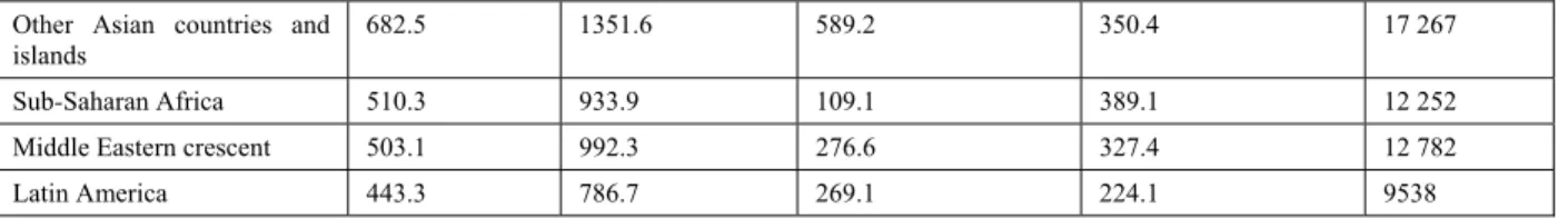
As the first decade of 21st century draws to a close, it is clear thatcardiovascular disease (CVD) is still a ubiquitous cause ofmorbidity and a leading contributor to mortality in the world [1,2]. Itis now widely realized that at present, the developing countries contributea greater share to the global burden of CVD than the developedcountries [3,4]. It is estimated that 5.3 million deathsattributable to CVD occurred in the developed countries in 1990,whereas the corresponding figure for the developing countriesranged between 8 to 9 million (i.e., a relative excess of 70%) [3,4]. Regional estimates of CVD mortality indicate that the difference would be even higher if the term "developed countries" is restrictedto established market economies only and excludes the formersocialist economies (Table 1). Table 1: Regional Differences in Burden of CVD (1990). DALY Indicates Disability-Adjusted Life Year
Region Population, millions CVD Mortality, thousands Coronary Mortality, thousands Cerebrovascular Mortality, thousands DALYs Lost, thousands Developed regions 1144.0 5328.0 2678.0 1447.9 39 118 Developing regions 4123.4 9016.7 2469.0 3181.2 108 802 Established market economies 797.8 3174.7 1561.6 782.0 22 058
Former socialist economies 346.2 2153.3 1116.3 665.9 17 060
India 849.5 2385.9 783.2 619.2 28 592
China 1133.7 2566.2 441.8 1271.1 28 369
*Address correspondence to Bashir M. Matata: Liverpool Heart & Chest Hospital NHS Foundation Trust, Thomas Drive, Liverpool, L14
Table 1: cont....
Other Asian countries and islands
682.5 1351.6 589.2 350.4 17 267
Sub-Saharan Africa 510.3 933.9 109.1 389.1 12 252
Middle Eastern crescent 503.1 992.3 276.6 327.4 12 782
Latin America 443.3 786.7 269.1 224.1 9538
Adopted from Murray and Lopez [3].
This high, yet inadequately recognized, contribution of developing countriesto the absolute burden of CVD is illustrated by the factthat 78% of the 49.9 million global deaths from all causes occurred in regions other than the established marketeconomies or former socialist economies (Table 2).
Table 2: Shows Regional Contributions to Mortality (1990). Values are Given as Percentage of World Total
Region All causes % CVD %
Established market economies 14 22
Former socialist economies 8 15
India 19 17
China 18 18
Other Asian countries and islands 11 9
Sub-Saharan Africa 10 7
Middle Eastern Crescent 9 7
Latin America 6 5
World 100 100
Adopted from Murray and Lopez [3].
During last decade of 20th century, the projected relative contribution of CVD deaths to total mortality was higherin the developed countries (nearly 49%) than that in the developing countries (nearly 23%). Yet the developing countries actually contributed68% to the total global deaths due to non-communicable diseasesand 63% of world mortality due to CVD. This is because the excess total mortality in the developing countries was translatedinto excess absolute CVD mortality due to the large populationsinvolved [3,4]. Moreover, a greater cause for concernis the early age of CVD deaths in the developing countries comparedwith the developed countries e.g. the proportion of CVD deaths occurring below the age of 70 years is 26.5%in the developed countries compared with 46.7% in the developing countries3-4and even largerfor India (52.2%) [3,4].
Therefore,the contribution of the developing countries to the global burdenof CVD, in terms of disability adjusted years of life lost,is 2.8 times higher than that of the developed countries (Table1).
Although there are inadequaciesand imperfections of cause-specific mortality ascertainmentmethods currently used in many developing countries, the conservative assumptionsmade by the analysts suggest that this pattern will become even more pervasive as the CVD epidemic accelerates in many developing regions of the world, even as it retains its primacy as the leading public health problem in the developed regions [5-8]. A considerable cause for alarm is the projected risein both proportional and absolute CVD mortality rates in thedeveloping countries over the next 25 years [6-9]. Reasons forthis anticipated acceleration of the epidemic includes genetic factors as well as demographic factors including lifestyle changes and nutritional transitions.
GENETIC FACTORS
It is increasingly recognized that CVD prevalence is a consequenceof the interaction between the distributionof relative genotype frequencies and environmental exposuresof a particular population [10,11]. It is suggested that distribution of such relative frequencies of genotypes involved in determining the distribution of individual susceptibilities to CVD is dependent on the number of segregatingsusceptibility genes, the number of alleles of each
gene, their relative frequencies, and the correlation between allelesof each gene and alleles of different genes [12-14]. There arehundreds of genes known to have functional allelic variationsthat might contribute to determining an individual’s susceptibilityto CVD and all functional variations in a particular gene are notexpected to be present in all populations [10-14]. Because new DNA variations arise in isolation and their chance, selection,and migration work as "filters" in each population to modifythe relative frequencies of genetic variations in evolutionarytime, different populations will have different combinationsof DNA variations [15,16]. Therefore different combinations of susceptibility genes will be involved in determining CVD risk in different individuals in different families and is always difficult to relate such different combinationsof susceptibility genes to the CVD risk. However, only few genetic studies of common multifactorial diseases recognizethe importance of this question [17,18].
In 2004, the INTERHEART study examined the influence of nine risk factors for CVD [19] and reported that smoking, diabetes, hypertension, obesity, diet, inactivity, no alcohol intake, ApoB: ApoA1 ratio and psychosocial factors accounted for 90-94% of population-attributable risk. Based on this model, the authors [19] suggested that populations with all these risk factors are 337 times more likely to suffer cardiac disease than populations with none. In addition to these risk factors (which may themselves be genetically determined), a positive family history increases coronary artery disease (CAD) and myocardial infarction (MI) risk to 2-3.9 fold [20].
Until recently, such attempts to identify the genetic associations used either candidate gene or linkage studies. The former examines variations in a low number of known, plausibly associated genes in affected cases and controls, and while linkage studies assess affected families/sibling pairs using microsatellite markers to define a genomic region linked to the phenotype. So far, these approaches have been applied with great success to identifying causative mutations in monogenic cardiovascular diseases, such as hypertrophic cardiomyopathy and long QT syndrome [21-24]. However, the complex interplay between environment and genetics demonstrated in INTERHEART made it clear that similar approaches are unlikely to identify the poorly penetrant and multiple causative genes that account for non-Mendelian diseases, such as CVD. Although in rare occasions CVD can also be inherited in a Mendelian fashion (predominantly in conditions leading to elevated LDL), this only accounts for a small proportion of incident cases [25], most of which are likely to be polygenic. Linkage studies of non-Mendelian CVD have provided some biased associations [26-34], conspicuously lacked reproducibility between cohorts, suffered from poor statistical power and lacked detailed genomic mapping provided by conventional microsatellite markers.
Recent technological advancement, coupled with greater understanding of the structure of the human genome derived from genome sequencing projects [35-37], now make unbiased whole-genome association studies (GWAS) possible [38]. The International Haplotype Mapping project [39] identified hundreds of thousands of single nucleotide polymorphisms (SNPs), assessed their degree of linkage disequilibrium (the degree to which a SNP predicts the DNA flanking it). It is reported that genotyping 0.008% of an individual's nucleotides (250 000-350 000 in total) is able to identify an individual genome [40]. This technological advance led the Wellcome Trust Case Control Consortium and others to perform SNP-based GWAS on patients with CAD compared with matched controls [41]. For example the most reproducible locus conferring increased risk of CAD is situated on chromosome 9 (locus 9p21.3) [42-44] and increases risk by approximately 1.2 for a single copy (1.5 in the 25% of the population who carry two copies) [45]. Interestingly, unlike other regions associated with surrogate risk factors for CAD, such as C-reactive protein (CRP) [46], adiposity [47] and left ventricle (LV) mass [48], the 9p21.3 locus does not affect such risk factors, suggesting that it promotes CAD in a non-canonical manner. However, studies are suggesting that SNP-based GWAS knowledge provides no additional benefit [49], despite the availability of genotyping via the internet. First because loci such as 9p21.3 confers effect by altering the regulatory region of DNA [42]; second the involved region that overlaps a non-coding RNA named ANRIL, only conserve in primates and not other mammals or lower organisms and third the associations of some loci (e.g. 9p21.3 locus) are also present in conditions such as dementia [50] and stroke [51], rather than specifically CAD. Therefore, incorporation of risk-conferring alleles such as 9p21.3 and others into a CAD prediction algorithm is still not clear and thus remains to be substantiated in terms of its true importance under the current models and on clinical parameters [52].
DEMOGRAPHIC FACTORS
In the secondhalf of the twentieth century, most developing countries experienceda major surge in life expectancy [53]. For example, the life expectancyin India rose from 41.2 years in 1951-1961 to 61.4 yearsin 1991-1996. This was principally due to a decline indeaths occurring in infancy, childhood, and adolescence.
This was alsorelated to more effective public health responses to perinatal, infectious,and nutritional deficiency disorders and to improved economicindicators such as per-capita income and social indicators such as female literacy in some areas. Although much remains to be done in these areas, the demographicshifts have augmented the ranks of middle-aged and older adults.
The increasing longevity provides longer periods of exposureto the risk factors of CVD resulting in a greater probability of clinically manifest CVD events [54]. The concomitant declineof infectious and nutritional disorders (competing causes of death)further enhances the proportional burden due to CVD and other chroniclifestyle-related diseases. This shift, representing a decline in deaths from infectious diseases and an increase in those dueto chronic diseases, is often referred to as the modern epidemiologicaltransition [8,9]. The ratio of deaths due to pre-transitional diseases (related to infections and malnutrition) to those caused by post-transitional diseases(e.g., CVD and metabolic
disorder) varies among regions and between countries, depending on factors such as the level of economic
developmentand literacy as well as availability and access to health care.
The direction of change towards a rising relative contributionof post-transitional diseases is, however, common to and consistent amongthe developing countries [9]. The experience of urban China, inwhich the proportion of CVD deaths rose from 12.1% in 1957 to35.8% in 1990, is illustrative of this phenomenon [55].
The United Kingdom itself is a diverse society with 7.9% of the population from minority ethnic groups (Africa, Middle-East, Indian Subcontinent, South America and Chinese region) [56]. The causes of the excess CVD and metabolic disorder morbidity and mortality in minority ethnic groups are incompletely understood by socio-economic factors.
However, the role of classical CVD risk factors is clearly important despite the patterns of these risk factors varying significantly by ethnic group. Moreover, the CVD epidemiology of African Americans does not represent well the morbidity and mortality experience seen in black Africans and black Caribbean’s, both in Britain and in their native African countries.
In particular, atherosclerotic disease and coronary heart disease are still relatively rare in the latter groups. This is unlike the South Asian Diaspora which has prevalence rates of CVD in epidemic proportions both in the Diaspora and on the subcontinent [56].
Data for population surveillance of CVD and metabolic disorders are limited in manycountries. The World Health Organization (WHO) has set up a rangeof projects aimed at improving the amount and quality of relevantdata [57]. The Surveillance of Risk Factors (SuRFs) project, launchedin 2003, presents chronic disease risk factor profiles from170 WHO member states. These data include patterns of physical inactivity, low fruit/vegetable intake,obesity, blood pressure, cholesterol, and diabetes [58].
The most recent report SuRF2 enables country comparisons for these data [59]. Fig. (1) shows data on the
percentageof adults in the different countries of SoutheastAsian Nations with body mass index (BMI) >30 kg/m2. The variation is marked and it is interesting to note that two of the poorest countries in the region, Laos and Myanmar, have severe obesity rates comparablewith some of the wealthiest. On the other hand, Singapore, the most developedcountry in the region does not suffer from obesity epidemic.
Although, the biological determinants of CVD and metabolic disorders in low and middle income countries are likelyto be similar to those in affluent countries, [60] the driversof these determinants are likely to differ. For example, rural-urbanmigration may be an important factor in promoting the adoptionof Western dietary habits and activity patterns, leading toan increased CVD risks.
Socioeconomic patterns of diseaserisk, so well established in affluent countries, are more complexin some low and middle income countries [60-62]. New opportunitiesto use large demographic surveillance projects as tools to study CVD and metabolic disorders are emerging rapidly as part on the work of INDEPTH (InternationalNetwork of field sites with continuous Demographic Evaluationof Populations and their Health in developing countries) [63].
Figure 1: Use of WHO web Global InfoBase [58,59]: Obesity (BMI > 30 kg/m2) in the Association of Southeast Asian Nations
in 2002.
Even with such studies of understanding of determinants for rising CVD/ metabolic disorder epidemic and
explaining such differences as to how rural-urban migration increases risksof obesity, diabetes, and CVD- will not be possible. One important caveat to looking at such datais to study the roleof impaired early growth, resulting from fetal and infant nutrition operating at different stages of the life course [64,65], an issue that particularly applies to when defining the causality of this problem.
NUTRITIONAL TRANSITION AND LIFE STYLE CHANGES
Another concern is that if population levelsof CVD risk factors rise as a consequence of adverse lifestylechanges accompanying industrialization and urbanization, therates of CVD mortality and morbidity could rise even higher than the rates predicted solely by demographic changes.
It is suggested that boththe degree and the duration of exposure to CVD risk factorswould increase due to higher risk factor levels coupled witha longer life expectancy. An increase in body weight (adjustedfor height), blood pressure, and cholesterol levels in Chinesepopulation samples aged 35 to 64 years, between the two phasesof the Sino-MONICA study (1984 to 1986 and 1988 to 1989) andthe substantially higher levels of CVD risk factors in urban population groups compared with rural population groups in Indiaprovide evidence of such trends [54,55,66,67]. A cross-sectional survey of urban Delhi and its rural environs revealed that a higher prevalence of CHD in the urban sample was associated with higher levels of body mass index, blood pressure, fasting blood lipids (total cholesterol, ratio of cholesterol to HDL cholesterol, triglycerides), and diabetes.54 The increasing use of tobacco in a number of developing countries will also translate into higher mortality rates of CVD, CHD and other tobacco-related diseases [68,69].
As reviewed by Drewnowski and Popkin [70] the globalavailability of cheap vegetable oils and fats has resulted in greatly increased fat consumption among many countries.
The transition now occurs at lower levels of the gross nationalproduct than previously and is further accelerated by rapidurbanization. For example, the proportion of upper-incomepersons who were consuming a relatively high-fat diet (>30%of daily energy intake) rose from 22.8% to 66.6% between 1989and 1993 in China. The lower- and middle-income classes also showed arise (from 19% to 36.4% in the former and from 19.1% to 51.0%in the latter) [68]. These countries, with a diet that istraditionally high in carbohydrates and low in fat, have shownan overall decline in the proportion of energy from complex carbohydratesalong with the increase in the proportion of fat [71]. The globalizationof food production and marketing is also contributing to theincreasing consumption of energy-dense foods poor in dietaryfibre and several micronutrients [71].
THE COMPLEXITY OF THE PROBLEM
The prior discussion in sections 1.1 -1.4 hence shows that CVD has a complex multifactorial aetiology leading to a reappraisal of the ways in which three key factors- genome, development and environment- influence the adult phenotype, including the individual’s susceptibility to disease. Neither genetic makeup nor exposures to adverse environments predict withcertainty the onset, progression, or severity of CVD. Diseasedevelops as a consequence of interactions between the "initial"conditions, coded in the genotype, and exposures to environmentalagents indexed by time and space [72-74] that are integratedby dynamic, regulatory networks at levels above the genome [75].
The interaction of an individual’s environmental experienceswith her/his genotype determines the history of her/his multidimensionalphenotype, beginning at conception and continuing through adulthood (Fig. 2).
At a particular point in time, each genotype has a range of possible phenotypes determined by the range of possible environmental histories. The phenotype of an individual to react to contemporary environments in a particular environmental niche, at a particular point in time, is influenced by the phenotype produced by previous genotype-phenotype combination. The figure illustrates this relationship, by collapsing an individual’s genotype-phenotype into single dimension, showing two of the many possible phenotype histories for a given genotype. Theconsequence of these interactions with exposures to environmentalagents indexed by time and space is that many individuals whohave a
genotype that predicts an increased risk of CVD will remain healthy because of exposures to compensatory
environments.The converse will also be true: individuals who have a genotypethat has a low risk of CVD might develop disease becauseof an adverse environmental history.
Figure 2: Adopted with Permission from Sing et al., [76].
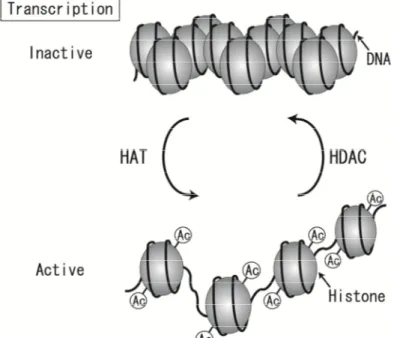
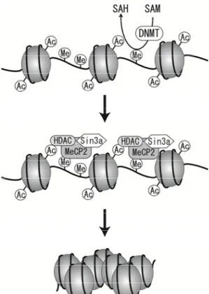
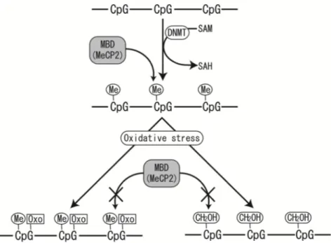
CVD research has revealedtens of high-risk environmental factors and hundreds of genes,each with many variations that influence disease risk. The phenotypic measuresof health are constantly being shaped, changed, and transposed as a consequence of epigenetic networks of cellular and organismaldimensions that change over the lifetime of the individual.At the level of the cell, these networks influence DNA methylationand repair; they also serve to organize coordinated responsesto heat-shock, oxygen deprivation, and other environmental changes [77]. The relationships between these subsystems influence the trajectoryof an individual’s phenotype to influence the expression of the participating genes [78-80] (Fig. 3).
This figure shows how a particular multigene genotype is connected to the domain of potential CVD phenotypes through the primary biochemical and physiological subsystems. The important role that biochemistry and physiology play inthe connections between the genome and disease phenotypes bringsinto question the utility of the overused, simplistic view thatthe genome produces an independent, isolated, and fixed one-wayflow of information from genome to phenotype. Studies have suggested that different ethnic groups that live in the same geographic areas and share similar environmental risks have different profiles of disease markers and prevalence, which may propose a genetic cause for differences in
disease susceptibility [81,82]. Yet, with some notable exceptions [83] few ancestry-specific alleles have been discovered that can explain particular pathologies. Other explanations of both inter-individual and ethnic differences in disease risk, therefore, need to be considered. Of note, high incidences of metabolic disease are found in those ethnic groups in which the average birthweight is low84 or the rates of gestational diabetes and maternal obesity are high [85].
Figure 3: Adopted with Permission from Sing et al., [76]. A Model for an Individual’s Propensity to Develop CVD such as
Coronary Artery Disease
Untangling the effects of genes from those of environmentally determined developmental processes is not straightforward. Importantly, fetal nutrition does not equate to maternal food intake, but rather is dependent on maternal metabolism, cardiovascular function and, particularly, placental function [86]. The long-lasting changes in developmental trajectory that underpin altered susceptibility to disease may arise, at least in part, from epigenetically mediated alterations in gene expression. Whereas compelling evidence supports both the developmental origins of health and disease and the underlying epigenetic mechanisms, [87]many features of the latter remain insufficiently understood. These elements include the differences among epigenetic mechanisms across species and between patterns of epigenetic modifications on paternal and maternal genomes, the mechanisms that regulate the establishment, stability and flexibility of epigenetic changes, and the precise connection between an epigenetic change, altered gene expression and the resultant phenotype for CVD epidemic in countries [72-80]. The hypothesis is currently recognised as "Developmental origins of Health and Disease” (DOHaD) and requires particular understanding before proceeding further with this subject.
DEVELOPMENTAL ORIGINS OF HEALTH AND DISEASE
DOHaD hypothesis states thatadverse intrauterine influences such as poor maternal nutrition leadto impaired fetal growth, resulting in low birth weight, short birthlength, and small head circumference. These adverse influences are postulated to also induce the fetus to develop adaptive metabolicand physiological responses. These responses, however, may lead to disordered reactions to environmentalchallenges as the child grows, with an increased risk of glucoseintolerance, hypertension, and dyslipidaemia in later life andadult CVD as a consequence [88-94]. Although some supportive evidencefor the hypothesis has been provided by observational studies, [95-99] it awaits further evaluation for a causal role. Ifit does emerge as an important risk factor for CVD, the populationsof developing countries will be at an especially enhanced riskbecause the vast numbers of poorly nourished infants whohave been born in the past several decades now suffer a threat through an over-nourished rich environment. The steady
improvementin child survival will lead to a higher proportion of such infantssurviving to adult life, when their hypothesized susceptibilityto vascular disease may manifest itself [100-109].
ORIGINS OF THE HYPOTHESIS- HISTORICAL PERSPECTIVE
The "early or fetal origin of adult disease hypothesis” originally proposed by Barkerand colleagues in Southampton, United Kingdom, suggested that environmental factors, particularly nutrition, act in earlylife to program the risks for the early onset of cardiovascularand metabolic disease in adult life and premature death [88,89, 93,94, 110,111]. Beforethe fetal origins hypothesis was articulated, an associationbetween early life events and later cardiovascular disease hadbeen proposed on more than one occasion. In 1934, Kermack et al. [112] demonstrated that death rates from all causes in theUnited Kingdom and Sweden fell between 1751 and 1930. The authors concluded that this was the result of better childhood livingconditions during this period. Subsequently, Forsdahl [113] reported that there was a correlation within different geographicalregions of Norway between coronary heart disease in 1964-1967and infant mortality rates some 70 years earlier.
Forsdahl [113] postulated that poverty may act through a nutritionaldeficit to result in a life-long vulnerability to disease with a more affluentadult life-style. In 1985, Wadsworth et al. [114] in the UnitedKingdom reported that adult blood pressure was inversely relatedto birth weight in men and women born in 1946. In 1986, Barkerand colleagues suggested that poor health and physique of mothers were important determinantsof the risk of stroke in their offspring [93]. Soon afterwards,they proposed that environmental influences, which impair growthand development in early life, result in an increased risk forischemic heart disease [94]. Thisthen led to a worldwide series of epidemiological studiesthat extended the initial observations on the association between pre- and postnatal growth and cardiovascular disease to include associations between early growth patterns and an increasedrisk for hypertension, impaired glucose tolerance, non-insulin-dependentor type-2 diabetes, insulin resistance, and obesity in adultlife [115-124].
THE THRIFTY PHENOTYPE HYPOTHESIS, DEVELOPMENTAL PLASTICITY AND PREDICTIVE ADAPTIVE RESPONSES
To explain the biological basis of the associationsobserved between early growth patterns and health outcomes in the epidemiological studies, number of mechanistic frameworks such as “thrifty genotype” [125,126] and then "thrifty phenotype"[127] derived from thrifty genotype, were proposed. “Thrifty genes” were proposed to be selected during evolution at a time whenfood resources were scarce and they resulted in a "fastinsulin trigger" and thus an enhanced capacity to store fat,which placed the individual at risk of insulin resistance andtype-2 diabetes [126].
In contrast the thrifty phenotype hypothesis suggested that when the fetal environment is poor, there isan adaptive response, which optimizes the growth of key bodyorgans to the detriment of others and leads to an altered postnatal metabolism, which is designed to enhance postnatal survivalunder conditions of intermittent or poor nutrition [128-129]. It was proposedthat these adaptations only became detrimental when nutritionwas more abundant in the postnatal environment, than it hadbeen in the prenatal environment [127].
Lucas suggests that thatthere are embryonic and fetal adaptive responses to a suboptimalintrauterine environment which result in permanent adverse consequenceseither via the induction, deletion, or impaireddevelopment of a permanent somatic structure or the physiological system [129]. In fact closing the critical window early in development allows the preservation of maternal strategy in offspring phenotype, which in humans benefits the mother by constraining offspring demand after weaning. The offspring gains by being buffered against environmental fluctuations during the most sensitive period of development, allowing coherent adaptation of organ growth to the state of the environment. The critical window is predicted to close when offspring physiology becomes independent of maternal physiology, the timing of which depends on offspring trait [130-133].All this highlight the relationship between intrauterine nutritional experiencesand subsequent health outcomes [134,135]. Researchers working with humans and animal models of human diseases often view the effects of early life events as the developmental plasticity. This embodies the idea that developmental plasticity is the ability of a single genotype to produce more than one alternative form of structure, physiological state, or behaviour in response to environmental conditions
[135-137]. Consistent with this, it is thought that CVD may be a consequence of fetal adaptations to under nutrition that are beneficial for short-term survival, even though they are detrimental to health in post reproductive life [112].
Although some effects of nutrition may be direct consequencesof alterations in substrate availability, McCance and
Widdowson demonstrated that early under nutrition had a permanenteffect on the subsequent growth of rats,
whereas later under nutritiononly had a transient effect [138].
It is clear from a range of diverse fields including evolutionaryecology and molecular biology that a given genotype can giverise to different phenotypes, depending on environmental conditions [139-141].
There are many different species where the impact of an environmentexperienced by one generation determines the development andbehaviour of the next generation. Female birds are able to altermany aspects of the composition of the egg in response to arange of environmental factors including food availability,levels of sibling competition, and the quality of their mates [139]. Such maternal effects can result in the effects of aspecific environmental factor persisting across several generations [135-137, 141].
If the effects of the past conditions produce mismatcheswith current, changed conditions, however, then developmental plasticity may have a detrimental effect on survival and reproductivesuccess [142]. Thus Bateson et al. [141] propose that for individualswhose early environment has predicted a high level of nutritionin adult life and who develop a large phenotype, the betterthe postnatal conditions the better will be their adult health.For individuals whose conditions in fetal life predicted pooradult nutrition and who develop a small phenotype, the expectedoutcomes may vary, although they are predicted to be worse offwhen there is a relative excess of nutrition in postnatal life.
There has also been a proposal to separate those homeostaticresponses that represent fetal adaptations to changes in the intrauterine environment and that may have long-term consequences, from those which need not confer immediate advantage but areinduced in the expectation of future adaptive changes [143]; thislatter group of responses has been defined as "predictive adaptive"[90,92,137,141]. In this model of predictive adaptive response, selection across generations operates to favour protection of those predictive adaptive responses thataid survival to reproductive age.
The programmed or plasticresponses made during development that have immediate adaptiveadvantage might also
act to limit the range of postnatal adaptive responses to a new environment and would be considered to be
"inappropriate" predictive adaptive responses. This generalmodel is therefore consistent with the original thrifty phenotypehypothesis which stated that fetal adaptations to a poor intrauterineenvironment may have adverse consequences if there is a relativeexcess of nutrition available in adult life.
The use of the term “predictive adaptive response” must be clarified because it is used in two very different ways in the literature. In a physiological context it refers to adjustments made by an individual in response to current conditions. For example, in conditions of severe intrauterine deprivation,there is the capacity to lose structural units
such as nephrons, cardiomyocytes, or pancreatic β-cells within developing organ systems. Such decreases in
structural and hence the life-longfunctional capacity of an organ system may be an inadvertentconsequence of a decrease in energy supply across the placentaor a selective trade off to maintain the development of moreimportant tissues, such as the brain [144-146].
In an evolutionary context it refers to changes in the characteristics of populations or species resulting from natural selection, mainly promoting Darwinian fitness and adaptive according to the evolutionary criteria of enhancing survival or reproductive success [147].
In case of fetal origins of disease, this would require that environmental conditions present early in life are predictive of the conditions the individual will encounter in the future over a range of timescales (Fig. 4).
It is suggested that at one extreme, rapid and reversible homoeostatic mechanisms counter an immediate challenge. Then, stressors or exposures during critical developmental periods can affect growth, tissue differentiation, and physiological set-points, affecting responses to environmental challenges for life. New evidence suggests that epigenetic mechanisms could contribute to such challenges [148,149].
Figure 4: Modes of Human Adaptability.
On a long timescale, the genomes of populations can change over many generations as a result of selection or drift, and there are many examples of responses to environmental change becoming integrated into the human genome [141,150-152]. Clinical medicine and public health research have focused largely on causation and intervention at the short-term end of this spectrum. In this context consideration of the outcomes of developmental plasticity acting over the intermediate timescale is now important [141]. In humans, development plasticity can induce responses that have short-term benefits for the mother or the fetus but on longer term costs reduced fitness leading to disease process [152,153]. It is suggested that when environmental conditions change strikingly between conception and adulthood, as has happened in most current human populations, the potential for a substantial mismatch is especially great, and this difference contributes to increased disease risk [135].
ENVIRONMENTAL CUES AFFECTING HUMAN DEVELOPMENT
These broad considerations are relevant to understanding of some critical variations such as developmental adaptations that permanently change structure, physiology, andmetabolism, thereby predisposing individuals to
cardiovascular, metabolic, and endocrine disease in adult life [154,155]. The human baby responds to under
nutrition, placental dysfunction and other adverse influences by changing the trajectory of his or her development and slowing growth. Although the fetus was thought to be well-buffered against fluctuations in its mother's condition, a growing body of evidence suggests that the morphology and physiology of the human baby is affected by the state of the mother [156,157].
It is possible therefore, that human development may involve induction of particular patterns of development by cues that prepare the developing individual for the type of environment in which he or she is likely to live. Individuals may be affected adversely if the environmental prediction provided by the mother and the conditions of early infancy prove to be incorrect [113].
Thus, people whose birth weights were towards the lower end of the normal range and who subsequently grows up in affluent environments are at increased risk of developing coronary heart disease, type-2 diabetes and hypertension [39,40, 156,158]. Those born as heavier babies and brought up in affluent environments enjoy a much reduced risk. The long-term influences may arise from cues acting from before conception to infancy [159].
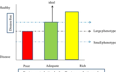
The ill effects of being small, which in the short term include high death rates and childhood illness, are usually treated as yet another inevitable consequence of adversity. However, a functional and evolutionary approach suggests that the pregnant women in poor nutritional condition may signal to her unborn baby that help it to cope with a shortage of food. When sufficiently high levels of nutrition are available after the development of a small phenotype has been initiated, marginal benefits of rapid growth may offset the costs [160], but they may also trigger the health problems arising in later life. This concept is illustrated in Fig. (5).
Large phenotype D is ea se fr ee Disease Adequate Rich Poor ideal Healthy Small phenotype
Although adaptive responses may explain some variation in human development, it would be implausible to argue that all responses to the environment should be explained in these terms. Under nutrition, stress or hypoxia may impair normal development. Babies with low birth weight have a reduced functional capacity and fewer cells [161]. The latter may be part of a general reduction in cell numbers or a selective trade-off in the development of tissues that are less important to the baby, such as the kidney [162]. Reduced numbers of nephrons at birth is a life-long deficit, as all nephrons are formed during a sensitive period of development in late gestation. The resulting increased functional demand on each individual nephron, for example by increased blood flow through each nephron, may lead to acceleration of the nephron’s death that accompanies normal ageing, with a consequent rise in blood pressure [163,164]. The diversity in past and present ecological conditions of humans is also likely to introduce complexity into the relationship between developmental prediction and later health outcome. For example, some populations may have adapted genetically to conditions of nutritional stress, especially seasonal food shortages, over a long time span, while others will have been buffered from such local evolutionary effects. The sharp increase in glucose intolerance leading to type-2 diabetes might arise from genetic differences between populations [165-167]. The possibility of a thrifty genotype well adapted to harsh conditions is not incompatible with the plastic induction of thrifty phenotypes from a pool of uniform genotypes. However, the hypothesis that differences in susceptibility to diabetes are explained by genetic differences would not readily account for the evidence from the Dutch famine of 1944-45 that glucose intolerance is induced by maternal malnutrition during the final three months of pregnancy [167]. However, persisting into adult life, insulin resistance leads to increasing blood glucose and type-2 diabetes develops, especially in people who have become overweight. 'Thrifty' handling of sugar becomes maladaptive if under nutrition in the womb is followed by excess in later life 168.
Figure 5: The Hypothetical Relationship Between Adult Health and Nutritional Level During Later Development for Two
Extreme Human Phenotypes that were Initiated by Cues Received by the Fetus. Reprinted by Permission from Macmillan Publishers Ltd: Bateson et al., Nature, 430: 419-421, 2004 [141].
Conversely, individuals with large bodies may be particularly at risk in harsh environments such as prison camps or during famines [153,168-169]. Especially striking is the evidence from a famine-exposed Ethiopian population, where the incidence of rickets was nine times greater in children who had been reported as having high birth weights than in age-matched control children [170]. No such differences were found in children with normal birth weights.
CONCLUSION
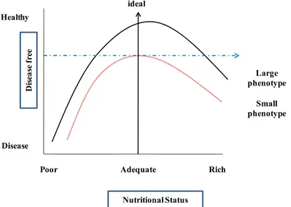
Numerous epidemiological and animal studies discussed so far have shown an association between altered maternal nutrition and cardiovascular or metabolic disease in the offspring. Namely, alterations in fetal nutrition (either under- or over-nutrition) may result during critical periods when offspring are most vulnerableto developmental adaptations that permanently change the structure, physiology and metabolism of the offspring, thereby predisposing individuals to metabolic and cardiovascular diseases in adult life.
Large phenotype D isea se f ree Disease Adequate Rich Poor ideal Healthy Small phenotype Nutritional Status
Today the most common maternal dietary imbalance in populationsis an excessive intake of dietary fat. There is growing body of evidence that significant health problems for women of reproductive age result from being overweight or obese due to overeating. Extensive studieshave shown that maternal over nutrition retards placental andfetal growth, and increases fetal and neonatal mortality inanimal models. Results of epidemiologicalstudies indicate that almost 65% of the adult population inthe U.S. is overweight (defined as a body mass index (BMI) >25 kg/m2), while 31% of the adult population is obese (definedas BMI > 30 kg/m2). Many overweight and obese
womenunknowingly enter pregnancy and continue overeating during gestation. These women usually gain more
weight during the firstpregnancy and accumulate more fat during subsequent pregnancies.Maternal obesity or over-nutrition before or during pregnancymay result in fetal growth restriction and increased risk ofneonatal metabolic syndrome and cardiovascular risk factors.
Previously, studies have demonstratedabnormalities in plasma lipids, vascular fatty acids, and evidence for reduced endothelium-dependent relaxation in adult offspringof rodent models fed on a lard-rich diet during pregnancy, suckling or lactation.However neither the designs of these studies carried out nor the fat intake mimic the typical high-fat Western diet and human situation. Moreover, to date no one has determined the role of early pharmacological intervention in mothers and its effects on offspring in terms of cardiovascular control using the animal model.
REFERENCES
[1] Nissen SE. Cardiovascular outcomes in randomized trials: should time to first event for "hard" end points remain the standard approach? J Am Coll Cardiol 2009; 54(25): 2363-65.
[2] Tai ES, Poulton R, Thumboo J, et al. An update on cardiovascular disease epidemiology in South East Asia. Rationale and design of the LIFE course study in CARdiovascular disease Epidemiology (LIFECARE). CVD Prevent Control 2009; 4(2): 93-102. [3] Murray CJL, Lopez AD. Global comparative assessments in the health sector. Geneva, Switzerland: World Health Organization
1994.
[4] Lopez AD. Assessing the burden of mortality from cardiovascular disease. World Health Stat Q 1993; 46: 91-6.
[5] Reddy KS, Yusuf S. Emerging epidemic of cardiovascular disease in developing countries. Circulation 1998; 97 (6): 596-601. [6] Thom TJ, Epstein FH, Feldman JJ, et al. Total Mortality and Mortality From Heart Disease, Cancer, and Stroke From 1950 to
1987 in 27 Countries: Highlights of Trends and Their Interrelationships Among Causes of Death. Washington, DC: US DHHS PHS, National Institutes of Health; NIH publication No. 1992; 92-3088.
[7] Whelton PK, Brancati FL, Appel LJ, et al. The challenge of hypertension and atherosclerotic cardiovascular disease in economically developing countries. High Blood Pressure Cardiovasc Prevent 1995; 4: 36-45.
[8] Pearson TA, Jamison DT, Tergo-Gauderies J. Cardiovascular disease. In: Jamison DT, Mosley WH, Eds. Disease Control Priorities in Developing Countries. New York, NY: Oxford University Press 1993.
[9] Omran AR. The epidemiologic transition: a key of the epidemiology of population change. Milibank Memorial Fund Q. 1971; 49: 509-38.
[10] Scriver CR, Byck S, Prevost L, Hoang L. PAH mutation analysis consortium. In: Chadwick DJ, Cardew G, Eds. Variation in the Human Genome (Ciba Foundation Symposium 197). Chichester, UK: John Wiley and Sons 1996: pp. 73-96.
[11] Weatherall D. The genetics of common diseases: the implications of population variability In: Chadwick DJ, Cardew G, Eds. Variation in the Human Genome (Ciba Foundation Symposium 197). Chichester, UK: John Wiley and Sons 1996: pp. 300-11. [12] Clark AG, Weiss KM, Nickerson DA, et al. Haplotype structure and population genetic inferences from nucleotide sequence
variation in human lipoprotein lipase. Am J Hum Genet 1998; 63 (2): 595-612.
[13] Fullerton SM, Clark AG, Weiss KM, et al. Apolipoprotein E variation at the sequence haplotype level: implications for the origin and maintenance of a major human polymorphism. Am J Hum Genet 2000; 67 (4): 881-900.
[14] Stengård JH, Clark AG, Weiss KM, Kardia S, et al. Contributions of 18 additional DNA sequence variations in the gene for apolipoprotein E to explaining variation in quantitative measures of lipid metabolism. Am J Hum Genet 2002; 71(3): 501-17. [15] Gluckman PD, Hanson MA. Living with the past: evolution, development, and patterns of disease. Science 2004; 305 (5691):
1733-36.
[16] Charles F. Sing, Jari H. Stengård, and Sharon L.R. Kardia. Genes, environment, and cardiovascular disease. Arterioscler Thromb Vasc Biol 2003; 23 (7): 1190- 96.
[17] Elahi MM, Asotra K, Matata BM, et al. Tumor necrosis factor alpha -308 gene locus promoter polymorphism: an analysis of association with health and disease. Biochim Biophys Acta 2009 (3);1792: 163-72
[18] Elahi MM, Gilmour A, Matata BM, et al. A variant of position -308 of the Tumour necrosis factor alpha gene promoter and the risk of coronary heart disease. Heart Lung Circ 2008; 17 (1): 14-8.
![Figure 1: Use of WHO web Global InfoBase [58,59]: Obesity (BMI > 30 kg/m 2 ) in the Association of Southeast Asian Nations](https://thumb-eu.123doks.com/thumbv2/123dokorg/4946154.52357/15.918.275.644.106.360/figure-global-infobase-obesity-association-southeast-asian-nations.webp)
![Figure 2: Adopted with Permission from Sing et al., [76].](https://thumb-eu.123doks.com/thumbv2/123dokorg/4946154.52357/16.918.270.656.507.765/figure-adopted-permission-sing-et-al.webp)
![Figure 3: Adopted with Permission from Sing et al., [76]. A Model for an Individual’s Propensity to Develop CVD such as](https://thumb-eu.123doks.com/thumbv2/123dokorg/4946154.52357/17.918.223.699.202.585/figure-adopted-permission-sing-model-individual-propensity-develop.webp)