ALK1 loss results in vascular hyperplasia in mice and humans
through PI3-kinase activation
Elisenda Alsina-Sanchís1,2,∆, Yaiza García-Ibáñez1,2,∆, Ana M. Figueiredo1,3, Carla Riera-Domingo1,2, Agnès Figueras1,2, Xavier Matias-Guiu2,4,5,6, Oriol Casanovas1,2, Luisa M. Botella7, Miquel A. Pujana1,2, Antoni Riera-Mestre8,9, Mariona Graupera1,3,10, 12* & Francesc Viñals1,2,11,12*
1 Program Against Cancer Therapeutic Resistance (ProCURE), Institut Català d’Oncologia (ICO), Hospital Duran i Reynals, Gran Via 199-203, 08907 L’Hospitalet de Llobregat (Barcelona), Spain; 2 Institut d’Investigació Biomèdica de Bellvitge (IDIBELL); 3 Vascular Signaling Laboratory, Institut d´Investigació Biomèdica de Bellvitge (IDIBELL), Gran Via 199-203, 08908 L’Hospitalet de Llobregat, Spain; 4 Servei d’Anatomia Patològica, Hospital Universitari de Bellvitge-IDIBELL; 5 Hospital Universitari Arnau de Vilanova, Lleida, Spain; 6 Universitat de Lleida; 7 Centro de Investigaciones Biológicas, Consejo Superior de Investigaciones Científicas (CSIC), Madrid, Spain; 8 HHT Unit, Internal Medicine Department, Hospital Universitari de Bellvitge-IDIBELL; 9 Departament de Ciències Clíniques, Facultat de
Fisiològiques, Campus de Bellvitge, Universitat de Barcelona, Avda. Feixa Llarga s/n, 08907 L’Hospitalet de Llobregat (Barcelona), Spain.
∆ Equal contribution
12 Joint last authors
Running title: PI3K inhibitors to treat HHT2 patients
* To whom correspondence should be addressed.
Mailing address: Dr. Francesc Viñals - Program Against Cancer Therapeutic Resistance (ProCURE), Institut Català d’Oncologia – IDIBELL, Hospital Duran i Reynals, Gran Via 199-203, 08908 L’Hospitalet de Llobregat, Barcelona, Spain.
E-mail: [email protected]; Dr. Mariona Graupera: Vascular Signaling
Laboratory, IDIBELL, Hospital Duran i Reynals, Gran Via 199-203, 08908
L’Hospitalet de Llobregat, Barcelona, Spain. E-mail: [email protected].
Keywords: PI3K, HHT, ALK1, BMP9, PTEN
Subject codes: Angiogenesis, Cell Signaling/Signal Transduction, Vascular Biology,
Vascular Disease
Figures: 6
Tables: 1
TOC category: Translational
ABSTRACT
Objective: Activin-receptor like kinase 1 (ALK1) is an endothelial cell-restricted
receptor with high affinity for bone morphogenetic protein 9 (BMP9) Transforming growth factor β (TGFβ) family member. Loss of function mutations in ALK1 cause a subtype of Hereditary Hemorrhagic Telangiectasia (HHT), a rare disease characterized by vasculature malformations. Therapeutic strategies are aimed at reducing potential complications due to vascular malformations, but there is no currently a curative treatment for HHT.
Approach and Results: In this work, we report that a reduction in ALK1 gene dosage
(heterozygous ALK1+/- mice) results in enhanced retinal endothelial cell proliferation
and vascular hyperplasia at the sprouting front. We found that BMP9/ALK1 represses VEGF-mediated phosphatidylinositol 3-kinase (PI3K) by promoting the activity of the phosphatase and tensin homolog (PTEN). Consequently loss of ALK1 function in endothelial cells results in increased activity of the PI3K pathway. These results were confirmed in cutaneous telangiectasia biopsies of HHT2 patients, in which we also detected an increase in endothelial cell proliferation linked to an increase on the PI3K pathway. In mice, genetic and pharmacological inhibition of PI3K is sufficient to
abolish the vascular hyperplasia of ALK1+/- retinas and in turn normalize the
vasculature.
Conclusions: Overall, our results indicate that the BMP9/ALK1 hub critically
mediates vascular quiescence by limiting PI3K signaling and suggest that PI3K inhibitors could be used as novel therapeutic agents to treat HHT.
Non-standard Abbreviations and Acronyms
ALK1: Activin-receptor like kinase 1 BMP: bone morphogenetic proteins
ERK: extracellular signal-regulated kinases FGF: fibroblast growth factor
HHT: Hereditary Hemorrhagic Telangiectasia PI3K: phosphatidylinositol 3-kinase
PTEN: phosphatase and tensin homolog TGFβ:Transforming growth factor β
INTRODUCTION
Blood vessels play essential roles in the transport of gases, nutrients, waste products and circulating cells in the healthy organism. While vessels are quiescent in adults, the growth of blood vessels is critical to development, growth and regeneration
1, 2. Angiogenesis, the formation of new blood vessels, consists of sprouting new
vessels from pre-existing ones and the eventual fusion of these with other sprouts or blood vessels to form new vascular connections. Vessel growth is stimulated by angiogenic factors, which are divided into: (1) activators, such as vascular endothelial growth factor-A (VEGF-A, hereafter referred to as VEGF), fibroblast growth factor 2 (FGF-2), and epidermal growth factor (EGF), which induce endothelial cell
proliferation and migration 1, 3; and (2) maturation factors, such as tumor necrosis
factor-α (TNF-α), interleukin-8 (IL-8) and some members of the transforming growth factor (TGF) β family, which prompt endothelial cells to cease proliferation,
reestablish basal membrane, and recruit mural cells 4, 5. Bone morphogenetic protein 9
(BMP9) is a member of the TGF-β family that selectively activates activin-receptor like kinase (ALK) 1, a serine-threonine kinase type I TGF-β receptor in endothelial
cells 6. Activation of ALK1 triggers the phosphorylation of Smad 1, 5 and 8, which in
turn form an active complex with Smad4, translocate to the nucleus, and stimulate the expression of genes such as inhibitors of differentiation (IDs), endoglin and Tmem100
7-9. BMP9 has been proposed as limiting VEGF and FGF-induced endothelial cell
proliferation 6, 10, 11. However, it is still unclear how ALK1 negatively regulates
proangiogenic cascades. Given the inhibitory role of ALK1 during vessel growth, it is not surprising that genetic and pharmacological blockage of ALK1 in vivo results in
Hereditary hemorrhagic telangiectasia (HHT), also known as Rendu-Osler-Weber syndrome, is a rare autosomal-dominant germline disease, with an incidence of
1:5,000, characterized by the local overgrowth of the vascular plexus 14. The
telangiectasia is the common name for vascular lesions in HHT patients 15, and
consists of an artery directly connected to a vein that generates a fragile site that can easily rupture and bleed. The telangiectasias are found on the skin of the face and hands, and the lining of the nose and mouth. Less common, but more severe, are the internal vascular lesions, which principally occur in lung, liver and the digestive tract, and may lead to hemorrhagic episodes, stroke or brain abscesses secondary to pulmonary arteriovenous malformations (AVMs); or arterial aneurysms and pulmonary arterial hypertension, or high-output heart failure secondary to liver
vascular malformations 16. HHT is divided into HHT types 1 and 2 on the basis of the
mutation responsible for the pathogenesis of the disease. While HHT1 arises from
inactivation mutations in ENG, the gene encoding the TGF-β co-receptor endoglin 17,
HHT2 is caused by mutations in ACVRL1, the gene encoding ALK1 18. Pulmonary and
cerebral arteriovenous malformations are more common in HHT1, while hepatic vascular malformations predominate in HHT2. Therapeutic strategies are aimed at reducing potential complications due to vascular malformations, but there is currently no curative treatment for HHT.
In this study we used heterozygous ALK1 mouse retinas and cultured endothelial cells to study how ALK1 represses vessel growth. We found that loss of ALK1 leads to increased stalk cell proliferation as a result of overactivation of PI3K signaling. By analyzing a small cohort of patients, we found that mutations in ALK1 result in increased activation of PI3K signaling in human telangiectasias compared with control vessels. These findings, together with the observation that blocking the
PI3K signaling pathway rescues ALK1-induced vascular hyperplasia, provide the proof of concept for therapeutic intervention with PI3K inhibitors for the treatment of HHT.
MATERIALS AND METHODS
Materials and Methods are available in the online-only Data Supplement.
RESULTS
Heterozygous loss of ALK1 results in retinal vascular hyperplasia
To gain insight into the molecular mechanism accounting for vascular malformations upon loss of ALK1 function in HHTs patients, we investigated vessel
growth in postnatal mouse retinas. ALK1-/- mice die in midgestation 12, 13, so we
studied heterozygous ALK1+/- retinas. Whole-mount isolectin B4 (IB4)-stained ALK1
+/-retinas showed no differences in vascular radial expansion or the number of branch points compared with wild-type littermates’ retinas at postnatal days 5 (P5), P7 and P9 (Fig. 1 and Suppl. Fig. 1). In contrast, a partial decrease in ALK1 levels resulted in vessel hyperplasia, as indicated by increased vessel width (Fig. 1). Vessel hyperplasia
in ALK1+/- retinas occurred in capillary areas located above veins at P5, P7 and P9,
whereas the effect was only observed in capillary areas located above arteries at P9 (Fig. 1). Together, these findings indicate that ALK1 signaling limits vessel width in sprouting angiogenesis but it is not necessary for EC migration and til/stalk election.
the phenotype of full inactivation of ALK1 in endothelial cells 19, demonstrating a dose-response effect of ALK1 in endothelial cells.
Coverage by mural cells regulates vessel diameter. Hence, the increase in vessel width induced by ALK1 inhibition could be a consequence of reduced coverage
by mural cells 20, 21. However, immunostaining with desmin, a marker for retinal
pericytes, did not reveal any obvious difference between wild-type and ALK1+/- retinas
(Suppl. Fig. 2). Next, we examined whether an increase in endothelial cell number could account for the increase in vessel width upon heterozygous loss of ALK1. To
this end, wild-type and ALK1+/- retinas were stained with Erg-1/2/3, an
endothelial-specific nuclear marker. Surprisingly, no differences were observed in the frequency of endothelial cells located in the inner retinal zone or in the first migratory endothelial
cell line (mainly tip cells) 22 (Fig. 2A and B). Conversely, an accumulation of 25%
more endothelial cells were observed in the sub-front (mainly formed of stalk cells) of
ALK1+/- retinas compared with wild-type retinas. To establish whether this greater
number was caused by an increase in endothelial cell proliferation, retinas were co-stained with Erg-1/2/3 and Edu (a proliferation marker) (Fig. 2A and C). Consistently, no differences were observed in the number of proliferative endothelial cells located in the internal vascular retinal area and in the first migratory endothelial line. Instead, a 41% higher frequency of proliferative endothelial cells was observed in the sub-front
of ALK1+/- retinas, indicating that ALK1 signaling restricts endothelial cell
BMP9 blocks VEGF-induced proliferation in HUVECs
To understand the mechanisms by which ALK1 prevents cell cycle progression in endothelial cells, we used cultured human umbilical vein endothelial cells (HUVECs) and studied the crosstalk between BMP9 and angiogenic signals such as VEGF. First, we stimulated HUVECs with VEGF, BMP9 or a combination of both, for 48 h or 72 h, followed by assessment of the total number of cells. While BMP9 alone did not change the growth of HUVECs, VEGF promoted a 37% increase in cell number after 48 h and a 326% after 72 h (Fig. 2D). However, VEGF failed to stimulate cell number in the presence of BMP9. To confirm that BMP9 blocks cell proliferation without stimulating cell death, we measured BrdU incorporation in quiescent HUVECs in the presence of BMP9 and VEGF. As expected, stimulation with VEGF led to a greater degree of BrdU incorporation in HUVECs (Fig. 2E) compared with non-stimulated conditions. We also found that BrdU incorporation was reduced by BMP9 stimulation in basal and VEGF-stimulated cells. BMP9 addition also reduced cell proliferation in complete medium (20% FCS). In agreement with the negative BMP9-ALK1 role, addition of LDN-212854, an ALK1 inhibitor increased BrdU incorporation in HUVEC alone and reverted the inhibition caused by BMP9 (Fig. 2E).
BMP9 dampens VEGF-induced activation of AKT and ERKs
Next, we analyzed the effect of 15 min or 4 h BMP9 pretreatment in VEGF-mediated activation of PI3K-AKT, ERK1/2 and p38. We used phosphoSMAD1/5 and total abundance of ID1 as readout of BMP9/ALK1 signaling (Fig. 3, Suppl. Fig. 3 and
Suppl. Fig. 4). Stimulation with VEGF for 10 and 30 min triggered the phosphorylation of AKT and ERK1/2. VEGF and BMP9 both stimulated p38 MAPK phosphorylation, with a synergistic effect upon incubation when both were used (Fig. 3 and Suppl. Fig. 4). Pretreatment with BMP9 for 15 min before VEGF incubation had no effect on AKT and ERK1/2 activation (Suppl. Fig. 4A). Instead, a 4-h preincubation with BMP9 blocked basal and VEGF-mediated phosphorylation of AKT (in Threonine 308 and Serine 473) and ERK1/2, without affecting total amount of AKT or ERK1/2 protein or phosphorylation of VEGFR2 (Fig. 3). This effect was dose-dependent, with a maximum effect achieved at a dose of 0.5 ng/ml of BMP9 in the absence of VEGF and of 5 ng/ml in the presence of VEGF (Suppl. Fig. 4C-E). Moreover, a minimum of 2 h time of preincubation with BMP9 was necessary to overrule the VEGF mediated activation of AKT and ERKs (Suppl. Fig. 4). We then sought to determine whether preincubation with VEGF also impaired the BMP9 canonical signaling pathway. However BMP9-induced phosphoSMAD1/5 and ID1 expression were not altered if the cells had been preincubated with VEGF for 1 h before adding BMP9 (Suppl. Fig. 5). Taken together, our data findings indicate that BMP9/ALK1 fine-tunes VEGF signaling, but this pro-angiogenic cue has no effect in modulating BMP9 signaling. Our results identify BMP9 as a negative modulator of proangiogenic signals in endothelial cells.
BMP9 inhibits PI3K signaling by increasing PTEN activity
The PI3K/PTEN axis 22, 23, but not ERK1/2 signaling 24, has been shown to
regulate endothelial cell proliferation in mouse retina angiogenesis. Furthermore, loss of PTEN in endothelial cells results in vascular hyperplasia similar to that observed in
ALK1+/- retinas 22. Therefore, we hypothesized that BMP9 fine-tunes PI3K signaling by regulating PTEN. To confirm this we first measured PTEN phosphatase activity. PTEN was immunoprecipitated from endothelial cells treated or not with BMP9 for 4 h and additional 30 min with VEGF, and PTEN activity was quantified by its capacity
to dephosphorylate PI(3,4,5)P3 to PI(4,5)P2. BMP9 treatment promoted a 48% increase
in PTEN phosphatase activity, and this effect was independent of the presence or not of VEGF (Fig. 4A). Next we measured protein abundance after BMP9 treatment. Our results showed that PTEN amount were maintained by a 4-h treatment with BMP9 but increased substantially (86%) when cells were treated with BMP9 for 24 h (Fig. 4B and Suppl. Fig. 6A). This effect was correlated with an increase in PTEN mRNA abundance induced by BMP9 (44% after 4 h and 184% after 24 h of BMP9 treatment) (Fig. 4C). To confirm that PTEN was important for the inhibitory effect of BMP9 on AKT, we blocked its expression in HUVECs by siRNA transfection. The siRNAs acting against PTEN reduced PTEN protein amount and led to hyperactivation of the PI3K/AKT pathway (Fig. 4D and Suppl. Fig. 6B). More importantly, PTEN depletion abolished the inhibitory effects of BMP9 on AKT phosphorylation in basal and VEGF-stimulated cells, confirming that PTEN plays a critical role in BMP9-induced repression of PI3K/AKT. Next we evaluated the effect of PTEN depletion on cell cycle progression in endothelial cells by assessment of the total number of cells. While VEGF failed to stimulate cell number in the presence of BMP9 in non silencing control cells, in the absence of PTEN BMP9 was not able to block VEGF-induced cell proliferation (Fig. 4E). We confirmed this result by incubating HUVEC with a PTEN inhibitor, SF1670. This compound also blocked the inhibitory effect of BMP9 on VEGF-induced endothelial cell number (Fig. 4F),further supporting that PTEN activity is necessary to mediate BMP9 effects.
Activation of the PI3K pathway in cutaneous telangiectasia biopsies of HHT2 patients
Having identified the mechanism of action of negative regulation by BMP9 signaling on pro-angiogenic signals, next we aimed to study the status of the PI3K pathway in samples from HHT2-affected individuals. To this end, we first examined publicly available gene expression data from nasal tissue of HHT2-affected and
control individuals (21 and 19, respectively 25). Using a rank-based algorithm 26, a
significant bias towards over-expression of genes linked to PI3K/AKT signaling (measured with three gene sets) was observed in telangiectasial tissue relative to normal tissue of HHT2 patients (P values < 0.01, Fig. 5A and Supplementary Table 1). Significant over-expression of these sets was also observed when comparing data of telangiectasial HHT2 tissue against normal tissue obtained from healthy individuals,
but no when comparing normal HHT2 against normal tissue of healthy individuals 25.
Next, we analyzed cutaneous telangiectasia biopsies from six HHT2 patients with mutations in the ACVRL1 gene (Table 1) and from three controls. We performed immunohistochemistry for CD34 (an endothelial cell marker), pAKT (S473),
pNDRG1 (Thr 346) and pS6 (S240/244) (markers of PI3K activation) 27, 28. As shown
in Fig. 5B and C we found a 53% increase of vessels that were positive for pAKT. Moreover, a low frequency of control vessels were positive for pNDRG1 (3%) (Fig. 5B and D). In contrast, in the telangiectasia cohort we found a five-fold greater frequency of vessels that were positive for pNDRG1. Similarly, while endothelial cells from control biopsies were negative for pS6 staining, we found a seven-fold greater
frequency of endothelial cells that were positive for pS6 (Fig. 5B and E). Interestingly, we found a statistical significant positive correlation between grade of positivity for pNDRG1 and nosebleed severity measured by the Epistaxis Severity Score (Table 1 and Suppl. Fig. 7).
Patients with HHT present enlarged abnormal vessels with high number of endothelial cells (Fig. 5B). We asked whether mutations in ALK1 also increased endothelial cell proliferation in vessels from HHT2 patients, as observed in our mouse model. We performed immunohistochemistry for Ki67, a proliferation marker, in our HHT2 patient’s group and compare it with controls. Results indicated that the mean proliferation index was 3.3x significantly higher in endothelial cells from telangiectasias of HHT2 patients than in control vessels (2.3 versus 0.7, respectively) (Fig. 5B and F).
Inhibition of PI3K prevents vascular hyperplasia induced by loss of ALK1 expression
Finally, we examined whether inhibition of PI3K signaling can revert the vascular hyperplasia phenotype induced by heterozygous loss of ALK1 in vivo. To this
end, we crossed ALK1+/- heterozygous animals with a constitutive mouse line in which
endogenous p110α/PI3K isoform is converted into kinase-dead protein (hereafter
p110αKD/WT)29. While homozygous p110αKD/KD mice die during embryonic
development as a consequence of vascular failure, heterozygous inactivation of p110α
results in mild reduction of radial expansion 30, an effect that was maintained in
reduced p110α activity rescued the hyperplasia induced by heterozygous loss of ALK1 (Fig. 6A and B). We validated these results by pharmacological inhibition of PI3K signaling with LY294002, a pan-PI3K inhibitor. Pups were treated with LY294002 at P6 and P7 (Fig. 6C), and their retinal vasculature was examined at P7. The hyperplasia induced by ALK1 heterozygosity was also rescued by pharmacological inhibition of PI3K activity (Fig. 6D and E). Taken together, these results confirm that BMP9/ALK1 regulates endothelial cell proliferation in vivo, at least partially, by inhibiting PI3K/AKT signaling.
DISCUSSION
In the present study we demonstrate that BMP9/ALK1 promotes vascular quiescence by inhibiting PI3K/AKT and ERK MAPK activation, and show that loss of ALK1 causes overstimulation of these signaling hubs. In recent years, the role of the BMP9-BMPRII-ALK1-endoglin-SMAD1/5 axis in endothelial cells has begun to be
clarified 31, 32. Indeed, BMP9/ALK1 inhibits cell proliferation and migration in
cultured endothelial cells 10, 33, 34 and in zebrafish 35. In mice, the blockade of
BMP9/ALK1 signaling by anti-BMP9 neutralizing antibody or by injection of the
extracellular domain of ALK1 increases vascular density 36. Also in mice, depletion of
ALK1 specifically in endothelial cells results in arteriovenous malformations as a
result of increased endothelial cell proliferation 19, 37-39. Our results in vitro and in vivo
confirm this anti-proliferative role of BMP9/ALK1 in sprouting angiogenesis. Our work also reveals that during vessel growth BMP9/ALK1 signaling does not regulate cell proliferation in all angiogenic endothelial cells in vivo but, rather, specifically in those cells located at the sprouting. Activation of Notch at the sprouting front results
in stalk cell cycle arrest 22 through a similar mechanism to that of BMP9/ALK1 (see below). It is therefore reasonable to speculate that Notch and BMP9/ALK1 cooperate to restrain stalk cell proliferation at this specific location. In keeping with this,
previous results have shown that ALK1, through SMAD1/5 40, 41 or SMAD2/3
activation 42, cooperates with the Dll4/Notch pathway to induce the stalk cell
phenotype.
Our in vitro and in vivo findings place PI3K/AKT and ERK MAPK at the center of the BMP9/ALK1 anti-proliferative response (Suppl. Fig. 9). Our results using HUVECs as a model have shown that BMP9/ALK1 blocks the PI3K/AKT and ERK activation induced by VEGF. In contrast, we have found that pharmacological inhibition of PI3K is sufficient to abolish ALK1-induced vascular hyperplasia in vivo. This observation, together with the fact that inhibitors of MEK1-ERKs in vivo fail to
block endothelial cell proliferation in wild-type retinal angiogenesis 24, suggest that
limiting PI3K signaling is the principal mechanism by which ALK1 stimulates vascular quiescence in vivo. The PI3K/AKT pathway stimulates cell proliferation,
migration and survival in endothelial cells downstream of many angiogenic cues 22, 43.
We have identified BMP1/ALK1 as a new upstream signal that regulates PI3K activity in sprouting angiogenesis. While most of the angiogenic cues activate the PI3K signaling pathway to execute their biological actions, we have found that BMP9/ALK1 represses this signaling cascade by stimulating the activity of PTEN, the
principal phosphatase that counteracts this signaling pathway in endothelial cells 43.
PTEN is a lipid and protein phosphatase whose function is regulated at multiple levels, including mRNA and protein expression, subcellular localization, and direct regulation of its phosphatase activity by post-translational modifications such as phosphorylation,
PTEN by BMP9/ALK1 in endothelial cells is also multifactorial. First, the observation that BMP9/ALK1 dampens AKT phosphorylation after a 2-h incubation with BMP9 suggests a direct regulation of PTEN catalytic activity. Recently, Ola et al. have
described that the phosphorylated Ser380/Thr382/Thr383-inactive form of PTEN 44,
was decreased in HUVECs and mLECs endothelial cells stimulated for 2 hours with
BMP9 39. Second, we have clear evidence that stimulation of endothelial cells by
BMP9/ALK1 increases mRNA and protein abundance of PTEN. This is not unique to BMP9/ALK1, as we have previously shown that Dll4/Notch also stimulates PTEN
expression to block stalk cell proliferation 22. Given the previously identified crosstalk
between ALK1 and Notch, the increase in PTEN amount upon BMP9 stimulation could be explained by an indirect response induced by Notch. However, we cannot rule out a direct effect of SMADs on PTEN promoter upon BMP9 stimulation. PTEN expression is also tightly regulated by miR, including in endothelial cells. Indeed, VEGF restrains PTEN expression amount by increasing the expression of the
miR-17-92 cluster in endothelial cells 46. Therefore, it is also possible that BMP9/ALK1
regulates PTEN abundance by inhibiting negative regulators of its expression. Taken together, we have identified a previously unknown interaction of BMP9/ALK1 and PTEN in endothelial cells, which seems critical to the pathogenesis of loss of ALK1 expression.
HHT is mainly caused by heterozygous mutations in endoglin (ENG) 17 or
ALK1 (ACVRL1) 18 genes. These are loss of function mutations that lead to reduced
BMP9/ALK1/endoglin/SMAD1/5 signaling and enhanced response to angiogenic
cues, and thereby to excessive abnormal angiogenesis 14. The telangiectasia arises
from a postcapillary hyperplastic venule that fuses directly with an arteriole, resulting
proliferation in the development of these enlarged abnormal vessels in HHT has not been studied in deep. Hashimoto et al. found higher Ki-67 index for endothelial cells
of sporadic brain AVMs compared with endothelial cells from control cortical vessels
47. In the case of HHT, Du et al. found increased endothelial cell proliferation in a
resection of intracranial AVM in a 26-day-old boy with HHT 48. Our data obtained in
patients, as well as the results obtained in mouse models, confirm that AVMs and increased endothelial cell proliferation are interrelated, suggesting that the AVM phenotype in HHT patients arises, at least in part, from an aberrant endothelial cells proliferation. In line with this stimulation of VEGF signaling, a promitogenic factor for endothelial cells, is required for the development of HHT. Indeed, although heterozygous ALK1 mice do not develop AVM, intracranial injection of VEGF in
ALK1+/- mice leads to abnormal AVM-like structures 49, and blocking VEGF
attenuates these vascular lesions 50. This is in agreement with the observation that
PI3K/AKT is a key signaling hub downstream of VEGF in angiogenesis 43. Our results
show for first time an overstimulation of the PI3K pathway on vessels of HHT2 patients. We found a statistically significant relationship between PI3K-activated vessels in the finger telangiectasia biopsy and the severity of nosebleeds measured by the Epistaxis Severity Score. This association suggests a more intense development of systemic telangiectasias (i.e., cutaneous or in nasal mucosa) when PI3K signaling is active. It is of particular note that we and others have found that the majority of venous malformations, the most common form of vascular malformation, are driven
by constitutively activating PI3K signaling 23, 51. Taken together, the results suggest
that overactivation of PI3K signaling is a common characteristic in vascular malformations. By using a mouse model for HHT, our findings also show that the genetic or pharmacological inhibition of PI3K prevents ALK1-induced vascular
hyperplasia in retinas. Our findings corroborate results by Ola et al. 39 in which homozygous deletion of ALK1 in endothelial cells results in arterial venous shuts also by up regulating PI3K signaling. These results suggest that PI3K inhibitors could be used as an alternative therapeutic approach to treating HHT patients. Inhibition of the PI3K signaling in the vasculature could therefore prevent the development of telangiectasias and improve nose bleeds in HHT patients. In keeping with this observation, a study has reported a clinical case of an HHT2 patient diagnosed with serous ovarian cancer who showed a drop in the frequency of epistaxis upon treatment
with a PI3K inhibitor (BKM120) 52. Therefore, we propose that the use of PI3K or
AKT inhibitors should be considered as alternative pharmacological strategies for treating HHT patients.
ACKNOLEDGEMENTS
Acknoledgements: We thank K. Pietras (Lund University, Sweden) and S. Paul Oh
(University of Florida, USA) for the ALK1 heterozygous animals. We are grateful to the subjects who participated in this study.
Sources of funding: This study was supported by research grants from the Spanish
Ministerio de Economía y Competitividad (SAF2013-46063-R and SAF2017-85869-R) and the Generalitat de Catalunya (2014SGR364) to F.V. E.A. is a recipient of a predoctoral fellowship from the Ministerio de Economía y Competitividad. Work in M.G.’s laboratory is supported by research grants SAF2014-59950-P and SAF2017-89116-R from the Spanish Ministerio de Economía y Competitividad (Spain), 2014-SGR-725 from the Catalan Government, the People Programme (Marie Curie Actions; grant agreement 317250) of the European Union's Seventh Framework Programme
FP7/2007-2013/, the Marie Skłodowska-Curie (grant agreement 675392) of the European Union's Horizon 2020 research and innovation programme, the project CB16/12/00445 (CIBERONCO), and from la Fundació Bancària “La Caixa”.
Disclosures: Unrestricted grant from Roche to finance the ProCure Programme, which
was paid to the Catalan Institute of Oncology (2015).
REFERENCES
1. Carmeliet P. Mechanisms of angiogenesis and arteriogenesis. Nat Med.
2000;6:389-395
2. Adams RH, Alitalo K. Molecular regulation of angiogenesis and
lymphangiogenesis. Nat Rev Mol Cell Biol. 2007;8:464-478
3. Carmeliet P, Jain RK. Molecular mechanisms and clinical applications of
angiogenesis. Nature. 2011;473:298-307
4. Jain RK. Molecular regulation of vessel maturation. Nat Med. 2003;9:685-693
5. Gaengel K, Genove G, Armulik A, Betsholtz C. Endothelial-mural cell
signaling in vascular development and angiogenesis. Arterioscler Thromb Vasc Biol. 2009;29:630-638
6. David L, Mallet C, Mazerbourg S, Feige JJ, Bailly S. Identification of bmp9
and bmp10 as functional activators of the orphan activin receptor-like kinase 1 (alk1) in endothelial cells. Blood. 2007;109:1953-1961
7. Valdimarsdottir G, Goumans MJ, Rosendahl A, Brugman M, Itoh S, Lebrin F,
Sideras P, ten Dijke P. Stimulation of id1 expression by bone morphogenetic protein is sufficient and necessary for bone morphogenetic protein-induced activation of endothelial cells. Circulation. 2002;106:2263-2270
8. Somekawa S, Imagawa K, Hayashi H, Sakabe M, Ioka T, Sato GE, Inada K,
Iwamoto T, Mori T, Uemura S, Nakagawa O, Saito Y. Tmem100, an alk1 receptor signaling-dependent gene essential for arterial endothelium
differentiation and vascular morphogenesis. Proc Natl Acad Sci U S A. 2012;109:12064-12069
9. Nolan-Stevaux O, Zhong W, Culp S, Shaffer K, Hoover J, Wickramasinghe D,
Ruefli-Brasse A. Endoglin requirement for bmp9 signaling in endothelial cells reveals new mechanism of action for selective anti-endoglin antibodies. PLoS One. 2012;7:e50920
10. Scharpfenecker M, van Dinther M, Liu Z, van Bezooijen RL, Zhao Q, Pukac
L, Lowik CW, ten Dijke P. Bmp-9 signals via alk1 and inhibits bfgf-induced endothelial cell proliferation and vegf-stimulated angiogenesis. J Cell Sci. 2007;120:964-972
11. Townson SA, Martinez-Hackert E, Greppi C, Lowden P, Sako D, Liu J, Ucran
JA, Liharska K, Underwood KW, Seehra J, Kumar R, Grinberg AV. Specificity and structure of a high affinity activin receptor-like kinase 1 (alk1) signaling complex. J Biol Chem. 2012;287:27313-27325
12. Oh SP, Seki T, Goss KA, Imamura T, Yi Y, Donahoe PK, Li L, Miyazono K,
ten Dijke P, Kim S, Li E. Activin receptor-like kinase 1 modulates transforming growth factor-beta 1 signaling in the regulation of angiogenesis. Proc Natl Acad Sci U S A. 2000;97:2626-2631
13. Urness LD, Sorensen LK, Li DY. Arteriovenous malformations in mice
lacking activin receptor-like kinase-1. Nat Genet. 2000;26:328-331
14. Choi EJ, Kim YH, Choe SW, Tak YG, Garrido-Martin EM, Chang M, Lee YJ,
Oh SP. Enhanced responses to angiogenic cues underlie the pathogenesis of hereditary hemorrhagic telangiectasia 2. PLoS One. 2013;8:e63138
15. Guttmacher AE, Marchuk DA, White RI, Jr. Hereditary hemorrhagic telangiectasia. N Engl J Med. 1995;333:918-924
16. Govani FS, Shovlin CL. Hereditary haemorrhagic telangiectasia: A clinical and
scientific review. Eur J Hum Genet. 2009;17:860-871
17. McAllister KA, Grogg KM, Johnson DW, Gallione CJ, Baldwin MA, Jackson
CE, Helmbold EA, Markel DS, McKinnon WC, Murrell J, et al. Endoglin, a tgf-beta binding protein of endothelial cells, is the gene for hereditary haemorrhagic telangiectasia type 1. Nat Genet. 1994;8:345-351
18. Johnson DW, Berg JN, Baldwin MA, Gallione CJ, Marondel I, Yoon SJ,
Jackson CE, Attisano L, Kucherlapati R, Porteous ME, Marchuk DA. Mutations in the activin receptor-like kinase 1 gene in hereditary haemorrhagic telangiectasia type 2. Nat Genet. 1996;13:189-195
19. Tual-Chalot S, Mahmoud M, Allinson KR, Redgrave RE, Zhai Z, Oh SP,
Fruttiger M, Arthur HM. Endothelial depletion of acvrl1 in mice leads to arteriovenous malformations associated with reduced endoglin expression. PLoS One. 2014;9:e98646
20. Park SO, Lee YJ, Seki T, Hong KH, Fliess N, Jiang Z, Park A, Wu X,
Kaartinen V, Roman BL, Oh SP. Alk5- and tgfbr2-independent role of alk1 in the pathogenesis of hereditary hemorrhagic telangiectasia type 2. Blood. 2008;111:633-642
21. Mahmoud M, Allinson KR, Zhai Z, Oakenfull R, Ghandi P, Adams RH,
Fruttiger M, Arthur HM. Pathogenesis of arteriovenous malformations in the absence of endoglin. Circ Res. 2010;106:1425-1433
22. Serra H, Chivite I, Angulo-Urarte A, Soler A, Sutherland JD,
Arruabarrena-Aristorena A, Ragab A, Lim R, Malumbres M, Fruttiger M, Potente M, Serrano M, Fabra A, Viñals F, Casanovas O, Pandolfi PP, Bigas A, Carracedo A, Gerhardt H, Graupera M. Pten mediates notch-dependent stalk cell arrest in angiogenesis. Nat Commun. 2015;6:7935
23. Castillo SD, Tzouanacou E, Zaw-Thin M, Berenjeno IM, Parker VER, Chivite
I, Milà-Guasch M, Pearce W, Solomon I, Angulo-Urarte A, Figueiredo A, Dewhurst R, Knox R, Clark G, Scudamore C, Badar A, Kalber T, Foster J, Stuckey D, David A, Phillips W, Lythgoe M, Wilson V, Semple R, Sebire N, Kinsler V, Graupera M, Vanhaesebroeck B. Somatic activating mutations in pik3ca cause sporadic venous malformations in mice and humans. Science Translational Medicine. 2016
24. Zhu T, Sennlaub F, Beauchamp MH, Fan L, Joyal JS, Checchin D, Nim S,
Lachapelle P, Sirinyan M, Hou X, Bossolasco M, Rivard GE, Heveker N, Chemtob S. Proangiogenic effects of protease-activated receptor 2 are tumor necrosis factor-alpha and consecutively tie2 dependent. Arterioscler Thromb Vasc Biol. 2006;26:744-750
25. Torring PM, Larsen MJ, Kjeldsen AD, Ousager LB, Tan Q, Brusgaard K. Long non-coding rna expression profiles in hereditary haemorrhagic telangiectasia. PLoS One. 2014;9:e90272
26. Subramanian A, Tamayo P, Mootha VK, Mukherjee S, Ebert BL, Gillette MA,
Paulovich A, Pomeroy SL, Golub TR, Lander ES, Mesirov JP. Gene set enrichment analysis: A knowledge-based approach for interpreting genome-wide expression profiles. Proc Natl Acad Sci U S A. 2005;102:15545-15550
27. Firestone GL, Giampaolo JR, O'Keeffe BA. Stimulus-dependent regulation of
serum and glucocorticoid inducible protein kinase (sgk) transcription, subcellular localization and enzymatic activity. Cell Physiol Biochem. 2003;13:1-12
28. Murray JT, Campbell DG, Morrice N, Auld GC, Shpiro N, Marquez R, Peggie
M, Bain J, Bloomberg GB, Grahammer F, Lang F, Wulff P, Kuhl D, Cohen P. Exploitation of kestrel to identify ndrg family members as physiological substrates for sgk1 and gsk3. Biochem J. 2004;384:477-488
29. Foukas LC, Claret M, Pearce W, Okkenhaug K, Meek S, Peskett E, Sancho S,
Smith AJ, Withers DJ, Vanhaesebroeck B. Critical role for the p110alpha phosphoinositide-3-oh kinase in growth and metabolic regulation. Nature. 2006;441:366-370
30. Graupera M, Guillermet-Guibert J, Foukas LC, Phng LK, Cain RJ, Salpekar A,
Pearce W, Meek S, Millan J, Cutillas PR, Smith AJ, Ridley AJ, Ruhrberg C, Gerhardt H, Vanhaesebroeck B. Angiogenesis selectively requires the p110alpha isoform of pi3k to control endothelial cell migration. Nature. 2008;453:662-666
31. Atri D, Larrivee B, Eichmann A, Simons M. Endothelial signaling and the
molecular basis of arteriovenous malformation. Cell Mol Life Sci. 2014;71:867-883
32. Tillet E, Bailly S. Emerging roles of bmp9 and bmp10 in hereditary hemorrhagic telangiectasia. Front Genet. 2015;5:456
33. Lamouille S, Mallet C, Feige JJ, Bailly S. Activin receptor-like kinase 1 is
implicated in the maturation phase of angiogenesis. Blood. 2002;100:4495-4501
34. David L, Mallet C, Vailhe B, Lamouille S, Feige JJ, Bailly S. Activin receptor-like kinase 1 inhibits human microvascular endothelial cell migration: Potential roles for jnk and erk. Journal of cellular physiology. 2007;213:484-489
35. Roman BL, Pham VN, Lawson ND, Kulik M, Childs S, Lekven AC, Garrity
DM, Moon RT, Fishman MC, Lechleider RJ, Weinstein BM. Disruption of acvrl1 increases endothelial cell number in zebrafish cranial vessels. Development. 2002;129:3009-3019
36. Ricard N, Ciais D, Levet S, Subileau M, Mallet C, Zimmers TA, Lee SJ, Bidart
M, Feige JJ, Bailly S. Bmp9 and bmp10 are critical for postnatal retinal vascular remodeling. Blood. 2012;119:6162-6171
37. Chen W, Sun Z, Han Z, Jun K, Camus M, Wankhede M, Mao L, Arnold T,
Young WL, Su H. De novo cerebrovascular malformation in the adult mouse after endothelial alk1 deletion and angiogenic stimulation. Stroke. 2014;45:900-902
38. Baeyens N, Larrivée B, Ola R, Hayward-Piatkowskyi B, Dubrac A, Huang B,
Ross TD, Coon BG, Min E, Tsarfati M, Tong H, Eichmann A, Schwartz MA. Defective fluid shear stress mechanotransduction mediates hereditary hemorrhagic telangiectasia. J Cell Biol. 2016;214:807-816
39. Ola R, Dubrac A, Han J, Zhang F, Fang JS, Larrivée B, Lee M, Urarte AA,
Kraehling JR, Genet G, Hirschi KK, Sessa WC, Canals FV, Graupera M, Yan M, Young LH, Oh PS, Eichmann A. PI3 kinase inhibition improves vascular malformations in mouse models of hereditary haemorrhagic telangiectasia. Nat Commun. 2016;7:13650
40. Larrivee B, Prahst C, Gordon E, del Toro R, Mathivet T, Duarte A, Simons M,
Eichmann A. Alk1 signaling inhibits angiogenesis by cooperating with the notch pathway. Dev Cell. 2012;22:489-500
41. Moya IM, Umans L, Maas E, Pereira PN, Beets K, Francis A, Sents W,
Robertson EJ, Mummery CL, Huylebroeck D, Zwijsen A. Stalk cell phenotype depends on integration of notch and smad1/5 signaling cascades. Dev Cell. 2012;22:501-514
42. Aspalter IM, Gordon E, Dubrac A, Ragab A, Narloch J, Vizan P, Geudens I,
Eichmann A, Gerhardt H. Alk1 and alk5 inhibition by nrp1 controls vascular sprouting downstream of notch. Nat Commun. 2015;6:7264
43. Graupera M, Potente M. Regulation of angiogenesis by pi3k signaling networks. Exp Cell Res. 2013;319:1348-1355
44. Song MS, Salmena L, Pandolfi PP. The functions and regulation of the pten
tumour suppressor. Nat Rev Mol Cell Biol. 2012;13:283-296
45. Worby CA, Dixon JE. Pten. Annual Review of Biochemistry. 2014;83:641-669
46. Chamorro-Jorganes A, Lee MY, Araldi E, Landskroner-Eiger S,
Fuertes M, Sahraei M, Quiles Del Rey M, van Solingen C, Yu J, Fernandez-Hernando C, Sessa WC, Suarez Y. Vegf-induced expression of mir-17-92 cluster in endothelial cells is mediated by erk/elk1 activation and regulates angiogenesis. Circ Res. 2016;118:38-47
47. Hashimoto T, Mesa-Tejada R, Quick CM, Bollen AW, Joshi S, Pile-Spellman
J, Lawton MT, Young WL. Evidence of increased endothelial cell turnover in brain arteriovenous malformations. Neurosurgery. 2001;49:124-131
48. Du R, Hashimoto T, Tihan T, Young WL, Perry V, Lawton MT. Growth and
regression of arteriovenous malformations in a patient with hereditary hemorrhagic telangiectasia. Case report. J Neurosurg. 2007;106:470-477
49. Hao Q, Zhu Y, Su H, Shen F, Yang GY, Kim H, Young WL. Vegf induces
more severe cerebrovascular dysplasia in endoglin than in alk1 mice. Transl Stroke Res. 2010;1:197-201
50. Han C, Choe SW, Kim YH, Acharya AP, Keselowsky BG, Sorg BS, Lee YJ,
Oh SP. Vegf neutralization can prevent and normalize arteriovenous malformations in an animal model for hereditary hemorrhagic telangiectasia 2. Angiogenesis. 2014;17:823-830
51. Castel P, Carmona J, Grego-Bessa J, Berger MF, Viale A, Anderson KV,
Bague S, Scaltriti M, Antonescu CR, Baselga E, Baselga J. Somatic pik3ca mutations as a driver of sporadic venous malformations. Science Translational Medicine. 2016
52. Geisthoff UW, Nguyen HL, Hess D. Improvement in hereditary hemorrhagic
telangiectasia after treatment with the phosphoinositide 3-kinase inhibitor bkm120. Ann Hematol. 2014;93:703-704
Highlights
- ALK1 receptor negatively regulates endothelial cell proliferation by repressing PI3K signaling through PTEN activation.
- Loss of ALK1 function in endothelial cells of HHT2 patients results in increased activation of PI3K signaling, enhanced endothelial cell proliferation, and in turn vascular hyperplasia.
- We have also found a correlation between PI3K-activated vessels in the telangiectasias and the severity of epistaxis.
- Genetic or pharmacological inhibition of PI3K is sufficient to abolish the vascular hyperplasia induced by heterozygous loss of ALK1 in mouse retinas, providing the proof of concept for therapeutic intervention with PI3K inhibitors for the treatment of HHT.
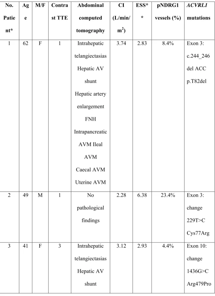
TABLE 1. HHT2 patients characteristics No. Patie nt* Ag e M/F Contra st TTE Abdominal computed tomography CI (L/min/ m2) ESS* * pNDRG1 vessels (%) ACVRL1 mutations 1 62 F 1 Intrahepatic telangiectasias Hepatic AV shunt Hepatic artery enlargement FNH Intrapancreatic AVM Ileal AVM Caecal AVM Uterine AVM 3.74 2.83 8.4% Exon 3: c.244_246 del ACC p.T82del 2 49 M 1 No pathological findings 2.28 6.38 23.4% Exon 3: change 229T>C Cys77Arg 3 41 F 3 Intrahepatic telangiectasias Hepatic AV shunt 3.12 2.93 4.4% Exon 10: change 1436G>C Arg479Pro
Hepatic artery enlargement NRH Uterine AVM * 4 70 F 0 Hepatomegaly Hepatic AV shunt Hepatic artery enlargement Intrapancreatic AVM Left renal artery aneurysm 3.7 6.59 24.2% Exon 10: change 1436G>C Arg479Pro * 5 49 M 0 Intrahepatic telangiectasias Intrapancreatic AVM Gastro-omental arteries aneurysms 2.9 4.57 13.5% Exon 10: change 1436G>C Arg479Pro 6 51 M 1 Hepatomegaly 3.3 6.05 16.4% Exon 10:
Hepatic AP shunt Hepatic artery enlargement 1450C>T Arg484Trp Male (M) or Female (F)
ESS: Epistaxis Severity Score
Epistaxis frequency: Daily (D) or weekly (W) TTE: Transthoracic echocardiography
CI: Cardiac Index on TTE
NRH: Nodular regenerative hyperplasia FNH: Focal nodular hyperplasia
AVM: Arteriovenous malformation
AV shunt: Arteriovenous shunt (hepatic artery to hepatic vein) AP shunt: Arterioportal shunt (hepatic artery to portal vein)
Figure 1 WT ALK1 +/-P5-Retinas A P7-Retinas 25 12.5 0 V essel width ( µm) **** p=0.051 15 10 5 0
No. branch points / 10
4 µ m 2 WT ALK1+/- WT ALK1 +/-Vein V essel width ( µm) 15 10 5 0 WT ALK1 +/-15 10 5 0
No. branch points / 10
4 µ m 2 WT ALK1 +/-Artery D C WT ALK1 +/-WT ALK1 +/-P9-Retinas E F 20 10 0 V essel width ( µm) *** WT ALK1 +/-15 10 5 0
No. branch points / 10
4 µ m 2 WT ALK1 +/-Vein V essel width ( µm) 15 10 5 0 WT ALK1 +/-** 15 10 5 0
No. branch points / 10
4 µ m 2 WT ALK1 +/-Artery B A V A A V V V V A A A A V 25 12.5 0 V essel width ( µm) WT ALK1 +/-15 10 5 0
No. branch points / 10
4 µ m 2 WT ALK1 +/-Vein V essel width ( µm ) 15 10 5 0 WT ALK1 +/-15 10 5 0
No. branch points / 10
4 µ
m
2
WT ALK1 +/-Artery
Figure 1. Heterozygous loss of ALK1 results in retinal vascular hyperplasia.
(A) Whole-mount visualization of blood vessels by isolectin B4 staining of wild type (WT) or ALK1+/- mice at P5. Red and green islets show venous and arterial selected regions where
quantification analysis was done. A indicates artery and V indicates vein. (B) Quantification of vessel width and number of branch points in veins and arteries of wild-type (n=8) and ALK1 +/-(n=7) retinas at P5. (C) Whole-mount visualization of blood vessels by isolectin B4 staining of wild type (WT) or ALK1+/- mice at P7. (D) Quantification of vessel width and number of branch points in veins and arteries of wild-type (n=28) and ALK1+/- (n=27) retinas at P7. (E) Whole-mount visualization of blood vessels by isolectin B4 staining of wild type (WT) or
ALK1+/- mice at P9. (F) Quantification of vessel width and number of branch points in veins
and arteries of wild-type (n≥3) and ALK1+/- (n=10) retinas at P9. Error bars indicate the standard error of the mean. Statistical significance of two-tailed Mann-Whitney U tests: **, p<0.01; ***, p<0.001; ****, p<0.0001.
Figure 2 Cell Number ( x 10 4) 6 0 ** ** Control BMP9 VEGF BMP9+ VEGF Control BMP9 VEGF BMP9+ VEGF 48h 72h D WT ALK1 +/-* * * * * * * * * * * * * * * * * * * * * * * * * * * * * * * * * * * * * * Erg Erg Edu Edu
Erg: Edu Erg: Edu
Erg: Edu : IB4
Erg: Edu : IB4
B C
E
Control BMP9 VEGF BMP9+ VEGF
BrdU positive cells / total cells ( %)
40 20 0 * * * 20% FBS 20% FBS + ALKi p=0.054 ** *** ** *
Num. Erg positive cells
40
20
0
First line
WT ALK1+/- WT ALK1
+/-Second line First line
WT ALK1+/- WT ALK1 +/-Second line * 25 12.5 0
% Edu+Erg+ positve cells / total Erg cells
20% FBS + BMP9 20% FBS + BMP9+ ALKi *** ** A 4 2
Figure 2. ALK1 negatively regulates endothelial cell proliferation in vitro and in vivo.
(A) Representative images of wild-type and ALK1+/- retinas immunostained with Erg-1/2/3 (green), Edu (blue) and isolectin B4 (red). Lines indicate the separation (20 µm) between the two areas of quantification: first line (tip and first neighboring cells) and second line (second and third neighboring cells). Scale bars, 20 µm. Yellow and white asterisks indicate Erg- and Edu-positive cells respectively located on the second line. (B) Bars show quantification of endothelial nuclei per unit area assessed by Erg positivity in wild-type (n=11) and ALK1 +/-(n=6) retinas at P7. (C) Quantification of percentage of proliferative endothelial cells (double Edu+/Erg+ with respect to total Erg+) in wild-type (n=11) and ALK1+/- (n=6) retinas at P7. (D) HUVECs were incubated for 48 h or 72 h in medium 199 with 0.5% serum, in the presence or absence of 10 ng/ml VEGF, 10 ng/ml BMP9 or 10 ng/ml VEGF and 10 ng/ml BMP9, after which the cell number was counted. Each data point represents the mean of at least three independent experiments. (E) Bars show quantification of proliferation of quiescent HUVECs plated for 24 h in the absence or presence of 10 ng/ml VEGF, 10 ng/ml BMP9 or 10 ng/ml VEGF and 10 ng/ml BMP9, or exponential HUVECs (with 20% FCS) plated for 24 h in the absence or presence of 0.5 µM LDN-212854 and in the absence or presence of 10 ng/ml BMP9. Cells were pulsed with BrdU for 4 h and subjected to immunostaining analysis. Results shown are the means of four independent experiments. Error bars indicate the standard errors of the mean. Statistical significance of two-tailed Mann-Whitney U tests: *, p<0.05; **, p<0.01; ***, p<0.001.
- - + + - + - + VEGF 30’ 4 h BMP9 Pretreatment * ** ** p-AKT p-ERK1/2 ** ** ** BC E - - + + - + - + p-E RK T-E RK (A .U .) 3 2 1 0 4 p-A KT /T -A KT (A .U .) 0. 5 1. 0 - - + + - + - + * p-p38 p-VEG FR 2 / T-VEG FR 2 ( A.U .) 0. 5 1. 0 p-VEGFR2 ** - - + + - + - + VEGF 30’ 4 h BMP9 Pretreatment - - + + - + - + p-p38 / T -p38 (A .U .) 3 2 1 0 4 F p-S MA D1 /5 / T-SMAD1/5 (A.U.) 0 2 4 6 8 10 p-SMAD1/5 ** - - + + - + - + D
p-VEGFR2 T-VEGFR2 p-AKT
(Ser473)
T
-AKT
p-ERK1/2 T-ERK1/2 p-p38 T-p38 p-SMAD1/5 T-SMAD1/5 ID1 TUBULIN
p-AKT
(Thr308)
0
Figure 3. BMP9 inhibits VEGF-mediated activation of AKT and ERK.
(A) Growth factor-depleted HUVECs were pretreated with vehicle or 10 ng/ml BMP9 for 4 h,
stimulated with VEGF for 30 min, and immunoblotted using the indicated antibodies. A representative blot of 5 independent experiments is shown. (B) Bars show quantification of the relative immunoreactivity of phosphoVEGFR2 normalized with respect to total VEGFR2. The mean of five independent experiments is shown. (C) Bars show quantification of the relative immunoreactivity of phosphoAKT (Ser473) normalized with respect to total AKT assessed as the mean of five independent experiments. (D) Bars show quantification of the relative immunoreactivity of phosphoERK1/2 normalized with respect to total ERK1/2. The mean of five independent experiments is shown. (E) Bars show quantification of the relative immunoreactivity of phosphoSMAD1/5 normalized with respect to total SMAD1/5. The mean of five independent experiments is shown. (F) Bars show quantification of the relative immunoreactivity of phospho-p38MAPK normalized with respect to total p38 MAPK. The mean of five independent experiments is shown. Error bars indicate the standard errors of the mean. Statistical significance of two-tailed Mann-Whitney U tests: *, p<0.05; **, p<0.01.
A Basal BMP9-4h BMP9-24h Protein Basal BMP9-4h BMP9-24h PTEN / RPL32 (% of basal) 300 150 0 * mRNA *** Control BMP9 VEGF BMP9+ VEGF Control BMP9 VEGF BMP9+ VEGF Unrelated siRNA
PTEN p-AKT Tubulin ID1
PTEN siRNA T -AKT Control BMP9 VEGF BMP9+ VEGF Control BMP9 VEGF BMP9+ VEGF Unrelated siRNA PTEN siRNA D E 0 10 20 30 40 50 - - + + - + - + VEGF 30’ 4 h BMP9 Pretreatment PTEN Activity (% of PI(3,4,5)P3 converted) Control BMP9 VEGF BMP9+ VEGF Control BMP9 VEGF BMP9+ VEGF DMSO PTEN inhibitor 70 kDa- 70
kDa-55 kDa- 55 kDa- 17
Number of cells (% of control) 0 100 200 300 400 0 100 200
300 Number of cells (% of control)
F *** *** ** p=0.057 p=0.1 *
Figure 4. BMP9 effects are mediated by PTEN.
(A) Growth factor-depleted HUVECs were pretreated with vehicle or 10 ng/ml BMP9 for 4 h
and stimulated or not with VEGF for 30 min. Cells were lysed, PTEN immunoprecipitated and PTEN phosphatase activity measured by evaluating the conversion of PI(3,4,5)3 to PI(4,5)2 by ELISA. Results are expressed as the percentage conversion of initial PI(3,4,5)3 to PI(4,5)2. The mean of four independent experiments is shown. (B) Growth factor-depleted HUVECs were treated with vehicle or 10 ng/ml BMP9 for 4 or 24 h and immunoblotted using appropiated antibodies. Bars show quantification of PTEN protein amount in basal situation (n=10) or induced by BMP9 4 h (n=5) or 24 h (n=10) and normalized with respect to tubulin. (C) Bars show quantification of PTEN mRNA induced by BMP9 stimulation for 4 or 24 h and normalized with respect to RPL32 gene. The mean of four independent experiments is shown. (D) HUVECs were transiently transfected with an unrelated control siRNA (unrelated siRNA) or PTEN siRNA, as described. After 48 h, cells were depleted of growth factors for 16 h. Cells were pretreated or not with 10 ng/ml BMP9 for 4 h, and then stimulated for 30 min in the absence or presence of 10 ng/ml VEGF. Cell were lysed and immunoblotted using the indicated antibodies. A representative blot of five independent experiments is shown. (E) After transfection with siRNAs, 1 x 10⁴ cells were seeded in 24-well plastic plates in normal medium. The next day the medium was changed to M199 with 0.5% FCS and the cells were treated or not with 10 ng/ml of BMP9, 10 ng/ml of VEGF or a combination of BMP9 and VEGF. After 48 h the cell number was assessed. Each data point represents the mean of at least eight independent experiments. (F) HUVECs were incubated in medium 199 with 0.5% serum, in the presence or absence of 10 ng/ml VEGF, 10 ng/ml BMP9, 10 ng/ml VEGF and 10 ng/ml BMP9, and in the absence or presence of 0.5 µM SF1670 PTEN inhibitor. After 72 h the cell number was counted. Each data point represents the mean of at least four independent experiments. Error bars indicate the standard errors of the mean. Statistical significance of two-tailed Mann-Whitney U tests: *, p<0.05; **, p<0.01; ***, p<0.001.
% o f p S 6 p o si ti ve e n d o th e li a l ce ll s 0 10 20 30 40 % o f p N D R G 1 p o s iti v e v e s s e ls 0 10 20 30
Control HHT2 patients Control HHT2 patients Control HHT2 patients
* * % o f p A K T p o s it iv e v e s s e ls 0 20 40 60 0 2 4 6 8 Control HHT2 patients
% of proliferative endothelial cells
* CD34 pNDRG1 pS6 pAKT Ki67 HHT2 patient 5 Control B A C > > D E F > > Figure 5
Figure 5. Increased activation of PI3K signaling and endothelial cell proliferation in HHT2 cutaneous telangiectasia biopsies.
(A) GSEA output plots showing significant over-expression of genes sets corresponding to
PI3K-AKT-mTOR canonical annotations (Hallmark PI3K-AKT-MTOR signaling and PID PI3KCI-AKT) or genes significantly over-expressed by oncogenic PI3KCA mutations. The detailed results of each set are provided in Supplementary Table 1. The GSEA enrichment scores and the nominal P values are shown. The red-shadow areas mark the leading peaks that contribute to the associations. (B) CD34, pNDRG1, pS6, pAKT and Ki-67 (brown nuclei,
arrows) staining of a control and a HHT2 patient. Scale bars, 100 µm (C) Quantification of
the percentage of pAKT-positive vessels in controls (n=3) and HHT2 patients (n=5). (D) Quantification of the percentage of pNDRG1-positive vessels in controls (n=3) and HHT2 patients (n=6). (E) Quantification of the percentage of pS6-positive endothelial cells in controls (n=3) and HHT2 patients (n=5). (F) Quantification of the percentage of Ki-67-positive endothelial cells in controls (n=3) and HHT2 patients (n=6). Error bars indicate the standard errors of the mean. Statistical significance of two-tailed Mann-Whitney U tests: *, p<0.05.
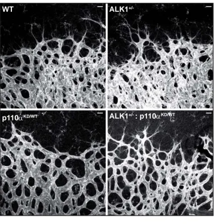
Figure 6 WT ALK1 +/-p110αKD/WT ALK1+/- : p110αKD/WT A 30 15 0 V e ssel width ( µm) WT ALK1 +/-p110 αKD/WT ALK1 +/- : p1 10α KD/WT ** ** ** 30 15 0 V e ssel width ( µm) WT ALK1 +/-WT ALK1 +/-* * WT WT ALK1 +/-ALK1 +/-Vehicle Vehicle PI3Ki PI3Ki B C E * P0 P6 (6:00pm)P7 (10:00am)P7 (2:00pm) PI3Ki PI3Ki Isolation
Figure 6. In vivo inhibition of PI3K blocks vascular hyperplasia in ALK1+/- retinas.
(A) Whole-mount visualization of blood vessels by isolectin B4 staining wild-type, ALK1+/-,
p110αKD/WT and double ALK1+/-/p110αKD/WT retinas at P7. (B) Quantification of vessel width in
veins of wild-type (n=4), ALK1+/- (n=8), p110αKD/WT (n=4) and double ALK1
+/-/p110αKD/WT
(n=6) retinas. (C) Scheme of pharmacologic approach to inhibit PI3K with LY294002. (D) Isolectin B4 stained wild-type and ALK1+/- treated with vehicle or LY294002 at P6 and P7. (E) Quantification of the vessel width of retinas shown in C (n≥4 per treatment and genotype). Error bars indicate the standard errors of the mean. Statistical significance of two-tailed Mann-Whitney U tests: *, p<0.05; **, p<0.01.