To my parents,
Malathi and Thangavel,
and my beloved brother,
for their endless support.
ABSTRACT
Estrogen signaling plays a vital role in breast, ovarian and endometrial cancers. The actions of estrogen are mainly mediated by classical estrogen receptors, ERα and ERβ that belongs to the nuclear receptor superfamily. In recent years, a class of membrane-associated estrogen receptors are found to mimic the functions of classical ERs, including genomic as well as non-genomic signaling. These non-genomic signaling events include pathways that are usually thought of as arising from transmembrane growth factor receptors and G protein-coupled receptors (GPCRs). GPCRs belong to a superfamily of cell surface signaling proteins. GPCRs represent the most significant family of validated pharmacological targets in medical biology. A member of the GPCR family, named GPER, mediates rapid biological responses to estrogen in diverse normal and cancer cells, as well as transformed cell types. The identification and characterization of GPER will lead to understand the mechanisms underlying complex biological pathways and identify potentially new drug targets.
Here, we proposed a novel gel-free method to isolate and enrich GPER from crude lysate using home-made hydroxyapatite column (HTP). The HTP eluate was subjected to cellulose acetate (CA) filteration, followed by on-membrane protein digestion with different proteases and analyzed by MALDI MS. GPER was identified by peptide mass fingerprinting (PMF) after intensive data analysis. Sequence analysis reports 3 potential N-glycosylation in GPER. We manually validated 2 out of 3 glycosylation sites in GPER from the obtained MS/MS data and also validated the glycan moieties predicted by Glycomod. This approach is the first of its kind to identify GPER and characterize post-translational modifications (PTMs) by MS-based proteomic analysis. The proposed method is simple, robust and unique with great reproducibility. Finally, we designed and synthesized polymer nanoparticles (NPs) in an effort to capture GPER with high affinity and selectivity from crude lysate. PNIPAm-based NPs were synthesized by a free radical precipitation polymerization method with no control
over the functional monomer sequence. The NP binding affinity was evaluated against both truncated-GPER (short peptide epitopes) and GPER (whole protein). As the NPs were designed with complementary functionality against the peptides/protein, the NPs-peptide/protein binding will be through multipoint interactions. The initial qualitative results obtained by immunoblotting analysis revealed interesting hints on GPER’s competitive affinity towards NPs when probed against multiple antibodies. We anticipate to use this strategy as a sample purification step prior to MS-based proteomic analysis.
Key words: GPR30/GPER, breast cancer, MALDI MS, N-glycosylation, synthetic polymer nanoparticles.
ACKNOWLEDGEMENTS
This thesis has been written as a part of my three year PhD at Department of Chemistry and Chemical Technologies in the University of Calabria, Italy, between March 2012 and February 2015. My research project was funded by the European commission under grant agreement number 264772 (ITN-CHEBANA).
First and foremost, I praise God, the almighty for providing me this opportunity and granting me the capability to proceed successfully. This thesis appears in its current form due to the assistance, guidance and encouragement of several people. I would therefore like to offer my sincere thanks to all of them.
I would like to express my deep and sincere gratitude to Prof. Giovanni Sindona, my esteemed supervisor and the director of the department of Chemistry and Chemical Technologies, for accepting me as a PhD student, and for his warm encouragement and continuous support throughout my PhD. I could not have imagined having a better supervisor for my PhD study.
I am deeply grateful to my cosupervisor, Prof.ssa Anna Napoli, whose support and motivation from the initial to the final level enabled me to develop an understanding of the subject. Her understanding and thoughtful guidance have provided a good basis for this thesis.
I would like to thank Dott.ssa Donatella Aiello for her advises and friendly assistance with various problems time to time. I greatly appreciate her time in teaching me the knowhow about the MALDI instrument during my initial days.
I express my sincere thanks to Prof. Marcello Maggiolini for his critical suggestions, and Dott.ssa Assunta Pisano for providing me the samples for my research work.
Next, I cordially thank Prof. Kenneth J Shea, for accepting me as a visiting scholar and giving me the opportunity to work in SHEA lab (UCI). I really had a valuable and enjoyable experience working with his team. His excellent guidance, encouragement and motivation have been of great value in this thesis.
I especially want to express my huge thanks to Dr Keiichi Yoshimatsu, Post Doctoral Research Fellow in SHEA lab, who gave his continuous help and support by answering all of my questions related to the research work. He was my colleague and my best “buddy” at UCI.
I would like to extend my thanks to Prof. Jennifer Prescher for understanding my research needs and allowing me work in her lab, utilizing her analytical instruments. I share my thanks to Lidia Nazarova, graduate student in Prescher lab (UCI), for helping me with me all my research needs throughout my stay.
My warm and special thanks are due to Dr Yi Ge, my MSc course coordinator (Cranfield University), for giving me confidence and support on all aspects of my career after MSc. For me, he is more than a course tutor, without his personal advice this PhD would not have been possible.
I would like to thank Prof. Bartolo Gabriele, coordinator of OMPI curriculum and Prof. Roberto Bartolino, director of ‘Bernardino Telesio’ Doctoral school of science and technology, UNICAL.
I cannot finish without thanking my family. I warmly thank and appreciate my parents and my brother. They were always supporting me and encouraging me with their best wishes.
Finally, I would like to thank my dearest friend, Dhanya Dhanyalayam. She was always there cheering me up and stood by me through the good times and bad.
CONTENTS
Abstract………..i Acknowledgements……….iii Contents………...v List of Figures………..vii List of Tables……….ixList of Acronyms and Abbreviations……….…x
CHAPTER 1 GENERAL INTODUCTION ... 1
1.1 Receptors and ligands: an overview ... 1
1.1.1 Cell surface receptors ... 2
1.1.2 Internal receptors ... 6
1.2 G protein-coupled receptors ... 8
1.2.1 Structure of GPCRs ... 8
1.2.2 G proteins: types and functions ... 10
1.2.3 Classification of GPCRs ... 11
1.2.4 GPCR activation and signaling ... 12
1.2.5 Clinical impact of GPCRs... 15
1.3 GPCRs and cancer ... 15
1.3.1 Steroid receptors in cancer ... 18
1.3.2 Physiological importance of estrogen ... 19
1.3.3 GPER as a GPCR for E2 ... 20
1.3.4 GPER-mediated signaling ... 24
1.3.5 Clinical significance of GPER ... 26
1.4 Rationale for project ... 29
CHAPTER 2 MASS SPECTROMETRY-BASED GPCR PROTEOMICS: Isolation and Identification of GPER by Peptide Mass Fingerprinting ... 32
2.1 Mass spectrometry-based proteomics ... 33
2.2 Classical proteomics unfit for GPER analysis... 38
2.3 Method development for GPER isolation ... 40
2.4 Identification of GPER by peptide mass fingerprinting ... 44
CHAPTER 3 GPER POST-TRANSLATIONAL MODIFICATION: Analysis of N-glycosylation in GPER by MALDI-TOF/TOF mass spectrometry ... 49
3.1 Protein post-translational modifications ... 49
3.2 Glycosylation in GPER ... 53
3.3 Glycopeptide derivatization with dansyl chloride ... 55
3.4 GPER deglycosylation with endoglycosidases ... 65
CHAPTER 4 AFFINITY CAPTURE-RELEASE STRATEGY FOR GPER PURIFICATION: Design and synthesis of polymer nanoparticles with high affinity for GPER ... 67
4.1 Introduction to affinity purification ... 67
4.2 Engineered polymer nanoparticles for GPER purification ... 69
4.3 Solid phase peptide synthesis ... 71
4.4 HPLC and MALDI analysis of synthesized peptide ... 76
4.5 Preparation on synthetic polymer nanoparticles ... 78
4.6 Nanoparticle characterization ... 79
4.7 Interaction between peptides and nanoparticles by HPLC analysis ... 81
4.8 Cell culture and cell lysis ... 84
4.9 Interaction between protein and nanoparticles by western blot analysis ... 85
CHAPTER 5 SUMMARY AND OUTLOOK ... 90
BIBLIOGRAPHY ... 94
APPENDIX ... 123
LIST OF FIGURES
1.1 Lock and key model ... 2
1.2 Ligand-gated ion channel receptors ... 3
1.3 Enzyme-coupled receptors ... 4
1.4 G protein-coupled receptors ... 6
1.5 Internal receptors ... 6
1.6 Signal transduction through internal receptors ... 7
1.7 Structural representation of GPCR ... 9
1.8 GPCR activation/deactivation cycle ... 14
1.9 Chemical structure of estrone (E1), estradiol (E2) and estriol (E3) ... 19
1.10 Estrogen signaling pathway ... 21
1.11 GPER-mediated genomic and non-genomic signaling... 25
2.1 Classical proteomics workflow ... 35
2.2 Schematic representation of MALDI and ESI ... 36
2.3 Schematic representation of MALDI-TOF/TOF optics ... 37
2.4 Chemical structures of different MALDI matrices ... 37
2.5 HTP and C18 spin column model ... 42
2.6 GPER isolation from SkBr3 lysate using HTP spin column ... 43
2.7 Consistency of HTP enrichment method ... 44
2.8 Cellulose acetate spin filter model ... 45
2.9 On-membrane digestion schematic representation ... 46
2.10 Peptide mass fingerprint of GPR1 ... 47
2.11 GPER identification by peptide mass fingerprinting (MALDI MS) ... 48
3.1 Common post-translational modifications ... 50
3.2 Different types of glycosidic linkages ... 54
3.3 MALDI MS spectrum of peptic mixture (SkBr3 lysate) ... 56
3.4 MALDI MS spectrum showing possible hexose sugar ... 57
3.5 MS/MS spectra of precursor ions m/z 1472.54 and m/z 1309.49 before
dansylation... 58
3.6 MS/MS spectra after dansylation ... 59
3.7 MS/MS spectrum of m/z 1543.50 ... 61
3.8 MS/MS spectrum of m/z 679.37 ... 61
3.9 Oligosaccharide structures corresponding to NLSHPL ... 62
3.10 MALDI MS spectrum of α-chymotrypsin-digested sample (SkBr3 lysate) ... 63
3.11 MS/MS validation of oligosaccharide structure ... 64
4.1 Protein purification using affinity chromatography ... 68
4.2 Synthetic polymer nanoparticles for protein purification ... 70
4.3 Reaction showing FMOC introduction to amine ... 73
4.4 Nova-PEG rink amide resin (Novabiochem)... 74
4.5 Flow-chart for selecting cleavage cocktail for FMOC SPPS ... 75
4.6 HPLC chromatograms of synthesized peptides (P1, P2, P3, P4 and P5)... 77
4.7 MALDI MS spectra of synthesized peptides (P1, P2, P3, P4 and P5) ... 78
4.8 Preparation of PNIPAm-based synthetic polymer nanoparticles ... 79
4.9 1H NMR spectrum of 462 nm NP in CD3OD, 500 MHz, 298 K ... 80
4.10 13C NMR spectrum of 462 nm NP in CD3OD, 500 MHz, 298 K ... 81
4.11 Selected peptides for Peptide-NP interaction studies ... 82
4.12 Peptide-NP interaction chart ... 83
4.13 HPLC analysis of Peptide-NP binding affinity ... 84
4.14 Immunoblot showing detectable protein concentrations ... 86
4.15 Western blot analysis of Protein-NP binding affinity ... 88
LIST OF TABLES
1.1 G protein types and associated functions ... 10
1.2 Second messengers and their cellular activities ... 13
1.3 GPCRs associated with human cancers ... 16
1.4 Different ligands for GPER with tested affinity (Kd) values ... 23
1.5 Estrogen receptors expression in human cancer cell lines ... 27
3.1 Common types of post-translational modifications ... 51
3.2 Glycosylation sites in GPER ... 54
3.3 Possible glycopeptides predicted using Glycomod ... 60
3.4 Oligosaccharide structures predicted by Glycomod ... 63
4.1 Truncated-GPER sequence information based on extracellular, cytoplasmic, and transmembrane domains ... 72
4.2 Selected short peptide epitopes for solid phase peptide synthesis ... 73
4.3 Cleavage cocktail used for synthesized peptides ... 74
4.4 %yield of the synthesized peptides (P1, P2, P3, P4 and P5) ... 76
4.5 Cell lysates and their total protein concentration ... 85
LIST OF ACRONYMS & ABBREVIATIONS
2DE Two-dimensional gel electrophoresis 7TMRs 7-transmembrane receptorsAAc Acrylic acid
APS Ammonium per sulfate ARs Androgen receptors
BIS Bis-acrylamide
CA Cellulose acetate
cAMP cyclic-adenosine monophosphate
cGMP-PDE cyclic guanosine monophosphate phosphodiesterases CHCA α-Cyani-4-hydroxycinnamic acid
CID Collision induced dissociation CL2 Cytoplasmic loop - 2
CL3 Cytoplasmic loop – 3
CTGF Connective tissue fgrowth factor
Cys Cysteine
DDT Dichlorodiphenyltrichloroethane DHB 2,5-Dihydroxybenzoic acid
DH hydrodynamic diameter
DLS Dynamic light scattering
DMEM Dulbecco’s modified eagle medium DMF N,N-Dimethylformamide
DNA Deoxyribonucleic acid DNS-Cl Dansyl chloride
E1 Estrone
E2 Estradiol (17β-estradiol)
E3 Estriol
EDT Ethanedithiol
EDTA Ethylenediaminetetraacetic acid EGFR Epidermal growth factor receptor EGTA Ethylene glycol tetraacetic acid EL1 Endoplasmic loop - 1
EL2 Endoplasmic loop - 2 EL3 Endoplasmic loop - 3
ELISA Enzyme-linked immuno sorbent assay ERs Estrogen receptors
ERK Extracellular-signal-regulated kinase FBS Fetal bovine serum
FMOC-CL Fluorenylmethyloxycarbonyl chloride GDI Guanine nucleotide dissociation inhibitor GDP Guanosine diphosphate
GEF Guanine nucleotide exchange factor GLcNAc N-acetylgucoseamine
GPCR G protein-coupled receptor
GPER G protein-coupled estrogen receptor GPR30 G protein-coupled receptor 30 GRK G protein-coupled receptor kinase GTP Guanosine triphosphate
GTs Glycosyltransferases
HB-EGF Heparin-bound-epidermal growth factor
Hex Hexose
HexNAc N-acetylhexoseamine
hGPER human G protein-coupled estrogen receptor HPLC High performance liquid chromatography HTP Hydroxyapatite
IgG Immunoglobulin G
IP3 Inositol triphosphate
IR Infrared
LCST Lowest critical solution temperature m/z mass-to-charge ratio
MALDI Matrix-assisted laser desorption/ionization
Man Mannose
MAPK Mitogen-activated protein kinase
MC Missed cleavage
Met Methionine
MIP Molecularly imprinted polymer MMP Matrix metalloproteinase mRNA messenger Ribonucleic acid
MS Mass spectrometry
MWCO Molecular weight cut-off
NCBI National center for biotechnology information Nd:YAG Neodymium doped: yttrium aluminium garnet NMR Nuclear magnetic resonance
NPs Synthetic polymer nanoparticles OHT 4-hydroxytamoxifen P1 Peptide 1 P2 Peptide 2 P3 Peptide 3 P4 Peptide 4 P5 Peptide 5
PAGE Polyacrylamide gel electrophoresis PBS Phosphate buffered saline
PEG Poly ethylene glycol pI Isoelectric point
PI3K Phosphoinositide 3-kinase xii
PLC Phospholipase C
PMSF Phenylmethanesulfonyl fluoride PNGase Peptide-N-glycosidase
PNIPAm Poly(N-isopropylacrylamide) ppm parts per million
PTM Post-translational modification RGS Regulators of G protein
RPMI Rosewell park memorial institute medium SDS Sodium dodecyl sulfate
SERDs Selective estrogen receptor down regulators SERMs Selective estrogen receptor modulators SPE Solid-phase extraction
SPPS Solid phase peptide synthesis TBAm N-tert butylacrylamide TFA Trifluoroacetic acid
TIS Thioanisole TM1 Transmembrane - 1 TM3 Transmembrane - 3 TM4 Transmembrane - 4 TM7 Transmembrane - 7 TOF Time-of-flight UV Ultra violet
WB Western blot analysis
CHAPTER 1
GENERAL INTRODUCTION
1.1 Receptors and ligands: an overview
Cell membranes in eukaryotes are naturally equipped with thousands of receptors, of many different kinds. Eukaryotic cells also encase their cell organelles like nucleus, ribosomes, endoplasmic reticulum, Golgi apparatus, mitochondria, lysosome with internal membranes that play host to a bunch of intracellular receptors. In general, receptors are nothing but protein molecules, ingrained in either the cell membrane or the cytoplasm of a cell. As the name denotes, receptors are macromolecular structures that receive information.[1] More specifically, receptors enable cells to sense stimuli or physical
changes in the internal or external environment, so that the cells can adjust to new situations. Based on their physical presence, receptors can be easily put into two broad categories, cell surface receptors and internal receptors (cytoplasmic and nuclear receptors). Receptors that are found on the membrane of internal cell organelles are also categorized under intracellular receptors, and interestingly, they share functional similarities with cell surface receptors.
Receptor proteins recognize and respond to endogenous chemical signals. The chemical signals can act either at the plasma membrane or within the cytoplasm (or nucleus) of the target cell.[2] The signaling molecules that bind to the receptors are
referred to as ligands and can be endogenous or exogenous in origin. A ligand can be any small molecule such as light-sensitive compound, odorant molecule, hormone, pheromone, growth factor, cytokine, neurotransmitter, toxin, pharmaceutical drug, or peptide (small protein).[3][4] Irrespective of the nature of initiating signal, the cellular
responses are determined by the presence of receptors that specifically binds the signaling molecules.[2] Each receptor is unique and assigned to activate a specific cellular
biochemical pathway when triggered. Every single receptor will just tie to ligands of a specific structure. Receptor-ligand interaction can be compared to a lock and key system, where a lock will just accept a specifically fashioned key (Fig. 1.1).[5] On binding its
corresponding receptor, the ligand initiates or inhibits the receptor’s designated biochemical pathway.
Figure 1.1 Lock and key model.
1.1.1 Cell surface receptors
Human cells are constantly communicating with each other and the outside world through the specialized integral membrane proteins that are collectively known as cell surface receptors (membrane receptors, transmembrane receptors). Cell surface receptors bind to an external ligand molecule and perform signal transduction, converting an extracellular signal into an intracellular signal. By doing so, the cell surface receptors play a unique and significant role in cellular communications and signal transduction. Ligands that interact with cell surface receptors are mostly impermeant signal molecules that can’t enter the cell. Every cell surface receptor has three main components: an N-terminal ligand binding domain (extracellular domain), a hydrophobic membrane-spanning region, and a C-terminal cytoplasmic domain (intracellular domain) inside the cell. The extracellular domain usually includes the binding site for the ligand, while the intracellular domain activates a series of intracellular signaling events once the ligand binds. The size and extent of each of these domains vary extensively, depending on the type of receptor. So far, a wide range of these receptors have been identified and studied.[2] They are grouped into three main classes of receptors, namely: ligand-gated
ion channel receptors, enzyme-coupled receptors, and G protein-coupled receptors. The names of these receptor classes are defined by the mechanism used to transform external signals into internal ones - via ion channel opening, enzyme activation, or protein action,
respectively. Because cell surface receptors interact with signal molecules or ligands externally and permit them to affect cell function without actually entering the cell.[6]
Ligand-gated ion channel receptors (Fig. 1.2) are also known as ionotropic receptors. These receptors bind a ligand and open a channel that allows the flow of specific types of ions such as Na+, K+, Ca+ or Cl- across the cell membrane, which changes the membrane
potential, causing an electric current.[7] To form a channel, this type of cell surface
receptor have an extensive membrane-spanning region. In order to interact with the phospholipid fatty acid tails that form the crux of the cell membrane, many of the amino acids in the membrane-spanning region are hydrophobic in nature. In contrast, the amino acids that line up on the inside of the channel are hydrophilic to allow the passage of water or ions. These receptors are responsible for the rapid transmission of signals across synapses in the nervous system. Good examples of such receptors are the neurotransmitter receptors.[2] Although the ligand-gated ion channel receptors are found
mainly in the nervous system and other electrically excitable cells such as muscle cells, the other two types of cell surface receptors are found particularly in every cell type of the body.[7]
Figure 1.2 Ligand-gated ion channel receptors.[2]
Enzyme-coupled receptors (Fig. 1.3) are cell surface receptors, composed of an extracellular domain containing the ligand binding site and an intracellular domain, often associated with an enzyme. In some cases, the intracellular domain of such receptor itself is an enzyme whose catalytic activity is regulated by the binding of an extracellular chemical signal. As of 2009, only six types of such receptors are known and they are receptor tyrosine kinases, tyrosine kinase associated receptors, receptor-like tyrosine phosphatases, receptor serine/threonine kinases, receptor guanylyl cyclases, and histidine kinase associated receptors.[8] The great majority of them are protein kinases,
often tyrosine kinases, which phosphorylate intracellular target proteins, thereby changing the physiological function of the target cells.[2]
Figure 1.3 Enzyme-coupled receptors.[2]
The enzyme-coupled receptors normally have large extracellular and intracellular domains, but the membrane spanning-region consists of a single alpha-helical region of the peptide strand.[9] On binding their ligands externally, the receptors undergo
conformational change that activates the enzyme, which then turn on a variety of intracellular signaling pathways. They are discovered through their role in responses to extracellular signal proteins that regulates the growth, proliferation, differentiation and
survival of cells in animal tissues. Disorders of cell growth, proliferation, differentiation, survival and migration are fundamental to cancer, and abnormalities in signaling via enzyme-coupled receptors have a major role in the development of this class of diseases.[8]
G protein-coupled receptors (GPCRs) (Fig. 1.4) are the largest of all the cell surface receptors. GPCRs bind a ligand and activate a membrane-bound, trimeric GTP-binding protein (G protein). The activated G protein then interacts with either an ion channel (effector) or an enzyme in the cell membrane, initiating a sequence of other effects. All GPCRs share the structural feature of crossing the cell membrane seven times, but each receptor has its own specific extracellular domain containing the ligand binding site and intracellular domain with G protein binding site.[9] GPCRs are also referred to as
7-transmembrane receptors (7TMRs), heptahelical receptors, serpentine receptors or metabotropic receptors. Metabotropic receptors do not form an ion channel passage, instead, they are indirectly linked with ion channels on the cell membrane through signal transduction mechanisms.[10] Heterotrimeric G proteins have three subunits: α, β, and γ.
When a ligand binds to a G protein-coupled receptor in the cell membrane, a guanosine diphosphate (GDP) molecule associated with the α subunit is exchanged for guanosine triphosphate (GTP). The β and γ subunits dissociate from the α subunit, and a cellular response is triggered either by the α subunit or the dissociated β-γ complex. Hydrolysis of GTP to GDP terminates the signal.[9] Cell signaling using GPCRs occurs as a cyclic series of
events. These receptors mediate responses involving hormones, local mediators and neurotransmitters.[2] Because of their involvement in wide range of cellular processes,
GPCRs are typically an appealing target for the development of drugs to treat a number of diseases.[11] Hundreds of different GPCRs have been identified so far. Some of the well
know examples include the β-adrenergic receptor, metabotropic glutamate receptors, receptors for odorants in the olfactory system, and many types of receptors for peptide hormones.[11]
Figure 1.4 G protein-coupled receptors.[2]
1.1.2 Internal receptors
Internal receptors (Fig. 1.5), also known as intracellular receptors, are found in the cytoplasm or nucleus of the cell and are normally activated by cell-permeant, hydrophobic or lipophilic ligand molecules that can pass through the cell membrane.[2]
Figure 1.5 Internal receptors.[2]
To initiate signal transduction, these ligands must passively diffuse through cell membrane. On entering the cell, many of these molecules bind to proteins that act as regulators of mRNA synthesis to mediate gene expression. Gene expression is the cellular process of transforming the information in a cell’s DNA into a sequence of amino acids that ultimately forms a protein. When the ligand binds to the internal receptor, a conformational change exposes a DNA-binding site on the protein. The ligand-receptor complex moves into the nucleus, binds to specific regulatory regions of the chromosomal DNA, and promotes the initiation of transcription (Fig. 1.6).[12] As the ligand-receptor
complex makes it all the way to the nucleus of the cell, these receptors are often called nuclear receptors.[13] Some intracellular receptors are located primarily in the cytoplasm,
while others are in the nucleus. In either case, once these receptors are activated they can affect gene expression by altering DNA transcription. Internal receptors can directly influence gene expression without having to pass the signal on to other receptors or messengers. Intracellular receptors are used widely by some classic hormones such as thyroid and steroid hormones.[14][15]
Figure 1.6 Signal transduction through internal receptors.[9]
Among all the above discussed receptors, GPCRs are the most abundant class of receptors in the human body.[16] They play a crucial role in an incredible range of functions
in humans. More than one-half of all prescribed drugs achieve their effects by binding to GPCRs. However, only a small portion of GPCRs have been investigated as drug targets leaving a wide area to explore and understand.
1.2 G protein-coupled receptors
G Protein-coupled receptors constitute by far the largest and most distinct superfamily of cell membrane signaling proteins in eukaryotes, with their unique seven-transmembrane-helix structure. They transduce extracellular signals as exerted by a hormone or neurotransmitter to an intracellular effector pathway through the activation of heterotrimeric G proteins.[17] In human, nearly 800 different genes code for GPCRs,
which account for ~4% of the entire protein-coding genome.[18] GPCRs are virtually
expressed in all types of tissues in the body.[19] GPCRs involvement in numerous
physiological processes and diseases including tumor growth and metastasis have been well documented in many scientific reports over the years. GPCRs have become drug targets for several life-threatening diseases. They are often expressed in low levels and in specific cell types, which contributes to the fact that they are the most important family of protein receptors serving as targets in drug discovery. An increased understanding of these receptors has significantly affected modern medicine.[20] Presently, one-quarter of
the top 100 best-selling drugs are targeted mostly to GPCRs that bind amines. In 2012, the Nobel Prize in Chemistry was jointly awarded to Robert Lefkowitz and Brian Kobilka for their groundbreaking research work which gave the first insight on how GPCRs function.[21] Moreover, there have been at least seven other Nobel Prizes awarded for
some aspect of G protein-mediated signaling in the past.
1.2.1 Structure of GPCRs
GPCRs consist of a single, serpentine-like polypeptide chain of variable length (from 300 to 1000 amino acids) that is folded into a globular structure and embedded in
the cell membrane.[22] Seven segments of this molecule span the entire width of the
membrane explaining why GPCRs are sometimes called 7-transmembrane receptors (7TMRs). The intervening portions that connect the seven membrane spanning α-helices loop both inside and outside of the cell forming three intracellular and three extracellular loops (Fig. 1.7). The extracellular amino terminal segment and cytoplasmic carboxyl terminal segment are attached to the TM1 domain and TM7 domain, respectively. Both termini are highly variable in length, and the amino-termini can comprise different functional domains each of which is able to provide specific properties to the relevant receptor.[23] Some GPCRs bear amine-linked glycosylation sites near their amino terminal
segment. The three extracellular loops (EL1, EL2 and EL3) are considered to play an important role in structure stabilization and ligand binding, whereas, the cytoplasmic loops (CL2 and CL3) are mainly engaged in G protein recognition and activation.[22]
Figure 1.7 Structural representation of GPCR.
1.2.2 G proteins: types and functions
G proteins are specialized proteins with the ability to bind the nucleotides GTP and GDP. The G protein acts as a molecular switch by binding either GTP (active/on) or GDP (inactive/off). Some G proteins, such as the signaling protein Ras, are small protein with a single subunit. The G proteins activated by GPCRs are trimeric in structure consisting of an α-, β- and γ- subunit. There are more than 20 different α-subunits, 6 different β-subunits and 12 different γ-subunits, creating a large number of theoretical combinations.[24] However, only a small number of combinations form biological
complexes. In humans, 16 Gα genes encode 23 known Gα isoforms.[24][25] Based on
sequence similarities, Gα proteins are grouped into 4 classes including Gα(S), Gα(i/o),
Gα(q/11) and Gα(12/13).[26] The Gα subunit binds to GDP when inactive. The tightly associated
Gβγ complex functions as a single unit and facilitates the association of Gα to the cytoplasmic part of the GPCR. Moreover, it inhibits the release of GDP from Gα and acts as a guanine nucleotide dissociation inhibitor (GDI).[24] Each Gα and Gβγ subunits activate
and regulate specific pathways that are shown in Table 1.1. The βγ subunits of G protein can also act as second messenger molecules, although their actions are not completely characterized.
Table 1.1 G protein types and associated functions.
Type Pathways and functions References
Gα(S) Activates Ca2+ channels, stimulates adenylyl cyclase pathway and cyclic adenosine monophosphate (cAMP) production
[16] [27] Gα(i/o) Activates K+ channels, inhibits Ca2+ channels, inhibits adenylyl
cyclase and cAMP production
[16] [28] Gα(q/11) Stimulates phospholipase C (PLC) pathway [16] [29] Gα(12/13) - Diverse ion transporter interactions
- Regulates G protein RhoA and stimulates PDZ-Rho guanine nucleotide exchange factors (PDZ-RhoGEF)
[16] [24]
Gβγ complex
- Inhibits the release of GDP from Gα and acts as GDI - Regulates Ca2+ and K+ channels
- Regulates kinase and small G protein including Phosphoinositide 3-kinase-γ (PI3Kγ)
- Various other regulation pathways have been considered for downstream activation of Gβγ
[24]
1.2.3 Classification of GPCRs
There are many different approaches for classifying the GPCRs. Both physiological and structural features have been used to classify GPCRs. The most commonly used system of classification is that implemented in the GPCRDB database, which divide GPCRs into six classes (Class A-F).[30] This A-F system is designed for both vertebrate and
invertebrate GPCRs. Class A contain rhodopsin-like and biogenic amine receptors, with over 80% of all GPCRs in humans; Class B: Secretin-like; Class C: Metabotropic glutamate receptors; Class D: Pheromone receptors; Class E: cAMP receptors; and the much smaller Class F contain Frizzled/smoothened receptors. Here, Classes A, B, C and F are found in mammalian species while Class D receptors are found only in fungi and Class E are exclusive to Dictyostelium.[31] The above six classes are further divided into sub-divisions
and sub-sub-divisions based on the function of a GPCR and its specific ligand.[32] As some
classes of the A-F system do not exist in human, an alternative classification system called GARFS has been proposed for classifying mammalian GPCRs. In GARFS system, the receptors are grouped into five major classes based on phylogenetic analyses and named Glutamate (G, with 15 members), Rhodopsin (R, with 701 members), Adhesion (A, with 30 members), Frizzled/Taste2 (F, with 24 members) and Secretin (S, with 15 members). Only a few human receptors (nearly 23 protein sequences) could not be designated to any of the above five classes and these were thus categorized as “Other 7TMRs”. It is, however fairly straight forward to place most of these “other” receptors into any of the main classes or groups using sequence similarity only.[33]
1.2.4 GPCR activation and signaling
Generally, all GPCRs have three characteristic domains: a signal recognizing domain (extracellular), a signal transmission domain (transmembrane), and a signal response and amplification domain (intracellular).[34] GPCRs receive a wide range of
ligands such as lipid analogues, amino acid derivatives, small peptides, as well as stimuli from light (photons), taste, odor (pheromones). The ligand is docked in a binding pocket that is usually present on the extracellular side.[35] The mechanism by which GPCRs
transmit extracellular signals through the cell membrane to intracellular responses is mediated by heterotrimeric G proteins. Since GPCRs do not have intrinsic enzymatic activity, binding of a ligand to the external domain of GPCR triggers a conformational change in the receptor, specifically in an ionic interchange between the TM3 and TM4 domains, which leads to receptor activation. Thereby, transducing the ligand’s message mechanically to the G protein which is closely associated to the intracellular or cytoplasmic side of the receptor and leads to different downstream signaling events.[36]
Specific G proteins bind to specific GPCRs[37], it is hard to determine these pairings based
on primary amino acid sequence. The interaction appears to depend on the whole tertiary structure of the GPCR.[35]
On receiving a signal, G protein becomes active, detaches from the GPCR and binds to an enzymatic effector protein lodged in the membrane. Activation of a single G protein can affect the production of hundreds or even thousands of second messenger molecules. The G proteins function as amplifiers, inducing the effectors to produce cascades of secondary messenger molecules that activate other enzymes, creating a diverse range of physiological responses.[34] Effector/second messenger systems include
retinal cyclic guanosine monophosphate phosphodiesterases (cGMP-PDE), ion channels (potassium, calcium), and several phospholipases and adenylyl cyclase subtypes. A list of cellular activities controlled by the effector/second messenger systems are shown in Table 1.2.[35]
Table 1.2 Second messengers and their cellular activities.
Effector/second messenger system Cellular activities
cGMP-PDE - Conversion of light signal into electrical nerve activity in rod cells - Color vision in cone cells
Phospholipases - Autocrine and paracrine regulation - Protein kinase C activation
- Ion channel conductance - Neurotransmitter release - Smooth muscle contraction - Platelet activating factor synthesis Adenylyl cyclases - Gene transcription
- Mitogenesis - Metabolism - Growth factor
Most G proteins involved in GPCR signaling are heterotrimeric with α, β, and γ subunits. When a ligand activates the GPCR, it induces a conformational change allowing the receptor to act as GEF that exchanges GDP for GTP on the Gα subunit. GTP binding promotes the dissociation of Gα from Gβγ, and then, the free GTP-bound Gα subunit and Gβγ heterodimer can activate various effector proteins, thus propagating an intracellular signaling cascade (Fig. 1.8).[38] The signaling continues until the G proteins are inactivated
by a mechanism dependent on the intrinsic GTPase activity of the Gα subunit, which is facilitated by the direct binding of regulators of G protein signaling (RGS) to activated GTP-bound Gα.[39] In simple, the intrinsic GTPase activity of the Gα subunit inactivates Gα by
hydrolyzing GTP back to GDP. The inactivated GDP-bound Gα subunit reforms and inactivates G protein with Gβγ complex turning off other downstream events.[40] A single
activated GPCR may activate multiple G proteins, and each G protein may activate numerous effector proteins, resulting in a considerable amplification of the signal.[41][42]
Upon prolong stimulation however, the receptors eventually inactivate even if their activating ligands remain bound. In this case, a G protein-coupled receptor kinase (GRK) phosphorylates the cytosolic portions of the activated receptor.[43] Once the receptor is
phosphorylated in this way, it binds with high affinity to β-arrestin protein, which inactivates the receptor by preventing its interaction with G proteins and decreasing its response to ligands or agonists (desensitization).[42] β-arrestins also act as adaptor
proteins and recruit the phosphorylated receptors to clathrin-coated pits from where the receptors are endocytosed and afterwards they can either be degraded in lysosomes or activate new signaling pathways.[44]
Figure 1.8 GPCR activation/deactivation cycle.
1.2.5 Clinical impact of GPCRs
Through this sequence of events, GPCRs help regulate an incredible range of bodily functions, from sensation to growth to hormone responses. Hence, proper functioning of this ‘molecular switching system’ is essential to the health of every individual organism. When the system malfunctions, the results can lead to acute or chronic human diseases, a partial listing of which includes cardiovascular disease (β1 -
adrenergic receptor)[45]; asthma (β2 - adrenergic receptor)[46]; endometrial, ovarian and
breast cancer (membrane estrogen receptor)[47]; and strokes and cerebral hypoperfusion
(A2a - adenosine receptor)[48][49]. Other disease states directly linked to mutations in GPCRs
include retinitis pigmentosa (rhodopsin), female infertility (follicle-stimulating hormone receptor), nephrogenic diabetes insipidus (vasopressin receptor), familial exudative vitreoretinopathy (frizzled receptors), and dominant and recessive obesity (melanocortin receptors)[50].
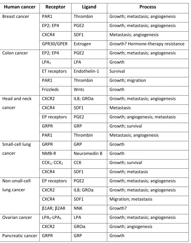
1.3 GPCRs and cancer
Miscommunication is the hallmark of cancer. Normally, our cells are in constant communication, deciding how to share resources, determining the best time to grow, and if necessary, the best time to quit. In contrast, cancer cells typically have corrupted these lines of communication, allowing them to grow without limits and greedily reserve resources for themselves. GPCRs are among the many different molecules of communication that are changed when a normal cell is transformed into a cancer cell. In multicellular organisms, GPCRs became indispensable to integrate and coordinate the function and proliferation of individual cell types.[51] As an aberration of the normal
relationships that organize cells coexistence, tumors commonly deceive cell-cell communication in order to expand and spread in the body. GPCRs represent critical elements in this process too.[52] An increasing number of studies link aberrant GPCR
expression and activation to numerous types of human malignancies.[52][53] For instance,
several GPCRs are overexpressed in different tumors[53] and GPCR variants can lead to
increased cancer risk. Some of the GPCRs that are more frequently implicated in human cancer are listed in Table 1.3.[52]
Table 1.3 GPCRs associated with human cancers.
Human cancer Receptor Ligand Process
Breast cancer PAR1 Thrombin Growth; metastasis; angiogenesis EP2; EP4 PGE2 Growth; metastasis; angiogenesis CXCR4 SDF1 Metastasis; angiogenesis
GPR30/GPER Estrogen Growth? Hormone-therapy resistance Colon cancer EP2; EP4 PGE2 Growth; metastasis; angiogenesis
LPA1 LPA Growth
ET receptors Endothelin-1 Survival
PAR1 Thrombin Growth; migration Frizzleds Wnts Growth
Head and neck cancer
CXCR2 IL8; GROα Growth; metastasis; angiogenesis CXCR4 SDF1 Metastasis
EP receptors PGE2 Growth; angiogenesis; metastasis GRPR GRP Growth; survival
PAR1 Thrombin Metastasis; angiogenesis Small-cell lung cancer GRPR GRP Growth NMB-R Neuromedin B Growth CCK1; CCK2 CCK Growth; survival CXCR4 SDF1 Growth; metastasis Non-small-cell lung cancer
EP receptors PGE2 Growth; metastasis; angiogenesis CXCR2 IL8; GROα Growth; metastasis; angiogenesis CXCR4 SDF1 Migration; metastasis
β1AR; β2AR NNK Growth?
Ovarian cancer LPA1-LPA3 LPA Growth; metastasis; angiogenesis CXCR2 GROα Growth; angiogenesis
Pancreatic cancer GRPR GRP Growth
CCK1; CCK2 CCK Growth Parathyroid gland
cancer
CASR Calcium Growth
Pituitary cancer TSH receptor TSH Growth; survival ACTHR ACTH Growth
Prostate cancer PAR1 Thrombin Growth; invasion
ETA Endothelin-1 Growth; survival; metastasis AT1 Angiotensin II Growth
EP2; EP4 PGE2 Growth; metastasis; angiogenesis LPA1 LPA Growth; invasion
B1; B2 Bradykinin Growth; survival; invasion GRPR GRP Growth; migration
Melanoma MC1R MSH Sensitivity to UV-induced DNA damage CXCR2 IL8; GROα Growth; metastasis; angiogenesis ETB Endothelin-1/3 Growth
Basal-cell carcinoma
Smoothened Sonic hedgehog Growth Testicular cancer LH receptor LH Growth Thyroid cancer TSH receptor TSH Growth
A very recent genomic characterization (1507 coding genes from 441 tumors) of somatic mutations with in the cancer genomes of multiple cancer types revealed an underestimated role for G protein signaling.[54] Moreover, emerging scientific reports
indicate that GPCRs have a crucial but often not fully appreciated role in cancer progression and metastasis. Malignant cells often hijack the normal physiological functions of GPCRs to proliferate autonomously, evade the immune system, increase their nutrient and oxygen supply, invade their surrounding tissues and disseminate to other organs.[52] GPCRs are also the target of key inflammatory mediators, therefore
providing a probable link between chronic inflammation and cancer.[52] In addition, GPCRs
have a central role in tumor-induced angiogenesis, and that tumor metastasis might involve the GPCR-guided migration of cancer cells to their target organs.[52] Abnormal
expression of GPCRs and/or their ligands is directly observed in cancer cells of various origins that abuse GPCRs signaling to directly stimulate growth, induce angiogenesis, inhibit apoptosis, promote spreading and induce immune-tolerance.[52][53] Therefore,
interfering with GPCRs and their downstream targets might provide an opportunity for the development of new, mechanism-based strategies for cancer diagnosis, prevention, and treatment.[52] Despite GPCRs represent one of the major pharmaceutical targets; it is
surprising that the clinical practice of cancer treatment includes only a few drugs that act on GPCR-mediated signaling.[51]
1.3.1 Steroid receptors in cancer
Numerous activated receptors are overexpressed in hormone-dependent and hormone-inhormone-dependent tumors and trigger multiple transduction pathways, which mediate relevant biological effects in diverse cancer cells.[47] Aberrant
signaling of steroid receptors play a role in several diseases, including hormone-dependent cancers such as breast, ovarian, endometrial and prostate cancer. Hormonal therapy is often the treatment of choice for breast and prostate cancers, as even in advanced cases the growth of cancer cells is still largely dependent on estrogens and androgens, respectively.[55][56] The steroid hormones activate their allied estrogen (ER)
and androgen (AR) receptors, which are transcription factors of the nuclear hormone receptor family.[57][58] As treatment continues some patients develop hormone-refractory
cancer lesions, which are characterized by their rapid growth and invasiveness.[58] The
aberrant activity of GPCRs might contribute to this progression from hormone-dependent to hormone-independent tumors, and might therefore represent suitable targets for the treatment of hormone-insensitive breast and prostate cancers.[52]
Cancers of female reproductive organs such as breast, ovarian and endometrial cancer are often dependent on steroid hormone, estrogen. These cancers tend to display variable expression of estrogen receptors (ERs) as well as various growth factor receptors
including epidermal growth factor receptor (EGFR). In addition to the cancers of female reproductive system, estrogen and ERs are reported to have roles in colon and prostate cancer.[59][60]
1.3.2 Physiological importance of estrogen
All living organisms produce hormones which act as chemical messengers in transmitting signals. Estrogen is a steroid hormone comprising a group of chemically similar compounds (Fig. 1.9) which include estrone (E1), estradiol (E2), and estriol (E3), and is the primary sex hormone in women, essential to the menstrual cycle. Steroid hormones, including 17β-estradiol (E2), regulate a wide range of physiological processes involved in the development and maintenance of an array of tissue types in mammals. Estrogen plays a key role in the development and general function of reproductive organs in women and has also shown to play a role in inflammation[61], cardiovascular
protection[62][63], neuroprotection[64], and maintenance of bone structure and strength[65].
Development of reproductive organs, regulation of estrus and menstrual cycling, and establishing pregnancies and maintaining pregnancy to term are the three classical roles of estrogen in female reproductive physiology.[66]
Figure 1.9 Chemical structure of estrone (E1), estradiol (E2) and estriol (E3). 19
The multiple biological actions elicited by estrogens are mainly mediated by the classical estrogen receptors, ERα and ERβ which belong to nuclear steroid hormone receptor superfamily.[67] ERα is critical to the development of mammary gland, as shown
in mice lacking ERα, the branching ductal structures typically seen in mature mammary glands are not seen at the end of puberty, and rather, mice have the rudimentary, non-branching mammary glands that they are born with.[68] Both ERα and ERβ are involved in
regulating the estrus cycle in mouse.[69] Estrogen is also critical to male fertility, shown by
ERα knockout mice, which are infertile due to breakdown of testicular structures.[69]
E2 also plays a well-defined role in osteoporosis, wherein E2 normally acts to prevent osteoclast-mediated bone loss and promotes bone formation by osteoblasts; when E2 levels are deficient, such as following menopause, the rate of bone loss increases, resulting in osteoporosis.[70] In a study conducted to monitor the role of E2 in
maintenance of bone density, a decrease in bone density is observed in patients with defective ERα or aromatase.[71]
Estrogen has been shown to play a role in neuroprotection in conditions like Parkinson’s disease and Alzheimer’s disease. In both conditions, postmenopausal hormone replacement therapy has been shown to be protective against disease onset, although E2 is not therapeutically useful after disease onset.[72][73] Estrogen can also
protect against stroke and cardiovascular disease in premenopausal women.[74][75] E2 also
induces rapid vasodilation via the release of nitric oxide and reduces the adhesion of inflammatory cells to atherosclerotic plaques.[76]
1.3.3 GPER as a GPCR for E2
It is universally appreciated that estrogen receptors, ERα and ERβ are primarily nuclear, and function as hormone-inducible transcription factors and induce estrogen-dependent gene transactivation.[77] However, the physical identity and nature of the
receptor(s) that manifest pre-genomic estrogen have been a matter of healthy debate.[77]
A number of recent reports has demonstrated the existence of membrane-associated 20
estrogen receptors that mimic the activity of the classical nuclear ERs.[78] These
membrane-associated ERs trigger diverse cellular functions by activating both genomic (transcriptional) and non-genomic (rapid) signaling.[79] Non-genomic signaling is less well
characterized and thought to involve a rapid mechanism and receptors located at the cell membrane. The rapid signaling events include pathways that involve a cross-talk between transmembrane growth factor receptors and G protein-coupled receptors.[79] The
existence of G protein-mediated signaling by estrogen and localization of estrogen binding sites to membranes suggested the possibility of a 7-transmembrane G protein-coupled receptor family member, G protein-protein-coupled receptor 30 (GPR30), being involved in certain aspects of estrogen function.[80][81] GPR30 has been implicated in mediating
both rapid and transcriptional events in response to E2 under certain circumstances (Fig. 1.10).[82] Several studies demonstrating estrogen pre-genomic signaling in
GPR30-positive, ER-negative cells indicate that GPR30 can act as a stand-alone (independent) receptor.
Figure 1.10 Estrogen signaling pathway. 21
GPR30 was first identified as an orphan 7TMR in different cells by multiple groups during 1996-1998.[83][84][85][86] Unlike ERs, which was isolated by a classical protein
chemistry strategy well suited for a soluble receptor, GPR30 was discovered by molecular cloning approaches that have been widely successful for identifying a large number of GPCR. Since, its ligand was unknown at that time, it was named after its significant homology to GPCR superfamily. Based upon its structural homology to angiotensin II receptors, and other chemotactic peptide receptors, it was presumed that the ligand for GPR30 was a peptide.[87] Furthermore, this receptor was found to be associated with local
ER expression in breast cancer cell lines.[85] Later in 2000, a study demonstrated that
estrogen rapidly activate extracellular signal-regulated kinase (Erk)-1 and Erk-2 in two breast cancer cell lines, MCF-7 (ERα+/ERβ+/GPR30+) and SkBr3 (ERα-/ERβ-/GPR30+), with the cell line SkBr3 expressing non-ERs.[88] These findings demonstrated that estrogen
might be a potential ligand for GPR30. This view was further confirmed by the observation that estrogen did not activate Erk-1/-2 in a breast cancer cell line MDA-MB-231 (ERα-/ERβ+/GPR30-) without GPR30 expression, whereas Erk-1/-2 was activated by estrogen after GPR30 transfection in the cells.[88] Therefore, GPR30 is necessary for the activation
of Erk-1/-2 by E2. So far, GPR30 has been detected in numerous human tissues or cell lines, such as heart, uterus, placenta, prostate, subcutaneous adipose, visceral adipose, arteries and vessels.[89]
GPR30 is now widely recognized as a receptor for E2. This receptor is included in the official GPCR nomenclature and was designated G protein-coupled estrogen receptor-1 (GPER) by the International Union of Basic and Clinical Pharmacology in 2007.[90] As a
GPCR, GPER has significantly different pharmacological properties and physiological roles than that of classical ERs. Rather than being a soluble receptor, GPER is a membrane receptor with seven transmembrane domains and is localized predominantly in the endoplasmic reticulum membrane.[91][92] Although, classic GPCRs are described as cell
membrane receptor which binds its ligand at cell surface, it is becoming accepted that some GPCR may be functionally expressed at intracellular sites.[93] This is particularly true
of GPCRs with lipophilic or endogenously produced ligands. Estrogen is a cell permeable hormone, which suggests intracellular localization of GPER possible. However, the subcellular localization of GPER is still an object of controversy as this receptor is not truly intracellular and sometimes observed on the cell membrane.[94][95] GPER does not directly
act as a transcription factor, but downstream signaling of GPER results in transcription of a variety of genes.[96][97] GPER also initiates a wide range of rapid signaling events, via
adenylyl cyclase[98], transactivation of EGFR through the release of heparin-bound
epidermal growth factor (HB-EGF)[88] and other pathways[99].
GPER has high affinity for E2, though not for other endogenous estrogens, such as E1 or E3.[94][100] 17α-estradiol and 17β-estradiol are two isomers of E2. Among the two,
17α-estradiol cannot bind GPER at all, neither can other steroid hormones, such as progesterone, testosterone, and glucocorticoid.[94] In addition, GPER can bind
GPR30-specific compounds, G-1 (GPR30-selective agonist)[101]; G-15 (GPER-specific synthetic
antagonist)[102]; and G-36 (GPER-specific synthetic antagonist)[103]. Selective estrogen
receptor down regulators (SERDs), such as ICI 182,780[98][94]; and selective estrogen
receptor modulators (SERMs), such as tamoxifen[98], reloxifene and 4-hydroxytamoxifen
(OHT)[104][105][106] are also found to bind GPER and mimic E2 effects. Also, a variety of
environmental estrogens, such as genistein, bisphenol A, zearalonone, nonylphenol, kepone, p,p'-DDT, 2,2',5',-PCB-4-OH and o,p'-DDE can bind GPER.[107] The affinities (Kd) of
GPER to different ligands are shown in Table 1.4.
Table 1.4 Different ligands for GPER with tested affinity (Kd) values. GPER Ligands Affinity (Kd)
E2 2.7 nM [94], 6 nM[100] E1 0.1% that of E2[94] E3 0.1% that of E2[94] G-1 11 nM[100] G-15 20 nM[102] ICI 182,780 ~10% that of E2[94] 23
Tamoxifen ~10% that of E2[94]
Genistein IC50 133 nM (~13% that of E2)[107] Bisphenol A 2-3% that of E2[107] Zearalonone 2-3% that of E2[107] nonylphenol 2-3% that of E2[107] kepone 0.25-1.3% that of E2[107] p,p'-DDT 0.25-1.3% that of E2[107] 2,2',5',-PCB-4-OH 0.25-1.3% that of E2[107] o,p'-DDE 0.25-1.3% that of E2[107]
1.3.4 GPER-mediated signaling
GPER is activated by E2, which also activates ERα and ERβ. As mentioned above, E2 initiates multiple intracellular signaling cascades. Although classical ERs have been demonstrated to be capable of mediating many of these responses, the signaling capabilities of GPER in response to estrogen have just begun to be described. GPER is capable of mediating both genomic and non-genomic responses induced by E2. Signaling pathways employed by GPER activation have not been fully elucidated yet. According to several published literatures, possible GPER-mediated signaling systems have been summarized in Fig. 1.11.[108]
Figure 1.11 GPER-mediated genomic and non-genomic signaling.[108]
Some of the initial reports demonstrated that the GPER does indeed couple to G proteins in breast cancer cells.[94][88] In studies of the GPER-mediated signaling, much of
the data have been obtained using the breast cancer cell lines, such as MCF-7 and SkBr3.[85][88] Briefly, E2 or other ligands with estrogenic properties may cross the cell
membrane and bind to GPER, which is predominantly expressed on the membrane of endoplasmic reticulum, and activate heterotrimeric G proteins. The Gαs subunit in the
activated trimeric G protein induces the activation of adenylyl cyclase, which results in the production of cAMP. On the other hand, the Gβγ subunits of the G protein activate Src tyrosine kinase, which binds to integrin α5β1 through an adaptor protein, Shc.[109] This
complex then activates matrix metalloproteinase (MMP), and the activated MMP cleaves the pro-HB-EGF, releasing free HB-EGF into the extracellular space. The HB-EGF
transactivates the EGFR via an autocrine/paracrine mechanism, leading to multiple downstream events, including activation of PLC, PI3K, and mitogen-activated protein kinase (MAPK).[82] Activated PLC produces inositol triphosphate (IP3), which further binds
to IP3 receptor and leads to intracellular calcium mobilization. The downstream signal of PI3K is Akt pathway. Main biological consequence of Akt activation is closely related to cancer cell growth; catalogued loosely into three aspects: survival, proliferation (increased cell number) and growth (increased cell size).[110] The activation of MAPK and
PI3K further results in expression of transcription factors such as c-fos.[96] The activated
EGFR can also induce extracellular-signal-regulated kinase (ERK) activation.[82][109] A
recent study reported that the activation of ERK through GPER after E2 stimulation results in the secretion of connective tissue growth factor (CTGF) into the extracellular space, and that this secretion is involved in the proliferation of breast cancer cells.[106] In
summary, the activation of GPER signaling cascades often leads to tumor promotion.
1.3.5 Clinical significance of GPER
Studies have demonstrated that GPER mediates rapid biological responses to estrogen in diverse normal, as well as transformed, cell types.[111][112] GPER gene
expression has been spotted in at least four types of human cancer cell lines (Table 1.5)[89], including breast cancer[94][85][88][113][114][106], endometrial cancer[115][104][116], ovarian
cancer[97][117][118] and thyroid cancer[105]. In human breast cancer, decreased GPER
expression is observed on both mRNA[119] and protein levels[113] when compared to
healthy tissues, and its expression level is positively correlated with ERα[119]. Whereas in
human endometrial cancer, GPER expression is up-regulated on both mRNA and protein levels when compared to the healthy tissues.[115] A number of experimental evidence
accumulates every year to prove that GPER is strongly associated with cancer proliferation, migration, invasion, metastasis, differentiation, prognosis, and drug resistance.
Table 1.5 Estrogen receptors expression in human cancer cell lines. Human cancer cell lines ERα ERβ GPER
Breast cancer cell lines
MCF-7 + + +
SkBr3 - - +
MDA-MB-231 - + -
T47D + + +
MDA-MB-468 - + +
Endometrial cancer cell lines
KLE - - +
RL95-2 + + +
Ishikawa + + +
HEC-1A - + +
Ovarian cancer cell line
BG-1 + + +
Thyroid cancer cell line
WRO + - +
Since breast cancer cells proliferate in response to E2, E2 antagonists have been used for breast cancer therapy. However, relapse and metastasis have frequently been observed during therapy involving E2 antagonists, suggesting the possibility that a signal pathway in response to E2 other than the ERs may be present in breast cancer cells. It has been noted that GPER acts as a receptor in an alternative pathway of E2 activation.[120]
Endocrine therapy is often the treatment of choice for breast cancer, including in advanced cases as long as they remain estrogen-dependent.[55] About two-thirds of all
breast carcinomas express ERα, and yet, tamoxifen is used to treat ERα-positive tumors.[111] Tamoxifen is an antagonist of ERα in breast tissue via its active metabolite,
4-hydroxytamoxifen. But, this antagonist acts as GPER agonist, which could significantly 27
influence the outcome of treatment.[121] 25% of all ERα-positive breast cancer patients do
not respond to tamoxifen therapy, instead, they develop hormone-refractory cancer lesions, which are characterized by their rapid growth and invasiveness.[111] Considering
the expression and signaling profile of GPER in breast cancer cells, it is clear that this receptor constitutes a target for anti-carcinogenic drug design and emphasizes the importance of evaluating the level of GPER expression in an ERα-positive cancer before using tamoxifen in endocrine therapy.
Moreover, women treated with tamoxifen against breast cancer display an increased incidence of endometrial cancer.[122] In endometrial cancer, GPER is considered
as a novel indicator of poor survival, as its high level expression is correlated with a more deteriorated cancer outcome.[123] Here again, GPER signaling is found to be involved in
the development of endometrial carcinoma by promoting proliferation and enhancing invasion.[115] Another recent study, proved a similar role of GPER in ovarian carcinoma.[124]
GPER was also involved in the stimulatory effects elicited by estrogen and ER antagonists in cancer-associated fibroblasts.[125] Together, these evidences support the hypothesis
that GPER represents an estrogen-responsive receptor that is overexpressed and functionally relevant in high-risk breast, endometrial, and ovarian carcinomas.[111] But,
the mechanism underlying the effect of GPER in estrogen-related cancer therapy is still unclear, and yet, there is no specific drug for blocking GPER action. It would be clearly important to clarify whether GPER is essential for certain cancer development and whether GPER is responsible for anti-estrogen therapy and chemotherapy resistance in these cancers.
Apart from cancer, reports have been published on other possible physiological roles of GPER in the nervous system as well as in reproduction, metabolism, bone, and in the cardiovascular and immune systems.[126] GPER has shown to play a role in insulin
secretion[127], vascular and myocardial function[128], renal disease and proteinuria [129][130].
1.4 Rationale for project
Science is not about simply accepting or denying findings of others, but about understanding, integrating, and communicating findings to advance current knowledge to the greatest possible degree. In the field of GPER research, a number of questions particularly with regard to rapid and chronic actions of GPER activators, inhibitors, partial agonists, or genetic GPER deficiency as well as potential roles of GPER in disease are still open.[131]
Despite showing a broad clinical significance, GPER holds several confusions and challenges that remains unclear and are to be addressed. Though GPER is recognized as an ER, mediating non-genomic effects induced by E2, some groups raise the most controversial question concerning whether GPER is an ER at all.[132][133][134] The ultimate
proof might have to come from a structural analysis of E2-bound GPER, but the first x-ray structures of any ligand-bound 7TM-GPCRs have only been solved very recently.[135] As a
GPCR, GPER has very different sequence and structure than the classical ERs, and also, membrane proteins are difficult to crystallize in order to generate x-ray structures. The binding pocket for estrogen and estrogen analogues in GPER is not specifically known.
Other unresolved puzzles include: What is the physiologic function of GPER in normal tissues as well as disease states? What are the overlapping and distinct functions of GPER with respect to ERα and ERβ? Does it initiate mostly independent responses? Is GPER expressed in the same or different cells and tissues compared to ERα and ERβ? How does it go to the membrane and which membrane after all? What are its structural and functional relationships to its neighbors in the GPCR family? Will drugs that selectively target GPER versus ERα and ERβ and vice versa be superior to drugs currently available for treating cancer, cardiovascular, neurological, renal and immune disorders. Sex differences of ER and GPER expression and their genomic and non-genomic functions as well as post-translational and epigenetic modifications such as methylation of ER DNA which may significantly affect its function also needs to be addressed.
In order to address some of the afore-mentioned challenges and to further advance our understanding on GPER’s mechanism of action, mass spectrometry-based proteomic approach was employed in the work presented in this thesis. This research was part of the project “Interaction of estrogen and estrogen receptors by MALDI-TOF/TOF” of the Initial Training Network - Chemical Bioanalysis (ITN-CHEBANA). The ITN is part of the Marie Curie Actions funded by the European commission. At the start of this research project, almost nothing was reported on the isolation and characterization of GPER by mass spectrometry, which was evident from the lack of published literature. By that time, most of the laboratories used molecular biological techniques for GPER research. Immunofluorescent and Western blot (WB) analyses were widely used to study expressed GPER. But today, we are first to report GPER identification by MALDI-TOF/TOF tandem mass spectrometry. For this thesis, we worked towards the development of a proteomics workflow for GPER investigation.
In Chapter 2, we discuss a bit about what proteomics is and how it could help in solving some of the puzzles associated with GPER, and move on to modern mass spectrometry-based tools to unravel complex physiological pathways.
Here, we demonstrate the hurdles in isolating membrane-bound GPER from crude lysate and propose a gel-free method using home-made hydroxyapatite (HTP) spin column to enrich and isolate GPER. During the study, we tested different proteolytic digestion conditions and made use of different proteases to pick the best one for GPER identification and characterization. The efficiency of the developed method for GPER isolation was verified by WB analysis with great reproducibility. This approach has proven to be successful as we were able to isolate and identify GPCRs including GPER by peptide mass fingerprinting (PMF).
In Chapter 3, we discuss on protein post-translational modifications (PTMs), and PTMs that are potentially found in GPER. For glycosylation study, we used the HTP method that we proposed in Chapter 2 for GPER isolation and carried out glycopeptide derivatization with dansyl chloride (DNS-Cl), followed by MS and MS/MS analyses. GPER
deglycosylation experiments were also performed to some extent and discussed in this chapter. From the obtained experimental data, we were able to validate 2 glycosylation sites and the predicted glycan structures, manually.
In Chapter 4, we introduce affinity capture-release strategy for GPER purification. This study revealed the possibility for developing synthetic antibodies for GPER. Here, we discuss about the design and synthesis of polymer nanoparticles to capture GPER with high affinity and selectivity among a mixture of proteins that are expressed in cancer cells. We evaluated the NPs-peptide/protein binding using HPLC and WB analyses. The initial results were interesting and we anticipate to use this strategy as a sample purification step prior to MS-based proteomic analysis.
Chapter 5 will bring the discussions together, summarizing the milestones achieved during the project. This chapter will also point some future directions that emerge from the results of this thesis, to achieve the long-term goal of studying molecular interactions of GPER with other receptors and ligands by tandem mass spectrometry.