Abstract. Epidemiological, clinical, biochemical and genetic research has revealed that renal cell cancer (RCC) etiology is hormone-related. It was shown that hormone receptors are abnormally expressed in RCC cells. Abnormal endocrine stimulation also plays a significant role in RCC pathophysi-ology. Cellular proliferation, migration, angiogenesis, and drug resistance in RCC is modulated by para- and autocrine hormonal stimulation. In particular, RCC overexpression of gonadotropin-releasing hormone and its receptor was reported. On the contrary, corticotropin releasing hormone was reported to inhibit RCC cell proliferation and regulate angiogenesis. Overexpression of luteinizing hormone also promotes RCC tumor angiogenesis. Estrogen receptor α overexpression increases the transcriptional factor activity of hypoxia induc-ible factor HIF-1α, but estrogen receptor β has a cancer suppressive role. Glucocorticoid receptors and androgen receptor are markers of indolent RCC and assigned tumor suppressive activity. Proopiomelanocortin is upregulated in VHL-mutated renal cell carcinoma via Nur77 transcrip-tion factor signaling. In RCC, follicle-stimulating hormone receptor promotes angiogenesis and metastatic formation via VEGF release. Mineralocorticoid receptor overexpression promotes cell survival and increases RCC cell proliferation. Vitamin D receptor expression is downregulated or absent in RCC and differentiate subtypes of renal cell tumors. RAR-β
promotes tumorigenesis but retinoic acid receptor γ expression correlates negatively with the TNM stage at diagnosis. Finally, progesterone receptor expression is negatively correlated with the cancer stage. Molecular data analysis revealed the possi-bility of renal cancer cell proliferation induction via hormone activated pathways. Inhibition of hormonal signaling may thus play a putative role in supportive therapies against this cancer type.
Contents 1. Introduction
2. Hypothalamus hormones 3. Pituitary gland hormones 4. Adrenal hormones 5. Sex hormones
6. Summary and conclusions 1. Introduction
Renal cell carcinomas (RCCs), tumors of epithelial origin, represent the majority of adult kidney neoplasms (1,2). In 2015, 61,560 new cases were expected in USA (3.6% of all new cancer) (3) and more than 10,380 in the UK. According to data in the Surveillance, Epidemiology, and End Results Registry, RCC is locally advanced in 53% of cases, region-ally advanced in 20% and metastatic in 22%. Corresponding 5-year survival rates are 90, 61 and 10%, respectively (4,5). Due to RCC chemo- and radio-resistance and the limited number of available targeted therapies, overall response rates (ORR) and overall survival (OS) are still unsatisfactory (6,7). Clear cell RCC (ccRCC) represents 85% of all renal cell cancers and arises from the proximal tubules. Both sporadic and inherited ccRCCs are associated with mutations in the von Hippel-Lindau (VHL) tumor suppressor gene. Mutation or inactivation (methylation) of the VHL gene with a subsequent second somatic event, often the deletion of the short arm of chromosome 3 (3p-), is associated with the development of ccRCC, and results in the deregulation of hypoxia-inducible pathways (8). The von Hippel-Lindau tumor suppressor protein (pVHL) acts as a HIF repressor by directly binding to hypoxia-inducible factor 1-alpha (HIF-1α) and targeting it to the ubiquitin-dependent degradation pathway. Mutations of VHL result in constitutive stabilization of the HIF-1α subunit, leading to highly angiogenic malignancy (9). This phenom-enon occurs due to overexpression of vascular endothelial growth factor (VEGF), its receptors (VEGFR1, VEGFR2 and VEGFR3), and platelet-derived growth factor (PDGF) recep-tors in response to constitutive activation of HIF-1α (10,11).
Some early clinical observations led to the hypothesis that RCC is a hormone-dependent tumor (12); these observations include the significant gender difference of RCC occurrence
Hormone signaling pathways as treatment
targets in renal cell cancer (Review)
ANNA M. CzARNECKA1, MAGDALENA NIEDzwIEDzKA1,2, CAMILLO PORTA3 and CEzARY SzCzYLIK1 1Department of Oncology, Military Institute of Medicine, warsaw; 2Faculty of Medicine, Medical University of warsaw, warsaw, Poland; 3Medical Oncology, IRCCS San Matteo University Hospital Foundation, Pavia, Italy
Received January 4, 2016; Accepted February 14, 2016 DOI: 10.3892/ijo.2016.3460
Correspondence to: Dr Anna M. Czarnecka, Laboratory of Molecular Oncology, Department of Oncology, Military Institute of Medicine, Szaserow 128, 04-141 warsaw, Poland
E-mail: [email protected]
Key words: renal cancer, clear cell cancer, estrogen, vitamin D receptor
(twice as common in men as in women) (12,13), a correlation between the cessation of gonadal activity and RCC develop-ment, and a reported regression of metastatic renal cancer during administration of progestin or androgen (occurring more often in men than in women). It was subsequently suggested that steroid receptor signaling pathways may become new targets for anticancer treatment. The efficacy of progesterone-based therapy, employed after nephrectomy, is supported by a large number of clinical observations. Primary reports seem to confirm that hormone treatment could be considered as a supportive treatment for metastatic RCC. Nevertheless, when RCC tumors are not hormone-dependent, as based on receptor expression, patients should only be treated with targeted therapy or immunotherapy, with or without radiotherapy (12,14-16). Multiple endocrine factors related with ccRCC development and progression remain unidentified. The development of relevant tumor induction models is thus instrumental to proposing strategies for analyzing signaling pathways and verifying gene expression data.
The role of hormone-related factors in RCC etiology was first hypothesized over 20 years ago. Even today, epidemiolog-ical evidence is still more profound than molecular. In classic epidemiological studies, it was defined that, for women, ccRCC risk was inversely related to age at first given birth (OR=0.7; for ≥25 vs. <25 years), and that hysterectomies doubled ccRCC risk (OR=2.3). A negative association between ccRCC and age at menarche was also shown, suggesting a link between steroid sex hormones and ccRCC (17). Moreover, high parity was shown to be associated with an increased risk of ccRCC, while oral contraceptive use was associated with a reduced risk. women with 5 or more births had a 2-fold increase in ccRCC risk when compared to those with 1 or 2 births (18). Some clinical studies concluded that due to significant gender differences, the occur-rence of RCC will be twice as common in men as in women, for whom it will vary after the cessation of gonadal activity. These studies also showed that the majority of cases are diagnosed in adults aged 50-70 years (12,13,19). At the same time clinical biochemical research revealed that serum levels of luteinizing hormones (LH), follicle-stimulating hormones (FSH), thyroid-stimulating hormones (TSH), luteotropic hormones (prolactin, PRL), human chorionic gonadotropin (beta-HCG) hormones, and parathyroid hormones (PTH) were significantly modulated in patients with urogenital tumors including RCC. After nephrectomy, PTH was frequently suppressed in these patients (20). Prolactin elevation was found in 45% of ccRCC patients regardless of the stage of the disease. At the same time, aberrant TSH and FSH were shown to be indicative of distant metastasis in ccRCC carriers. Serum PTH was also found to be decreased in patients with tumor dissemination. The overexpression of human gonad-otropin-releasing hormone (GnRH) and their receptors has been demonstrated in ccRCC (21). Inhibition of somatostatin (growth hormone-inhibiting hormone, or GHIH) release from the pituitary gland was also shown to control the proliferation of tumor cells through a family of G protein-coupled recep-tors [somatostatin receptor (SSTR)1-SSTR5] via the autocrine and paracrine modes. The indirect antitumor effects of SSTRs include anti-angiogenic actions (22,23). Moreover, parathyroid hormone-related protein (PTHrP), a cytokine-like polyprotein, was determined to be a survival factor for human ccRCC: its
expression was found to be negatively regulated by the VHL tumor suppressor gene at the level of messenger RNA (mRNA) stability (9), and induced phosphorylation of Akt at S473 via activated integrin-linked kinase (ILK), which in turn acted as either a phosphoinositide-dependent kinase (PDK2) or a facilitator protein to phosphorylate Akt, with nuclear factor kappa B (NF-κB) serving as the downstream Akt target (24). The examples mentioned above underscore the possibility of alternative pathway activation in ccRCC via hormones and illustrate the need to explore the molecular basis of phenomena observed in clinics (25,26). Functional cell biology analysis has also recently provided new data. Most recently, a three-dimensional in vitro angiogenic system consisting of microvascular endothelial cells was used to study the influ-ence of hormones on neovascularization and high levels of LH were found to promote angiogenesis in ovarian cancer models via the PI3K/AKT-mTOR pathway (27). Moreover, multiple clinical ccRCC research papers, on disease progres-sion and treatment response, incorporate patient endocrine status in prognostic scores. The most notable current results indicate that hypothyroidism can serve as a predictive marker of therapy outcome in patients with metastatic RCC (28). In summary, ccRCC patients without paraneoplastic syndromes and no endocrinologic disease have frequent abnormalities in steroid and peptide hormones and expression of their receptors in renal cancer tumor masses, which in consequence influence disease progression (29,30).
2. Hypothalamus hormones
Growth hormone-releasing hormone (GHRH). GHRH antag-onists suppress the growth of ccRCC lines xenografted into nude mice. The antitumor effects of GHRH antagonists are exerted in part through the inhibition of the secretion of GH from the pituitary gland and the resultant reduction in levels of the hepatic insulin-like growth factor I (IGF-I). The main effects of GHRH antagonists are exerted directly on tumors, as the principal action of GHRH antagonists in vivo appears to be the direct suppression of autocrine and/or paracrine production and the expression of the genes encoding IGF-I (IGF1) and IGF-II (IGF2) in tumors (31). GHRH ligands are present in cancer cells and might function as autocrine and/or paracrine growth factors. Pituitary-type GHRH receptors and their splice variants are also found in tumor samples (32). The presence of the GHRH ligand has also been demonstrated in cancer cells, suggesting that GHRH could be a growth factor in RCC (31). GHRH antagonists JV-1-38 and Mz-4-71 inhibit the growth of orthotopic Caki-1 human RCC and inhibit the development of metastases in lung and lymph nodes (31,33). The receptors for GHRH antagonists on Caki-1 tumors are distinct from binding sites detected in the pituitary gland (34). More recently in ACHN, A498 and 786-0 human RCC cells GHRH antagonists MIA-602, MIA-604, MIA-606 and MIA-690 inhibited the proliferation of these cells in nude mouse xenograft models (35). More data are still needed. Somatostatin (growth hormone-inhibiting hormone-SS, GHIH or SRIF). The presence of transcripts for somatostatin receptor (SSTR) subtypes 1, 2, 3 and 4 was proven in RCC tissues (36) and human proximal tubular epithelial cells (PTEC). PTECs
express somatostatin, but mitogens, epidermal growth factors (EGFs), and hydrocortisone inhibit PTEC somatostatin secretion; however, direct stimulation by adenylate cyclase (i.e. forskolin) and fetal bovine serum induce secretion of somatostatin in PTEC cell cultures. These findings raise the possibility that renal-derived somatostatin modulates tubular cell function via autocrine and paracrine mechanisms (37). Nevertheless, phase II trials of somatostatin analogue admin-istration (SSA) did not result in the control of RCC growth. Consequently, the use of SSA in advanced RCC does not seem to be a relevant therapeutic option (38); therefore, the role of SS in ccRCC requires further elucidation.
Corticotropin-releasing hormone (corticotropin-releasing factor, corticoliberin CRH or CRF). Corticotropin-releasing hormone (CRF) acts via signaling mediated by corticotropin-releasing hormone receptors (CRHRs). The effects of Urocortin (Ucn) are also exerted through the activation of CRFRs and the involvement of the Ucn-CRFR system in pathophysi-ological conditions, including the regulation of angiogenesis and the inhibition of its proliferation was described in RCC. Suppression of neovascularization through VEGF reduction and tumor cell cycling inhibition is modulated mainly through the activation of CRHR2 (39). The direct activity of cortico-tropin-releasing hormones on ccRCC cells was only described recently. Corticotropin-releasing hormone-binding protein (CRHBP) gene downregulation is 33-fold on mRNA level in RCC tissues compared to control paired normal tissues. This CRHBP downregulation is also correlated with aggressiveness of RCC tumors (40).
Thyrotropin-releasing hormone (prolactin-releasing Hormone-TRH, TRF or PRH). Thyrotropin-releasing hormone [stimulates the release of thyrotropin (thyroid-stimulating hormone, TSH)] and prolactin from the anterior pituitary, but may also be taken up by kidney cells (41). Specific DNA hyper-methylation of CpG sites on TRH gene was only reported over last years in RCC (42), and no further clinically relevant data is available at this time.
Gonadotropin-releasing hormone (Luteinizing hormone-releasing hormone-GnRH or LHRH). Gonadotropin-hormone-releasing hormone (GnRH), also known as follicle-stimulating
hormone-releasing hormone (FSH-RH), luteinizing hormone-hormone-releasing hormone (LHRH), gonadoliberin, and luliberin mediates release of follicle-stimulating hormone (FSH) and luteinizing hormone (LH) from the anterior pituitary but expression of LHRH receptors was investigated in surgically removed speci-mens of RCC and in human RCC cell lines (A-498, ACHN and 786-0), and positive staining (expression) was found in each of the cases. In the tumor samples, LHRH receptor expression was found to be very high. It was therefore hypothesized that inhibitors of LHRH receptors (i.e. AN-201 or AEzS-108), which bind with high affinity to LHRH receptors, can be targeted against ccRCC tumors with overexpression of these receptors (43,44). In Caki-1 cell line-based xenograft models, GnRH antagonists [i.e. Cetrorelix (SB-75)] were tested and shown to effectively inhibit the growth of RCC tumors. It was subsequently proposed that this group of compounds should be considered in therapies for patients with metastatic ccRCC (45). The whole genome gene expression profile of LHRH-activated ccRCC cells is not currently known.
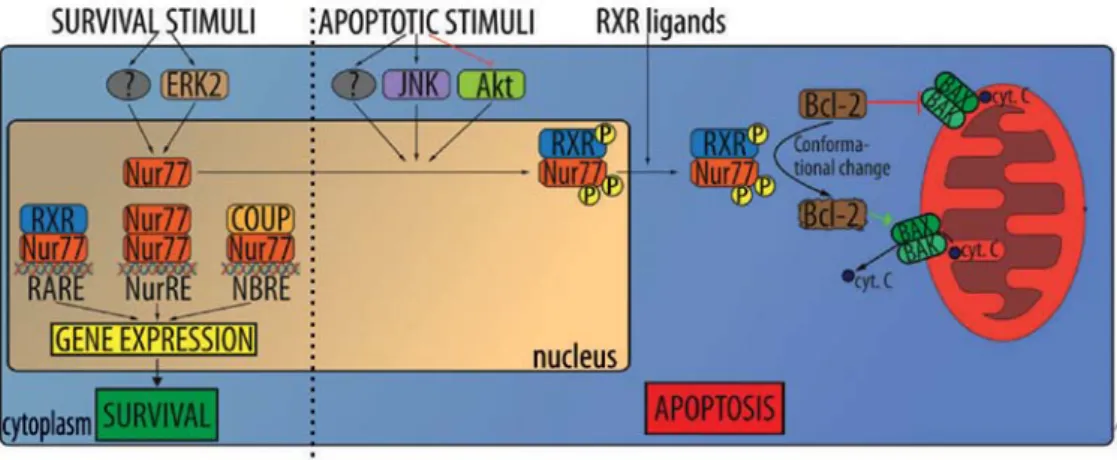
Proopiomelanocortin (POMC). It was shown that proopi-omelanocortin (POMC), an adrenocorticotropic hormone precursor, is upregulated in VHL-mutated RCC. Regulatory mechanism in which proopiomelanocortin (POMC) and nuclear receptor subfamily 4 group A member 1 (NR4A1)/ Nur77 are upregulated in VHL-mutated RCC was identified. Nur77, a member of the orphan steroid receptor superfamily, is believed to be activated by HIF under hypoxic conditions, and in turn regulate the production of the peptide hormone precursor POMC (46). Nur77 was identified as a critical tran-scription factor responsible for POMC overproduction and to be directly regulated by HIF. HIF-1α (but not HIF-2α) binds to a HIF-responsive site in the Nur77 promoter region, acti-vating the expression of Nur77 during hypoxic (VHL-mutant) conditions. Mutation or deletion of the HIF binding site in the Nur77 promoter region significantly decreased activation of a Nur77 and its target genes. The treatment of cells with Nur77 antisense oligonucleotides reduces POMC transcription under hypoxic conditions. In contrast to the normal control tissue the tumor tissues produce abnormally high amounts of Nur77 and POMC. Taken together, these results strongly suggest that Nur77 is both necessary and sufficient for hypoxia-dependent transcription of POMC (Fig. 1) (46).
3. Pituitary gland hormones
Growth hormone (human growth hormone, somatotropin-HGH, GH). Growth hormones (GH) stimulate proliferation and differentiation of normal human cells but have also been shown to be involved in the development of malignant tumors by leading to excess IGF-I production in the liver, as well as having direct effects via GH receptors (GHR) expressed in a variety of tumors, including RCC, colorectal and breast cancer (12,47). Both GH and IGF-I have been shown to act as oncogenes by inducing mitogenic and anti-apoptotic effects in a variety of tumors and cancer-derived cell lines via endocrine and/or autocrine/paracrine mechanisms. It should be noted that the expression of IGF-IR is increased by the activation of oncogenes such as SV40 T antigen and c-MYB, but decreased by the activation of tumor suppressor genes such as p53 and wT1 (48). Oncogenic transformation seems to be responsible for the local expression of IGF-IR and GHR in tumor tissues including RCC. It has been reported that acromegaly patients have an increased risk of developing malignant tumors, although several epidemiological studies have shown that RCC rarely co-occurs with acromegaly (49). Several epidemiological and experimental studies have proposed the hypothesis that elevated GH/IGF-I levels are associated with oncogenic processes in RCC, thyroid tumors, and colon cancer, and play a crucial role in tumorigenesis in acromegaly and/or the growth in multiple tumors (50). Sekizawa et al (49) described a case in which a 56-year-old man diagnosed with acromegaly was also found to have multiple tumors (ccRCC, colon cancer, follicular thyroid tumor and GH-producing pituitary adenoma). This is a rare case, as the association of acromegaly with multiple tumors other than in MEN1 has not been reported elsewhere in the literature. Nevertheless several large-scale epidemiological studies on the co-incidence of neoplastic diseases and acro-megaly have shown coincidence with RCC.
Adrenocorticotropic hormone (ACTH). The biosynthesis of adrenocorticotropic hormone (ACTH), also known as corticotropin is controlled by a multiple transcription factors through the promotion of proopiomelanocortin (POMC), the precursor to ACTH and other enzymes involved in the synthesis of steroid hormones in pituitary cells. Hypoxia acti-vates the hypothalamic-pituitary-adrenal (HPA) axis, resulting in an increase of ACTH and ACTH receptor expression; this suggests that oxygen fluctuation may influence the release of cellular hormones in the tumor niche (46).
Thyroid-stimulating hormone (TSH). Patients with thyroid disease (TD) and abnormal TSH levels, including on-nodular TD, solitary nodules, multinodular TD, thyroid cancers [with either the presence or absence of anti-thyroglobulin (TgAb)], and anti-thyroid peroxidase (TPOAb) or anti-thyroid-stimu-lating hormone (TSH) receptor autoantibodies, are at higher risk of developing kidney cancer (OR=3.40) compared to the general population (51). whereas, hypothyroidism is associ-ated with longer progression-free survival (PFS) in sunitinib and sorafenib treatments (25,28). The severity of vascular endothelial growth factor receptor tyrosine kinase inhibitor (TKI) therapy-associated hypothyroidism (TSH >10 mIU/l)
is associated with improved treatment efficacy and survival outcomes in patients with metastatic RCC (52). This year meta-analysis suggested that development of hypothyroidism during TKI therapy is not clearly predictive of efficacy in patients with metastatic RCC or advantage in overall survival (OS) (53).
Follicle-stimulating hormone (FSH). Follicle-stimulating hormones (FSHs) are released under the influence of gonadotropin-releasing hormones (GnRH). The FSH receptor (FSHR), which was expected to be expressed only in the ovary and testis, was recently detected in the blood vessels of many solid tumors, including RCC (54,55). FSHR expression was evaluated in the endothelium of 1,336 primary solid tumors, representing 11 tumor types, comprised mostly of genitouri-nary malignancies and 64 RCC cases. The FSHR expression in the neo-vasculature of tumors found it almost exclusively in peripheries, in a region <1 cm inside or outside of the tumor in 70% of cases, but ~30% of samples had equal FSHR expres-sion in total tumor mass. FSHR expresexpres-sion was not detected in the blood vessels of nonmalignant tissues (55,56).
Based on functional studies it was suggested that FSHR may contribute to neoangiogenesis, which would make it an interesting target for therapeutic and imaging purposes, as well as a potentially useful prognostic and/or predic-tive biomarker in genitourinary cancers. FSHR may also contribute to the development of metastases due to its position on the luminal surface of the endothelium. Moreover, FSHR may play a role in tumor intravasation, allowing RCC cells to penetrate through the endothelium and into circulation (57). In addition, FSHR expression at the periphery of tumors (55), where the tumor interacts with the stroma, may suggest its role in metastases. The epithelial-to-mesenchymal transition (EMT) is believed to be critical to the formation of metastases, while the interaction of stromal elements with tumor cells at the tumor periphery is thought to contribute to EMT. FSHR has also been proposed as a marker of tumor endothelium in solid tumors (56). Expression of FSH receptor (FSHR) expres-sion was shown to be effective prediction markers of tumor vasculature response to sunitinib treatment. The percentage of FSHR-stained vessels was on average 5 times higher for patients who responded to treatment than the control group, and almost 8 times higher than in the non-responsive group. However, no significant differences were detected in the total density of vessels between these 3 groups, nor was a significant correlation found between FSHR expression and tumor grade. Nevertheless, a far greater number of FSHR-positive vessels were detected in patients who responded to the treatment. The response threshold between the two groups of patients was defined at 23% FSHR-positive vessels. Not only was a higher density of FSHR-expressing vessels observed in the primary tumors of patients who responded to sunitinib treatment, but the von willebrand factor (vwF) was detected in these vessel (FSHR+/vwF+) (30). Plausible mechanisms that could
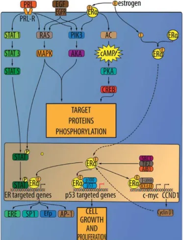
explain this correlation have been hypothesized. One possible mechanism is that FSHR stimulation by FSH leads to VEGF secretion by the ECs, which in turn stimulates VEGFR2 on the ECs as an autocrine mechanism. In fact, it has been observed that the binding of FSH to FSH receptors in ovarian granulosa cells induces an increase in hypoxia-inducible factor
1α protein levels, which consequently leads to upregulation of VEGF production (Fig. 2) (57). It was later suggested that FSHR expression in the blood vessels of ccRCC primary tumors can be used to predict if patients will respond to sunitinib treatment (56). Another possible explanation for the correlation between FSHR expression and sunitinib efficacy is that FSHR activates one or more of the other kinases known to be inhibited by sunitinib like c-Kit, PDGFR, CSF-1R, FLT3 or RET (30).
Treatment. The FSH-FSHR signaling axis can be targeted with drugs in several ways, and GnRH antagonists that decrease FSH levels are currently available. Futhermore, the use of FSH-neutralizing antibodies could potentially cause an even greater reduction in FSH levels. FSHRs may be targeted by the use of molecules with drug- or radio-immunoconjugates. In order to do so, several classes of antagonizing compounds have been identified, most of which are negative allosteric modulators. An example is the thiazolidinone analogues, whose modifications in the chemical structure of the thiazolid-inone backbone allow it to divide its generation into positive, mixed, and negative allosteric modulators of FSHR (30). Several studies have explored the use of FSHR allosteric modulators in vivo. Recently, ADX61623, another negative allosteric modulator of FSHR, was investigated. ADX61623 inhibited some elements of FSHR signaling, such as proges-terone synthesis and cAMP production, but failed to inhibit estradiol synthesis (56). FSHR-antagonizing molecules need to be examined further, as they may play a role in targeting FSHR in RCC and other tumors.
Prolactin (PRL). Prolactin (PRL) is measured by its patho-logical range in patients with RCC. Even patients without paraneoplastic syndromes experience frequent changes that are directly caused by RCC (20), although only a single case of hyperprolactinemia induced by RCC has thus far been reported (58).
4. Adrenal hormones
Glucocorticoid receptors (GR). In normal kidneys, glucocor-ticoid receptors (GRs) are expressed in proximal tubules and glomeruli. In RCC, high GR expression is a positive prognostic marker. Initial studies, conducted in the early 1980s, and that discovered the presence of GRs in kidney tumors, were based on ligand-binding assays (59,60). GRs were found to be overexpressed in 66% of ccRCC cases, 26% of pRCC cases, 14% of oncocytomas and 6% of chRCC cases. Moreover, GR expression serves as a favorable marker and correlates with low nuclear grade and stage. A significant correlation between GR expression and OS in RCC patients may be hypothesized. The majority of patients with ccRCC-expressing GRs were still alive by the end of the follow-up, in contrast to those with expression-negative tumors (59,61). As a result, the correla-tion of GR expression with less aggressive tumor behavior was reported and the anti-proliferative role of GR signaling in RCC was suggested. Suppression of transcription factors, including cAMP response element-binding protein (CREBs), nuclear factor kappa-light-chain-enhancer of activated B cells (NF-κBs), signal transduction activator of transcription (STATs), activator protein 1 (AP-1s), p53s, CCAAT-enhancer-Figure 2. The effect of FSH stimulation on RCC cells.
binding proteins (C/EBP), and SMADs was suggested as inhibitory mechanisms mediated by GR signaling (59,62). In RCC, two main isoforms of GR were analyzed. GR-α, a predominant isoform, exhibits steroid binding activity. GR-β has a lower expression in normal kidney tissues, but is overexpressed in inflammatory blood cells localized in the tumor (59). In RCC, GRs have been shown to bind not just glucocorticoids (63), but with progesterone, diethylstilbestrol, testosterone, and aldosterone as well, albeit with low affinity. In addition, progestin or medroxyprogesterone easily binds to GRs. As a result of medroxyprogesterone acetate treatment RCC tumor regression was reported (64).
Treatment. Efficacy of RU486 (Mifepristone), a glucocorticoid receptor antagonist, on TRAIL-induced apoptosis in human RCC, Caki-1, A498 and ACHN cell lines was measured. RU486 is known as an anti-progesterone and anti-glucocorticoid agent. Its low dose has no effect on apoptosis, but sensitizes Caki-1 cells to TRAIL-induced apoptosis. As a result, RU486 enhances TRAIL-mediated apoptosis through the downregu-lation of Bcl-2 and c-FLIP(L) as well as CHOP-mediated DR5 upregulation. TRAILs bind to their receptors (death receptors DR4 and DR5) to form death-inducing signal complexes (DISC) by recruiting caspase-8, which in turn increases apop-tosis through the activation of caspase-3. In addition, TRAILs activate mitochondrial apoptotic pathways: TRAILs release cytochrome c to the cytosol via disruption of mitochondria membrane permeability, allowing it to form apoptosomes. These responses were also commonly observed in a variety of other cancer cells, including SK-Hep-1 (hepatocarcinoma cells) and HT29 (colon cancer cells) (62). In addition, RU486 slightly inhibited XIAP expression, increased active cleaved caspase-3 and PARP cleavage, induced cytoplasmic histone-associated DNA fragments, and blocked chromatin in the nuclei, indicating apoptosis. Moreover, RU486-mediated TRAIL-induced apoptosis acts independently of GR and PR signaling, through CHOP-mediated DR5 expression and the downregulation of Bcl-2 and c-FLIP expression. As TRAILs induce apoptosis only in tumor cells, they may be a promising cancer treatment, and medications such as RU486 may be a novel strategy for the recovery of TRAIL sensitivity in cancer cells otherwise resistant to TRAIL (62). In a study conducted by Arai et al (65), the inhibitory effects of dexamethasone (DEX) on the growth of RCC in vivo and in vitro were examined and suppression of NF-κB activation was measured. DEX binds more powerfully to the glucocorticoid receptor than cortisol. DEX has long been used to suppress inflammatory reactions in patients with advanced cancers by activating transcription factor NF-κB and its target genes IL-6, IL-8 and VEGF. All the RCC cell lines tested responded to DEX treatment, but RCC growth suppression was more remarkable in vivo than in vitro. Caki-1 cells expressed a low level of GR protein, and GR was translocated into the nucleus. Moreover, after 6 weeks of treatment, mean tumor volume statistically decreased. H&E staining showed less inflammatory cells and necrotic tissues in DEX groups as well. Intracellular IL-6, as well as those in the conditioned medium, was downregulated in all cell lines following treatment. Concentrations of IL-8 in the conditioned medium were remarkably decreased in NC65 cells, while VEGF secretion was lowered by 30-65% in all RCC lines,
with the inhibition of the nuclear translocation of NF-κB (65). After therapy with dexamethasone, cases of complete regres-sion (CR) occurred, and the cases included patients with metastases located in the brain and lungs. Dexamethasone treatment may thus be a candidate for RCC supportive therapy. This compound was shown to inhibit expression and signaling mediated by NF-κB, VEGF, IL-6 and IL-8. Cortisone therapy was also reported to be successful in some cases. Over 10 years of complete remission (CR) was demonstrated in patients with retroperitoneal lymph node involvement and liver metastases. This observation supports the thesis that GR and its agonists may play an important role in anticancer ccRCC therapy (66). Using dexamethasone in combination with kinase inhibitors was examined (59).
Mineralocorticoid receptors (MR). Mineralocorticoid recep-tors (MR) in the kidneys are expressed in Henle's loop, distal tubules and collecting ducts. Due to its compartment-specific expression in the kidneys, MR was suggested as a diagnostic marker for oncocytomas and chromophobe RCC. MR was indicated as both a highly specific and sensitive marker of the distal nephron cells and its derived neoplasms (67). Aside from MR, 11beta-HSD2 [11β-Hydroxysteroid dehydrogenase (HSD-11β or 11β-HSD)] was also investigated as a RCC subtype marker. Expression of both MR and 11beta-HSD2 was detected in the distal nephrons of normal kidneys. MR and 11beta-HSD2 were highly expressed in 90% of chromo-phobe RCCs and 93% of oncocytomas, thus reflecting their histogenetic origin. No MR staining was detected in ccRCC, since its absence corresponds with its proximal tubule origin. Only 2.6% cases of ccRCC showed focal positivity for 11beta-HSD2, whereas all papillary RCCs were negative (67). In functional MR studies, aldosterone binding was observed to be more significantly decreased in clear cell RCCs than in normal tissues, both in the cytosol and in the nucleus (59).
Recent studies carried out in the US and Eastern Europe have shown that increased activity of the renin-angiotensin-aldosterone system (RAAS) is one of the major risk factors influencing genetic changes and is mainly observed in cases of ccRCC. Epidemiological evidence suggests that RAAS-related hypertension and obesity increase the risk of RCC development (68), while epidemiological evidence from the MRC Blood Pressure Unit in Glasgow has shown that pharmacological suppression of the RAAS lowers the risk of developing renal cancer in hypertensive patients as opposed to their non-hypertensive counterparts. In experimental models, the inhibition of angiotensin-converting enzymes restricted the growth of human renal cancer cells in a mouse xenograft system; and in vitro, restored the sensitivity of mouse Renca cells to the growth-suppressing effects of TGFβ. Moreover, in several experimental animal models of hypertension, overactivity of the RAAS was accompanied by renal tubular hyperplasia. One potential mechanism to explain this is the influence of the RAAS on the expression and activity of the K-ras cellular oncogene to promote tumor growth. King et al (68) also observed that the K-ras isoform K-RAS4A is aldosterone-sensitive in renal cancer, and that its overexpres-sion contributes to the survival and increased proliferation of RCC cells in response to RAAS activation. This was demon-strated by adding spironolactone to RCC cultures, which
mineralocorticoid activation and K-RAS4A expression were shown to support RCC proliferation. A second study treatment involved transfection with siRNA, which also suppressed K-ras protein expression and decreased cell count by ~40-73% after 72 h. In addition, it was found that K-RAS4A acts through the Raf and Akt pathways to support the survival and growth of RCC cells. Both Raf and Akt proteins were mark-edly reduced following a decrease in K-RAS4A. According to Varela et al (69) there is evidence that K-ras may act in tandem with the SwI/SNF/PBRM1 complex to promote the formation of renal carcinoma. Kotelevtsev et al (70) demonstrated that the mineralocorticoid receptor is expressed in about half of human clear cell carcinomas. In this model, K-RAS4A expres-sion was induced by aldosterone treatment and its activation sustained by an as yet unproven tyrosine kinase that may have been involved with the epidermal growth factor or insulin-like growth factor 2 receptor; also, K-RAS4A links the phospha-tidylinositol 3-kinase pathway to ENaC activity. Thus, K-ras is also involved in cell growth promotion via aldosterone-sensitive growth responses (70).
Vitamin D receptors (VDR). Vitamin D receptor (VDR) signaling regulates multiple target genes and promotes cell differentiation, angiogenesis, and angiogenic proliferation in multiple tissues, including the kidneys. Long-term vitamin D serum levels were suggested to be inversely correlated with renal cancer development risk. VDR was shown to be overex-pressed in several malignant neoplasms, including metastatic RCC tumors, and in poorly differentiated and sarcomatous RCCs of Fuhrman grade IV in particular (59,71). The rela-tionship of 1,25-Dihydroxyvitamin D3 receptors (VDR) to histological features in RCC was further investigated (72). VDR expression was shown to be absent in the proximal tubules. In contrast, tumors originating from the distal nephron tested positive for VDR, including the majority of papillary RCCs, chromophobe RCCs and oncocytomas. Positive VDR staining could help in efforts to differentiate between papillary RCC and clear cell RCC with papillary features (73). VDR immu-nohistochemistry results can help classify RCC tumors (73). Generally, ccRCC tests negative for VDR. In addition, ccRCC exhibits decreased VDR mRNA levels when compared to normal kidney tissues, and VDR staining is limited only to the peripheral region of the tumor (73). Analysis of RCC samples exhibited expression of 1,25-dihydroxyvitamin D3 receptors in 81% of the tumors. Absence or loss of the receptor was associated with low differentiated sarcomatoid tumors with a poor prognosis. On the other hand, the expression of VDR receptors in the receptor-positive tumors did not correlate with clinical stage or pathological grade or RCC (72). The expres-sion of 1,25-(OH)2D3 receptors was also analyzed in normal
kidney tissues and primary RCC samples. In 83% of RCC cases, 1,25-(OH)2D3 receptor expression was found, and in
65% of the cases the receptor was overexpressed. The mean expression of the 1,25-(OH)2D3 receptor in RCCs is
signifi-cantly lower than in normal kidneys. The lower 1,25-(OH)2D3
receptor expression may be due to lack of differentiation in the malignant-transformed renal cells. A functional analysis of the 1,25-(OH)2D3 receptor in both normal and RCC tissues
A possible correlation between 1,25-(OH)2D3 receptor
expres-sion in primary tumors and the late development of lymph node metastases was also found (74). In an earlier study, no significant difference between expression of VDR in ccRCC and control tissues was found (75). Such a discrepancy could be explained by the variable degree of differentiation in the tumors analyzed in these two studies.
Subsequent physiological studies have revealed the compli-cated role vitamin D plays in renal tissue. According to several biochemical studies, even when exogenous vitamin D supple-mentation was present, the formation of VDR-DNA complexes in RCC was decreased. This impairment was shown to be secondary to deregulated VDR heterodimerization with RXR (retinoid X receptor) in tumor cells. It was shown that the expression of RXR-γ in RCC was correlated with OS of RCC patients (59). Another study discovered that VDR mRNA was almost undetectable in clear cell RCC as compared to normal kidney tissue, and was accompanied by the underexpression of CYP2R1, CYP27B1 and CYP27A1 enzymes (76). No correla-tion between concentracorrela-tions of VDR receptors and cancer cell DNA-ploidy was found. The mean VDR concentration used was 8.4 fmol/mg of protein (range, 2.8-15.9) in diploid tumors and 7.0 fmol/mg of protein (range, 0-27.8) in DNA aneuploid tumors (11 out of 22) (74).
Treatment. Vitamin D exerts its anticancer activity by inducing apoptosis and inhibiting cell proliferation. This effect has been demonstrated in many cancer models, including malignant melanoma, breast cancer, osteogenic sarcoma and acute myelogenic leukemia (77). In one report, proliferation of the RCC cell line, derived from a pulmonary metastasis, was inhibited by calcitriol treatment (72). In another study, RCC tumor growth was inhibited, pulmonary and hepatic metastates were decreased, and the OS of mice was prolonged in relation to the dose of vitamin D provided (78). Although vitamin D-based supplementation therapy shows promising results, its implementation is impeded by its toxic hypercal-cemic effect (59). Bypassing this toxicity is possible by using alternative vitamin D derivatives. At the same time, alkylating derivative 1,25-dihydroxyvitamin D3-3-bromoacetate was investigated in vitro and in vivo. This study showed that 1,25(OH)2D3-3-BE had a significantly more potent effect on
the inhibition of human RCC cell line proliferation than an equivalent concentration of normal vitamin D. In addition, an increase in the apoptosis rate was observed with a reduction in cyclin A expression (79).
RAR (retinoic acid) receptors and RXR (retinoid X) receptors. Expression of retinoic acid receptors (RARs) and retinoid X receptors (RXRs) are cell type-specific. RAR and RXR are expressed in proximal tubules and renal interstitial cells. RAR-β mRNA was found to be constrictively expressed in normal kidney cells. Furthermore, RAR and RXR expres-sion was reported in podocytes (74). Expresexpres-sion of RXR-β
was reported in proximal tubules and interstitial cells, while RXR-α and RXR-γ were present mostly in the nuclei of proximal tubule cells (80). In the analysis of 49 RCC tumors, RXR-α expression was detected in 70% of the cases, RXR-β
in 47% of the cases, and RXR-γ in 85% of the cases (80). Only RXR-γ expression was found to be inversely correlated with the TNM stage, because patients with RXR-γ-positive tumors were observed to have prolonged OS (59). RAR-β probably contributes to tumorigenesis in RCC because the deletion of its gene on the short arm of chromosome 3 is frequently found in RCC and other cancers. RAR-β mRNA is often not detectable in RCC cell lines. This also suggests minimal inhibition or resistance to 13-cis-RA treatment (59).
Treatment. In clinical studies, the effect of retinoic acid on RCC was tested in patients with multiple metastases. In a large trial, responses to therapy with either IFN-α 2a alone or in combination with 13-cis-retinoic acid (13-CRA) was evaluated. This study showed that 19% of patients on combined treatment did not show progression after 24 months. Progression-free (PFS) and overall survival (OS) rates for patients were significantly longer when treated with combined IFN-α 2a and 13-CRA therapy (81). In another study, 3 different treatment arms were analyzed: i) a treat-ment group given a combination of IFN-α 2a, IL-2, and fluorouracil; ii) a treatment given a combination of IFN-α 2a, IL-2, fluorouracil, and 13-CRA; and iii) a control group given vinblastine and IFN-α. The results showed that group 1 and 2 had a significantly longer progression-free period and overall survival rate, although no significant difference in efficacy between them was demonstrated (82). According to both of the studies mentioned above, retinoid treatment can have a beneficial effect, at least for a subgroup of RCC patients. 5. Sex hormones
Estrogen receptors (ER). Two isoforms of estrogen receptors (ER) are known and are encoded on different chromosomes: ER-α and ER-β. ER-α is mainly expressed in reproductive organs, while ER-β is expressed in genitourinary human tissues in the central nervous system. Both types of estrogen receptors are also expressed in normal renal interstitial stromal cells. In tumor samples, ERs are found in stromal tumors, cystic nephromas and angiomyolipomas (59). The relative concentrations of these receptors in the renal tumor is as follows: progesterone > estrogen > androgen > glucocorticoid > mineralocorticoid receptors (Fig. 3) (83). The affinity of the ER-β isoform to bind with 17-β-estradiol is similar to ER-α. At the same time, androgens and phytoestrogens are bound with greater affinity by ER-β (84).
Clinical studies. Initial studies in estrogen and RCC covered the procedure for experimental induction of cancers. The procedure covers the supply of female sex hormones to male mice, or male Syrian hamsters, as well as in female guinea pigs, after ovariectomy, during low progesterone secretion, or before reproductive maturity. Endocrine balancing following the resection of ovaries has been seen to delay the occur-rence of tumors in various organs, including the kidneys. Moreover, environmental exposure to both xeno-estrogen and estrogen is associated with cancer development. RCC can be experimentally induced in animal models by exposure to high levels of estrogen, which might suggest the involve-ment of estrogen receptors in the etiology of renal cancer
and possibly, xeno-estrogen as well. In normal human kidney and RCC cytosols, both estradiol and progesterone receptors have been found, although a lower binding capacity of human renal cancer cytosols compared to that of normal kidney cytosols was reported, perhaps due to a nuclear translocation of the receptors in the neoplastic tissue (12). Early studies of ER in renal cancer showed that the expression of ERs in RCC was highly variable (85). ERs were detected in only 30% of the tumors and 40% of normal kidney tissues, but in some experiments ERs were not detected in any RCC samples. At the same time, ER expression in the interstitial cells of the human kidney, in both adults and children, has also been reported. ER- and PR-positive stroma, described as Müllerian-like, was also found in normal kidneys and meta-plastic kidney tissues (86). A recent immunohistochemical study covering 182 RCCs of different subtypes found that ER immunoreactivity was demonstrated in 1.1% of patients, one with ccRCC, and the other, chromophobe RCC (77). Expression of ER and PR in some human renal tumors has led to suggestions that some of these benign tumor types, particularly mixed epithelial-stromal tumors (MEST), cystic nephromas, or angiomyolipomas with epithelial cysts (AMLEC), may be related to excessive exogenous estrogens. At the same time, the incidence of renal cancer appears to be gender-related, since it is twice as high in men than it is in women. In addition, ER-α genetic polymorphism in the kidneys also seems to play an important role in the develop-ment of renal cancer (87).
as angiomyolipomas have been reported to increase in size during pregnancy or as a secondary effect of oral contra-ceptive therapy. In animal studies, tumors similar to that of human angiomyolipomas have also been observed to grow with estrogen therapy and to regress on tamoxifen. In hamster animal models, renal epithelial tumors have been shown to be inducible with estrogen. Additionally, ovarian-like stromas are reported to be quite common; but on the other hand, they do not constitute the exclusive type of stroma in most tumors of a specific type, and in many tumors no ovarian-like stromas are even present. In early clinical reports, the presence of steroid receptors in renal tissue extracted from nephrectomy specimens was correlated with responses to progesterone-based therapy. In this trial, tumors obtained from 23 patients were examined. In the cytosol fraction of cancer cells, 61% were found to contain ER and 61% were found to contain PR. Moreover, 39% of the tumors tested positive for both ER and PR, while 17% tested negative for both ER and PR. ER and PR were also examined in the nuclear fraction of 3 renal cancers, and estradiol nuclear receptors were found in 2, while progesterone nuclear recep-tors were found in 3. Next, treatment response was evaluated. with few exceptions, patients were treated with the following progestins: medroxyprogesterone acetate (MPA) with R5020, and 17,21-dimethyl-19- norpregna-4,9-diene-3,20-dione, after nephrectomy. The best responders to progestin treatment were those patients with hormone-dependent tumors (ER+
PR- or vice versa). These patients did not develop metastases
for up to 22 months following nephrectomy. Other patients, also treatment responders, also achieved disease stabilization (SD) with prolonged OS (12). In another study that included primarily perimenopausal female patients (and 1 male patient with a history of diethylstilbestrol treatment after prostatic adenocarcinoma), ER expression in RCC was observed. The excessive growth of RCC in these patients was suggested to be stimulated by therapeutic hormones, overproduction of estrogen, or perimenopausal hormonal abnormalities (88). Functional cell biology. A novel role for estrogen-induced cathepsin D in hamster kidneys during tumorigenesis that involves renal tubular damage followed by cell proliferation may contribute to renal tumor formation. Primary estrogen-induced RCCs and their metastases showed significantly increased levels of all 3 cathepsin D isoforms. A concomi-tant increase of cathepsin D, along with estrogen receptor proteins, indicates that cathepsin D gene expression is under the control of estrogen and is possibly mediated via induced renal estrogen receptors. Cathepsin D is considered to be an early estrogen-response gene. Oncogenes such as c-Myc, c-Fos and c-Jun (and all early estrogen-response genes) are over-expressed in the kidneys after only four months of estrogen treatment. Either tamoxifen or dihydrotestosterone (DHT) prevented the rise in cathepsin D and estrogen receptor content observed after estrogen treatment alone (89). Numerous in vitro studies have suggested the possibility that potentially reactive intermediates of estrogen may be the causative factors contributing to renal cell injury during chronic and prolonged exposure to estrogen (18,90). Estrogen-mediated cytotoxicity
ER-α. Single nucleotide polymorphisms of the ER-α gene in RCC samples were investigated. Six different polymorphic loci of this gene were analyzed in 113 RCCs to determine their frequency. The results showed that the distribution of genotypes of codon 10 varied between RCC patients and healthy controls, and the relative risk of this genotype was calculated as HR=2.51. Analysis of DNA from pairs of cancerous and normal tissues detected genotypic changes in 9% of cancer samples on (and exclusively) exon 1 (codons 10 and 87) of ER-α. This leads to the conclusion that codon 10 polymorphism on exon 1 of ER-α may be involved in RCC development. No differences were observed between men and women in the distribution of codon 10 genotypes in the control group. No association was observed between gender, age, or stage of renal cancer with polymorphisms of other genes such as p53 (91,92). Estrogen receptor-α
(ER-α) was recently found to be a novel proteasomal degradation target of the pVHL E3 ligase. Overexpression of VHL suppresses ER-α expression in RCC, whereas downregulation of pVHL can increase ER-α expression (8). Overexpression of ER-α may increase HIF-1α transcription factor activity. In VHL-deficient cells, the expression of ER-α and HIF-1α is retained, and blocking ER-α using its inhibitor could suppress the proliferation of VHL-deficient cells as effectively as hypoxia-induced growth suppression. The experiment also showed the anti-proliferative effect of faslodex (ER-α inhibitor) in VHL-deficient cells by inducing p53 expression (8). It was also shown that after binding to ER-α, the estrogen complex promotes the transcription of growth-related factors that enhance gene expression and mitosis and promotes proliferation, leading to cancer devel-opment and tumor progression (93).
ER-β. Previous studies indicated that ER-β has anti-prolifera-tive and apoptosis-inducing functions (69). In RCC ER-β was suggested as tumor suppressor. ER-β is highly expressed in RCC cell lines, and estrogen-activated ER-β reduced growth hormone downstream signaling activation of the AKT, ERK, NF-κB, MMP9 and JAK signaling pathways but also increased apoptotic cascade activation (caspase-3, -8, -9, BID activation and reduced Bcl-2 and survivin expression). ER-β signaling increases RCC cell migration via induction of the VEGFa/ HIF2α pathway. Moreover, in RCC tumors, infiltrating neutro-phils modulate the expression of ER-β and in turn promote RCC migration (94). ER-β has higher expression in normal renal tissue than in tumorous tissues. In contrast, as no ER-α
expression was observed in these cell lines, only ER-β was activated through estrogen stimulation. Estrogen treatment significantly decreased the proliferation, migration, invasion, and increased apoptosis of 786-O (high endogenous ER-β), and ER-β siRNA-induced silencing attenuated the estrogen-induced effects. Ectopic expression of ER-β in A498 (with low endogenous ER-β) increased sensitivity to estrogen. Therefore, estrogen is believed to activate the ER-β suppressive function, resulting in the elimination of cancer cells (93) which leads to different RCC incidence rates between males and females. It also implies that ER-β may be a useful prognostic marker
for RCC progression and a novel developmental direction for improved RCC treatment (95).
Treatment. Chronic treatment with diethylstilbestrol (DES) and polydiethylstilbestrol phosphate produced renal tumors in male hamsters. The presence of renal tumors results in increased activity of hepatic glucuronyl transferase that dimin-ishes DES in chronically treated hamsters. The antiestrogen nafoxidine, administered along with DES, completely inhibits tumor formation (96). Ten patients with advanced RCC were given combined chemoendocrine treatment with tegafur and tamoxifen. One out of 2 patients with ER-positive tumors and 3 out of 4 patients with ER-negative tumors responded favor-ably to this treatment (97).
Androgen receptors (AR). In normal kidneys, androgen receptors (AR) are constitutively expressed in the proximal and distal tubules and localized in cell nuclei. They are also focally expressed in some Bowman's capsule cells. Expression of AR is higher in adjacent normal kidneys than in RCC tissues. AR expression in RCC is negatively correlated with pT stage and Fuhrman's grade (97). In RCC tumors, ARs are detectable in clear cells, papillaries and chromophobe RCCs. No difference of AR immunoreactivity was detected between histological subtypes (77). Upregulated expression of ARs was shown as a favorable marker in RCC (77,98). In another study by Brown et al (99), which included primary clear cell RCCs and their metastases, AR immunoreactivity was mainly present in primary tumors, but not in their respec-tive metastases. AR expression was higher in adjacent normal kidneys (90.9%) than in RCC tissues or control group human ccRCC cell lines. Specifically, there were 40.7% AR-positive cases in pT1 compared with 8.0% in pT3, and 50.0% of grade I cases were found to be AR-positive compared with 12.9% in grade III. AR expression was more abundant in primary RCC tissues (12.5%) than in their respective metastases (0%). There was no significant difference found in AR-positive rates between male and female RCC patients from the same subgroups who had the same pT stages or Fuhrman's grades. Immunohistochemical analysis of 182 RCC tumors for ER, PR, and AR expression in relation to associations with histological subtype, pT stage, grading, gender and impact on disease-free survival was conducted. AR expression was found in 27 of 182 tumors (14.8%), 24 males and 3 females. AR expression was significantly associated with a lower stage and grade, moderate, or high differentiation of tumor cells. Outcome expectancy decreased with de-differentiation and tumor growth. AR-positive RCCs showed a significantly better prognosis (77). Another study discovered AR to be inversely correlated with TNM stage pT1 tumors being AR-positive for 27% of cases, in contrast to pT3 tumors being 4% positive. Additionally, the presence of AR was inversely correlated with nuclear grade. Thus, patients with AR-positive tumors were observed to have a longer progression-free condition and overall survival rate (77). Recently, high AR expression was associated with favorable prognostic factors, such as low pT stage and low histologic Fuhrman's grade among the sample of 120 primary RCCs (100,101). In functional studies androgen receptors (AR) were shown to induce HIF2α/VEGF signals that potentially drive RCC progression. Anti-AR targeting
inhibits RCC cell migration and invasion (102). Normal kidney cells that were transformed into cancerous versions had decreased AR expression rates or more localized cell nuclei. Observations of generally high levels of AR in hamster renal tumors are consistent with the finding that the growth rate of transplanted primary renal tumors is stimulated by testos-terone propionate (83). Dihydrotestostestos-terone-specific receptors were present in all RCC samples examined (20 of 20) and in 13 of 14 normal renal parenchyma samples. Testosterone receptors were found in a smaller number of cases. Moreover, significantly higher levels of the dihydrotestosterone receptor were found in high-stage compared to low-stage tumors (103). In summary, the utility of AR in RCC as a prognostic, diag-nostic or therapeutic factor is uncertain and requires further investigation and observation.
Treatment. Hormonal manipulation in patients with RCC seems to increase survival in patients with these receptors. The survival rate of patients with 1 or more receptors was signifi-cantly higher than that of patients with no receptors (104). Ahmed et al (105) investigated the efficacy of flutamide (an anti-androgenic drug) treatment in Phase II disease-oriented drug trials on patients with advanced bidimensional RCC. Of 25 treated cases, 1 experienced partial cancer remission and 2 were deemed progression-free. Flutamide showed no benefits in patients with disseminated RCC. Sixty-two specimens were evaluated for steroid binding sites: 33 of 62 specimens showed no hormone-binding sites, and only 12 cases exhibited androgen binding.
Progesterone receptors (PR). In normal human kidneys, 30% of investigated tissue samples were positive for progesterone receptors (PRs) in the mesangial cells of glomeruli, in intersti-tial stromal cells and in several tubules (86). Two predominant isoforms of PR were found, PR-α and PR-β, both of which are derived from 1 gene due to alternative promoter usage. Their DNA binding and steroid hormone activities are similar, although higher transcriptional activating potential was observed in the case of PR-β (59). The PR was found in 40% and 30% of normal and carcinomatous kidney tissue, respec-tively (105). PR expression was decreased in 10% of tumors and increased in only 1% of patients, one with ccRCC and one with pRCC (77). Less frequent than ER, PR was found in stromal cells of benign renal carcinomas: angiomyolipomas, cystic nephromas, oncocytomas, mixed epithelial and stromal tumors, and also in chromophobe RCC (86). Expression of PR in tumor stroma was also reported in benign renal tumors as well as in normal kidneys and metaplastic nodules (86). In general, PR appears to be a highly specific marker for chro-mophobe RCC. It is also a highly specific and sensitive marker for oncocytomas. In particular, PR immunoreactivity is more abundant in oncocytomas than in chromophobe cancer, which can be used to distinguish between these two tumor types. Moreover, PR expression is not detectable in other subtypes of RCC tumors, such as pRCC or ccRCC with eosinophilic cytoplasm (59).
Treatment. Estradiol receptor (ER) and progesterone receptor (PR) expression was evaluated in 27 RCCs in an attempt to predict the response to progestational therapy. Patients whose
with ER-PR-renal cancer showed negative results in follow-up therapy (12). In the study of McDonald et al (13), PR was measured in eight RCCs compared to nine normal renal tissues and one melanoma tissue sample. PR was identified in all of the samples, with the exception of one RCC. Three patients, all of whom had receptor-positive tumors, were treated with medroxyprogesterone acetate for metastatic disease. In one of these patients, an objective response to treatment was achieved. Nakano et al (104) also demonstrated that hormonal manipulation in patients with one, or more receptors, results in a significantly higher survival rate.
6. Summary and conclusions
In the past several decades, clinical observations and molecular studies have led to the hypothesis that RCC is a hormone-dependent tumor. Steroid receptors are transcrip-tion factors that control cell differentiatranscrip-tion, proliferatranscrip-tion and death (12). Active hormone receptors are found to be abnor-mally expressed in RCC cells, while abnormal endocrine stimulation is thought to influence cell proliferation, migration and angiogenesis. The expression of steroid receptors varies between normal kidney tissues and RCC tumors (59). To date, the study of these receptors in RCC has been confined to estrogen (ER) and progesterone receptors (PR), but the employment of novel molecular biology and cell biology tech-niques have supplemented the data on steroid hormone RCC dependence. More recently, immunocytochemistry, tissue and protein microarray platforms, mass spectrometry, quantitative reverse real-time PCR, whole genome cDNA analysis, and DNA sequencing have served as functional studies of steroid hormone receptors in renal cancers (68,106). The molecular role of each hormone in RCC pathophysiology is currently still being elucidated, in order to provide a precise model of hormonal interactions with oncogenesis. RCC patients without any paraneoplastic syndromes have experienced frequent changes in peptide hormone balance, which is either directly or indirectly caused by renal cancer tumor masses and which may in turn influence disease biology. On the basis of immu-nohistochemistry staining, specific hormones can be used as potential biomarkers of progression of oncogenesis, or to aid in the identification of tumor types. Yet, another targeted approach would be to use hormonal receptors for treatment, so as to inhibit hormonal activity with chemical inhibitors. The employment of techniques such as protein and tissue micro-array technology, whole genome micro-arrays, mass spectrometry, DNA sequencing, and cell cultures has helped to reveal the expression and role of steroid receptors and their signaling pathways.
Starting with hypothalamus hormones, the presence of the GHRH ligand has been demonstrated in cancer cells, suggesting that GHRH could be a growth factor. In turn, GHRH antagonists exhibit antitumor effects by suppressing the growth of ccRCC lines xenografted into nude mice and inhibiting the growth of orthotopic Caki-1 human RCC, as well as the development of metastases in lung and lymph nodes. The main action of GHRH antagonists in vivo appears to be the direct suppression, through specific binding sites, of
auto-and IGF-II (IGF2) in tumors. The overexpression of GnRH and its receptor has been found in ccRCC. As LHRH receptor expression was found to be very high in RCCs, its inhibitors can be targeted to ccRCC tumors expressing these receptors for therapy. GnRH antagonists effectively inhibited the growth of tumors in the Caki-1 cell line xenografts of nude mice. As a result, this group of compounds was proposed as a therapy for patients with metastatic or recurrent ccRCC. The role of somatostatin is still unproven and requires further elucidation. On the one hand, in Phase II trials somatostatin analogue administration did not result in the control of RCC growth. However, the findings did raise the possibility of using renal-derived somatostatin to modulate tubular cell function through the family of G protein-coupled receptors via autocrine and paracrine modes. It was proposed that the indirect antitumor effects of somatostatin receptors are anti-angiogenic actions. CRH and Ucn were reported to suppress neovascularization through the reduction of VEGF and the inhibition of tumor cell cycling. POMC was found to be constitutively upregulated in VHL-mutated renal cell carcinoma via Nur77: a critical transcription factor responsible for POMC overproduction that is directly regulated by HIF-1α during hypoxic conditions.
Biochemical analyses revealed that serum levels of pituitary gland hormones are also significantly modulated in patients with urogenital tumors. Elevated GH and IGF-I levels are associated with oncogenic transformation in a variety of tumors affecting the general population, and induce mitogenic and anti-apoptotic effects; for instance, in RCC, thyroid tumors, and colon cancer, due to local expression of IGF-IR stimulated by excess production in the liver and direct GHR effects in the tumor tissues. These mechanisms can be easily observed in acromegaly patients, who can have multiple cancers simultaneously. ACTH and ACTH receptors are stimulated by the HPA axis in hypoxia, as their production is controlled by the main transcription factor Nur77, the same as in POMC production. Retinoic acid was shown to inhibit Nur77 in the prevention of Cushing's syndrome. TSH and PRL are measured by pathological range in patients with RCC, and are sensitive to indicating distant metastasis in ccRCC carriers. Moreover, hypothyroidism is associated with longer PFS in sunitinib and sorafenib treatments. Prolactin eleva-tion was found in 45% of ccRCC patients, and its level was unrelated to the stage of the disease. Serum PTH was found to be decreased in patients with tumor dissemination. The FSHR was identified in the tumor endothelium of many genitouri-nary malignancies, including RCC, in which its expression was mostly observed as being equally located throughout the tumor's neovasculature. According to one hypothesis, FSH signaling could induce VEGF in tumor endothelium, which could contribute to the development of metastatic disease. On the other hand, it makes FSHR an appealing target for therapeutic and imaging purposes and as a prognostic biomarker in genitourinary cancers. FSHR antagonists, such as thiazolidinone analogues, neutralizing antibodies, and negative allosteric modulators should be further examined in clinical trials. GRs are abundantly expressed in ccRCC tumors. Their expression was correlated with a low nuclear grade and tumor stage. High GR expression is considered to
be a marker of less aggressive RCC tumors. Prolonged OS was reported in RCC patients with GR-positive tumors. GR signaling in RCC cells likely results in the suppression of other transcription factors induced by signaling in cancer cells. MR appears to be a specific and sensitive marker of the distal nephron and its derived carcinomas: chromophobe RCCs and oncocytomas. In the case of ccRCC, MR staining was rarely detected. Therefore, MR serves as a marker for the expres-sion of subtypes of the major types of renal cell neoplasms. In addition, recent studies have also shown that increased activity of the renin-angiotensin-aldosterone system (RAAS), mostly due to hypertension and obesity, is one of the major risk factors influencing genetic changes, and is mostly observed in clear cell forms of RCC. Inhibition of angiotensin-converting enzymes restricts the growth of human renal cancer cells in a mouse xenograft system, and in several experimental animal models of hypertension, overactivity of the RAAS is accom-panied by renal tubular hyperplasia. A potential mechanism for this is the influence of isoforms of the K-RAS oncogene, whose overexpression contributes to the survival and increased proliferation of renal cancer cells in response to activation of the RAAS. The expression of VDR is inversely correlated with RCC development risk. The downregulation or loss of receptor expression was only reported in poorly differentiated sarco-matoid tumors with a poor prognosis. However, the amount of the receptor in the receptor-positive tumors did not relate to the other clinical and pathological features of the patients. Vitamin D, depending on dosage, inhibited cancer growth, prolonged overall survival in mice, and reduced hepatic and pulmonary metastates. Because of the vitamin D hypercal-cemic toxic effect, alternative vitamin D-like molecules have been explored and have shown promising results; these include alkylating derivatives of 1,25(OH)2D3. RAR and RXR are
normally expressed in proximal tubules and interstitial cells. RAR-β is involved in solid tumorigenesis, as it is associated with the deletion of the short arm of chromosome 3, where it has been mapped. RAR-β mRNA was not detected in renal cancer cell lines, suggesting either resistance to or minimal inhibition with 13-cis-RA treatment. RXR-γ was found to be a favorable marker in RCC, with an inverse correlation with clinical and pathological stages. Patients with RXR-γ-positive tumors were observed to have prolonged OS. Moreover, reti-noid treatment with 13-cis-RA resulted in prolonged PFS and OS in RCC patients.
Both ER and PR have been found in cytosols of human RCC, but with a lower binding capacity of cancer cytosols compared to that of normal kidneys, likely due to a nuclear translocation of the receptors in the neoplastic tissue. In clinical studies, positive responses to progestin treatment were demonstrated in patients with hormone-dependent tumors (ER+ PR- or vice versa), and in those who either did
not develop metastases within 18 to 22 months of nephrec-tomy or had at least achieved stabilization of the disease and a longer period of survival. The question of the influence of exogenous estrogens is still unclear. First, the expression of ER and PR in some benign human renal tumors has led to suggestions that they may be related to excessive exogenous estrogens, as human kidneys are traditionally thought to be unresponsive to estrogen. Furthermore, angiomyolipomas have been reported to increase in size during pregnancy or
secondary oral contraceptive therapy. RCC can be experi-mentally induced by exposure to estrogens in animal models. Moreover, renal cancer incidence seems to be gender-related, with an incidence that is 2 times higher in men than in women. The data strongly suggest an environmental, exogenous influence of estrogens in RCC etiology, but more research is still needed. The frequency of ER expression in human RCCs was highly variable in different RCC studies. Many reports provide evidence that ER is most often not expressed in RCCs. Moreover, cathepsin D was found to be estrogen-induced in hamster kidneys, leading to tumorigenesis by the mediation of renal tubular damage following reparation of cell proliferation that contributes to RCC formation. In primary estrogen-induced renal tumors and their metastases, significantly elevated levels of all 3 cathepsin D isoforms were detected. ER-α was found to be a novel proteasomal degradation target of the pVHL E3 ligase, thus downregula-tion of pVHL (in VHL-deficient cells) can increase ER-α
expression, which in turn can increase the transcription factor activity of HIF-1α by binding to it and then promoting tumor progression. Using ER-α inhibitors (i.e. faslodex) could suppress the proliferation of VHL-deficient cells, as could inducing p53 expression. ER-β might also play a tumor suppressive, anti-proliferative role. Estrogen-activated ER-β
reduced growth hormone downstream signaling pathways and also increased apoptotic cascade activation. Estrogen treatment significantly decreased the proliferation, migra-tion, invasion, and increased apoptosis of cell lines with high endogenous ER-β. Therefore, estrogen is believed to activate ER-β's suppressive function, resulting in the elimination of cancer cells; this could explain different RCC incidence rates between males and females. It is implied that ER-β may be a useful prognostic marker for RCC progression and a novel direction for RCC treatment. AR expression was found to be higher in adjacent normal kidneys than in RCC tissues, and was slightly higher in primary RCC tissues than in their respective metastases (no expression). Normal kidney cells that were transformed into cancerous versions had decreased AR expression rates or more localized cell nuclei. They were also negatively associated with pT stage and Fuhrman's grade, although results did not show any significant differ-ence between male and female RCC patients in the subgroups which had the same pT stage or Fuhrman's grade. In contrast, androgen receptor-induced HIF2α/VEGF signals that drive RCC progression and AR targeting inhibited RCC cell migration and invasion. Additionally, significantly higher levels of the dihydrotestosterone receptor were found in high-stage compared to low-high-stage tumors. PR immunoreactivity serves as a highly specific and sensitive diagnostic marker for chromophobe RCC and oncocytomas, which could be used for distinguishing between these 2 subtypes. PRs are also found in clear cell RCCs, but to a lesser extent. The increased expression of PRs is a favorable prognostic marker in RCC. Prolonged estrogen treatment augments the levels of specific progesterone binding. PRs were found, although less frequently than ER, in mostly ovarian-like stromal cells of benign renal neoplasms. Patients whose tumors were posi-tive either for ER or PR (or both) had favorable outcomes from progestational therapy. The above-mentioned examples underscore the possibility of alternative pathway activation