Original article
Definitions and reliability assessment
of elementary ultrasound lesions in
giant cell arteritis: a study from the
OMERACT Large Vessel Vasculitis
Ultrasound Working Group
Stavros Chrysidis,1 Christina Duftner,2 Christian Dejaco,3,4
Valentin S Schäfer,5 Sofia Ramiro,6 Greta Carrara,7 Carlo Alberto Scirè,7,8 Alojzija Hocevar,9 Andreas P Diamantopoulos,10 Annamaria Iagnocco,11
Chetan Mukhtyar,12 Cristina Ponte,13 Esperanza Naredo,14 Eugenio De Miguel,15 George A Bruyn,16 Kenneth J Warrington,17 Lene Terslev,18 Marcin Milchert,19 Maria Antonietta D'Agostino,20 Mattew J Koster,17 Naina Rastalsky,21
Petra Hanova,22 Pierluigi Macchioni,23 Tanaz A Kermani,24 Tove Lorenzen,25
Uffe Møller Døhn,18 Ulrich Fredberg,25,26 Wolfgang Hartung,27 Bhaskar Dasgupta,28 Wolfgang A Schmidt29
To cite: chrysidis S, Duftner c, Dejaco c, et al. Definitions and reliability assessment of elementary ultrasound lesions in giant cell arteritis: a study from the OMeract large Vessel Vasculitis Ultrasound Working group. RMD Open 2018;4:e000598. doi:10.1136/ rmdopen-2017-000598 ►Prepublication history and additional material for this paper are available online. to view these files, please visit the journal online (http:// dx. doi. org/ 10. 1136/ rmdopen- 2017- 000598).
Sc and cDu contributed equally. received 11 October 2017 revised 4 March 2018 accepted 6 March 2018
For numbered affiliations see end of article.
Correspondence to
Dr christina Duftner; christina. duftner@ gmx. at
AbstrAct
Objectives to define the elementary ultrasound (US)
lesions in giant cell arteritis (gca) and to evaluate the reliability of the assessment of US lesions according to these definitions in a web-based reliability exercise.
Methods Potential definitions of normal and abnormal
US findings of temporal and extracranial large arteries were retrieved by a systematic literature review. as a subsequent step, a structured Delphi exercise was conducted involving an expert panel of the Outcome Measures in rheumatology (OMeract) US large Vessel Vasculitis group to agree definitions of normal US appearance and key elementary US lesions of vasculitis of temporal and extracranial large arteries. the reliability of these definitions on normal and abnormal blood vessels was tested on 150 still images and videos in a web-based reliability exercise.
Results twenty-four experts participated in both Delphi
rounds. From originally 25 statements, nine definitions were obtained for normal appearance, vasculitis and arteriosclerosis of cranial and extracranial vessels. the ‘halo’ and ‘compression’ signs were the key US lesions in gca. the reliability of the definitions for normal temporal and axillary arteries, the ‘halo’ sign and the ‘compression’ sign was excellent with inter-rater agreements of 91–99% and mean kappa values of 0.83–0.98 for both inter-rater and intra-rater reliabilities of all 25 experts.
Conclusions the ‘halo’ and the ‘compression’ signs
are regarded as the most important US abnormalities for gca. the inter-rater and intra-rater agreement of the new OMeract definitions for US lesions in gca was excellent.
InTROduCTIOn
Giant cell arteritis (GCA) is the most common primary systemic vasculitis, occurring
predominantly in Caucasian populations.1
Key messages
What is already known about this subject?
► Ultrasound (US) of temporal, axillary and other arteries is increasingly used for confirming a suspected diagnosis of giant cell arteritis (gca) in clinical practice.
► although several prospective studies comparing US with the clinical diagnosis and/or results of temporal artery biopsy reveal a good diagnostic performance, its diagnostic value has been questioned particularly because of a lack of data on reliability.
What does this study add?
► this study includes the first systemic literature review on US definitions of normal and abnormal temporal and extracranial arteries in suspected gca.
► this is the first study that provides definitions of the normal US appearance and key elementary lesions of vasculitis of temporal and extracranial large arteries based on international expert consensus.
► inter-rater and intra-rater reliabilities for reading stored US images and videos of normal and vasculitic temporal and axillar arteries applying the consensus-based definitions are excellent.
GCA mainly involves large and medium-sized arteries, predominantly branches of the external carotid arteries such as the temporal arteries, and the aorta and its large branches such as the subclavian and axillary arteries. Temporal artery biopsy has been regarded as the gold standard for decades; however, biopsy is invasive, and it lacks sensitivity, particularly in extracranial large vessel
(LV)-GCA.2 Imaging techniques including ultrasound
(US), MRI and positron emission tomography-CT are increasingly being used in diagnosis of GCA and may in future replace biopsy in many cases.3 4 Notably, US is less invasive, reveals a higher sensitivity, particularly in extrac-ranial disease, and results become available faster.5 Early diagnosis and treatment of patients with GCA are impor-tant since patients may develop irreversible ischaemic complications, including vision loss and stroke. The implementation of fast track clinics that involve US as a point-of-care test for patients with suspected GCA has led to a decrease of permanent vision loss.6 7 A recently published multicentre study showed that a diagnostic algorithm including US is cost-effective compared with a conventional strategy focusing on biopsy only.2
GCA is characterised by inflammatory infiltration of the artery wall resulting in the so-called ‘halo’ sign, first described in 1995, which is a hypoechoic (dark) thick-ening of the vessel wall as visualised by US.8 In contrast to the healthy artery, the inflammatory wall thickening
is not compressible upon application of pressure with the US probe. This feature has recently been termed the ‘compression’ sign.9
Several studies have been conducted thus far to inves-tigate the accuracy, construct and criterion validity of US in the diagnosis of GCA, and four meta-analyses
of these studies have been published until now.10–13
Despite the growing body of evidence supporting the utility of US in GCA, standardised definitions of the elementary normal and abnormal appearance and their reliability are lacking. Therefore, an Outcome Measures in Rheumatology (OMERACT) Large Vessel Vasculitis (LVV)-US Working Group was formed in order to agree on the US lesions suggestive of GCA as well as to test the reliability of these definitions.
The first aim of this study was to retrieve currently avail-able definitions of US key elementary lesions describing vasculitis in temporal and extracranial large arteries by a systematic literature review (SLR). Second, we intended to produce consensus-based definitions of normal and GCA characteristic appearances of temporal and extra-cranial large arteries as detected by US, using a Delphi process among international experts. This Delphi process included definitions of the US appearance of (1) normal, (2) arteriosclerotic and (3) vasculitic temporal and axil-lary arteries and (4) a consensus on which anatomical structures and findings should be considered when performing US in suspected GCA. The third aim was to test the inter-rater and intra-rater reliabilities of the definitions of each elementary US lesion in GCA using a web-based exercise.
MeThOds study design
The study design followed the stipulated OMERACT methodology in accordance with previous studies of the OMERACT US Working Group for defining disease
Figure 1 Normal temporal artery branch: (A) longitudinal view; (B) transverse view; (C) longitudinal view before compression; (D) longitudinal view with compression; (E) transverse view before compression and (F) transverse view with compression. The arrows are indicating the artery.
How might this impact on clinical practice?
► these consensus-based definitions provide clinicians with a clear guideline on how to evaluate US findings in suspected gca.
► they provide a basis for future trials in gca including US as an inclusion criterion and evaluating US as an outcome parameter.
► this study shows that images and videos of US examinations of temporal and axillary arteries in suspected gca can be stored and reliably re-evaluated by experts.
characteristic lesions and testing reliability of US in other rheumatic diseases.14–16 The OMERACT LVV-US Working Group was formed at the Annual American College of Rheumatology (ACR) meeting Boston, Massachusetts, USA, in 2014.
sLR to identify previously applied us definitions of LVV
According to the OMERACT standard operating procedures, a SLR was conducted to identify defini-tions of normal and abnormal US appearance of large arteries applied in previous studies. Details on the key question, search, data synthesis and quality assessment are provided in the online supplementary material. In brief, two authors (CDu and CDe) searched the PubMed, EMBASE and the Cochrane Library data-bases using Medical Subject Headings (MeSH) terms, full text and truncated words (see online supplemen-tary material for full search strategy) from the incep-tion dates (1946, 1974 and 1993, respectively) to 23 November 2014. The following inclusion criteria were applied: (1) number of patients enrolled ≥20
patients and (2a) full research articles of prospective or retrospective studies on diagnostic accuracy of US in suspected LVV (ie, cranial and extracranial LV-GCA, Takayasu arteritis (TAK) and idiopathic aortitis as these exhibit similar US pathologies) using an appropriate reference standard (ie, clinical diagnosis, published criteria and/or positive temporal artery biopsy) or (2b) cross-sectional studies assessing LVV by US in patients with established GCA, polymyalgia rheumatica (PMR) or TAK. Data were extracted using a predefined template. The Quality Assessment of Diagnostic Accu-racy Studies-2 and Quality In Prognosis Studies tools were used to assess quality of diagnostic accuracy and prognostic studies, respectively17 18 (see online supple-mentary tables S4 and S6).
delphi consensus on definitions of LVV elementary us appearances
The group decided to focus the Delphi exercise on US key lesions for GCA only, because of the paucity of US data in TAK and idiopathic aortitis.
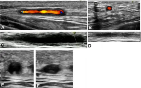
Figure 2 Abnormal temporal artery branch: (A) longitudinal view with ‘halo’ sign; (B) transverse view with ‘halo’ sign; (C) longitudinal view before compression; (D) longitudinal view with compression (‘compression’ sign positive); (E) transverse view without compression and (F) transverse view with compression (‘compression’ sign positive).
Figure 3 Axillary artery: (A) longitudinal view of normal artery; (B) transverse view of normal artery; (C) longitudinal view with ‘halo’ sign and (D) transverse view with ‘halo’ sign.
Based on the results from the SLR, the steering
committee (CDu, CDe and WAS) developed a WORDTM
-based written questionnaire that included 25 statements. Of these 25 statements, 3 addressed the definitions of the appearances of normal and arteriosclerotic temporal and extracranial large arteries; 15 statements addressed 5 definitions of the ‘halo’ sign, stenosis (temporal and extracranial large arteries), occlusion, ‘compression’ sign (temporal arteries) and vessel wall pulsation (temporal arteries) and 7 statements addressed the requirements for diagnosis of vasculitis by US.
Twenty-five physicians experienced in US and/or LVV were invited by email to participate. They were from 14 countries (Austria, Czech Republic, Denmark, France, Germany, Italy, Norway, Poland, Portugal, Slovenia, Spain, The Netherlands, UK and USA). The group consisted of 22 rheumatologists, 1 internist and 2 physicians in the last year of rheumatology training. Nine, six, four, two and four participants have performed >300, 101–300, 51–100, 21–50 and <20 diagnostic GCA US examinations, respectively. Sixteen were currently offering a diagnostic GCA US clinic. The participants were asked to rate each definition using a level of agreement or disagreement for each statement according to a 1–5 Likert scale with 1=strongly disagree, 2=disagree, 3=neither agree nor disagree, 4=agree and 5=strongly agree. A Likert score of 4 or 5 was considered as agreement. Only when statements achieved a score of >75%, a consensus was considered for appropriately defining the category. Statements satisfying these requirements were used for the definition of the most important US elementary appearances for the diag-nosis of vasculitis. Those statements with already achieved agreement, but suggestions for an improved wording in the first Delphi round were rephrased according to the experts’ comments and reappraised in the second round. Statements with a <75% agreement in the first round were not further taken to the second round.
The questionnaire also included a rating of the impor-tance of the different US elementary appearances for the diagnosis of cranial and extracranial LVV using a Likert scale as mentioned above. Up to two reminders were sent out to the experts if they had not responded within the given time limit. The answers of the first Delphi round were summarised with the percentage of agreement to each statement. For the second Delphi round, all comments of the panelists were anonymised and re-sent together with a questionnaire revised by the steering committee to those experts who had responded in the first round. At a face-to-face meeting of the expert panel (‘round 3´), held at the 2015 San Francisco ACR Meeting, the wording of one definition was slightly revised.
Inter-rater and intra-rater web-based reliability exercise
All members of the OMERACT LVV-US Working Group were asked to submit 16 representative still images and 20 representative videos (figures 1-3): eight still images and eight videos represented normal anatomical segments (common temporal artery, frontal branch,
parietal branch and axillar arteries) in longitudinal and transverse planes; and the eight other still images and eight videos represented the same segments exhibiting the ‘halo’ sign. Four additional videos showed a positive and a negative ‘compression’ sign of the temporal artery branches in longitudinal and transverse views, respec-tively. All pathological images and videos originated from patients with active disease who met the expanded ACR classification criteria of GCA, and in whom diag-nosis was confirmed either by temporal artery biopsy or on a clinical basis, including US and follow-up.19 The images and videos were collected by a facilitator of the group (SC) who constructed an electronic database using REDCap (Research Electronic Data Capture; Vanderbilt University, Nashville, Tennessee, USA) hosted by a server from the Italian Society for Rheuma-tology.20
From 550 submitted images and videos, 150 images and videos were selected by the facilitator for the web-based reliability exercise: 20 videos of axillary arteries, 20 still images of axillary arteries, 45 videos of temporal arteries, 45 still images of temporal arteries and 20 videos of the ‘compression’ sign applied to temporal arteries. The distribution between longitudinal/transverse views and normal/pathological vessels was as follows: temporal artery still images and videos: transverse 56, longitudinal 54, pathological 57 and normal 53. Axillary artery still images and videos: transverse 18, longitudinal 22, patho-logical 19 and normal 21. A link with the web-based exer-cise was sent to the same physicians who participated in the Delphi process, asking them to apply the definitions agreed in the Delphi exercise to decide whether each still image or video was suggestive of vasculitis according to the definitions. Two weeks after the first evaluation, the participants received the same images and videos in a different order for evaluating the intra-rater agreement.
All images and videos were anonymised for patients’ data, the centre where the image was obtained, US machine settings/producer and intima-media thickness (IMT) measurements. Images and videos from patients were only submitted from countries without restrictions for patient image transfer.
statistical analysis
In the SLR and in the Delphi process, only descriptive statistics were used. Intra-rater and inter-rater reliabil-ities were calculated using the kappa coefficient (κ). Intra-rater reliability was assessed by Cohen’s κ, and inter-rater reliability was studied by calculating the mean κ on all pairs (ie, Light’s κ).21 Kappa coefficients were interpreted according to Landis and Koch with κ values of 0–0.2 considered poor, 0.2–0.4 fair, 0.4–0.6 moderate,
0.6–0.8 good and 0.8–1 excellent.22 The percentage
of observed agreement (ie, the percentage of observa-tions that obtained the same score) and prevalence of the observed lesions were also calculated. Analyses were performed using R Statistical Software (Foundation for Statistical Computing, Vienna, Austria).
ResuLTs
sLR on definitions of key elementary us lesions describing vasculitis
Out of 2960 articles screened, 39 studies were finally included (see online supplementary figure S1). Some of these studies addressed more than one key objective (and are reported in the following as if they were sepa-rate articles). Twenty-four articles focused on diagnostic accuracy of US in GCA9 23–45 (main study characteristics, detailed results including risk of bias and provided defi-nitions of US key elementary lesions are summarised in online supplementary tables S2–4), studies investigated the value of US for the prediction of GCA outcome46–48 (online supplementary tables S5–6), 13 studies reported the possible role of US for monitoring disease activity23 27 28 30 33 36 37 39 40 46–49 (online supplementary table S7) and 14 cross-sectional studies assessed LVV by US in patients with GCA, PMR and TAK4 30 37 49–59 (online supplementary table S8). All diagnostic accuracy studies evaluated the role of US for the diagnosis of cranial GCA, two of them also included patients with extra cranial LV-GCA.37 44 In seven reports, arterial involvement of patients with PMR was addressed,23 27 36 50–53 and two cross-sectional studies assessed by US the involvement of LV in patients with TAK.58 59 No diagnostic accuracy study was identified for TAK and isolated idiopathic aortitis.
Most US studies in patients with GCA and PMR tested the ‘halo’ sign (n=36)4 9 23–44 46–57 as a key elementary lesion defining vasculitis. Other US signs of vascu-litis reported (mostly in combination with the ‘halo’ sign) were stenosis (n=21),4 9 23–25 30 32 33 36–38 41 46 49–56 occlusion (n=18),4 9 23 30–32 36 38 41 46 49–56 the ‘compres-sion’ sign (n=2)9 45 and a conspicuous vessel wall pulsa-tion by M-mode (n=1).31 Cut-off values of the IMT for the definition of the ‘halo’ sign were provided in nine studies,4 27 38 39 43 50 53 54 57 ranging from 0.3 to 1 mm for temporal arteries and from 1.3 to 2 mm for extracranial large arteries. For TAK, the term ‘Macaroni ‘sign has been used in two studies describing the same pathology as the ‘halo’ sign.58 59 Stenosis, occlusion and arterial dilatation have also been addressed as US key elementary signs in patients with TAK.58 59
No separate definitions for the distinction between acute and chronic vasculitic lesions have been published, neither for GCA nor for TAK.
delphi exercise
Twenty-four of the 25 invited participants responded to the first Delphi questionnaire (96% response rate). All 24 participants also responded to the second round of the Delphi questionnaire (100% response rate).
In round 1, a consensus was achieved on nine defini-tions on normal temporal and extracranial large arteries, arteriosclerosis, ‘halo’ sign, stenosis of temporal and extracranial large arteries, occlusion, ‘compression’ sign (temporal arteries) and US assessment of the ‘compres-sion’ sign (temporal arteries) (table 1). A definition of the ‘halo’ sign not including the measurement of the
IMT was preferred by the group, because of the high vari-ance of proposed cut-off values for temporal and extra-cranial large arteries found in the SLR and the lack of validated data at that time.4 27 38 39 43 50 53 54 57
In round 2, three definitions (arteriosclerosis, ‘halo’ sign and stenosis of temporal arteries) were redefined, voted and agreed upon. The statements on vessel wall pulsation (definition and assessment) and the assessment of the ‘halo’ sign by measurement of vessel wall thick-ness did not reach the threshold for consensus. At the OMERACT LVV-US face-to-face group meeting (‘round 3’), the second part of the definition on ´stenosis in temporal arteries´ was rephrased from ‘… before or behind the stenosis’ to ‘… proximal or distal to the stenosis’. The final definitions for normal and patho-logical cranial and extracranial vessels are described in
table 1.
The ‘halo’ sign and ‘compression’ signs were deemed to be the most important US signs for cranial and extra-cranial GCA with 100% and 83.3% agreement, respec-tively. Of the panelists, 95.8 % thought that the ‘halo’ sign needs to be present to meet the minimum require-ment for vasculitis.
Web-based exercise on still images and videos
Eighteen members from 13 different countries had submitted images and videos including five different US brands (Hitachi, Esaote, GE, Siemens and Philips) using linear transducers with maximum grey scale frequencies of 15, 18 or 22 MHz. Twenty-five group members partic-ipated in the web-based exercise in round 1, and 25/25 participants (100%) performed the exercise in round 2.
The reliability of the 25 participants was excellent with mean inter-rater agreements for all still images and videos of 91–99% and mean Light’s κ values of 0.83–0.98 for inter-rater reliability (table 2) depending on the lesions and sites assessed. Also, the examined intra-rater reliability with a mean agreement of 91–99% and a mean Cohen`s kappa values of range 0.83–0.98 (table 3) was excellent. The inter-rater and intra-rater reliabilities performed all with κ >0.8 irrespective of the view (longi-tudinal or transverse, still images or videos) or anatom-ical segments.
dIsCussIOn
Many previous studies have investigated US as a diag-nostic tool for GCA using different definitions for normal and abnormal findings. This study now provides expert consensus-based definitions for US in LVV that can be applied in future studies. The consensus-based defini-tions revealed excellent inter-rater and intra-rater relia-bilities when tested on images and videos of patients.
Although we included all types of LVV as possible search terms in the SLR, the Delphi as well as reliability exercise was focused on GCA only, as the SLR revealed insufficient data to provide a solid basis for the consensus process. It is, however, the clinical experience of the
Table 1 Statements on definitions (n=8) and conduction (n=1) of ultrasound (US) elementary appearances in large vessel vasculitis agreed upon through a Delphi survey
Domain Definition Agreement (%) Delphi round
US appearance of normal temporal
arteries
Pulsating, compressible artery with anechoic lumen surrounded by mid-echoic to hyperechoic* tissue. Using US equipment with high resolution, the intima-media complex presenting as a homogenous, hypoechoic or anechoic echostructure delineated by two parallel hyperechoic margins (‘double line pattern’) may be visible.
95.7 1
normal
extracranial large arteries
Pulsating, hardly compressible artery with anechoic lumen; the intima-media complex presents as a homogenous, hypoechoic or anechoic echostructure delineated by two parallel hyperechoic margins (‘double line pattern’), which is surrounded by mid-echoic to hyperechoic tissue.
100 1
arteriosclerotic
arteries Heterogeneous and in part hyperechoic, irregularly delineated and eccentric vessel wall alteration. 95.8 2 ‘halo’ sign Homogenous, hypoechoic wall thickening, well delineated towards the luminal
side, visible both in longitudinal and transverse planes, most commonly concentric in transverse scans.
91.3 2
stenosis in
temporal arteries A stenosis is characterised by aliasing and persistent diastolic flow by colour Doppler US. The maximum systolic flow velocity determined within the stenosis by pulsed wave-Doppler US is ≥2 times higher than the flow velocity proximal or distal to the stenosis.
95.8
100 23
stenosis in extracranial large arteries
Typical vasculitic vessel wall thickening with characteristic Doppler curves
showing turbulence and increased systolic and diastolic blood flow velocities. 75 1 occlusion Absence of colour Doppler signals in a visible artery filled with hypoechoic
material, even with low pulse repetition frequency and high colour gain.
87.5 1
‘compression’ sign of temporal arteries
The thickened arterial wall remains visible upon compression; the hypoechogenic vasculitic vessel wall thickening contrasts with the mid-echogenic to
hyperechogenic surrounding tissue.
78.3 1
US assessment of ‘compression’
sign of temporal arteries
The compression sign should be assessed by applying pressure via the transducer until the lumen of the temporal artery occludes and no arterial pulsation remains visible.
91.3 1
*The term ‘midechoic’ is equivalent to the term ‘isoechoic’.
Table 2 Inter-rater agreements for the ‘halo’ and ‘compression’ signs in temporal and axillary arteries
Section Lesion (mean prevalence, %)* Agreement (mean, %) Agreement(range) Light‘s κ (mean) Light‘s κ (range)
‘halo’ (all images & videos) 51.4 94 82–100 0.89 0.65–1
‘halo’ (all images) 54 98 89–100 0.95 0.78–1
‘halo’ (all videos) 49.3 92 77–100 0.84 0.54–1
‘halo’ temporal arteries (images & videos) 53.2 94 78–100 0.87 0.58–1
‘halo’ temporal arteries (images) 57.5 97 84–100 0.94 0.69–1
‘halo’ temporal arteries (videos) 50 91 74–100 0.83 0.49–1
‘halo’ axillary arteries (images & videos) 46 97 80–100 0.93 0.58–1
‘halo’ axillary arteries (images) 45 99 90–100 0.98 0.80–1
‘halo’ axillary arteries (videos) 47 94 70–100 0.88 0.34–1
‘compression’ sign (videos) 53.6 92 70–100 0.83 0.34–1
experts that US abnormalities in patients with TAK look similar. Future US studies in TAK and idiopathic aortitis are necessary to gather more data on US key lesions also in these LVV entities.
The OMERACT Group agreed that ‘halo’ sign and ‘compression’ sign should be regarded as the primary elementary US signs of cranial and/or LV-GCA without including stenosis or occlusion. The ‘halo’ sign has been applied in most published studies.4 9 23–44 46–57 The ‘compression’ sign was only addressed by two studies from one research group so far.9 45 However, it has shown good diagnostic performance and is feasible in daily practice. It is a method to better visualise the ‘halo’ sign. In early studies, the presence of stenosis helped to increase the sensitivity of temporal artery US.10 23 On the other hand, many sonographers feel that stenosis may reduce the specificity of the examination.2 Furthermore, due to far higher resolution of modern US equipment, a ‘halo’ sign can now usually be visualised in stenotic vessel areas, and temporal artery occlusions in GCA usually occur together with the non-compressible ‘halo’ sign’.5
It was also agreed not to include the measurement of IMT for the definition of the ‘halo’ sign, as at the time of the Delphi process only proposals for cut-off values but no studies for validating cut-off values were available. Several previous studies had proposed a wide range of cut-off values for the diameter of a halo sign, for example, 0.3–1 mm for temporal arteries and 1.3–2 mm for extra-cranial large arteries.4 27 38 39 43 50 53 54 57 A study investigating patients with newly diagnosed active GCA and healthy controls has been recently performed by members of the group (WAS and VSS) for calculating IMT cut-off values in normal temporal and axillary arteries.60 The role of IMT measurements for diagnosis and monitoring is yet uncertain and needs to be addressed by future studies.
The inter-rater and intra-rater agreements of the web-based exercise were excellent. Images and videos were submitted by participating experts as in previous OMERACT-related US exercises.14–16 61 Images and videos for the present web-based exercise were taken from
patients with newly diagnosed active GCA since US signs in patients with established disease resolve rapidly with treatment.40 Reliability data for 12 sonographers reading videos from the international multicentre TABUL study have now been published.2 Videos from that study were randomly chosen from all stored videos of the study, irre-spective of their quality, whereas the quality of images and videos in the OMERACT study may have been better as the members submitted material which they deemed representative. Sonographers of the TABUL study were less experienced than sonographers of the present OMERACT study. Kappa values for the intra-rater reli-ability in the TABUL study were 0.69–0.81. Inter-rater reliability was only provided as intraclass correlation coef-ficients (ICCs). Notably, the reliability of 14 pathologists reading temporal artery biopsy specimens was similar when compared with the 12 sonographers (ICC 0.61 vs 0.62).
We asked the experts to submit images from GCA cases and controls which include patients with arterio-sclerosis. Few of the control cases indeed had arterioscle-rotic changes; however, we did not specifically question in our rating to distinguish between arteriosclerosis and non-arteriosclerotic controls. We were therefore unable to conduct a separate analysis in this regard. We did not score images and videos with stenosis or occlusions.
In conclusion, an international expert consensus was reached using OMERACT methodology for the defi-nitions of normal US appearance and abnormalities seen in the temporal and axillary arteries in GCA. This OMERACT exercise (along with the previously reported TABUL study) shows that images and videos of US scans of inflamed temporal and axillary arteries can reliably document the characteristic and diagnostic abnormali-ties in patients with suspected GCA. Our study supports the use of US abnormalities, including both images and videos, as an inclusion criterion for future GCA trials. Confidence is increasing in the use of US in mainstream clinical practice, and it may be incorporated into future guidelines for GCA diagnosis.
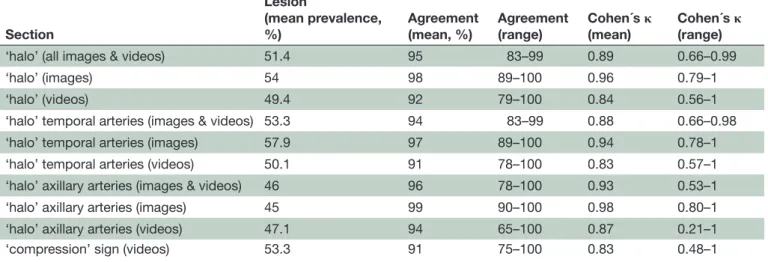
Table 3 Intra-rater agreements for the halo’ and ‘compression’ signs in temporal and axillary arteries
Section
Lesion
(mean prevalence,
%) Agreement (mean, %) Agreement(range) Cohen´s κ (mean) Cohen´s κ (range)
‘halo’ (all images & videos) 51.4 95 83–99 0.89 0.66–0.99
‘halo’ (images) 54 98 89–100 0.96 0.79–1
‘halo’ (videos) 49.4 92 79–100 0.84 0.56–1
‘halo’ temporal arteries (images & videos) 53.3 94 83–99 0.88 0.66–0.98
‘halo’ temporal arteries (images) 57.9 97 89–100 0.94 0.78–1
‘halo’ temporal arteries (videos) 50.1 91 78–100 0.83 0.57–1
‘halo’ axillary arteries (images & videos) 46 96 78–100 0.93 0.53–1
‘halo’ axillary arteries (images) 45 99 90–100 0.98 0.80–1
‘halo’ axillary arteries (videos) 47.1 94 65–100 0.87 0.21–1
The next step in the OMERACT validation process is the inter-rater and intra-rater reliability test of these defi-nitions for normal and vasculitic arteries in patient-based exercises.
Author affiliations
1Department of rheumatology, Hospital of Southwest Jutland, esbjerg, Denmark 2Department of internal Medicine, clinical Division of internal Medicine ii, Medical University of innsbruck, innsbruck, austria
3Department of rheumatology, Medical University of graz, graz, austria 4Department of rhematology, Hospital of Bruneck, Bruneck, italy
5iii. Medical clinic, Department of Oncology, Hematology and rheumatology, University Hospital Bonn, Berlin, germany
6leiden University Medical center, leiden, the netherlands 7epidemiology Unit, italian Society for rheumatology (Sir), Milan, italy 8Department of rheumatology, University of Ferrara, Ferrara, italy
9Department of rheumatology, University Medical centre ljubljana, ljubljana, Slovenia
10Martina Hansens Hospital, Bærum, Oslo, norway
11Dipartimento Scienze cliniche e Biologiche - reumatologia, Università degli Studi di torino, torino, italy
12Department of rheumatology, norfolk and norwich University Hospital, norwich, UK
13Hospital de Santa Maria - cHln, lisbon academic Medical centre, lisbon, Portugal
14Hospital Universitario Fundación Jiménez Díaz, Madrid, Spain 15University Hospital la Paz, Madrid, Spain
16Mc groep Hospitals, lelystad, the netherlands 17rheumatology, Mayo clinic, rochester, Minnesota, USa
18copenhagen center for arthritis research (cOPecare), copenhagen, Denmark 19Department of rheumatology and internal Medicine, Pomeranian Medical University, Szczecin, Poland
20Hôpital ambroise Paré, Boulogne-Billancourt, France 21St. elizabeth's Medical center, Boston, Massachusetts, USa
22Department of rheumatology, First Faculty of Medicine, charles University of Prague, Prague, czech republic
23arcispedale Santa Maria nuova, reggio emilia, italy
24Department of rheumatology, University of california, los angeles, california, USa
25Diagnostic centre region Hospital Silkeborg, Silkeborg, Denmark 26Department of rheumatology, Odense University Hospital, Odense, Denmark 27asklepios Medical center, Bad abbach, germany
28Southend University Hospital nHS Foundation trust & anglia ruskin University, Southend-on-Sea, UK
29Medical centre for rheumatology, immanuel Krankenhaus Berlin, Berlin, germany
Contributors WaS, cDe, cDu and Sc designed the study. cDe and cDu organised the Delphi exercise. WaS and Sc organised the web-based exercise. Sc build the database, collected all images and selected the study images. Sc was responsible for data entry and data precession together with caS, gc and Sr who did the statistical analyses. WaS, cDu and Sc wrote the manuscript. all authors contributed to the acquisition of data and have read and revised the manuscript.
Funding the authors have not declared a specific grant for this research from any funding agency in the public, commercial or not-for-profit sectors.
Competing interests none declared.
Patient consent not required.
ethics approval ethical approval for the web-based exercise was obtained from the Data agency of the Southwest Hospital in esbjerg, Denmark.
Provenance and peer review not commissioned; externally peer reviewed.
data sharing statement there are not additional unpublished data from the study.
Open Access this is an Open access article distributed in accordance with the creative commons attribution non commercial (cc BY-nc 4.0) license, which permits others to distribute, remix, adapt, build upon this work non-commercially, and license their derivative works on different terms, provided the original work is properly cited and the use is non-commercial. See: http:// creativecommons. org/ licenses/ by- nc/ 4. 0/
© article author(s) (or their employer(s) unless otherwise stated in the text of the article) 2018. all rights reserved. no commercial use is permitted unless otherwise expressly granted.
RefeRences
1. Lawrence RC, Felson DT, Helmick CG, et al. Estimates of the prevalence of arthritis and other rheumatic conditions in the United States. Part II. Arthritis Rheum 2008;58:26–35.
2 Luqmani R, Lee E, Singh S, et al. The role of ultrasound compared to biopsy of temporal arteries in the diagnosis and treatment of giant cell arteritis (TABUL): a diagnostic accuracy and cost-effectiveness study. Health Technol Assess 2016;20:1–238.
3. Brack A, Martinez-Taboada V, Stanson A, et al. Disease pattern in cranial and large-vessel giant cell arteritis. Arthritis Rheum
1999;42:311–7.
4. Schmidt WA, Seifert A, Gromnica-Ihle E, et al. Ultrasound of proximal upper extremity arteries to increase the diagnostic yield in large-vessel giant cell arteritis. Rheumatology 2008;47:96–101. 5. Muratore F, Pipitone N, Salvarani C, et al. Imaging of vasculitis: state
of the art. Best Pract Res Clin Rheumatol 2016;30:688–706. 6. Patil P, Williams M, Maw WW, et al. Fast track pathway reduces
sight loss in giant cell arteritis: results of a longitudinal observational cohort study. Clin Exp Rheumatol 2015;33(Suppl 89):S103–6. 7. Diamantopoulos AP, Haugeberg G, Lindland A, et al. The
fast-track ultrasound clinic for early diagnosis of giant cell arteritis significantly reduces permanent visual impairment: towards a more effective strategy to improve clinical outcome in giant cell arteritis?
Rheumatology 2016;55:66–70.
8. Schmidt WA, Kraft HE, Völker L, et al. Colour Doppler sonography to diagnose temporal arteritis. Lancet 1995;345:866.
9. Aschwanden M, Daikeler T, Kesten F, et al. Temporal artery compression sign--a novel ultrasound finding for the diagnosis of giant cell arteritis. Ultraschall Med 2013;34:47–50.
10. Karassa FB, Matsagas MI, Schmidt WA, et al. Diagnostic performance of ultrasonography for giant-cell arteritis: a meta-analysis. Ann intern Med 2005;142:359–69.
11. Arida A, Kyprianou M, Kanakis M, et al. The diagnostic value of ultrasonography-derived edema of the temporal artery wall in giant cell arteritis: a second meta-analysis. BMC Musculoskelet Disord
2010;11:44.
12. Ball EL, Walsh SR, Tang TY, et al. Role of ultrasonography in the diagnosis of temporal arteritis. Br J Surg 2010;97:1765–71. 13. Duftner C, Dejaco C, Sepriano A, et al. Imaging in diagnosis,
outcome prediction and monitoring of large vessel vasculitis: a systematic literature review and meta-analysis informing the EULAR recommendations. RMD Open 2018;4:e000612.
14. Filippou G, Scirè CA, Damjanov N, et al. Definition and reliability assessment of elementary ultrasonographic findings in calcium pyrophosphate deposition disease: a study by the OMERACT calcium pyrophosphate deposition disease ultrasound subtask force. J Rheumatol 2017;44:1744–9.
15. Gutierrez M, Schmidt WA, Thiele RG, et al. International consensus for ultrasound lesions in gout: results of Delphi process and web-reliability exercise. Rheumatology 2015;54:1797–805.
16. Naredo E, D'Agostino MA, Wakefield RJ, et al. Reliability of a consensus-based ultrasound score for tenosynovitis in rheumatoid arthritis. Ann Rheum Dis 2013;72:1328–34.
17. Whiting PF, Rutjes AWS, Westwood ME, et al. Research and Reporting Methods Accuracy Studies. Ann Intern Med 2011;155:529–36.
18. Hayden JA, Côté P, Bombardier C. Evaluation of the quality of prognosis studies in systematic reviews. Ann Intern Med
2006;144:427–37.
19. Dejaco C, Duftner C, Buttgereit F, et al. The spectrum of giant cell arteritis and polymyalgia rheumatica: revisiting the concept of the disease. Rheumatology 2017;56:506–15.
20. Harris PA, Taylor R, Thielke R, et al. Research electronic data capture (REDCap)--a metadata-driven methodology and workflow process for providing translational research informatics support. J Biomed Inform 2009;42:377–81.
21. Light RJ. Measures of response agreement for qualitative data: some generalizations and alternatives. Psychol Bull 1971;76:365–77. 22. Landis JR, Koch GG. The measurement of observer agreement for
categorical data. Biometrics 1977;33:159–74. 23. Schmidt WA, Kraft HE, Vorpahl K, et al. Color duplex
ultrasonography in the diagnosis of temporal arteritis. N Engl J Med
24. Venz S, Hosten N, Nordwald K, et al. [Use of high resolution color Doppler sonography in diagnosis of temporal arteritis]. Rofo
1998;169:605–8.
25. LeSar CJ, Meier GH, DeMasi RJ, et al. The utility of color duplex ultrasonography in the diagnosis of temporal arteritis. J Vasc Surg
2002;36:1154–60.
26. Nesher G, Shemesh D, Mates M, et al. The predictive value of the halo sign in color Doppler ultrasonography of the temporal arteries for diagnosing giant cell arteritis. J Rheumatol 2002;29:1224–6. 27. Salvarani C, Silingardi M, Ghirarduzzi A, et al. Is duplex
ultrasonography useful for the diagnosis of giant-cell arteritis? Ann Intern Med 2002;137:232–8.
28. Schmid R, Hermann M, Yannar A, et al. [Color duplex ultrasound of the temporal artery: replacement for biopsy in temporal arteritis].
Ophthalmologica 2002;216:16–21.
29. Murgatroyd H, Nimmo M, Evans A, et al. The use of ultrasound as an aid in the diagnosis of giant cell arteritis: a pilot study comparing histological features with ultrasound findings. Eye 2003;17:415–9. 30. Pfadenhauer K, Weber H. Duplex sonography of the temporal and
occipital artery in the diagnosis of temporal arteritis. A prospective study. J Rheumatol 2003;30:2177–81.
31. Reinhard M, Schmidt D, Hetzel A. Color-coded sonography in suspected temporal arteritis-experiences after 83 cases. Rheumatol Int 2004;24:340–6.
32. Romera-Villegas A, Vila-Coll R, Poca-Dias V, et al. The role of color duplex sonography in the diagnosis of giant cell arteritis. J Ultrasound Med 2004;23:1493–8.
33. Karahaliou M, Vaiopoulos G, Papaspyrou S, et al. Colour duplex sonography of temporal arteries before decision for biopsy: a prospective study in 55 patients with suspected giant cell arteritis.
Arthritis Res Ther 2006;8:R116.
34. Bley TA, Reinhard M, Hauenstein C, et al. Comparison of duplex sonography and high-resolution magnetic resonance imaging in the diagnosis of giant cell (temporal) arteritis. Arthritis Rheum
2008;58:2574–8.
35. Ghinoi A, Zuccoli G, Nicolini A, et al. 1T magnetic resonance imaging in the diagnosis of giant cell arteritis: Comparison with ultrasonography and physical examination of temporal arteries. Clin Exp Rheumatol 2008;3(Suppl 49):S76–80.
36. Pérez López J, Solans Laqué R, Bosch Gil JA, et al. Colour-duplex ultrasonography of the temporal and ophthalmic arteries in the diagnosis and follow-up of giant cell arteritis. Clin Exp Rheumatol
2009;27(Suppl 52):S77–82.
37. Aschwanden M, Kesten F, Stern M, et al. Vascular involvement in patients with giant cell arteritis determined by duplex sonography of 2x11 arterial regions. Ann Rheum Dis 2010;69:1356–9.
38 Maldini C, Dépinay-Dhellemmes C, Tra TT, et al. Limited value of temporal artery ultrasonography examinations for diagnosis of giant cell arteritis: analysis of 77 subjects. J Rheumatol 2010;37:2326–30. 39. Habib HM, Essa AA, Hassan AA. Color duplex ultrasonography
of temporal arteries: role in diagnosis and follow-up of suspected cases of temporal arteritis. Clin Rheumatol 2012;31:231–7. 40. Hauenstein C, Reinhard M, Geiger J, et al. Effects of early
corticosteroid treatment on magnetic resonance imaging and ultrasonography findings in giant cell arteritis. Rheumatology
2012;51:1999–2003.
41. Pfenninger L, Horst A, Stuckmann G, et al. Comparison of histopathological findings with duplex sonography of the temporal arteries in suspected giant cell arteritis. Klin Monbl Augenheilkd
2012;229:369–73.
42. Black R, Roach D, Rischmueller M, et al. The use of temporal artery ultrasound in the diagnosis of giant cell arteritis in routine practice.
Int J Rheum Dis 2013;16:352–7.
43. Muratore F, Boiardi L, Restuccia G, et al. Comparison between colour duplex sonography findings and different histological patterns of temporal artery. Rheumatology 2013;52:2268–74.
44. Diamantopoulos AP, Haugeberg G, Hetland H, et al. Diagnostic value of color Doppler ultrasonography of temporal arteries and large vessels in giant cell arteritis: a consecutive case series. Arthritis Care Res 2014;66:113–9.
45. Aschwanden M, Imfeld S, Staub D, et al. The ultrasound compression sign to diagnose temporal giant cell arteritis shows an excellent interobserver agreement. Clin Exp Rheumatol
2015;33(Suppl 89):S113–5.
46. Schmidt WA, Moll A, Seifert A, et al. Prognosis of large-vessel giant cell arteritis. Rheumatology 2008;47:1406–8.
47. De Miguel E, Roxo A, Castillo C, et al. The utility and sensitivity of colour Doppler ultrasound in monitoring changes in giant cell arteritis. Clin Exp Rheumatol 2012;30(Suppl 70):S34–8.
48. Czihal M, Piller A, Schroettle A, et al. Outcome of giant cell arteritis of the arm arteries managed with medical treatment alone: cross-sectional follow-up study. Rheumatology 2013;52:282–6.
49. Schmidt WA, Natusch A, Möller DE, et al. Involvement of peripheral arteries in giant cell arteritis: a color Doppler sonography study. Clin Exp Rheumatol 2002;20:309–18.
50. Stammler F, Ysermann M, Mohr W, et al. [Value of color-coded duplex ultrasound in patients with polymyalgia rheumatica without signs of temporal arteritis]. Dtsch Med Wochenschr
2000;125:1250–6.
51. Schmidt WA, Gromnica-Ihle E. Incidence of temporal arteritis in patients with polymyalgia rheumatica: a prospective study using colour Doppler ultrasonography of the temporal arteries.
Rheumatology 2002;41:46–52.
52. Pfadenhauer K, Esser M, Berger K. Vertebrobasilar ischemia and structural abnormalities of the vertebral arteries in active temporal arteritis and polymyalgia rheumatica--an ultrasonographic case-control study. J Rheumatol 2005;32:2356–60.
53. Stammler F, Grau C, Schnabel A. [Value of colour Doppler ultrasonography in relation to clinical pretest probability in giant cell (temporal) arteritis]. Dtsch Med Wochenschr
2009;134:2109–15.
54. Förster S, Tato F, Weiss M, et al. Patterns of extracranial involvement in newly diagnosed giant cell arteritis assessed by physical examination, colour coded duplex sonography and FDG-PET. Vasa
2011;40:219–27.
55. Czihal M, Zanker S, Rademacher A, et al. Sonographic and clinical pattern of extracranial and cranial giant cell arteritis. Scand J Rheumatol 2012;41:231–6.
56. Czihal M, Tatò F, Rademacher A, et al. Involvement of the femoropopliteal arteries in giant cell arteritis: clinical and color duplex sonography. J Rheumatol 2012;39:314–21.
57. Ghinoi A, Pipitone N, Nicolini A, et al. Large-vessel involvement in recent-onset giant cell arteritis: a case-control colour-Doppler sonography study. Rheumatology 2012;51:730–4.
58. Taniguchi N, Itoh K, Honda M, et al. Comparative ultrasonographic and angiographic study of carotid arterial lesions in Takayasu's arteritis. Angiology 1997;48:9–20.
59. Cantú C, Pineda C, Barinagarrementeria F, et al. Noninvasive cerebrovascular assessment of Takayasu arteritis. Stroke
2000;31:2197–202.
60. Schäfer VS, Juche A, Ramiro S, et al. Ultrasound cut-off values for intima-media thickness of temporal, facial and axillary arteries in giant cell arteritis. Rheumatology 2017;56:1479–83.
61. Terslev L, Naredo E, Iagnocco A, et al. Defining enthesitis in spondyloarthritis by ultrasound: results of a Delphi process and of a reliability reading exercise. Arthritis Care Res 2014;66:741–8.