junction (CEJ), with exposure of the root surfa-ce, leading to pain, unfavorable aesthetic appe-arance and root caries. Surgical root coverage is the only therapeutic choice if cause-specific measures are insufficient to correct the defor-mity of gingival mucose1.
Epiregulin (Epi) is a broad specificity EGF (epidermal growth factor) family member with the unique characteristic to transmit a more po-tent mitogenic signal than EGF itself by binding all possible ErbB receptor complexes2,3, thus, sti-mulating cell proliferation in various cellular li-nages, such as keratinocytes and fibroblasts4,5.
Gum wound healing is important for both pe-riodontal pathologies and surgery: for this rea-son, it is conceivable a beneficial effect of Epi as treatment of gingival recession, considering the mounting evidence suggesting also a crucial role of Epi in mediating proliferation and migration of gingival epithelial cells and fibroblasts6.
The aim of the present study was to design an innovative nanotechnological approach to deliver Epi directly to the gingival pocket, where its po-sitive effect on cell proliferation and migration is needed.
Nanotechnology has gained significant clinical interest in recent years, and nanoparticles (NPs) have become very attractive to the pharmaceutical and biomedical fields as drug delivery vehicles7.
NPs can deliver a plethora of drugs, vaccines or biological macromolecules. By acting as drug de-livery systems, NPs allow both a targeted admini-stration of the active component to specific organ or cells and a controlled release of the drug7-9.
Moreover, biodegradable polymeric NPs repre-sent a useful delivery device as they can protect drug moieties from enzymatic disruption and provide sustained drug release over a certain time in a controlled manner, reducing side effects and dosage of therapeutic agents8,10-12.
Abstract. – OBJECTIVE: Epiregulin is a mem-ber of the epidermal growth factor (EGF) family produced by keratinocytes: the aim of this study was to investigate the ability of biocompatible nanoparticles loaded with such growth factor to increase human keratinocytes proliferation.
MATERIALS AND METHODS: Different PLGA (Poly-d,l-lactide-co-glycolide)-nanoparticles (NPs) formulations have been characterized in size and zeta potential by dynamic light scat-tering (DLS) analysis. The ability of the different PLGA-NPs formulations to adhere onto dental surfaces has been tested, and epiregulin-en-riched PLGA-NPs has been produced. Epireg-ulin release from NPs has been tested by en-zyme-linked immunosorbent (ELISA) assay and the proliferative effects of epiregulin-NPs on hu-man keratinocytes have been evaluated.
RESULTS: DLS analysis revealed a different size distribution depending on the PLA/PGA (poly lactic acid/poly glycolic acid) ratio used. 50:50 PLGA-NPs exhibited the smaller size and the best dental adhe-sive ability. Moreover, such epiregulin-loaded NPs was able to increase cell proliferation.
CONCLUSIONS: Direct dental pocket drug de-livery implies the NPs solution loading onto the dental surface at the cement-enamel junction level: 50:50 PLGA-NPs, with their small size and excellent adhesive ability, represent an interest-ing tool to deliver epiregulin directly where there is the need for epithelial proliferation. These re-sults describe a possible strategy for periodon-tal pocket delivery of Epiregulin-loaded PL-GA-NPs and might provide a new approach for the treatment of gingival recession, where gingi-val epithelium proliferation is needed.
Key Words:
Epiregulin, Gingival recession, PLGA nanoparticles.
Introduction
Gum recession is defined as the shift of the marginal tissue apical to the cement-enamel
M. RIZZI
1, M. MIGLIARIO
2, V. ROCCHETTI
2, S. TONELLO
1, F. RENÒ
11Innovative Research Laboratory for Wound Healing, Health Sciences Department, University of Eastern Piedmont, Novara, Italy.
2Dental Clinic, Health Sciences Department, University of Eastern Piedmont, Novara, Italy.
Epiregulin-loaded PLGA nanoparticles
increase human keratinocytes proliferation:
preliminary data
Poly-d,l-lactide-co-glycolide (PLGA) is one of the most successfully used polymer to develop drug delivery systems, thanks to its attractive pro-perties, such as favorable mechanical characteri-stics, biodegradability and biocompatibility, and FDA (US Food and Drug Administration) appro-val for human usage.
PLGA-NPs are biodegradable in the body: they undergo hydrolysis, leading to the production of the original monomers lactic acid and glycolic acid, that are metabolized via the Krebs cycle8,9,12,13.
In the present study, epiregulin-enriched PL-GA-NPs was produced and characterized. Once identified the formulation assuring the best adhe-sion on the dental surface, epiregulin-loaded NPs was produced and tested to evaluate growth fac-tor release and its ability to modulate cell prolife-ration in an in vitro model (human keratinocytes (HaCaT) cells).
Materials and Methods
Teeth Collection
Healthy adult teeth were obtained from “S.C.D.U. Odontoiatria e Stomatologia”, of the “Maggiore della Carità” Hospital (Novara, Italy) after routine extraction with written informed consent obtained from patients. The study was approved by the Ethical Committee of “Maggiore della Carità” Hospital in Novara, ALS of Biella, Novara, Vercelli and Verbano-Cusio-Ossola (stu-dy n. CE 63/11).
Soon after extraction, teeth were immersed in a 0.2% chlorhexidine solution for 30 minutes and then stored at 4°C in phosphate-buffered saline (PBS) solution (pH = 7.4) during the study period.
PLGA-nanoparticles Production
PLGA-NPs were prepared by a modified double solvent evaporation method14. Briefly, 60 mg of PLGA (50:50, 65:35, 75:25) crystals (Sigma-Aldrich, Saint Luis, MO, USA) were dissolved in 1 ml of dichloromethane (DCM) (Sigma-Aldrich, St. Luis, MO, USA) at room temperature. To produce control NPs, 50 µl of 1% polyvinyl alcohol (PVA) (Sigma-Aldrich, St. Luis, MO, USA) aqueous solution were added to PLGA and the solution was sonicated for 1 min. After that, 5 volumes of 1% PVA aqueous solution were carefully added to the resulting emulsion in order to maintain phase separation. A further 2 min sonication was performed to obtain the final emulsion that was evaporated
overnight under fume hood, to remove DCM. The resulting NPs were washed 5 times in di-stilled water by centrifugation at 13000 rpm for 5 min, resuspended in water and stored at 4oC.
NPs containing epiregulin were produced as above by adding 50 µl Epi (human recombinant epiregulin, E. coli derived, R&D Systems, Min-neapolis, MN, USA, 50 ng/ml), instead of PVA, to the PLGA solution dissolved in DCM during the first step of the preparation.
Particle Size and Zeta Potential Measurement
Particle size and polydispersity index after water dispersion and the charge density exposed on the surface of PLGA-NPs were evaluated by using DLS (dynamic light scattering) technique. PLGA-NPs were dispersed in water (1 mg/ml) and measured through DLS analysis in order to determine the particle size distribution. DLS experiments were carried out using a Zetasizer Nano ZS instrument (Malvern Instruments Ltd, Malvern, UK), operating in a particle size range from 0.6 nm to 6 μm and equipped by a laser He-Ne with λ = 633 nm. Zeta potential analysis (ζ-potential), carried out to deter-mine the stability behavior of our NPs solutions, was performed using the same instrument.
NPs adhesion to Dental Surface
In order to evaluate the NPs adhesion onto the dental surface, teeth were photographed be-fore and after NPs loading, testing three different PLA/PGA (poly lactic acid/poly glycolic acid) ratio formulations (50:50, 65:35 and 75:25) in or-der to determine the best PLGA grade in terms of dental surface adhesion. The NPs suspension was positioned at the CEJ level, let dry and pho-tographed. NPs-loaded teeth were then incubat-ed in simulatincubat-ed saliva15 at 37°C for 3 hours in order to simulate the oral cavity real conditions. Then, digital pictures of each tooth were captured for comparison and NPs attachment areas were quantified using ImageJ software (U. S. National Institutes of Health, Bethesda, MD, USA). NPs covered area after incubation, was expressed as the percentage of the initial area at time zero ± standard deviation (SD).
Epiregulin Release Assay
As dental adhesion and particle size results indicate the optimal PLA/PGA polymer ratio to be 50:50, we produced 50:50 PLGA-NPs contain-ing Epiregulin and analyzed its release profile by enzyme-linked immunosorbent assay (ELISA)
(Uscn Life Sciences Inc., Wuhan, China). Briefly, Epi-enriched NPs have been incubated in simu-lated saliva at 37°C and at fixed time points (3h, 6h, 1, 2, 10 days) simulated saliva aliquots of 100 µl for each sample have been assayed following manufacturer’s instructions; the optical density (O.D.) was read at 450 nm on a microplate reader. Results were expressed as mean values ± standard deviation (SD).
Cell Culture
Spontaneously immortalized keratinocytes (HaCaT, CLS Cell Lines Service GmbH, Ep-pelheim, Germany), isolated from human adult skin16, were grown in culture flask (75 cm2) in DMEM medium (Euroclone, Milan, Italy) supple-mented with 10% heat-inactivated foetal bovine serum (FBS) (Euroclone, Milan, Italy), penicillin (100 U/ml), streptomycin (100 mg/ml) and L-glu-tamine (2 mM) (Euroclone, Milan, Italy) in a hu-midified atmosphere containing 5% CO2 at 37°C.
Cell Proliferation Assay
To evaluate the proliferative effects of epiregu-lin released from PLGA-NPs, HaCaT cells were seeded in 24-well plates at a density of 2x10⁴ cel-ls/well and incubated in the presence or absence of PLGA-NPs supernatant, obtained after Epi-lo-aded and unloEpi-lo-aded NPs solution centrifugation. Such samples were diluted 1:4 in Dulbecco’s Modified Eagle’s Medium (DMEM) before cell treatment. As NPs supernatant main component is represented by water, cells were also treated with the same volume of water in order to exclude vehicle interference. After 72h of incubation, cel-ls were fixed in 3.7% formaldehyde-3% sucrose solution and stained with 1% toluidine blue solu-tion. Stained samples were photographed at 10X magnification, using an optical microscope (Leica ICC50HD, Leica Microsystems Wetzlar GmbH, Wetzlar, Germany) and cell proliferation was evaluated by counting cells in 10 random fields in three samples for each experimental condition. Results were expressed as cells/mm2 ± standard deviation (SD).
Statistical Analysis
Unpaired Student’s t-tests were used for sta-tistical analysis. Stasta-tistical evaluation was per-formed with the Prism 4.0 statistical software (GraphPad Software Inc., La Jolla, CA, USA). Probability values of p<0.05 were considered sta-tistically significant.
Results
NPs Characterization
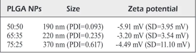
NPs size distribution analysis by DLS approa-ch revealed a different size distribution depending on the formulation (i.e. PLA/PGA ratio). In parti-cular, the 50:50 PLGA-NPs formulation displayed the smallest size among the three formulations analyzed (Table I). In general, particle aggrega-tion is less likely to occur for charged particles (high zeta potential) due to electric repulsion. The zeta potential found here for the different NPs for-mulations suggest that there was no difference in the surface charge characteristics of these NPs, but might allow particles aggregation. Neverthe-less, these data do not indicate that NPs suspen-sion is unstable, as the hydrophilicity of PLGA is sufficient to maintain the particles suspended.
PLGA NPs Adhesion to Dental Surface
NPs ability to adhere to the dental surface is an essential feature allowing their direct placement at the CEJ level; for this reason, the different PL-GA-NPs formulations ability to adhere on teeth was evaluated.
As shown in Figure 1, 50:50 PLGA-NPs ex-hibited the best dental adhesion ability. Indeed, after 3h of incubation in artificial saliva, 50:50 PLGA-NPs were still present onto the dental sur-face covering an area of 87.6 ± 1.2% compared to time zero (NPs freshly applied onto the teeth). On the contrary, the other PLGA formulations (65:35 and 75:25) have shown a statistically significant reduction (p<0.001) in the attachment area.
As 50:50 PLGA-NPs displayed the better adhe-sion ability on dental surface, growth factor load-ed NPs were producload-ed using such scaffold.
In vitro Epiregulin Release
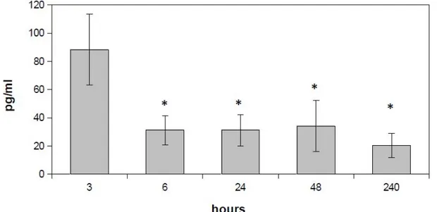
Quantification of epiregulin release from PL-GA-NPs performed by ELISA assay indicates that Epi-enriched NPs incubated in simulated saliva at 37°C were able to produce a peak in Epi release after 3 hours of incubation (Figure 2). At this time point, 88 ± 25 pg/ml of Epi have been detected,
Table I. Size and zeta potential. Size and zeta potential of the different PLGA-NPs formulations analyzed by dynamic light scattering.
PLGA NPs Size Zeta potential 50:50 190 nm (PDI=0.093) -5.91 mV (SD=3.95 mV) 65:35 220 nm (PDI=0.235) -3.20 mV (SD=3.54 mV) 75:25 370 nm (PDI=0.617) -4.49 mV (SD=11.10 mV)
while a significant reduction (p<0.05) and a sta-bilization of the Epi release levels occurred at the following time points.
Epiregulin Released from PLGA NPs Stimulates Cell Proliferation
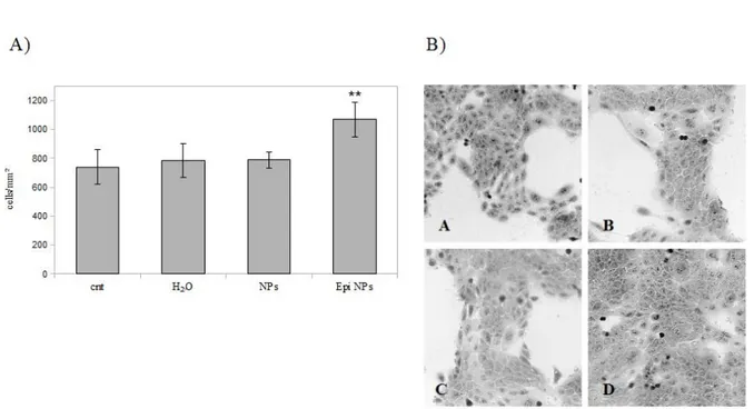
As shown in Figure 3, a statistically signifi-cant (p<0.01) increase in cell proliferation occurs only in the samples treated with Epi-enriched PL-GA-NPs supernatant while unloaded NPs
super-natant did not affect cell proliferation. Therefore, even if epiregulin released by NPs is very low, it was sufficient to induce an increase in cell proli-feration compared to control conditions.
Discussion
Gingival recession or marginal tissue recession is a pathological condition defined as the location
Figure 1. NPs adhesion to dental surface. A) Digital pictures of teeth immediately after NPs loading (T0), and after 3 hours (T1) of incubation in simulated saliva. B) Quantification of NPs attachment areas on the different PLGA formulations. Data are expressed as percentage ± SD of the initial filled area (**p<0.001).
Figure 2. Epiregulin release assay. Quantification of epiregulin release from 50:50 PLGA-NPs performed by ELISA assay (*p<0.05).
of the marginal tissue apical to the cement-ena-mel junction with exposure of the root surface.
Gum recession is a quite common condition af-fecting a large number of persons worldwide, in-cluding both dentally aware populations and tho-se with limited access to dental care. Its etiology is multifactorial and its management is generally based on a thorough assessment of the causative factors and the degree of involvement of tissues.
From a clinical point of view, different surgical procedures exist to manage such condition.
When gum recession is treated by a surgi-cal approach, techniques used for root coverage mainly focus on tissue displacement to increase the width of keratinized tissue, in order to solve poor aesthetics or root hypersensitivity1.
One of the major drawbacks of surgery is re-presented by the presence of wounds, along with creeping attachment of free gingival grafts and possible complications.
Literature data show that localized drug deli-very systems might represent a deli-very interesting tool for the treatment of periodontal diseases as they can lead to lower incidence of side effects and enhanced patient compliance17,19.
Epiregulin is a member of the EGF growth factor family known to stimulate keratinocytes proliferation19. As it can also regulate epithelial cells and fibroblast proliferation, it might also
play a crucial role in gingival tissue wound he-aling, finally leading to the reconstruction of the damaged extracellular matrix and to fill damaged connective tissue forming the granulation tissue6. An innovative nanotechnological approach for the treatment of gum recession could be repre-sented by the direct delivery and release of Epi directly in the periodontal pocket.
To obtain such effect, polymeric NPs could represent an interesting vehicle, as their size re-present an important determinant for drug release and NPs degradation, finally affecting the effi-cacy of the therapeutic agent14,20-22.
In this study, different PLA/PGA ratio for-mulations were tested. DLS analysis demonstra-ted that 50:50 PLGA-NPs exhibit the lowest size compared to 65:35 and 75:25 formulations, sugge-sting a more efficiently cellular uptake than larger size formulations.
The degradation rate of NPs depends on the hy-drophilicity of the polymer. As lactic acid is more hydrophobic than glycolic acid, it slows down the degradation process of lactide-rich PLGA copoly-mers21. Therefore, 50:50 PLGA-NPs also display the fastest degradation rate in vivo, compared to the other PLGA formulations, which is a desirable result for patients13,21.
As direct dental pocket drug delivery implies the NPs solution loading onto the dental surface
Figure 3. Cell proliferation. A) Cellular proliferation of treated cells assessed by manual count. Results are expressed as me-ans ± SD (**p<0.01). B) Optical microscopy images of HaCaT cells grown in presence or absence of NPs supernatant. A= cnt,
at the CEJ level, their ability to adhere onto dental surface is a characteristic of a major significance.
In the present study, different PLGA-NPs for-mulations ability to adhere onto the dental surface in experimental conditions simulating the oral ca-vity condition were tested, in order to identify the best NPs to be loaded with epiregulin.
50:50 PLGA-NPs showed the lowest particle size, associated to the better adhesion ability; for these reasons they were chosen as the vehicle for epiregulin.
The in vitro Epi release from PLGA-NPs reve-aled a peak in growth factor release after 3 hours of incubation in simulated saliva. Epi released from NPs was tested for its ability to modulate keratinocytes proliferation, demonstrating that, although released levels were low, a significant cell proliferation stimulation occurs.
Results described herein agree with a previous study [4] indicating that an epiregulin dose as low as 0.05 ng/ml is sufficient to induce a significant in-crease in cell proliferation in human keratinocytes.
Conclusions
Even if more in vitro and in vivo investigations are needed, preliminary data described herein suggest that 50:50 Epi-enriched PLGA-NPs could provide a new tool for the treatment of gingival recession.
Acknowledgments
Authors thanks Dr. Fabio Carniato from the Depart-ment of Sciences and Technological Innovation, Uni-versità del Piemonte Orientale “A. Avogadro’’ for the precious technical help in NPs characterization. Conflicts of interest
The authors declare no conflicts of interest.
References
1) TugnaiT a, Clerehugh V. Gingival recession-its
si-gnificance and management. J Dent 2001; 29: 381-394.
2) Shelly M, PinkaS-kraMarSki r, guarino BC, WaTerMan h,
Wang lM, lyaSS l, aliMandi M, kuo a, BaCuS SS, Pier -Ce Jh, andreWS gC, yarden y. Epiregulin is a potent
pan-ErbB ligand that preferentially activates hetero-dimeric receptor complexes. J Biol Chem 1998; 273: 10496-10505.
3) koMuraSaki T, Toyoda h, uChida d, MoriMoTo S.
Epi-regulin binds to epidermal growth factor receptor and ErbB-4 and induces tyrosine phosphorylation of epidermal growth factor receptor, ErbB-2, ErbB-3 and ErbB-4. Oncogene 1997; 15: 2841-2848. 4) ShirakaTa y, koMuraSaki T, Toyoda h, hanakaWa y, yaMa
-Saki k, TokuMaru S, SayaMa k, haShiMoTo k. Epiregulin,
a novel member of the epidermal growth factor fa-mily, is an autocrine growth factor in normal human keratinocytes. J Biol Chem 2000; 275: 5748-5753. 5) MoriTa S, ShirakaTa y, ShiraiShi a, kadoTa y, haShi
-MoTo k, higaShiyaMa S, ohaShi y. Human corneal
epithelial cell proliferation by epiregulin and its cross-induction by other EGF family members. Mol Vis 2007; 13: 2119-2128.
6) kiM JM, Bak eJ, Chang Jy, kiM ST, Park WS, yoo
yJ, Cha Jh. Effects of HB-EGF and epiregulin on
wound healing of gingival cells in vitro. Oral Dis 2011; 17: 785-793.
7) hanS Ml, loWMan aM. Biodegradable
nanopar-ticles for drug delivery and targeting. Curr Opin Solid State Mater Sci 2002; 6: 319-327.
8) lü JM, Wang X, Marin-Muller C, Wang h, lin Ph,
yao Q, Chen C. Current advances in research and
clinical applications of PLGA-based nanotechno-logy. Expert Rev Mol Diagn 2009; 9: 325-341. 9) danhier F, anSorena e, SilVa JM, CoCo r, le BreTon
a, PréaT V. PLGA-based nanoparticles: an
over-view of biomedical applications. J Control Relea-se 2012; 161: 505-522.
10) VaSir Jk, laBhaSeTWar V. Biodegradable
nanopar-ticles for cytosolic delivery of therapeutics. Adv Drug Deliv Rev 2007; 59: 718-728.
11) Sah h, ThoMa la, deSu hr, Sah e, Wood gC.
Con-cepts and practices used to develop functional PLGA-based nanoparticulate systems. Int J Na-nomedicine 2013; 8: 747-765.
12) ghaSeMian e, VaTanara a, rouholaMini naJaFaBadi a,
rouini Mr, gilani k, daraBi M. Preparation,
cha-racterization and optimization of sildenafil citrate loaded PLGA nanoparticles by statistical factorial design. Daru 2013; 21: 68.
13) Makadia hk, Siegel SJ. Poly Lactic-co-Glycolic Acid
(PLGA) as biodegradable controlled drug delivery carrier. Polymers 2011; 3: 1377-1397.
14) PraBha S, Zhou WZ, PanyaM J, laBhaSeTWar V. Size
dependency of nanoparticles-mediated gene transfection: Studies with fractionnated nanopar-ticles. Int J Pharm 2002; 244: 105–115.
15) dong Z, Pu-liang Z, Xiao-Jing P, Bin l, Jin-Qing W.
Corrosion performance of medical titanium alloys in three different physiological electrolytes. J Clin Rehab Tissue Eng Res 2009; 13: 6689-6692. 16) BoukaMP P, PeTruSSeVSka rT, BreiTkreuTZ d, hornung J,
MarkhaM a, FuSenig ne. Normal keratinization in a
spontaneously immortalized aneuploid human ke-ratinocyte cell line. J Cell Biol 1988; 106: 761-771. 17) Jain n, Jain gk, JaVed S, igBal Z, Talegaonkar S, ah
-Mad FJ, khar rk. Recent approaches for the
tre-atment of periodontitis. Drug Discov Today 2008; 13: 932-943.
18) TariQ M, iQBal Z, ali J, BaBooTa S, Talegaonkar S, ah -Mad Z, Sahni Jk. Treatment modalities and
evalua-tion models for periodontitis. Int J Pharm Investig 2012; 2: 106-122.
19) draPer Bk, koMuraSaki T, daVidSon Mk, nanney lB.
Epi-regulin is more potent than EGF or TGFalpha in pro-moting in vitro wound closure due to enhanced ERK/ MAPK activation. J Cell Biochem 2003; 89: 1126-1137. 20) reJMan J, oBerle V, Zuhorn iS, hoekSTra d.
Si-ze-dependent internalization of particles via the
pathways of clathrin- and caveolae-mediated en-docytosis. Biochem J 2004; 377: 159-169. 21) Bala i, hariharan S, kuMar Mn. PLGA
nanoparti-cles in drug delivery: the state of the art. Crit Rev Ther Drug Carrier Syst 2004; 21: 387-422. 22) gan Q, Wang T, CoChrane C, MCCarron P.
Modula-tion of surface charge, particle size and morpho-logical properties of chitosan-TPP nanoparticles intended for gene delivery. Colloids Surf B Bioin-terfaces 2005; 44: 65-73.