UNIVERSITA’ DEGLI STUDI DEL MOLISE
DIPARTIMENTO DI MEDICINA E SCIENZE DELLA SALUTE
DOTTORATO DI RICERCA IN
SCIENZE PER LA SALUTE
SSD:BIOCHIMICA CLINICA E BIOLOGIA MOLECOLARE CLINICA
Synergistic effects of dietary bioactive
compounds and investigation of Nrf2/HO-1
axis in HIV-1 transgenic rat
PhD Candidate:
Sergio Davinelli
PhD Supervisor:
PhD Coordinator:
Prof. Giovanni Scapagnini
Prof. Guido Maria Grasso
XXVI ciclo
INDEX
ABSTRACT
5
1. INTRODUCTION
7
1.1 Food synergy: approaching a new generation of
nutraceuticals
7
1.2 Mitochondrial function, aging and bioactive food
components
8
1.3 Neuroprotective effects of L-carnosine and EGCG 11
1.4 Relevance of Nrf2/HO-1 axis during the oxidative
imbalance in HIV-1 infection
13
2. AIM OF THE STUDY
17
3. EXPERIMENTAL PROCEDURES
19
3.1 Materials
19
3.1.1 HUVEC cell culture
19
3.1.2 Neuronal culture
19
3.1.3 Animals
20
3.2 Methods
21
3.2.1 MitoTracker® Red staining
21
3.2.2 Histochemical staining for senescence
21
associated β-galactosidase (SA-β-gal)
3.2.3 Tat and gp120 immunohistochemistry
22
3.2.4 Nitrotyrosine detection by
23
immunohistochemistry
3.2.5 SIRT1 activity assay
23
3.2.6 Heme Oxygenase Activity Assay
23
3.2.7 Cell Viability
24
3.2.8 Quantitative real-time PCR for mitochondrial DNA
25
3.2.9 Measuring the mRNA expression of
mitochondrial biogenesis factors via
25
quantitative real-time PCR
3.2.10 Real Time Quantitative RT-PCR to measure
HO-1 and Hsp72 gene expression
25
3.2.11 RNA isolation and quantitative real-time
RT–PCR in different tissues of HIV-1 TG rat
26
3.2.12 Western Blot Analysis for HO-1
27
3.2.13 Western blot in HIV-1 TG rat
27
3.2.14 Statistical analysis
28
4. RESULTS
29
4.1 Enhancement of mitochondrial biogenesis by resveratrol and
equol
29
4.1.1 Combination of resveratrol and equol induces mitochondrial
biogenesis in HUVEC
29
4.1.2 Induction of SIRT1 by the association of resveratrol and
equol
30
4.1.3 The association of resveratrol and equol activates
mitochondrial biogenesis factor
31
4.2. Synergistic Effect of L-Carnosine and EGCG in modulating HO-
1 and Hsp72
32
4.2.1 Synergistic effect of EGCG/L-Car supplementation in
preventing neuronal cell death
32
4.2.2 Stimulation of the HO-1 and Hsp72 Pathways by EGCG/L-Car
supplementation
34
4.2.3 L-Car contributes to an increase in the levels of HO-1
36
4.3 Altered expression pattern of Nrf2/HO-1 axis during
accelerated-senescence in HIV-1 transgenic rat
38
4.3.1 Detection of HIV-1 Transgene Expression
38
4.3.2 Increased nitrosative stress in the brain of HIV-1 TG rat
39
4.3.3 Protein decline of Nrf2 and HO-1 in spleen and liver of HIV-TG
rat
40
4.3.4 Reduced Nrf2 and HO-1 protein levels in multiple brain areas
of HIV-1 TG rat
43
4.3.5 SA-β-gal expression is enhanced in the HIV-1 TG rat
45
5. DISCUSSION
49
ABSTRACT
Considerable evidence suggests that combinatorial action of dietary bioactive compounds may be useful to prevent or reduce senile features. Therefore, a synergistic multi-target approach in dietary intervention may be effective in slowing down the aging process and increase healthy aging. Functional foods and nutraceuticals can exert specific anti-aging benefits such as improvement in mitochondrial function or induce neuroprotective effects to counteract the deleterious consequences of oxidative stress. In this project, we evaluated a novel treatment strategy by combining two bioactive dietary constituents (resveratrol and equol) to determine their effect on mitochondrial function. The combined use of both compounds increased mitochondrial mass, mitochondrial DNA content, SIRT1 enzymatic activity and induced mitochondrial biogenesis factors such as PGC1-α, TFAM and NRF-1. Therefore, identification of this novel synergism may provide a new perspective for future treatments aiming to modulate the mitochondrial activity. Next, we investigated the combined effect of L-Carnosine and EGCG, two bioactive dietary compounds that have received particular attention because of their potential role in modulating oxidative stress associated with aging. We demonstrated that the neuroprotective effects of EGCG and L-Carnosine are achieved through the modulation of HO-1/Hsp72 systems. Our results indicate that the combined administration of EGCG and L-Carnosine reduces the neuronal damage caused by oxidative stress. Since chronic oxidative stress plays a central role in the pathogenesis of many diseases, including HIV-1 associated disorders, the last part of the project aimed to investigate the age-related patterns of Nrf2 and HO-1 in different
brain regions and tissues of HIV-1 transgenic rat. The Nrf2/HO-1 axis constitutes a crucial cell survival mechanism to counteract oxidative stress and several recent studies have shown that bioactive food compounds can modulate Nrf2/HO-1 pathway. However, in the context of HIV-1 infection its role remains largely uncharacterized. In HIV-1 transgenic rat, we observed a significant reduction in the protein levels of Nrf2 and HO-1, suggesting a weakening in the cytoprotection exerted by Nrf2/HO-1 system. Moreover, the declined protein function of Nrf2 and HO-1 was accompanied by the acquisition of premature senescence phenotype in HIV-1 transgenic rat. Dietary inducers of Nrf2 and HO-1 may provide a novel strategy for restoring this pathway and mitigate oxidative stress during HIV-1 infection.
1. INTRODUCTION
1.1 Food synergy: approaching a new generation of
nutraceuticals
The literal meaning of synergy is ‘working together’ but a useful definition is ‘an effect seen by a combination of substances that is greater than would have been expected from a consideration of individual contributions’ (Heinrich, 2004). In addition, combining the words “nutrition” and “pharmaceutical,” the word “nutraceuticals” refers to foods or food products that clinical evidence suggests may provide health and medical benefits, including for prevention and treatment of disease. Such products may be categorized as dietary supplements, specific diets, herbal products, or processed foods such as cereals, soups, and beverages. Dietary supplements can be extracts or concentrates and are found in many forms, including tablets, capsules, liquids, and powders. Vitamins, minerals, herbs, or isolated bioactive compounds are only a few examples of dietary ingredients in the products. Functional foods are designed as enriched foods close to their natural state, providing an alternative to dietary supplements manufactured in liquid or capsule form. Examples of nutritional and pharmacological synergies are increasingly widespread in the scientific literature (Jia, 2009; Ma, 2009; Ulrich-Merzenich, 2009), and it should be noted that many single-target drugs cannot fully correct a complex disease condition or a pathological process (Keith, 2005). Isolated nutrients have been extensively studied in well-designed, long-term, large randomised clinical trials, typically with null and sometimes with harmful effects. For this reason, synergy assessment has become a key area in
nutritional studies. In particular, dietary phytochemicals such as quercetin, genistein, curcumin and catechins tend to increase the therapeutic effect by modulating one or more targets of the signal transduction pathway, by increasing the bioavailability of the other drug or, by stabilizing the other drug in the system (HemaIswarya, 2006). Flavonoids derived from almond skin acted synergistically with vitamins C and E to enhance resistance of human and hamster LDL cholesterol to oxidation (Chen, 2005). However, it is often argued that the bioavailability of dietary phytochemicals in target tissues is to low but there are numerous studies showing that by combining compound mixtures the bioavailability of certain bioactive food products can be improved. For example, piperin, the major alkaloid found in black pepper has been shown to improve the oral bioavailability of otherwise poorly absorbable compounds. One example is the combination of piperin and curcumin, leading to an improved bioavailability of curcumin, an inflammatory and anti-cancer phytochemical from turmeric (Gertsch, 2011). A recent work showed that pomegranate juice polyphenols, peel polyphenols, and oil exhibit synergy in inhibition of cancer cell proliferation (Lansky, 2005). Clearly, more work is needed to provide novel insights into the mechanisms by which specific food components work synergistically but may enhance the understanding of diet and health.
1.2 Mitochondrial function, aging and bioactive
food
components
Mitochondria play a central role in the cell metabolism and mitochondrial dysfunction has been recognized as an important contributor to an array of
human pathologies. Mitochondrial dysfunction is particularly associated with the onset and progression of many age-related disorders such as neurodegenerative and cardiovascular diseases as well as metabolic disorders and age-related muscle wasting (Harman, 1972; Trifunovic, 2008; Lanza, 2010). The major components of the mammalian system of oxidative phosphorylation (OXPHOS) are the four complexes of the respiratory chain, and since OXPHOS complexes I-IV transfer electrons and consume most of the cellular oxygen, it is assumed that OXPHOS is the main cellular producer of reactive oxygen species (ROS) (Orrenius, 2007). The rate of the oxidant generation by mitochondria is a critical factor in aging, and the rate of peroxide generation increases with age (Richter, 1988). Moreover, mitochondrial biogenesis includes the cellular processes involved in the synthesis and degradation of the organelle and mitochondrial renewal is impaired in aging causing accumulation of damage to mitochondria (Gomez-Cabrera, 2012). Several regulatory factors are implicated in the modulation of mitochondrial biogenesis and peroxisome proliferator-activated receptor- γ (PPAR- γ) coactivator 1 α (PGC-1 α) has emerged as a master regulator for the mitochondrial transcription and translation machinery (Scarpulla, 2008). In addition, PGC1-α appears to act as a central coordinator of multiple transcription factors (Wu, 1999). In particular, it has been shown that PGC1-α is able to strongly interact and co-activate the nuclear respiratory factor 1 (NRF-1) (Puigserver, 2003). Furthermore, NRF-1 is implicated in the interaction with several mitochondrial genes including the mitochondrial transcription factor A (TFAM), one of the most important mammalian transcription factors for mitochondrial DNA (mtDNA) (Kaufman, 2007).
Noteworthy, recent evidence has highlighted NAD+-dependent protein deacetylase sirtuin 1 (SIRT1) as a critical factor for the regulation of mitochondrial function. Indeed, SIRT1 with PGC1-α and its regulatory circuit have been recognized to have a direct involvement in the control of mitochondrial biogenesis and metabolism (Aquilano, 2010). Therefore, PGC1-α constitutes an energy sensing cellular platform that controls mitochondrial function and its network provides a link between mitochondria and ageing that may be useful as an anti-ageing strategy. Several nutraceuticals have been found to attenuate the progression of age-related dysfunction (Prasain, 2010) and there are a variety of dietary strategies to ameliorate mitochondrial function in ageing (Ferrari, 2004). Flavonoids (apigenin, kaempferol, luteolin, myricetin, quercetin), grape/wine polyphenols, vitamin E, chlorophyllin (water-soluble chlorophyll analogue) and other phenols can protect membrane polyunsaturated fatty acids from oxidation, avoiding mitochondrial and other biomembrane disruptions (Ferrari, 2004). For instance, among isoflavones (members of the class of flavonoids) equol (the main active product of daidzein metabolism) has recently attracted scientific interest since its antioxidant activity may be useful in treating age-related diseases (Rüfer, 2006), however its influence on mitochondria is still poorly understood. On the contrary, resveratrol is a naturally occurring polyphenol with wide-ranging health benefits including well-established properties in promoting mitochondrial biogenesis during ageing (Ungvari, 2011). Therefore, studies to identify new nutritional interventions focused on improving mitochondrial function will be extremely beneficial to prevent mitochondrial decline.
1.3 Neuroprotective effects of L-carnosine and EGCG
Aging is associated with disruption of cerebral function and increased susceptibility to neuronal loss. Nutrition has been recognized as an important factor in the modulation of disease and longevity and several studies suggest that consumption of diets rich in functional foods may help to counteract age-related cognitive declines and the risk of developing neurodegenerative disease (Kapoor, 2009; Ferrari, 2004; Eussen, 2011; Perry, 2011). Moreover, oxidative stress has long been linked to the neuronal cell death and it is associated with certain neurodegenerative conditions (Andersen, 2004). (-)-Epigallocatechin-3-gallate (EGCG) and Carnosine (β-alanyl-histidine, L-Car) are among the very few nutraceuticals able to cross the blood brain barrier (Lin, 2007; Tanida, 2005). L-Car is an endogenous dipeptide found at high concentrations in glial and neuronal cells throughout the brain (Quin, 1992). L-Car has been studied extensively and even though its functional role is still not completely understood many biological functions and therapeutic actions have been proposed. Several studies indicate that L-Car has useful features and significant neuroprotective actions by acting as anti-oxidant and free-radical scavenger (Kohen, 1988; Preston, 1998). For instance, nitrosative stress induced in astrocytes by LPS and INF-γ clearly increases the expression of inducible nitric oxide (iNOS) but the addition of L-Car markedly reduced iNOS expression, confirming a protective effect of the dipeptide (Calabrese, 2005). In addition to its ability to quench an excess of ROS, L-Car has also been proposed as a metal chelating agent with beneficial effects in the context of neurodegenerative diseases (Trombley, 1998; Baran, 2000; Hipkiss, 2007). L-Car was suggested to be useful for preventing accumulation of aging
features by interfering with the glycation processes and preventing the crosslinking of glycoxidised proteins to physiological macromolecules (Rashid, 2007; Brownson, 2000). Noteworthy, many studies have shown the protective effect of L-Car in rescuing cells from amyloid-β (Aβ) neurotoxicity (Bellia, 2011). EGCG also has been reported to have antioxidant, anti-glycating, metal-chelating, and neuroprotective activities (Jeong, 2004; Lee, 2007; Weinreb, 2009; Davinelli, 2012). EGCG is a member of the catechin family and it is a major polyphenolic constituent of green tea leaves (Levites, 2003). There are numerous epidemiological studies that have emphasized the health-promoting effects of EGCG and recently this compound has attracted scientific attention as a potential nutritional strategy to counteract age related chronic disorders and to improve longevity (Khan, 2007). In particular, reports demonstrated that EGCG was found to elevate the activity of two major antioxidant enzymes, superoxide dismutase (SOD) and catalase in mice striatum. Furthermore, it has been reported that EGCG is able to inhibit the nuclear translocation of nuclear factor-kappa β (NF-kβ) in human neuroblastoma SH-SY5Y cells (Weinreb, 2009). Dietary antioxidants, such as L-Car and EGCG have recently demonstrated to be neuroprotective through the modulation of proteins such as heat shock protein 70 (Hsp70) and heme oxygenase-1 (HO-1) (Calabrese, 2011; Bellia, 2005). Hsp72 is the major stress inducible form of cytosolic Hsp70 and one of the best studied chaperones of the Hsp70 family which consists of at least 12 members (Tavaria, 1996). Hsp72 confers a cytoprotective role against various environmental stresses (Velazquez, 1984) and several neuroprotective effects have been shown against a variety of insults (Brown, 2007). A wide array of
exogenous factors and physiological signals are able to induce transcription and translation of Hsp72, including inflammatory response and oxidative stress (Dybdahl, 2005; Calabrese, 2001). During stressor exposure different intracellular signals are involved in the expression/release of Hsp72 which is coupled to activation of signalling pathways such as Toll-like receptors, adrenoreceptors, and NF-kB (Ortega, 2012) inducing a plethora of adaptive responses. HO-1 is one of the phase II enzymes and it is involved in the metabolism of heme for iron reutilization and oxidative stress tolerance (Poss, 1997). Specifically, this stress-inducible intracellular enzyme plays a pivotal role in the degradation of heme into carbon monoxide (CO), ferrous iron, biliverdin, and bilirubin (Chung, 2008). The cytoprotective effect of HO-1 may be attributed to its by-products, i.e. bilirubin, CO and free iron (Ryter, 2006). Moreover, the products of heme degradation have strong influence on inflammatory processes and the induction of HO-1 pathway has been shown to act as a powerful defensive mechanism for tissues exposed to an oxidant challenge. Therefore, the induction of Hsp72 and HO-1 by bioactive food components may be crucial to reduce or prevent the neuronal damage.
1.4 Relevance of Nrf2/HO-1 axis during the oxidative
imbalance in HIV-1 infection
Substantial advances have been made over the past three decades to elucidate the complex pathobiological events related to the acquired immune deficiency syndrome (AIDS) caused by human immunodeficiency virus-1 (HIV-1). Multiple studies have shown that constitutive production of inflammatory mediators and radical species may significantly contribute to the severity and
progression of AIDS (Turchan-Cholewo, 2009; Deeks, 2011; Le Saux, 2012). Deregulation of inflammatory processes is a hallmark of HIV-1 infection (Appay, 2008) which may further increase the sustained cellular redox imbalance observed in HIV-1 infected patients (Gil, 2003). Indeed, several authors have recently claimed the crucial role of oxidative stress in the pathogenesis of HIV-1-related diseases (Lassiter, 2009; Husain, 2009). In particular, it was reported that HIV-1 proteins such as envelope glycoprotein gp120 (gp120) and transactivator of transcription (Tat) cause oxidative stress and cellular dysfunction in tissues and cells that are not generally infected by the virus (Price, 2005; Fan, 2013). It has also been proven that viral proteins released extracellularly may interact with uninfected cells and play a critical role to promote inflammation or affect the intracellular redox balance (Nath, 2002). Therefore, chronic inflammation and persistent alterations of redox signaling during HIV-1 infection contribute to the premature occurrence of age-related comorbidities, including liver disease and neurocognitive impairment (Bathia, 2012). The rise of HIV-1-associated non-AIDS conditions attributed to highly active antiretroviral therapy (HAART) and commonly observed in the elderly suggests a new and fascinating paradigm describing HIV-1 infection as amodel of accelerated aging (Pathai, 2013). Although the premature aging of the immune system of HIV-1 carriers has been demonstrated (Desai, 2010), convincing evidence to support the role of oxidative stress theory of aging in HIV-1 are still lacking. Although consistent oxidative imbalance affects the body during HIV-1 infection (Ngondi, 2006; Mandas, 2009 ), very limited experimental data are available. The deregulation of redox-sensitive stress response may be due to multiple factors
such as exposure to circulating "virotoxins" in the extracellular environment (Nath, 2002), presence of oxidized proteins in several brain regions, including prefrontal cortex and hippocampus (Mollace, 2001; Mattson, 2005), or the virus making the liver more vulnerable to oxidative stress (Lin, 2013). Recently, the essential role of nuclear factor erythroid 2-related factor 2 (Nrf2) in the context of HIV-1 infection has obtained growing attention and interest (Deramaudt, 2013). Nrf2 acts as a sensor of oxidative and electrophilic stress and serves as a master switch in the networks coordinating the induction of a battery of protective genes that are crucial for cytoprotection and cell survival (Niture, 2013). In addition, HO-1 plays a pivotal role in the maintenance of cellular redox homeostasis (Scapagnini, 2004) and its expression is mainly regulated by Nrf2. These two systems are essential components of the cellular stress response and they display combined critical biological activities to efficiently counteract oxidative stress and inflammation (Paine, 2010). Noteworthy, HO-1 activity is a potent defense factor for HIV-1 infection and a direct correlation between HO-1 induction and inhibition of HIV-1 replication has been shown (Devadas, 2006). Several recent studies have shown that nutritional compounds can modulate the activation of Nrf2/HO-1 axis (Scapagnini, 2011) and these compounds may have a positive impact on this pathway during HIV-1 infection. A recent study showed that EGCG supplementation significantly improved the changes associated with Tat-induced oxidative stress. The HIV-1 Tat activates the NF-kB signaling transduction pathway, which is necessary for viral replication. By increasing the nuclear levels of Nrf2, EGCG decreased the levels of NF-kB and ROS production in cells transfected with a
Tat plasmid. These data suggest that the Nrf2 signaling pathway is the primary target for the prevention of increased viral gene expression Tat-induced HIV-1 by EGCG which reduces NF-kB (Zhang, 2012). Thus, these studies suggest that bioactive food compounds have the ability to activate the Nrf2 pathway, increases the activity of antioxidant enzymes such as HO-1 and may be useful to modulate Nrf2/HO-1 system during HIV-1 infection.
2. AIM OF THE STUDY
Emerging evidence suggests that combinatorial action of numerous biologically active compounds may be a valuable source in a variety of therapeutic applications. Moreover, the benefits of multi-target action are well established in a variety of pathological models. Therefore, identification of novel synergisms may provide a new perspective for future treatments aiming to modulate crucial biological pathways involved in the pathomechanism(s) of many disorders. Many dietary supplements and nutraceuticals may be useful to augment the efficacy of pharmacological approaches or provide physiological benefit to improve age-related decline. Aging process is characterized by a general decline in cellular activity and it is also associated with a decrease in mitochondrial function correlated to the onset and progression of age-related pathologies. In this context, it should be noted that oxidative stress play a central role in age-related cognitive declines and in the risk of developing neurodegenerative disease. Persistent alterations of redox signaling contributes to many of the abnormalities associated with the pathogenesis of many diseases, including HIV-1 related disorders. In addition, the partial successful of HAART caused an increase in the life expectancy of HIV-1 infected patients leading to frailty syndrome and age-related diseases during HIV-1 infection. The Nrf2/HO-1 axis constitutes a crucial cell survival mechanism to counteract oxidative stress and its role in HIV-1 remains largely uncharacterized. For all these reasons, we first examined whether the combination of two polyphenols (resveratrol and equol) would be more effective to modulate mitochondrial function. We next investigated the combined effect of two known nutraceuticals such as L-Car and EGCG on the
activation of two stress-responsive pathways to achieve cytoprotection against oxidative stress-induced cell damage.Finally, in order to design future treatments during the redox imbalance caused by HIV-1 infection, we further characterized the Nrf2/HO-1 system, which is the main pathway associated with the oxidative stress response and mainly modulated by dietary compounds.
3. EXPERIMENTAL PROCEDURES
3.1 Materials
3.1.1 HUVEC cell culture
HUVEC were obtained from Lonza and maintained in endothelial basal medium (EBM-2) supplemented with growth factors (Lonza). Cells were grown at 37°C in 5% CO2 and serial passages were performed when the cells reached a 80% confluence. As described by Grillari et al. (Grillari, 2000) at around 30 passages when the cells exhibited the irreversible growth arrest, they were used for the experimental procedures. HUVEC cells were treated with resveratrol (trans-3, 4, 5,-trihydroxystilbene) (purity 98%) purchased from Cayman Chemical Company and equol ((3S)-3-(4-Hydroxyphenyl)-7-chromanol) (purity 98%) purchased from INDOFINE Chemical Company.
3.1.2 Neuronal culture
The embryos were extracted from a 17-day pregnant Wistar rat (Harlan-Sprague-Dawley). The fetuses were sacrificed and the heads placed in high glucose phosphate buffered saline (PBS) (Sigma). The brains were then quickly dissected under a stereomicroscope to isolate brain cortex, which was then cut into small fragments and exposed to papain, activated in the presence of cysteine and ethylen-diaminotethracetic acid (EDTA), for 9 minutes at 37°C. The fragments were mechanically dissociated through a fire-polished Pasteur pipette to obtain single cells suspension. The cell suspension was layered onto a gradient consisting of 2 ml Earle's balanced salt solution (Gibco) containing 20 mg of bovine serum albumin and 20 mg of trypsin
inhibitor (ovomucoid), and then centrifuged. The pellet was successively resuspended in high-glucose Dulbecco's modification of Earle's medium containing 10% heatinactivated fetal bovine serum (Gibco,) and antibiotics (100 IU/ml of penicillin and 100 μg/ml of streptomycin) (ICN-Biomedicals). The cells were then counted and tested for viability using the trypan blue exclusion test (viability was > 99%). Finally, the cells were seeded onto 25 cm2 T-flasks (Corning) previously coated with poly-D-lysine. After 48 hours, 1 μM cytosine arabinoside was added to the cells to inhibit glial cell growth. Cell cultures were incubated at 37°C in a humid 5% CO2 and 95% air
environment. This protocol produced an enriched neuronal culture. For the cell treatment we used the following compounds: 3,4,5-Trihydroxybenzoic acid (2R,3R)-3,4- dihydro-5,7-dihydroxy-2-(3,4,5-trihydroxyphenyl)-2H-1-benzopyran- 3-yl ester, (TEAVIGOTM, purity min. 94 %) and L-Car (Sigma, purity min. 98 %).
3.1.3 Animals
The HIV-1 transgene (NL4-3Δgag/pol) construction and generation of the HIV-1 TG have been previously described in detail (Reid, 2001). Age-matched male HIV-1 TG rats and genetic background control Fisher 344 (F344) were used and experiments were designed to study the following age groups: young (3-months-old), adult (12-months-old), and aged (23-months-old). The animals were housed under pathogen free conditions and food and water were provided ad libitum. Rats were anaesthetized with an overdose of CO2 and sacrificed. Spleen and liver were quickly removed, washed with cold PBS and immediately frozen at -80 °C until use. Whole brain was rapidly
excised and the hemispheres were either directly snap-frozen or further dissected on ice to prepare the following brain regions: hippocampus, cortex and cerebellum. The left and right sides of the brain were randomly chosen for either protein or gene expression analyses and kept at -80 °C until processed.
3.2 Methods
3.2.1 MitoTracker® Red staining
The HUVEC cells were subdivided into three groups, which were treated, as previously tested (Csiszar, 2009) with resveratrol, 10 μM for 48 hours, equol 10 μM (Kamiyama, 2009) for 48 hours, and with the composition containing resveratrol and equol 10 μM for 48 hours, respectively. The mitochondrial mass in HUVEC was determined selectively loading mitochondria with Mitotracker fluorescent red dye (Invitrogen). Fluorescent calcein (green) and Hoechst 33258 (blue) dyes were used to stain the cytoplasm and the nuclei, respectively. The mitochondrial density-area was calculated with respect to cytoplasmatic volume using Zeiss AxioVision imaging software (Zeiss). Only cells with intact cytoplasmatic calcein stain were included in the analysis.
3.2.2
Histochemical staining for senescence associated
β-galactosidase (SA-β-gal)
SA-β-gal activity was detected by a histochemical procedure, as previously described (Debacq-Chainiaux, 2009). Briefly, the tissues and the brain were frozen and embedded in tissue freezing medium (TBS, Fisher Scientific). Sections were cut on a cryostat and mounted onto Superfrost/Plus microscope
slides (Fisher Scientific). Sections were fixed in 1% formaldehyde for 1 min, washed in PBS, and stored at -80 °C until used for SA-β-gal staining. Slides were immersed in X-Gal staining solution (pH 6) and after overnight incubation in the dark at 37 °C, sections were washed in PBS and viewed under bright field. SA-β-gal activity was determined by the detection of stained blue-green tissues.
3.2.3 Tat and gp120 immunohistochemistry
Tissues from TG and Fisher 344 Sprague–Dawley control rats were fixed in 10% neutral buffered formalin and embedded in paraffin. Five-microgram tissue sections (spleen and lymph nodes) were used and a modified avidin/biotin method was used for immunohistochemical localization of HIV gene products (Wiley, 1986). Paraffin sections were processed and treated with avidin/biotin blocking solution (Vector Laboratories) and non-immune sera appropriate for blocking the secondary antibody at a 1:5 dilution. Primary antibodies included mouse anti-HIV-1 gp120 monoclonal antibody (NEN), diluted 1:150; mouse anti-HIV-1 Tat monoclonal antibody (NEN), diluted 1:100. Biotinylated secondary antibodies were incubated for 2 h at room temperature with anti-mouse IgG (rat-absorbed) (Vector Laboratories), at dilutions of 1:200, and labeled with Vecta Stain Elite ABC kit (Vector Laboratories), followed by addition of 3,3′-diaminobenzidine tetrahydrochloride (DAB) peroxidase (Sigma).
3.2.4 Nitrotyrosine detection by immunohistochemistry
The brain was rapidly removed intact and frozen at −80°C. Nitrotyrosine was detected immunohistochemically in brain sections as an indicator of the presence of peroxynitrite and other nitrosating agents, as previously described (Scott, 1999). Sections were incubated overnight with 1:500 dilution of primary anti-nitrotyrosine antibody (Upstate Biotechnology), and specific labeling was detected with avidin-biotin peroxidase complex (Vectastain Elite ABC kit, Vector Laboratories.).
3.2.5 SIRT1 activity assay
Nuclear SIRT1 activity was evaluated in cells treated with resveratrol, equol and resveratrol + equol as described previously by Ferrara et al. (Ferrara, 2008). We measured SIRT1 using a deacetylase fluorometric assay kit (Sir2 Assay Kit, CycLex, Ina). The final reaction mixture (100 μL) contained 50 mM Tris–HCl (pH 8.8), 4 mM MgCl2, 0.5 mM DTT, 0.25 mA/mL Lysyl endopeptidase, 1 μM Trichostatin A, 200 μM NAD, and 5 μL of extract nuclear sample. All determinations were performed in triplicate.
3.2.6 Heme Oxygenase Activity Assay
Microsomes from harvested cells were added to a reaction mixture containing NADPH, glucose-6-phosphate dehydrogenase, rat liver cytosol as a source of biliverdin reductase, and the substrate hemin. The mixture was incubated in the dark at 37°C for 1 hour and the reaction was stopped by the addition of 1 ml of chloroform. After vigorous vortex and centrifugation, the extracted
bilirubin in the chloroform layer was measured by the difference in absorbance between 464 and 530 nm (e= 40 mM-1cm-1).
3.2.7 Cell Viability
Cell viability was determined in cortical neurons treated for 12 hours with EGCG 25 μM, L-Car 25 μM, or with EGCG 25 μM + LCar 25 μM, followed by incubation for 2 hours in the presence of glucose oxidase (50 mU/ml). After treatment with glucose oxidase, cells were washed and exposed to complete medium containing 1% Alamar blue for 5 hours according to manufacturer’s instruction (Serotec). After the incubation period, optical density in the medium of each well was measured using a plate reader (Molecular Devices). The intensity of the red color is proportional to the viability of cells, which is calculated as difference in absorbance between 570 nm and 600 nm and expressed as percentage of untreated cells.
3.2.8 Quantitative real-time PCR for mitochondrial DNA
Total DNA was extracted from HUVEC cells using the DirectPCR lysis reagent (Viagen Biotech). The number of mtDNA copies was quantified by qRT-PCR according to the protocols of Adabbo et al. (Adabbo, 2009). Two different housekeeping genes were used (cytochrome oxidase III and β-actin) for normalization. mtDNA per nuclear genome was calculated as the ratio of cytochrome oxidase III (mitochondrial) DNA to β-actin (nuclear) DNA. Quantification was performed using the ΔΔCT method.
3.2.9 Measuring the mRNA expression of mitochondrial
biogenesis factors via quantitative real-time PCR
The qRT-PCR technique was used to determine the effect of resveratrol, equol and of the mixture of the two compounds (resveratrol + equol) (10 μM for 24 h) on mRNA expression of mitochondrial biogenesis factors such as Nrf-1, TFAM and PGC1-α in HUVEC cells using Light Cycler technology (Roche Molecular Biochemicals). The total RNA was isolated with Mini RNA isolation II Kit (Zymo Research) and was reverse transcribed using SuperScript III RNase H-free reverse transcriptase (Invitrogen). Efficiency of the PCR reaction was determined performing dilution series of a standard sample. Hypoxanthine phosphoribosyltransferase (HPRT) was used for internal normalization and quantification was performed using ΔΔCT method.
3.2.10 Real Time Quantitative RT-PCR to measure HO-1 and
Hsp72 gene expression
Total RNA from cell cultures was extracted using Trizol (Sigma, St. Louis, MI, USA). Single stranded cDNAs were synthesized incubating total RNA (1 μg) with SuperScript II RNase H reverse transcriptase (200 U), oligo-(dT) primer (100 nM), dNTPs (1 mM), and RNase-inhibitor (40 U) at 42 °C for 1 hour in a final volume of 20 μl. Reaction was terminated by incubating at 70 °C for 10 min. Forward (FP) and reverse (RP) primers were used to amplify HO isoforms. The expected amplification products for HO-1, HO-2 are 123 and 331 base pairs, respectively. Aliquots of cDNA (0.1 and 0.2 μg) and known amounts of external standard (purified PCR product, 102 to 108 copies) were amplified in parallel reactions. Each PCR reaction (final volume
20 μl) contained 0.5 μM of primers, 2.5 mM Mg2+
and 1 x Light cycler DNA master SYBR Green (Roche Diagnostics). PCR amplifications were performed with a Light-Cycler (Roche Molecular Biochemicals). Quantification was performed by comparing the fluorescence of PCR products of unknown concentration with the fluorescence of the external standards. For this analysis, fluorescence values measured in the log linear phase of amplification were considered using the second derivative maximum method of the Light Cycler Data Analysis software (Roche Molecular Biochemicals). Specificity of PCR products obtained was characterized by melting curve analysis followed by gel electrophoresis, visualized by ethidium bromide staining.
3.2.11 RNA isolation and quantitative real-time RT–PCR in
different tissues of HIV-1 TG rat
Total RNA was extracted from spleen, liver, whole brain, hippocampus, cortex and cerebellum homogenates, using TRIzol (Invitrogen) and QIAamp Rneasy Mini Kit (Qiagen), and DNase I-digested (Invitrogen). Quantity and quality of RNA were determined using a NanoDrop spectrophotometer (Thermo Scientific). cDNA was synthesized using iScript™ cDNA Synthesis Kit (Bio-Rad). Aliquots of cDNA were used to perform a quantitative real-time RT-PCR using iQ™ SYBR® Green Supermix Kit (Bio-Rad) with an iQ™5 Multicolor Real-Time PCR Detection System (Bio-Rad). Primers (Nrf2, HO-1, Tat, GADPH, and β-actin) were designed for each gene using NCBI/primer-blast program and were synthesized by Sigma-Aldrich. Specificity of PCR products obtained was characterized by melting curve
analysis followed by gel electrophoresis. The fold-change in gene expression was quantified by the ΔΔCT method and the data are expressed as the relative level of the target gene in the HIV-1 TG rat normalized to the internal control (β-actin) and compared with F344 wild-type counterpart. All reactions were carried out in triplicate.
3.2.12 Western Blot Analysis for HO-1
After treatment with ECGC and/or L-Car, samples of neurons were analysed for HO- 1 protein expression using a western immunoblot technique. An equal amount of proteins (30 μg) for each sample was separated by SDS-polyacrylamide gel electrophoresis and transferred to nitrocellulose membranes. Proteins were estimated by using bicinchoninic acid reagent. The nonspecific binding of antibodies was blocked with 3% nonfat dried milk in Tris buffered saline (TBS-T). Membranes were then probed with a polyclonal rabbit anti-HO-1 antibody (Stressgen Victoria, BC) (1:1000 dilution in TBS-T, pH 7.4), for 2 hours at room temperature. After three washes with TBS-TBS-T, blots were visualized using an amplified alkaline phosphatase kit from Sigma (Extra-3A), and the relative density of bands was analyzed by the use of an imaging densitometer (model GS-700; Bio-Rad).
3.2.13 Western blot in HIV-1 TG rat
The spleen, liver, whole brain, hippocampus, cortex and cerebellum were homogenized in RIPA lysis buffer in the presence of a protease inhibitor cocktail (Sigma-Aldrich). Extracts were subjected to centrifugation at 10,000 rpm for 20 min. Protein concentration was determined using the Bradford
protein assay. Equal amounts of protein extract were electrophoresed (12% SDS-polyacrylamide) and electrotransferred onto PVDF membrane (Bio-Rad). Membranes were blocked with 5% non-fat milk dissolved in PBS containing 0.1% Tween-20 (PBST) for 1 h. The membranes were incubated overnight at 4°C with primary antibodies and the immunoblots were performed by using the following antibodies: anti-rat Nrf2 (rabbit polyclonal, 1:1000 diluition, Abcam), anti-rat HO-1 (rabbit polyclonal, 1:1000 diluition, Abcam), anti-rat β-actin (rabbit polyclonal, 1:1000 diluition, Cell Signalling). Membranes were washed with PBST incubated with respective HRP conjugated secondary antibodies (Santa Cruz, Inc.). After further washing in PBST, the membranes were detected using an ECL chemiluminescent substrate kit (Invitrogen) and exposed to Kodak X-ray film. The bands were quantified by ImageJ software, and normalised to β-actin, which served as an internal control.
3.2.14 Statistical analysis
In cells treated with EGCG and L-Car, differences were analyzed by using one-way analysis of variance combined with the Bonferroni test. Values were expressed as the mean ± S.E.M., and differences between groups were considered to be significant at p < 0.05. In cells treated with resveratrol and equol, differences between various treatments were analysed by unpaired Student’s t-tests with P values <0.01 considered highly significant and P < 0.05 considered significant. In the experiments involving HIV-1 TG rat, the values presented in the graphs are the means for at least three experiments. Results are expressed as mean ± standard error of the mean (SEM) of three or
more experiments. Student’s “t” test was used to assess differences between groups; a P value of <0.05 was considered significant.
4. RESULTS
4.1 Enhancement of mitochondrial biogenesis by
resveratrol and equol
4.1.1 Combination of resveratrol and equol induces
mitochondrial biogenesis in HUVEC
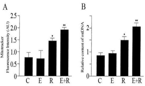
We examined the combined treatment of resveratrol and equol on HUVEC cells to identify their effects on the mitochondrial biogenesis. In particular, the primary goal of this investigation was to determine whether combining resveratrol and equol would increase the expression of key factors involved in mitochondrial biogenesis. The Mitotracker staining showed that mitochondria were located in the perinuclear region in HUVEC (data not shown). Treatment with resveratrol increased significantly the density-area ratio in Mitotracker-labeled endothelial cells as compared to the cytoplasmatic volume (Fig. 7A). The enhanced mitochondrial biogenesis in cells treated with both compounds simultaneously was also confirmed by the increased cellular mtDNA content (Fig. 7B). Overall, we found that co-treatment with resveratrol and equol positively affect mitochondrial biogenesis.
Figure 7: The combined treatment of resveratrol and equol strongly increased the number of mitochondria in HUVEC cells. (A) Mitotracker fluorescent intensities
were analysed to assess the mitochondrial biogenesis. (B) Relative mitochondrial
DNA (mtDNA) content was estimated by qRT-PCR. Representative data of at least
3 experiments each performed in triplicate. (*= P < 0.05, **= P < 0.01). C: control;
E: equol; R: resveratrol; E+R: equol + resveratrol.
4.1.2 Induction of SIRT1 by the association of resveratrol and
equol
Analysis of the effect of a combined administration of resveratrol and equol showed an increase in SIRT1 enzymatic activity in HUVEC endothelial cells (Figure 8). Although equol was much less effective to induce SIRT1, the effect of a combination of resveratrol and equol was greater than the response achieved by the single compounds (Figure 8).
Figure 8: Increase of SIRT1 enzymatic activity by combined administration of resveratrol and equol. Fluorimetric SIRT1 activity assay to determine the effect achieved in HUVEC by the
combined exposure to equol and resveratrol. Representative data of at least 3 experiments
each performed in triplicate. (*= P < 0.05, **= P < 0.01). AFU: arbitrary fluorescence units;
C: control; E: equol; R: resveratrol; E+R: equol + resveratrol.
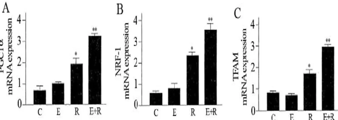
4.1.3 The association of resveratrol and equol activates
mitochondrial biogenesis factor
PGC1-α is involved in regulating the expression of mtDNA via increased expression of TFAM which is co-activated by NRF-1 (Handschin, 2009). We used qRT-PCR measurements to analyse the expression of PGC1-α, NRF-1 and TFAM. The results indicate that these mitochondrial biogenesis factors were strongly increased by combined treatment of resveratrol and equol (Fig. 9).
Figure 9: Effect of resveratrol and equol on mRNA expression of PGC1-α (A), NRF-1 (B), TFAM (C) in HUVEC. qRT-PCR measurement to assess the mRNA
expression of the mitochondrial biogenesis factors. Representative data of at least 3
experiments each performed in triplicate. (*= P < 0.05, **= P < 0.01). C: control;
E: equol; R: resveratrol; E+R: equol + resveratrol.
4.2. Synergistic Effect of L-Carnosine and EGCG in
modulating HO-1 and Hsp72
4.2.1 Synergistic effect of EGCG/L-Car supplementation in
preventing neuronal cell death
To assess the neuroprotective activity of EGCG, L-Car and of a mixture of the two compounds, cell viability was determined in mouse cortical neurons treated with different doses (0-100 μM) of the two compounds for 12 hours (Fig. 1). A significant decrease of viability of about 25% was observed only at the 100 μM dose of EGCG. We therefore decided to use the combination of the two compounds (25 μM each) before exposing the cells for 2 hours to glucose oxidase (50 mU/ml).
Figure 1: Cell viability of mouse cortical neurons. The cells were treated for 12 hours with
15, 25, 50 and 100 μM of EGCG or L-Car *P<0.01.
The glucose oxidase generates hydrogen peroxide at a constant rate and it is known to produce cellular injury in vitro (Chang, 1996). Treatment of cells for 2 hours with glucose oxidase resulted in 27% of residual cell viability (Fig. 2). Exposure of cells for 12 hours to 25 μM EGCG reduced glucose oxidase mediated damage, rising cell viability to 51% (24% more than the glucose oxidase treatment alone). L-Car at 25 μM concentration was less effective in protecting cells from oxidative damage, giving a viability of 33% (7% more than the glucose oxidase treatment). Remarkably, the association of the two compounds protected cells in a synergistic way, giving a rate of neuronal survival of 76% (49% more than the glucose oxidase treatment, which is more than the expected additive effect of 31%).
Figura 2: Cell viability of mouse cortical neurons exposed to glucose oxidase. The cells
were treated for 2 hours with 50 mU/ml of glucose oxidase after exposure for 12
hours with 25 μM of EGCG, L-Car or EGCG + L-Car.* P<0.01; **P<0,001.
4.2.2 Stimulation of the HO-1 and Hsp72 Pathways by
EGCG/L-Car supplementation
We assessed the ability of EGCG and L-Car to elicit the HO-1 and the Hsp72 pathways by measuring HO-1 and Hsp72 gene expression through quantitative real-time PCR. HO-1 and Hsp72
mRNA steady state levels were measured following administration of increasing doses of EGCG and L-Car (Fig. 3).
Figura 3: Stimulation of HO-1 and Hsp72 mRNA levels. Mouse cortical neurons were
treated with increasing doses of EGCG and L-Car (5, 15, 25, 50 or 100 μM) and
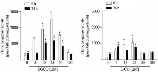
Compared to the not-inducible HO-2 paralog gene, EGCG elicits a dose-dependent increase of HO-1 mRNA, which reaches the maximum (about 8 fold) at 25 μM, and decreases subsequently at 50 and 100 μM. LCar is instead much less active in inducing HO-1 gene expression at the same concentrations, and stimulates HO-1 maximum expression at 25 μM. An opposite behaviour is observed for Hsp72 gene expression which is normalized according to the levels of the paralog not inducible gene Hsc70. EGCG administration gives a dose dependent enhancement of Hsp72 expression at the highest concentration of 50 μM and 100 μM. The last concentration is also associatedto a 25% decrease in cell viability (see Fig. 1). The 25 μMdose, known to have no effects on cell viability, does not enhance Hsp72 gene expression in a statistically significant way. L-Car at the same concentration induces an increase of Hsp72 of about 8- fold (Fig. 3). To confirm that the increase in HO-1 gene expression corresponded to an equivalent increase in HO-1 activity, we measuredthe amount of bilirubin, as indicator of HO-1 activity, after 6 and 24 hours of EGCG and L-Car treatments in presence of glucose oxidase. We observed an increase in HO-1 activity that was comparableto the enhanced mRNA expression, confirming the functional significance of the data obtained by the real time PCR determinations(Fig. 4).
Figura 4: Heme oxygenase activity of mouse cortical neurons. The amount of bilirubin
was measured after 6 and 24 hours on cells treated with 5, 15, 25, 50 or 100 μM of EGCG or L-Car. * P<0.01.
4.2.3 L-Car contributes to an increase in the levels of HO-1
To estimate the contribution of HO-1 in the synergistic neuroprotective effect of EGCG and L-Car we repeated the first experiment by adding also Tin protoporphirin IX (ZnPP), a compound known to potently and specifically inhibit HO-1 activity. As shown in Fig. 5, the inhibition of HO-1 activity caused by ZnPP reduced the neuroprotective effects of the combination of the two compounds by 35%, bringing it to the level obtained with EGCG alone.
Figura 5: Cell viability of mouse cortical neurons exposed to HO-1 inhibitor (ZnPP). The
cells were exposed for 2 hours into 50 mU/ml glucose oxydase after treatment for
12 hours with 25 μM of EGCG, L-Car, EGCG + L-Car or 10 μM ZnPP. *P<0.01; **P<0,001.
Determination of HO-1 by western blotting after EGCG and L-Car administration showed that EGCG strongly increased HO-1 expression while L-Car was much less effective. Similar results were already showed by quantitative real-time PCR. Interestingly their combination enhanced HO-1 protein levels in a synergistic way at 25 μM dose of treatment and decreased HO-1 expression at higher concentration (50 μM) of the compounds (Fig. 6). Therefore, a contribution of L-Car could be observed also in terms of cooperative increase in the levels of HO-1, which is primarily induced by EGCG.
Figura 6 Protein levels of HO-1 estimated by densitometry of bands of western blot.
Neurons were treated with 25 μM of EGCG or L-Car and with, 25 or 50 μM of EGCG + L-Car. Levels of HO-1 are normalized to β-actin. * P<0.01. ** P<0.001.
4.3 Altered expression pattern of Nrf2/HO-1 axis during
accelerated-senescence in HIV-1 transgenic rat
4.3.1 Detection of HIV-1 Transgene Expression
We performed a RT-PCR followed by gel electrophoresis (Fig. 10) to demonstrate the expression of HIV-1 transcript that encodes Tat in the HIV-1 TG rat. PCR was conducted to use a set of primers that only gives rise to PCR product if the HIV-1 transcript encoding for the Tat gene was expressed.
Figure 10: Detection of HIV-1 Tat by RT-PCR in HIV-TG rat. Splenocytes were
extracted from A) Fischer, F344 and B) HIV-1 TG rat.Formalin-fixed paraffin-embedded 5 sections of spleen from line 1 TG rats were analyzed by immunohistochemistry for Tat and gp120. All two proteins were evident in cells within the lymph-node tissues (Fig. 11).
gp120
Tat
Figure 11: Detection of HIV-1 gp120 and Tat protein in HIV-1 TG rats. HIV-1 Tat and
gp120 detected from lymph-node tissues by immunocytochemical analysis
4.3.2
Increased nitrosative stress in the brain of HIV-1 TG rat
Figure 12 shows the results of immunohistochemical detection of nitrotyrosine in representative brain sections from Fischer F344 and HIV-1 TG rat . Nitrotyrosine, an indicator of peroxynitrite formation and other nitrosating agents, was detected in the brain HIV-1 TG rat, whereas no staining was observed in brain regions of F344.
a) Fischer 344 rat b) HIV-1 Tg rat
Figure 12: Nitrotyrosine staining in brain of F344 and HIV-1 TG rats. Representative
photomicrograph of a brain section taken from a) wild-type F344 rat and b) HIV-1
TG rat. Intense positive staining of nitrosative species was found in HIV-1 TG rat.
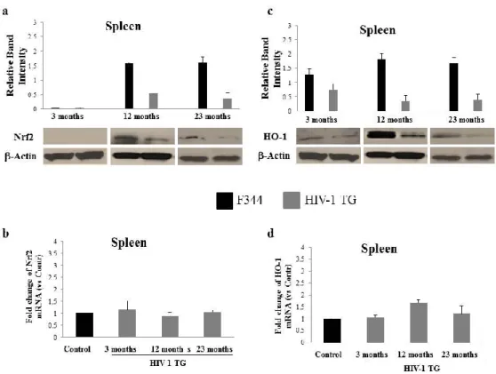
4.3.3 Protein decline of Nrf2 and HO-1 in spleen and liver of HIV-TG rat
As reported by Reid et al. (Reid, 2001) viral transcripts and proteins were identified in several tissues of HIV-1 TG rat, including spleen and liver. To gain insight into the role of Nrf2 in the pathogenesis of HIV-1, we investigated its protein content in spleen extract from 3, 12 and 23 months-old rats by western blot analysis. Nrf2 protein was undetectable in the spleen of both wild-type and HIV-1 TG 3 months-old animals. Consistently, Nrf2 protein was significantly lower in the 12 and 23 months-old HIV-1 TG rats as compared tocontrol animals (Fig. 13a). In contrast, the Nrf2 RNA levels quantified by qRT-PCR did not exhibited any substantial changes in the spleen from 3, 12 and 23 months-old HIV-1 TG rats compared to F344 animals (Fig. 13b), which excludes impairment in the transcriptional regulation of Nrf2. We next examined whether the pattern detected in Nrf2 levels was accompanied by a corresponding trend in HO-1 expression. Similar to the above findings, HO-1 protein declines significantly in each age group of HIV-1 TG rat spleen as compared to non-transgenic controls (Fig. 13c). Unexpectedly, HO-1 RNA levels were still present in all three transgenic groups with a slight increase at 12 months (Fig. 13d). These results suggest that in the spleen of HIV-TG rat, the downregulation of Nrf2 and HO-1 may be controlled primarily at the post-transcriptional level.
Figure 13: Nrf2 and HO-1 expression in the spleen of HIV-1 TG and F344 rats of varying ages. Western blot and densitometric analysis of total Nrf2 (a) and HO-1
(c) from spleen homogenates of young group: 3 months old; middle-aged
group: 12 months old; and aged group: 23 months old (P<0.05 ). (b) RNA
quantification of Nrf2 and HO-1 (d) from spleen of 3, 12, 23 months old
rats. β-actin is used as internal control (P<0.05 ). Black bars: F344 rat; Grey bars: HIV-1 TG rat.
Since liver performs key metabolic functions, including anti-oxidation and detoxification and Nrf2 provide an essential contribution to these processes, we investigated its hepatic protein level in HIV-1 TG rat. Noticeably, elevated Nrf2 protein was observed at 3 months in HIV-TG rat despite the absence of increased concomitant RNA expression (Fig. 2a, 2b), suggesting that an enhanced rate of protein translation may occur in young animals to counteract the stress due to the presence of the HIV-1 transgene. With increasing age, the levels of Nrf2 protein decreased compared to the control animals and in the
liver of older HIV-1 TG animals, Nrf2 protein became undetectable (Fig. 14a). Figure 14b shows that the Nrf2 RNA level in liver of 23 months-old HIV- 1 TG rat was higher than that of middle-aged rats. This result supports the idea that in the liver, Nrf2 protein degradation occurs more quickly during the aging process of HIV-TG rat, even with the increased production of RNA in 23 months-old compared to 12 months-old. To further verify whether the expression of HIV-1 transgene affects the expression of one of the key enzymes involved in the regulation of the liver antioxidant stress, we determined the age-related level of HO-1 between HIV-1 TG animals and their wild-type counterparts. As shown in Fig. 14c, HO-1 protein content was undetectable at 3 months in both experimental groups, while its level was markedly decreased in 12 months-old and at 23 months-old compared to F344 animals. Interestingly, RNA level was subjected to a strong decrease in middle-aged HIV-1 TG rat, similarly to the protein level, and to an increase in the older age (Fig. 14d). Even considering that the rate of HO-1 protein synthesis may remain constant between 12 and 23 months-old in HIV-1 TG rat, the enhanced RNA content at 23 months-old may indicate an adaptive transcriptional response in counteracting the negative effects of HIV-1 transgene.
Figure 14: Age-related expression of Nrf2 and HO-1 at protein and RNA levels in liver of HIV-1 TG and F344 rats. Western blot and densitometric analysis of total
hepatic Nrf2 (a) and HO-1 (c) proteins from young, middleaged and aged group
(P<0.05 ). (b) Nrf2 and HO-1 (d) RNA levels from 3, 12, 23 months old rats.
β-actin is used as internal control (P<0.05 ). Black bars: F344 rat; Grey bars:
HIV-1TG rat.
4.3.4 Reduced Nrf2 and HO-1 protein levels in multiple brain
areas of HIV-1 TG rat
The pro-inflammatory and pro-oxidant effects of HIV-1 related proteins in the brain are well known. Therefore, we next investigated the in vivo impact of HIV-1 on the expression of Nrf2 in the brain, by measuring its levels in whole brain, cortex, cerebellum and hippocampus extracts. Our results indicate a substantial Nrf2 protein decrease in HIV-1 TG rat, particularly in the whole brain, cerebellum
and hippocampus at 23 months-old as well as in the cortex of middle-aged and older HIV-1 TG rat (Fig. 15b, 15c, 15d).
Figure 15: Nrf2 protein levels in the brain of HIV-TG and F344 rats of varying ages.
Western blot and densitometric analysis of total Nrf2 in whole-brain (a),
cerebellum (b), cortex (c) and hippocampus (d) from 3, 12, 23 months old rats
(P<0.05 ). Black bars: F344 rat; Grey bars: HIV-1 TG rat.
The qRT-PCR quantification revealed differential Nrf2 RNA expression with age in all HIV-1 TG analysed brain regions with a tendency to increase at late age, (Fig. 16).
This observed RNA profile in HIV-1 TG animals may be a late transcriptional response to the presence of HIV-1 transgene.
Figure 16: RNA expression of Nrf2 in the brain. Nrf2 RNA in different brain regions:
whole-brain (a), cerebellum (b), cortex (c) and hippocampus (d) from young,
middle-aged and aged group (P<0.05 ). Black bars: F344 rat; Grey bars: HIV-1
TG rat.
For a more comprehensive evaluation, we next examined whether HO-1 protein levels were negatively affected during aging in HIV-1TG rats. Results of the western blot assay showed that middle-aged and aged HIV-1 TG groups exhibited significantly lower HO-1 content than control F344 rats (Fig.17).
Figure 17: HO-1 protein levels in the brain of HIV-TG and F344 rats of varying ages.
Western blot and densitometric analysis of HO-1 in whole-brain (a), cerebellum
(b), cortex (c) and hippocampus (d) from 3, 12, 23 months old rats (P<0.05 ).
Black bars: F344 rat; Grey bars: HIV-1TG rat.
In addition, Fig. 18 indicates the RNA levels of HO-1 in various brain regions of HIV-TG rat; the data displayed different expression patterns, showing an increase in prevalence at 23 months-old. Taken together, the results provide compelling evidence that the chronic expression of HIV-1-related proteins causes defective protein expression of HO-1 and a shared transcriptional signature at 23 months-old among multiple brain areas.
Figure 18: RNA expression of HO-1 in the brain. HO-1 RNA in different brain regions:
whole-brain (a), cerebellum (b), cortex (c) and hippocampus (d) from young,
middle-aged and aged group (P<0.05 ). Black bars: F344 rat; Grey bars: HIV-1
TG rat.
4.3.5 SA-β-gal expression is enhanced in the HIV-
1
TG rat
Up to this point our data indicate that the oxidative state observed in the HIV-1 rat might be linked to the agedependent reduction of Nrf2 and HO-HIV-1 proteins. Considering that persistent oxidative stress promotes the occurrence of premature senescent phenotype, we next assessed SA-β-gal expression in HIV-1 TG rat. SA-β-gal positive tissue areas showed substantial increase in expression in 12 months-old HIV-1 TG rats with respect to age-matched F344 animals (Fig. 19). As compared with the control, no considerable variation of SA-β-gal activity was found in spleen, liver and brain of young HIV-1 TG rat (data not shown). These results suggest that the declined protein function of
Nrf2 and HO-1 was accompanied by the acquisition of premature senescence phenotype in HIV-1 TG rat.
Figure 19: Staining for senescence-associated-galactosidase (SA-β-gal) in HIV-1 TG and F344 rats. Representative staining from middle-aged group of (a) spleen F344
rat; (b) spleen HIV-TG rat; (c) liver F344 rat; (d) liver HIV- 1 TG rat; (e) brain
5. DISCUSSION
Recently, synergy assessment has become a key area in medicine research in order to enhance efficiency of treatments and affect not only one single target, but several targets. Numerous nutraceuticals have been found to target and attenuate the progression of age-related dysfunction (Prasain, 2010). Currently, there are a variety of dietary strategies to ameliorate mitochondrial function in ageing (Ferrari, 2004). Although resveratrol and equol have recently attracted scientific interest for a wide-ranging of health benefits, their influence on mitochondria is still poorly understood. Indeed, the first aim of the project was to investigate the effects of resveratrol and equol on mitochondrial biogenesis using the two compounds individually and in combination. Several studies were conducted showing the synergistic effect of resveratrol with different compounds (Csaki, 2009) but the combination with equol has never been tested on the mitochondrial function. The analysis of Mitotracker intensity showed that resveratrol induced an increase in mitochondrial mass compared to non-treated cells. Equol alone was not effective in terms of augmenting the mitochondrial mass, however the combined treatment (resveratrol + equol) was more effective respect to resveratrol alone. It is important to point out that mitochondrial dysfunction tend to induce a wide range of adaptations of nuclear gene expression, named the retrograde response (Butow, 2004). Typical of this adaptive process (mitohormesis) (Calabrese, 2012; Calabrese, 2007; Tapia, 2006) is the up-regulation of mitochondrial biogenesis (Biswas, 1999). A robust adaptive response may further explain the increase in mitochondrial mass. Moreover, It has been consistently demonstrated that activation of SIRT1 stimulates
mitochondrial biogenesis and resveratrol has been utilized as a SIRT1 activator to regulate mitochondrial function (Lagouge, 2006). Since equol exhibits a wide range of biological properties, it may be a sirtuin-targeting nutraceutical to prevent mitochondrial decline. In this study, the combination of resveratrol with equol is associated with the activation of SIRT1. In addition, PGC1-α is a key component in modulating mitochondrial function and interacts with transcription factors such as NRF-1and TFAM. Our results indicate that these mitochondrial biogenesis factors were increased by combined treatment of resveratrol and equol. Collectively, these data demonstrate that the combination of two known natural products, resveratrol and equol exerts a synergistic effect on mitochondrial function because stimulates the mitochondrial biogenesis more than the single compounds alone. Clearly, more work is needed to provide novel insights into the mechanisms by which resveratrol and equol synergize to regulate the mitochondrial dynamics. However, the co-administration of these agents may be a possible nutraceutical and/or anti-ageing strategy for a number of mitochondria-associated disorders.
Recently, a growing number of studies have indicated that the dietary antioxidants may be beneficial for neuronal recovery and survival in neurodegenerative disorders (Calabrese, 2009). Over the past years, the involvement of the HO-1 pathway in anti-degenerative mechanisms has received considerable attention (Piantadosi, 1997; Dorè, 2002). Notably, HO-1 induction occurs together with the induction of other heat shock proteins during various physiopathological conditions, generating potent protective
system against brain oxidative injury (Calabrese, 2004). The second aim of the project was to provide evidence that the combination of EGCG and L-Car elicits two different pathways with synergistic neuroprotective effects and increasing viability of neuronal cells. Our results demonstrated that the neuroprotective effects of EGCG and L-Car are achieved through the targeting of HO-1/Hsp72 systems. the effect of a combination of EGCG and L-Car was greater than the response achieved by the single compounds alone and over the expected additive effect of EGCG and L-Car. The synergistic action of these two agents supports the idea of a crosstalk between the HO-1 and the Hsp72-mediated pathways which potentiates the efficacy of the two compounds used alone. Moreover, the hypothesis is further reinforced since we assumed that the cytoprotection obtained through the expression of Hsp72 is less effective when HO-1 is blocked with its inhibitor ZnPP. Because of this crosstalk, combination of drugs activating the single pathways might have a more pronounced antioxidant effect in a number of physiopathological conditions where the sensing of redox status is imbalanced. These findings imply that HO-1/Hsp72 system might work as a promising therapeutic approach with potential for clinical usefulness. To our knowledge, this is the first time that the antioxidant activities of the selected natural agents EGCG and L-Car were investigated in combinations. Our study demonstrates that combined administration of EGCG and L-Car possesses a more apparent antioxidant activity compared with each of them alone. Therefore, our findings indicate that the synergistic antioxidant effects of EGCG and L-Car may be a successful approach in the prevention of brain aging. Finally this