International PhD
PLANT HEALTH TECHNOLOGIES AND PROTECTION OF AGROECOSYSTEMS
XXVII CYCLE 2012-2014
Sustainable approaches to control Calonectria diseases in ornamental nursery
This thesis is presented for the degree of Doctor of Philosophy by
ALESSANDRO CINQUERRUI
COORDINATOR TUTOR
This research was supported by MIUR project number PON01_01611 (SO.PRO.ME: “Sustainable production of potted plants in Mediterranean environment”).
a
Contents
1. Introduction: Calonectria disease in the Mediterranean basin ... 1
2. The genus Calonectria: history and taxonomy ... 4
2.1. Importance of Calonectria ... 7
2.2. Epidemiology ... 10
2.3. Morphology ... 13
2.4. Mating compatibility and strategies... 15
2.5. Multigene phylogeny ... 16
2.6. Calonectria morganii complex ... 18
2.6.1. Cylindrocladium scoparium ... 18
2.7. Calonectria scoparia complex ... 21
2.7.1. Calonectria mexicana ... 22
2.7.2. Calonectria pauciramosa ... 23
2.7.3. Calonectria polizzii ... 26
2.7.4. Calonectria pseudomexicana ... 27
2.7.5. Calonectria tunisiana ... 28
2.8. Calonectria disease control ... 30
2.8.1. Chemical control ... 31
2.8.2. Soil fumigation and solarization ... 38
2.8.3. Biological control of Calonectria spp. ... 43
3. Integrated Pest Management ... 48
3.1. Introduction: definition of biological control ... 50
3.2. BCAs: mechanisms of action... 52
4. Thesis aims ... 59
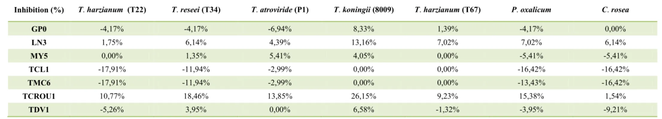
5. In vitro antagonism of BCAs against Calonectria species. ... 66
5.1. Materials and methods ... 66
5.2. Results... 68
5.3. Discussion ... 73
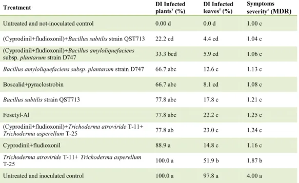
6. Biological and chemical control of Calonectria leaf spot on Callistemon viminalis ... 74
6.1. Materials and methods ... 74
b
6.3. Discussion ... 82
7. Biological and integrated control of Calonectria leaf spot on Metrosideros excelsa “aurea” and Callistemon “Captain Cook” ... 84
7.1. Materials and methods ... 84
7.2. Results... 88
7.3. Discussion ... 100
8. Biological and integrated control of Calonectria stem rot on Dodonaea viscosa ... 102
8.1. Materials and methods ... 102
8.2. Results... 107
8.3. Discussion ... 116
9. Biological and integrated control of Calonectria pauciramosa on Polygala myrtifolia ... 118
9.1. Materials and methods ... 118
9.2. Results... 122
9.3. Discussion ... 125
10. Effects of label and sub-label rates of dazomet and metham-sodium on survival of Calonectria spp. ... 126
10.1. Materials and methods ... 126
10.2. Results ... 129
10.3. Discussion ... 135
11. Conclusions ... 137
Acknowledgements ... 139
1 1. Introduction: Calonectria disease in the Mediterranean basin
In last decades, ornamental plant nurseries have occupied an increasingly important role in the agricultural economy, in particular in southern Italy. For this reason, trade of plants between countries is becoming more frequent. These activities could favour the introduction of new pathogens. Among these,
Calonectria spp. represent a serious threat for the nursery production in several
countries, among which also Italy (Koike et al., 1999; Polizzi and Crous, 1999; Schoch et al., 2001b; Henricot and Beales, 2003; Lane et al., 2006; Pérez-Sierra
et al., 2007; Chen et al., 2011).
The species belonging to the genus Calonectria cause several symptoms, such as damping-off, cutting rot, crown and root rot, stem canker, leaf blight and petiole rot on a wide range of plant hosts.
Although in the Mediterranean basin Calonectria species were little known until few years ago, today they are a serious threat for crop. The first disease caused by Calonectria has been described by Booth and Murray in 1960. He identified a leaf spot of Hedera helix caused by Calonectria hederae G. Arnaud ex C. Booth. et D. Murray. Later, in 1964, a damping-off of rose cutting caused by
Ca. morganii Crous, Alfenas et M.J. Wingf (as Cy. scoparium) was detected by
Storey.
In Italy, symptoms of root and petiole rot caused by Cy. spathiphylli Schoult., El-Gholl et Alfieri were detected, for the first time, on Spathiphyllum cv. Mauna Loa by Carrai and Garibaldi in 1990.
In South Italy Ca. pauciramosa is the most important species among these nectroid fungi. This species, found more frequently, is responsible for different symptoms on several plant-host, such as leaf spots on Arbutus unedo, Acacia
retinodes, Feijoa sellowiana, Dodonaea viscosa, Pistacia lentiscus, Brahea armata, Agonis flexuosa (Polizzi and Catara, 2001; Vitale and Polizzi, 2007;
Polizzi et al., 2007a; Polizzi et al., 2010), crown rot and stem blight on plants of
Pistacia lentiscus (Vitale and Polizzi, 2007), defoliation and stem blight on
plants of Eugenia myrtifolia (Polizzi et al., 2009a), crown and root rot on
2
sellowiana (Polizzi et al., 2006b; Vitale et al., 2008; Vitale et al., 2009a; Polizzi et al., 2009a).
In 1999, Polizzi and Crous published the first report of a new disease of
Polygala myrtifolia caused by Ca. pauciramosa C.L. Schoch et Crous (= Cy. pauciramosum C.L. Schoch et Crous) in Italy and Europe. Initially this fungus
had been identified as Ca. morganii after isolations from myrtle plants (Polizzi and Azzaro, 1996).
Ca. pauciramosa has a centre of origin in Central and South America and is well
established in Australia, Europe, South Africa and the USA. Variability in the DNA sequence data from a series of Ca. pauciramosa isolates from Australia, California, Italy, South Africa and South America illustrated the possibility that more than one introduction of this species could have occurred into Italy from South Africa or other countries (Schotch et al., 2001a).
Since the early 1990s, Ca. pauciramosa was reported as agent causal of several diseases in most ornamental nurseries in the areas of southern Italy (Polizzi and Catara, 2001; Polizzi et al., 2006a; Vitale et al., 2008; Polizzi et al., 2009a, 2010), where it causes considerable losses to ornamental plants.
More recently, Polizzi et al. (2006b) have identified Ca. morganii (as Cy.
scoparium) as causal agent of leaf spot, blight and crown rot on mastic trees
(Pistacia lentiscus). Then, damping-off and leaf spot caused by Ca. morganii on different cultivars of bottlebrush cuttings were described in Italy (Polizzi et al., 2007b). More recently, Polizzi et al. (2009b) have linked Ca. morganii to leaf spot and twig blight on mallee honey myrtle in Italy.
Furthermore, subsequent studies conducted by Vitale and Polizzi in 2008 have shown the co-existence of Ca. pauciramosa and Ca. morganii in the same host. In 1994, in Hampshire Buxus sempervirens was identified as host of a new species of the genus Calonectria. Symptoms of blight were observed on this plant and the causal agent was identified in Ca. pseudonaviculata L. Lombard, M.J. Wingf et Crous (as Cy. pseudonaviculatum Crous, J.Z. Groenew et C. F. Hill) based on DNA sequence and morphological data (Henricot et al., 2000; Crous et al., 2002).
3 More recently, in December 2002 Henricot and Culham (2002) described this species as Cy. buxicola Henricot. More recently, it has been proposed to retain the name of Cy. buxicola against Ca. pseudonaviculata (Henricot et al., 2012). Although the origin of this new species is unknown, it was hypothesized that it was first introduced into Europe, where it is widespread in many countries including Austria, Belgium, Croatia, Czech Republic, France, Germany, Italy, the Netherlands, Slovenia, Spain and Switzerland (Crepel and Inghelbrecht, 2003; Brand, 2005; CABI, 2007; Henricot et al., 2008; Saracchi et al., 2008; Benko Beloglavec et al., 2009; Varela et al., 2009; Cech et al., 2010; Šafránková et al., 2012) and then to New Zealand (EPPO, 2004). Cy. Buxicola represents the main causal agent of blight of different species of Buxus, such as
B. macrophylla, B. sempervirens, B. sinica and B. colchica (Henricot et al.,
2008; Gorgiladze et al., 2011).
Symptoms of peg, pod, and root necrosis of peanuts, but also leaf spot, damping-off, blight, crown and root rot of several plant hosts are caused by another species of the genus Calonectria, such as Ca. ilicicola Boedijn et Reitsma (= Cy. parasiticum Crous, M.J. Wingf. et Alfenas) (Crous, 2002). Based on molecular data, Ca. ilicicola has been identified for the first time as causal agent of crown and root rot on potted of Laurus nobilis plants (Polizzi et
al., 2012). In the Isles of Scilly (UK) a wilting disease on L. nobilis was first
identified caused by Cy. ilicicola, known as Ca. lauri, but incorrectly linked to the sexual morph Ca. ilicicola. Later, based on molecular comparison of the ex type strain, Crous et al. (1993b) proved the linkage between Ca. ilicicola and the asexual morph Cy. parasiticum. Furthermore, Ca. lauri was isolated from leaves of Ilex quaquifolium in France and Netherlands and from root of B.
sempervirens in Belgium (Lechat et al., 2010).
More recently, three new cryptic species, such as Ca. polizzii, Ca.
pseudomexicana and Ca. tunisiana, have been described based on multigen
phylogenetic analyses, morphological characters and mating compatibility (Lombard et al., 2010b, 2011).
Ca. polizzii L. Lombard, Crous et M.J. Wingf was identified for the first time on Arbutus unedo and Callistemon citrinus plants showing leaf spot symptoms, but
4 its pathogenicty was confirmed in 2011 by Lombard et al. Currently, it is present as plant pathogen in the Mediterranean basin, in particular in Tunisia and in Italy (Lombard et al., 2011; Aiello et al., 2013; Vitale et al., 2013b). Furthermore, two new Calonectria species, such as Ca. pseudomexicana L. Lombard, G. Polizzi, et Crous and Ca. tunisiana L. Lombard, G. Polizzi, et Crous were reported for the first time in Tunisia in an ornamental nursery (Lombard et al., 2011). These species reside in Calonectria scoparia complex and are closely to Ca. mexicana. The presence of Ca. mexicana in the Mediterranean basin, and in particular in Africa, was reported for the first time by Lombard et al. in 2011.
These three cryptic species, such as Ca. polizzii, Ca. tunisiana and Ca.
pseudomexicana, are responsible for crown and root rot and leaf spot on
seedlings of Callistemon spp., Dodonaea viscosa, Metrosideros spp. and Myrtus
communis (Vitale et al., 2013b).
2. The genus Calonectria: history and taxonomy
The genus Calonectria (Ca.) was first identified in 1867 by De Notaris, with Ca.
daldiniana as the type (Rossman, 1979a). Subsequently, this species was
incorrectly confronted with Ca. pyrochroa because it was identified based only on the teleomorph morphology (Rossman, 1979a). The fungi of this genus are characterized by having an ascocarp wall structure that is brightly colored, changing to blood-red in 3 % KOH solution, warty to scaly (Rossman, 1993; Rossman et al., 1999). In addition, it is important to stress that Cylindrocladium (Cy.) is the anamorph of this genus (Rossman, 1993; Rossman et al., 1999). The presence of the anamorph allows the identification of some specimens to species level (Schoch et al., 2000b; Crous, 2002), considering to the restricted morphological characteristics of the teleomorph (Rossman, 1979b, 1983).
Morgan in 1892 identified for the first time the anamorph genus
Cylindrocladium, based on Cy. scoparium, found as a saprobe in a pod of Gleditsia triacanthos. This genus is characterized by branched conidiophores
5 More lately, Crous (2002) mentioned that some species belonging to this genus are pathogenic to numerous plants with a wide distribution in sub-tropical and tropical regions.
Taxonomically, the genus Calonectria belongs to the family of Nectriaceae, one of three family of the order Hypocreales (Rogerson, 1970; Rossman, 1983; Rossman, 1996; Rossman et al., 1999; Hirooka et al., 2012). The Nectriaceae is characterized by having uniloculate ascomata that are orange to purple and not immersed in well-developed stromata (Rossman et al., 1999).
Calonectria is distinguished from the others 20 genera of this family by its Cylindrocladium anamorph and its importance as plant pathogens (Lombard et al., 2010a).
After the first description of seven Cylindrocladium species and their teleomorph (Boedijn and Reitsma, 1950), Rossman in 1983 identified five species, in which it was described the novel Ca. ophiospora but not its anamorph state.
As a matter of fact, the anamorph morphology is an important characteristic in order to distinguish species of Calonectria (Peerally, 1991a). More lately, 10
Calonectria species and corresponding Cylindrocladium anamorphs were
identified. In addition, 16 Cylindrocladium species without any associated teleomorph were reported (Lombard et al., 2010b).
The genus Cylindrocladiella was mistakenly identified as having
Cylindrocladium-like species with small conidia (Boesewinkel, 1982) and Nectricladiella teleomorphs, to synonymy with Cylindrocladium (Schoch et al.,
2000b).
According to Crous and Wingfield (1994), the anamorph characteristics are most important in the taxonomy of Calonectria spp. Accordingly, in their monograph on Cylindrocladium they detected 22 Cylindrocladium species, by associating with them 16 Calonectria species, of which five species were assigned to the genus Cylindrocladiella based on morphological characters of the holomorph.
In one of the most recent monograph, 28 Calonectria species were identified, all associated with Cylindrocladium anamorphs (Crous, 2002). Moreover, 18
6
Cylindrocladium species were recognized, although no information was found
on their teleomorph states. In detail, seven taxa, belonging to this latter group, were of doubtful authenticity. Until a few years ago, several authors reported a total of 109 Calonectria and 96 Cylindrocladium species (Crous, 2002; Crous et
al., 2004b, 2006; Gadgil and Dick, 2004; Lombard et al., 2009, 2010c).
At present, by searching on Index Fungorum (www.indexfungorum.org) it is possible found a total of 317 and 92 names records respectively for Calonectria and Cylindrocladium. A similar search on Mycobank shows a total of 325 and 115 names records respectively for Calonectria and Cylindrocladium (www.mycobank.org; Crous et al., 2004a; Robert et al., 2005).
Calonectria spp. are characterized by their yellow to dark red perithecia, with
scaly to warty ascocarp walls giving rise to long-stalked, clavate asci with 1– multi-septate ascospores and Cylindrocladium (Cy.) anamorphs (Rossman 1993; Crous, 2002; Lombard et al., 2010a).
The genus Cylindrocladium is characterized by branched conidiophores with stipe extension terminating in characteristics vesicles and producing cylindrical, 1-multispetate conidia (Crous and Wingfield, 1994; Crous, 2002).
The anamorph, that is the state most common in nature, showed different types of morphological characters for distinguishing Calonectria, such as vesicle shape, stipe extension length, conidial septation, and dimensions on a standardized medium under defined growth conditions (Boesewinkel, 1982; Peerally, 1991a; Crous and Wingfield, 1994; Schoch et al., 2001a; Crous, 2002). Moreover, it is important to stress that some intraspecific variation in vesicle shape and conidial dimension can be common, resulting in confusion in the taxonomic level (Crous and Peerally, 1996; Crous et al., 1998a).
Calonectria spp. differ three different morphological forms of conidia, of which
the macroconidia are present in all but Ca. multiseptata (Peerally, 1991a; Crous and Wingfield, 1994; Crous et al., 1998b; Crous, 2002). Mega- and microcondia are less frequent, so they are not considered as important characters to identify different species (Sobers, 1971; Crous and Wingfield, 1994; Crous and Seifert, 1998a; Crous, 2002).
7
Calonectria species have homothallic and heterothallic mating systems (Alfieri et al., 1982; Schubert et al., 1989; Crous and Wingfield, 1994; Crous, 2002).
Heterothallic Calonectria spp. have a biallelic heterothallic mating system with the female structures (protoperithecia) spermatised by conidia or hyphae of an opposite mating type strain (Schoch et al., 1999, 2000a, 2001a). Crous (2002) points out that some Calonectria spp. has retained the ability to recombine with other closely related Calonectria spp., although the progeny from these crosses have low levels of fertility. This has complicated the application of the biological species concept for Calonectria, although it has been useful for some species (Schoch et al., 1999; Lombard et al., 2010a, b).
2.1. Importance of Calonectria
After the first identification as a saprobe (Graves, 1915), Massey in 1917 detected the pathogenicity of Calonectria species, and in particular Anderson in 1919 proved the pathogenicity of Ca. morganii (as Cy. scoparium).
Initially, Calonectria species were recognized as causal agent of disease on 30 plant families (Booth and Gibson, 1973; French and Menge, 1978; Peerally, 1991a; Wiapara et al., 1996; Schoch et al., 1999). More recently, Crous has reported Calonectria species as causal agents of disease symptoms on about 100 plant families and 335 plant host species, among which important forestry, agricultural and horticultural crops (Crous, 2002).
In Europe and in Asia, Calonectria species are capable to cause diseases on horticulture crops, especially in garden and ornamental commercial nurseries (Polizzi and Crous, 1999; Polizzi, 2000; Crous, 2002; Henricot and Culham, 2002; Pérez-Sierra et al., 2007; Polizzi et al., 2007a, b; Hirooka et al., 2008; Polizzi et al., 2009a, b, c; Vitale et al., 2009a). In detail, plants belonging to the families of Anacardiaceae, Aquifoliaceae, Araceae, Araliaceae, Arecaceae,
Asteraceae, Buxaceae, Ericaceae, Myrtaceae, Polygaceae, Rhamnaceae and Rosaceae are attacked by these pathogens, with several disease symptoms such
as crown rot, collar rot and root rot, leaf spots, and cutting rot (Massey, 1917; Anderson, 1919; Storey, 1964; Aragaki et al., 1972, 1988; Peerally, 1974; de Prest and Poppe, 1988; Carrai and Garibaldi, 1990; Uchida and Kadooka, 1997;
8 Litterick and McQuilken, 1998; Polizzi and Crous, 1999; Polizzi 2000; Crous, 2002; Polizzi and Catara, 2001; Henricot et al., 2000; Henricot and Culham, 2002; Henricot and Beales, 2003; Poltronieri et al., 2004; Lane et al., 2006; Polizzi et al., 2006a, b; Pérez-Sierra et al., 2006, 2007; Polizzi et al., 2007a, b; Vitale and Polizzi, 2007; Aghajani et al., 2008; Henricot et al., 2008; Hirooka et
al., 2008; Vitale et al., 2008; Polizzi et al., 2009c; Vitale et al., 2009a; Lechat et al., 2010; Alfenas et al., 2013b).
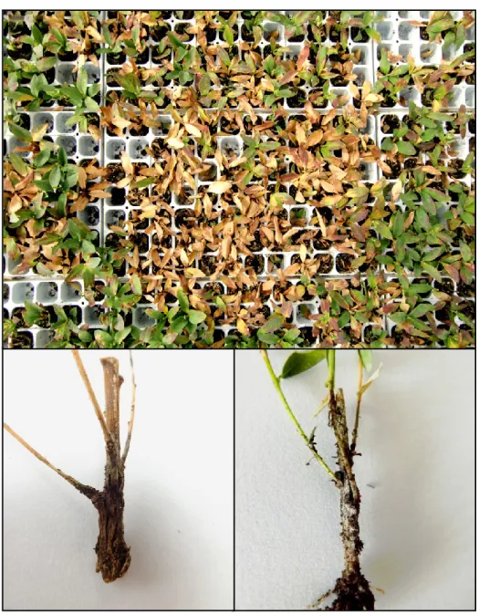
Figure 1 - Leaf spots on Metrosideros excelsa “aurea” (a), Callistemon spp. (b,c), Metrosideros excelsa (d), Dodonaea viscosa (e)
a
b
c
9 In agriculture, several economically important crops have been reported as susceptible plants to infections of Calonectria species. In particular, Calonectria infections are very common in the family of Fabaceae and Solanaceae. Ca.
ilicicola, Ca. pyrochroa, Ca. brassicae (as Cy. gracile) were reported as
responsible for Cylindrocladium black rot on Arachis hypogeal (peanut), red crown rot of Glycine max (both in USA) and Cylindrocladium tuber rot of
Solanum tuberosum (Brazil), respectively (Bell and Sobers, 1966; Beute and
Rowe, 1973; Rowe et al., 1973; Sobers and Littrell, 1974; Rowe and Beute, 1975; Phipps et al., 1976; Johnson, 1985; Dianese et al., 1986; Berner et al., 1988, 1991; Culbreath et al., 1991; Porter et al., 1991; Varon AF de, 1991; Hollowell et al., 1998; Kim et al., 1998; Boedijn and Reitsma, 1950; Bolkan et
al., 1980, 1981). Other diseases associated to Calonectria species on agricultural
crops include root rot and leaf diseases of fruit bearing and spice plants (Jauch, 1943; Wormald, 1944; Sobers and Seymour, 1967; Nishijima and Aragaki, 1973; Milholland, 1974; Krausz and Caldwell, 1987; Hutton and Sanewski, 1989; Anandaraj and Sarma, 1992; Risède, 1994; Jayasinghe and Wijesundera, 1996; Risède and Simoneau, 2001; Vitale and Polizzi, 2008), post-harvest diseases of fruits (Fawcett and Klotz, 1937; Boedijn and Reitsma, 1950; Sepiah, 1990; Fitzell and Peak, 1992; Vaidya and Rao, 1992; Sivapalan et al., 1998), root and crown rot of Medicago sativa (alfalfa) (Ooka and Uchida, 1982; Hwang and Flores, 1987) and sheath net blotch of Oryza sativa (Crous, 2002).
These plant pathogens also cause diseases on some forestry plant families, such as Fabaceae, Meliaceae, Myrtaceae and Pinaceae. In particular, they are responsible for symptoms of cutting rot (Crous et al., 1991; Crous, 2002; Lombard et al., 2009, 2010d; Alfenas et al., 2013a), damping-off (Batista, 1951; Cox, 1953; Terashita and Itô, 1956; Sharma and Mohanan, 1982; Sharma et al., 1984; Crous et al., 1991; Brown and Ferreira, 2000; Crous, 2002; Taniguchi et
al., 2008) leaf diseases (Cox, 1953; Hodges and May, 1972; Barnard, 1984;
Sharma et al., 1984; El-Gholl et al., 1986; Peerally, 1991a,b; Crous et al., 1993b; Crous and Wingfield, 1994; Crous et al., 1998b; Schoch and Crous 1999; Schoch et al., 1999; Booth et al., 2000; Park et al., 2000; Crous and Kang, 2001; Gadgil and Dick, 2004; Lombard et al., 2010b, 2011; Alfenas et al.,
10 2013a), shoot blight (Sharma et al., 1984; Crous et al., 1991, 1998b; Crous and Kang, 2001), stem cankers (Cox, 1953; Sharma et al., 1984, 1985; Crous et al., 1991; Lombard et al., 2009) and root rot (Cox, 1953; Hodges and May, 1972; Cordell and Skilling, 1975; Mohanan and Sharma, 1985; Crous et al., 1991; Lombard et al., 2009; Alfenas et al., 2013a, b).
Whereas, in many cases, Calonectria species cause disease in seedling and cutting production, on the other in few cases Cylindrocladium species cause leaf disease e shoot blight resulting in defoliation of trees leading to loss of vigour (Hodges and May, 1972; Sharma et al., 1985; Booth et al., 2000; Park et al., 2000; Crous and Kang, 2001; Crous, 2002; Old et al., 2003; Rodas et al., 2005).
2.2. Epidemiology
As mentioned before, species of Calonectria (Ca.) are commonly reported as responsible for several disease symptoms, including crown, root and stem rot, stem or crown canker, blight, root and pod rot (Crous, 2002).
At first, several species of this genus were reported as saprobe (Graves, 1915). Moreover, after its first identifications as causal agent of disease on rose, Cy.
scoparium was reported as responsible for several disease symptoms, such as
damping-off, root rot, crown canker, fruit rot, stem lesions, tuber rot, etc. (Massey, 1917; Anderson, 1919; Crous et al., 1991).
Calonectria species are polycyclic fungi. They are able to differentiate
chlamydospores, which form microsclerotia, in soil and senescing plant tissue. Microsclerotia represent the primary inoculum of Calonectria spp. in the soil (Phipps et al., 1976), where they are present in the infected plant material, such as leaves or stems, fall to the ground and release them in the soil.
Bugbee and Anderson in 1963 (a) showed that the pathogen formed microsclerotia in infected needles. Cy. scoparium produced microsclerotia in substomatal chambers in leaf spot on azalea 10 days after inoculation (Reis and Chaves, 1967).
Although microsclerotia aren’t capable to survive well in the soil if the water content is low (Sung et al., 1980), they have vitality than about 15 years even
11 without host plant (Thies and Patton, 1970; Sobers and Littrell, 1974), as long as there is an adequate soil moisture level (Pataky and Beute, 1983).
Microsclerotia are able to survive in the soil with an optimal temperature of 25 °C, even without soil moisture (Almeida and Bolkan, 1981). Anderson in 1919 reported the presence of microsclerotia at depth of up to 66 cm below the soil surface.
Some authors performed experiments to evaluate the effect of heat on the vitality of microsclerotia. Certain levels of heat produced by an Egedal bed steamer are able to reduce microsclerotia of Cy. floridanum present at 5-10 cm soil depths. Moreover, at the same time, microsclerotia present at or below 15 cm have maintained their vitality (Dumas et al., 1998). In fact, moisture, soil temperature and depth of soil are three important factors that can determine the level of survival of microsclerotia (Phipps and Beute, 1977, 1979; Roth et al., 1979; Taylor et al., 1981; Pataky and Beute, 1983; Crous, 2002; Kuruppu et al., 2004). Moreover, it was demonstrated that solarization effectively suppressed
Ca. pauciramosa microsclerotia (Polizzi et al., 2003). Subsequently, Vitale et al. (2013a) confirmed that short-term soil salarization is capable to suppress Calonectria microsclerotia in nurseries and demonstrated that different
solarizing materials, such as ETFE, could improve this technique.
The infection can also start with the germination of conidia or ascospores. The presence of water or humidity, represented by rain and irrigation practices, plays a key rule during this process, creating an environment favorable to the development of infection. Moreover, the onset of disease caused by these pathogens is favoured by conditions of nutrient stress of seedlings (Arentz, 1991).
In ornamental nurseries, the movement of water is one of the main factors in the spread of conidia originating from microsclerotia, onto potting mixes and plants (Vitale et al., 2013b).
On host plant, conidia are able to germinate rapidly, as followed reported. Indeed, Sharma and Mohanan 1990 asserted that conidia of Cy.
quinqueseptatum germinate faster in vivo than in vitro. It was demonstrated that
12 (Bolland et al., 1985), because this pathogen penetrates directly, without need to appressoria over stomata for stomatal penetration (Sharma and Mohanan, 1990). This type of penetration is spread in many Calonectria species, as observed for
Cy. pauciramosum. Some Calonectria species are able to produce phytotoxin
that influence the virulence of pathogen. It was reported that Cy. scoparium and and Cy. pterididis were able to produce the phytotoxin Cyl-2 (Hirota et al., 1973; Nikolskaya et al., 1995).
13 2.3. Morphology
For many years, the description of fungal species has been performed taking into consideration morphological and phenotypic characters (Brasier, 1997; Taylor
et. al., 2000; McNeill et al., 2005). However, nowadays this is an old method
that cannot allow to distinguish different Calonectria species due to their morphological similarity (Lombard et al., 2010c; Lombard et al., 2011; Chen et
al., 2011). Thus, biological and phylogenetic characters are the basis for the
identification of new Calonectria species (Rossman, 1996; Brasier, 1997; Taylor
et al., 2000; Crous et al., 2004b, 2006).
Also, the characterization of the genus Calonectria and of its anamorph
Cylindrocladium has been based on morphology of its species (Lombard et al.,
2010a).
Until the 1990s, sexual compatibility and mostly morphological characteristics were used to identify different species (Boedijn and Reitsma, 1950; Peerally 1991a; Crous et al., 1992; Crous and Wingfield, 1994; Crous, 2002), thanks to which several complexes, such as Ca. scoparia complex (Schoch et al., 1999),
Ca. brassicae (as Cy. gracile) complex (Crous et al., 2004b) and Ca. kyotensis
complex (Crous et al., 2006) were identified.
Vesicle shape, stipe extension length and macroconidial septation and dimensions are characteristics of the anamorphs, widely used for identifications (Boesewinkel, 1982; Peerally, 1991a; Crous and Wingfield, 1994; Crous, 2002). In particular, on the one hand some authors stated that vesicle morphology was one of the most important taxonomic criteria to distinguish species of
Cylindrocladium (Sobers, 1968; Peerally, 1973; EI-Gholl et al., 1986; EI-Gholl et al., 1989; Peerally, 1991a), but on the other, some authors asserted that this
characteristic was highly variable (Hunter and Barnett, 1978; Rossman, 1983). More lately, some studies demonstrated that this criterion is reliable when examined on carnation-leaf agar (CLA), under pre-determined conditions of incubation (Fisher et al., 1982; Crous et al., 1992). Besides, ascospore septation and dimensions, ascospore number within the asci and perithecial colour are the most important morphological characteristics of the teleomorph used for identifications. In detail, it is important to emphasize that perithecia of
14
Calonectria species shouldn’t be used in identifications because they are
morphologically very similar (Crous and Wingfield, 1994; Crous, 2002).
Calonectria spp. has widely been studied by culturing colonies on carnation leaf
agar medium (CLA) (Fisher et al., 1982; Crous, 2002). More recently, the diagnosis of the genus Calonectria is based on the use of synthetic nutrient-poor agar (SNA; Nirenburg, 1981; Lombard et al., 2009, 2010) and minimal salt agar (MSA; Guerber and Correll, 2001; Halleen et al., 2006; Lombard et al., 2010b) with sterile toothpicks, which have been used to induce the morphological characters (Lombard et al., 2010b).
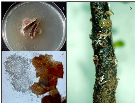
Figure 3 - Perithecia, asci and ascospore of Calonectria pauciramosa on PDA medium (a,c); perithecia of Calonectria ilicicola on Laurus nobilis (b)
The use of malt extract agar (MEA) as growth medium is an effective method to determine the size of conidia and other features. In addition, optimal growth temperatures are generally determined on MEA at 5-35 °C in 5°C intervals in the dark, while colony colours were determine after 7 day on MEA at 25 °C in the dark (Vitale et al., 2013b).
In addition, substrate utilization and cell wall polysaccharide are biochemical techniques, which could be used in phenotypic characterization. Different
a b
15
Cylindrocladium species have been identified by using aminopeptidase
specificity (Stevens et al., 1990) and utilisation of nitrogen and carbon (Hunter and Barnett, 1978; Sharma et al., 1992). The identification of Cylindrocladium species and of its teleomorph is possible by using polysaccharides extracted from cell walls of Cylindrocladium (Ahrazem et al., 1997). However, some species belonging to the same complex cannot be separate based solely on the use of this method (Crous, 2002).
Relatively to the optimum growth temperature, most of the Calonectria species, such as Ca. morganii, Ca. pauciramosa and Ca polizzii, are considered eurithermal with a temperature range of less than 10°C and 30°C, while others, such as Ca. mexicana, Ca. spathiphylli and Ca. ilicicola are high-temperature species, growing well both at 10°C or above and 30°C or above. Instead, Ca.
pseudonaviculata has a thermal optimum between 5°C and 35°C (Crous et al.,
2002). This classification has been drawn up taking into account different growth temperatures between Calonectria species (Crous, 2002).
2.4. Mating compatibility and strategies
In the genus Calonectria the identification of new species required the use of mating strategies (Schoch et al., 1999; Crous, 2002), which has allowed the identification of about 18 homothallic and 34 heterothallic Calonectria species (Crous, 2002; Crous et al., 2004b; Gadgil and Dick, 2004; Crous et al., 2006). Moreover, it was found that the heterothallic species showed a biallelic mating system (Schoch et al., 1999).
In the genus Calonectria some species are self-sterile hermaphrodites, which required fertilization from an opposite mating type. This feature has been demonstrated by studies on female fertility of Cylindrocladium, as described for other heterothallic ascomycetes (Schoch et al., 1999, 2000a,b, 2001a; Leslie and Klein, 1996).
The application of biological species concept (BCS) has shown several issues, in particular when genetically isolated fungal strains retain the ancestral ability to recombine to produce viable progeny (Brasier, 1997; Taylor et al., 1999, 2000; Kohn, 2005). More narrowly, the formation of fertile perithecia requires the
16 presence of a further isolate that does not contribute to the genetic make-up of the progeny (Overmeyer, et al., 1996; Neubauer and Zinkernagel, 1995). Ca.
colombiana and Ca. zuluensis, two new species of Ca. scoparia complex, have a
homotallic mating system, which are distinct from Ca. pauciramosa with a biallelic, heterothallic mating system (Schoch et al., 2001a; Lombard et al., 2010a). Although Ca. polizzii is a species closely related to Ca. pauciramosa, its isolates were not able to mate with either of the tester strains of Ca.
pauciramosa or other Ca. pauciramosa isolates from different geographic
regions (Lombard et al., 2010a).
2.5. Multigene phylogeny
Although the morphological and phenotypic characters have been played an important role in the description of Calonectria species, nowadays it has been set aside with more focus on biological and phylogenetic characters (Rossman, 1996; Brasier, 1997; Taylor et al., 2000).
The identification of species, based on the morphological characters and sexual compatibility using standardized media (Boedijn and Reitsma, 1950; Peerally, 1991a; Crous et al., 1992; Crous and Wingfield, 1994; Crous, 2002), resulted in the identification of several species complexes, as many Cylindrocladium species are morphologically very similar.
The identification of the anamorph is based on the vesicle shape, stipe extension length and macroconidial septation and dimensions (Boesewinkel, 1982; Peerally, 1991a; Crous and Wingfield, 1994; Crous, 2002). Ascospore septation and dimensions, ascospore number within the asci and perithecial colour are the characteristics used to identify the teleomorph. As mentioned previously, perithecia of Calonectria species are morphologically very similar and these are not typically useful in identifications (Crous and Wingfield, 1994; Crous, 2002). One of the techniques, that can be used to separate the different Calonectria species, is the biochemical one, which can also be employed in the phenotypic characterization (Lombard et al., 2010c).
This technique allowed to distinguish different Cylindrocladium species, based on the use of aminopeptidase specificity (Stevens et al., 1990) and utilization of
17 nitrogen and carbon (Hunter and Barnett, 1978; Sharma et al., 1992). However, this method has been allowed the identification of some species within complex not easily discernible (Crous, 2002).
The taxonomy of this genus has been influenced by phylogenetic studies, which allowed to distinguish the anamorph from teleomorph. The first phylogeny of the genus Cylindrocladium was published by using β-tubulin sequence (Schoch
et al., 2001b). Thanks to application of molecular techniques and particularly
DNA sequence, it was possible the recognition of several cryptic species (Lombard et al., 2010c). The multi-gene approach is very important for studying the phylogenetic relationships of phenotypic closely related Calonectria spp. (Lombard et al., 2010b).
Although a serious of molecular approaches has been useful, such as total protein electrophoresis (Crous et al., 1993a; El-Gholl et al., 1993), isozyme electrophoresis (El-Gholl et al., 1992, 1997; Crous et al., 1998b), random amplification of polymorphic DNA (RAPD) (Overmeyer et al., 1996; Victor et
al., 1997; Schoch et al., 2000a; Risède and Simoneau, 2004) restriction
fragment length polymorphisms (RFLP) (Crous et al., 1993b, 1995, 1997b; Jeng
et al., 1997; Victor et al., 1997; Risède and Simoneau, 2001) and DNA
hybridization (Crous et al., 1993b, 1995, 1997a; Victor et al., 1997), important results in the taxonomy of Calonectria have been obtained by applying DNA sequence comparisons and associated phylogenetic inference (Lombard et al., 2010c).
The first distinction between two different species such as Cy. scoparium and
Cy. floridanum was obtained by employing 5.8S ribosomal RNA gene and
flanking internally transcribed spacers (ITS) sequences (Jeng et al., 1997). Although past studies have shown that the ITS gene region provides limited information to distinguish Calonectria spp. (Schoch et al., 1999, 2001b; Crous, 2002; Henricot and Culham, 2002; Crous et al., 2004b, 2006), it is still a very importance diagnostic tool (Vitale et al., 2013b). However, the β-tubulin (Schoch et al., 2001b) and histone H3 (Kang et al., 2001a) gene regions have been applied for the identification of cryptic species within Calonectria species complexes (Lombard et al., 2010a).
18 Although, at first, β-tubulin (BTUB) sequence data was the major phylogenetic study used in the taxonomy of Calonectria (Schoch et al., 2000b), subsequently taxonomic studies were developed by using multigene DNA sequence data relative to the nuclear ribosomal internal transcribed spacer (ITS), BTUB, Histone H3 (HIS3) and translation elongation factor 1-α (TEF-1α) (Crous et al., 1999; Schoch et al., 2000a,b; Crous and Kang, 2001; Kang et al., 2001a,b; Henricot and Culham, 2002; Crous et al., 2004b, 2006; Lombard et al., 2009, 2010). The first comprehensive multigene data set of Calonectria has been provided by Lombard et al. (2010d). He obtained these results by applying seven gene regions such as 28S large subunit (LSU), actin (ACT), β-tubulin, calmodulin (CAL), HIS3, ITS and TEF-1α, and was able to identify 11 species complexes.
More lately, translation elongation 1-alpha (TEF-1α) and calmodulin are the most common gene sequences used for the identification of the species (Crous et
al., 2004b; Lombard et al., 2010a).
2.6. Calonectria morganii complex
Species belonging to Calonectria morganii complex are characterised by having uniseptate macroconidia and vesicles varying from pyriform to obpyriform or ovoid to ellipsoidal. This complex includes Ca. cerciana L. Lombard, M.J. Wingf. and Crous, Ca. insularis C.L. Schoch and Crous, Ca. morganii, Ca.
sulawesiensis, Ca. hawksworthii (Peerally) L. Lombard, M.J. Wingf. and Crous, Ca. leucothöes (El-Gholl, Leahy and T.S. Schub.) L. Lombard, M.J. Wingf. and
Crous, Ca. variabilis Crous, B.J.H. Janse, D. Victor, G.F. Marias and Alfenas and Ca. brasiliensis (Peerally) L. Lombard, M.J. Wingf. and Crous and Ca.
hodgesii (Schoch et al., 2001a; Crous, 2002; Lombard et al., 2010d; Alfenas et al., 2013a).
2.6.1. Cylindrocladium scoparium
Cylindrocladium scoparium Morgan (teleomorph Calonectria morganii Crous, Alfenas and M. J. Wingf) is the type species of the anamorph genus Cylindrocladium Morgan. A polyphagous fungal species, widely reported as
19 causal agent of disease on over 30 plant families belonging to Gymnosperms and
Angiosperms (Both and Gibson, 1973; Bertus, 1976; Frence and Menge, 1978;
Peerally, 1991a; Wiapara et al., 1996). In addition, it was reported as plant pathogen both in forest and ornamental nurseries (Bugbee and Anderson, 1963a, b). It was isolated for the first time from a dead pod of honey locust in Ohio (Morgan, 1892).
This pathogen is able to cause different symptoms such as crown and root rot, leaf spots, damping-off, stem canker, cutting rot, needle blight, epicormic growth, death of trees seedling and shoot blight (Batista 1951; Cordell and Rowan, 1975; Cordell and Skilling, 1975; Ferreira, 1989; Neubauer and Zinkernagel, 1996). In addition, this species was reported as causal agent of wilt disease in Rhododendron and Azalea (Backhaus, 1994) and of fruit rot in post-harvest (Sivapalan et al., 1998).
Although Cy. scoparium has been reported from Africa (Doidge, 1950; Darvas
et al., 1978; Botha and Crous, 1992), South America (Palmucci et al., 1996;
Tozzetto and Ribeiro, 1996), Europe (Overmayer et al., 1996; Polizzi and Azzaro, 1996), Asia (Mohanan and Sharma, 1985; Srinivasan and Gunasekaran, 1995) and New Zeeland (Wiapara et al., 1996), its presence has only been confirmed from North and South America (Crous et al., 1993a) and, recently, in Europe (Polizzi et al., 2006b). Furthermore, recently, several studies have shown that many of these reports were incorrectly ascribed to Cy. scoparium (Schoch et al., 1999), because some isolates have proven to be Cy.
pauciramosum C.L. Schoch and Crous, which forms part of the Cylindrocladium candelabrum species complex (Schoch et al., 1999).
In Sicily, Cy. scoparium has been initially reported as causal agent of leaf spot, blight and crown rot on mastic tree seedlings (Pistacia lentiscus) (Polizzi et al., 2006b). The next year, Polizzi et al. (2007b) reported this pathogen as causal agent of damping-off and leaf spot on different Callistemon cultivars. More recently, in Sicily, it has been observed that Cy. scoparium caused leaf spot and shoot blight on malle honeymyrtle (Melaleuca acuminate F. Muell) (Polizzi et
al., 2009b). All these first reports confirm the spread of this pathogen in Sicilian
20
Calonectria morganii is newly described as the teleomorph of Cylindrocladium scoparium.
Ca. morganii Crous, Alfenas and M.J. Wingfield is characterized by having
perithecia solitary or in groups, yellow to orange, which become red-brown with age. In section, perithecia show apex orange to dark orange, body yellow to orange, base red-brown, globose to subglobose, 280-520 μm high, 280-400 μm diameter, apex turning pale red to red, body turning red, and base red-brown to dark red-brown (KOH+). The perithecia wall consists of an outside layer of textura globulosa, 30-60 μm wide, an inner layer of textura angularis of 15-25 μm wide and a center one that is composed of a thin-walled and hyaline. Asci are clavate, 75-100x 8-15 μm, tapering to a long thin stalk, and contain ascospores aggregated in the upper third of the ascus, hyaline, guttulate, fusoid with rounded ends, straight to slightly curved, 1-septate. Macroconidiophores consist of a stipe septate, hyaline, smooth, 60-110 x 6-7 μm, terminating in an ellipsoidal to pyriform or clavate vesicle, with a diameter of 6-8 μm. Conidiogenous apparatus is 60-110 μm long and 60-100 μm wide. Its primary branches are aseptate or 1-septate with dimensions of 11-40 x 4-5 μm, while the secondary and tertiary are aseptate with dimensions of 11-20 x 4-5 μm, 10-15 x 4-5 μm, respectively. Also, additional branches are 6-aseptate with dimensions of 10-15 x 3-4 μm. Each terminal branche produce 2-6 phialides, that are doliiform to reniform, hyaline, aseptate, with dimension of 9-15 x 3-4 μm. Conidia are cylindrical, rounded at both ends, straight, (40-)42-50(-66) x 3-4(-5) μm, 1-septate, lacking a visible abscission scar, held in parallel cylindrical clusters by colourless slime. Megaconidia and microconidia are unknown (Crous, 2002).
Cy. scoparium, on cultural medium such as Potato Dextrose Agar (PDA) or
Malt Extract Agar (MEA), presents colonies amber brown to buckthorn brown to sayal brown. Chlamydospores extensive, dense, distributed throughout medium, form microsclerotia. Its optimum growth temperature is 25-30 °C, with minimum above 5°C and maximum above 35°C (Crous, 2002).
21 2.7. Calonectria scoparia complex
The Calonectria scoparia species complex is characterized by species having small, 1-septate macroconidia and the formation of ellipsoidal to obpyriform terminal vesicles on the stipe extensions (Schoch et al., 1999, Crous, 2002; Lombard et al., 2010b). The complex was previously considered as having a biallelic, heterothallic mating system (Schoch et al., 1999, 2001a). The morphological similarities between Ca. morganii (= Cylindrocladium
scoparium) and Ca. scoparia (= Cy. candelabrum) didn’t allow their distinction.
Indead, these two Calonectria species are different: the anamorph state of Ca.
morganii is characterized by having ellipsoidal to pyriform vesicles and Ca. scoparia by having ellipsoidal to obpyriform vesicles (Crous et al., 1993a).
If on the one hand, incorporating DNA sequence data proved that Ca. morganii is limited to the Northern Hemisphere and Brazil (Crous et al., 1993a; Overmeyer et al., 1996; Schoch et al., 2000a), on the other Ca. scoparia constitutes part of a species complex consisting of four mating groups, each representing a different Calonectria species that includes Ca. pauciramosa (anamorph: Cy. pauciramosum), Ca. scoparia, Ca. pseudoscoparia, Ca.
mexicana (anamorph: Cy. mexicanum) and Ca. spathulata (Schoch et al., 1999). Ca. colombiana, Ca. zuluensis and Ca. polizzii increase the number of species of Ca. scoparia complex. Among these species, Ca. polizzii is morphologically
closely related to Ca. pauciramosa, from which it is considered different for its smaller 1-septate macroconidia (Lombard et al., 2010a).
More recently, two new species belonging to the genus Calonectria, such as
Ca. pseudomexicana L. Lombard, G. Polizzi and Crous and Ca. tunisiana L.
Lombard, G. Polizzi and Crous, were added to this complex (Lombard et al., 2011).
In 2013, a new species of Calonectria has been isolated from infected plants of
Metrosideros polymorpha. The isolated fungal pathogen has not previously
described, so that it has been identified as Calonectria metrosideri R.F. Alfenas, O.L. Pereira, P.W Crous and A.C. Alfenas, sp. nov (Alfenas et al., 2013b). Through phylogenetic analysis, this species was found closely related to other species of the Calonectria scoparia complex (Schoch et al., 1999; 2001b).
22 2.7.1. Calonectria mexicana
Calonectria mexicana has only been reported from soil samples collected in
Mexico (Schoch et al. 1999; Crous, 2002), until it has been observed for the first time its pathogenicity and reported as causal agent of leaf spot, crown rot and root rot on young plants of Dodonaea viscosa, Callistemon spp., Metrosideros spp. and Myrtus communis (Lombard et al., 2011).
Ca. mexicana C.L. Schoch and Crous (anamorph: Cylindrocladium mexicanum
C.L. Schoch and Crous) belong to the Ca. scoparia complex (Schoch et al., 1999). The distinction of this species from the other seven Calonectria spp. in the complex is based on their unique, widely ellipsoidal and papillate vesicles (Schoch et al., 1999, Lombard et al., 2010a; Chen et al., 2011).
Ca. mexicana is characterized by having perithecia solitary or in groups, orange
to red. In section its perithecia show apex and body orange to red, base red-brown, subglobose to ovoid, 400-500 μm high, 350-450 μm diameter, body turning dark red-brown, and base dark red-brown (KOH+). Perithecial walls consist of two thick-walled layers: outside layer of textura globulosa, 35-90 μm wide, becoming more compressed towards inner layer of textura angularis, 5-15 μm wide, becoming thin-walled and hyaline towards the center. Outer cells are 20-35 x 20-30 μm, while inner cells are 5-15 x 4-6 μm (Crous, 2002).
Asci 8-spored are calvate, 70-120 x 10-20 μm, tapering to a long thin stalk, and contain ascospores aggregated in the upper third of the ascus, hyaline, guttulate, fusoid with rounded ends, straight to slightly curved, 1-septate, (35-)40-55(-65) x 5-6(-7) μm (Crous, 2002).
Macroconidiophores consist of a stipe, a penicillate arrangement of fertile branches, a stipe extension and terminating in a broadly ellipsoidal vesicle with papillate apex, (7-)8-10(-12) μm of diameter. Conidiogenous apparatus is 40-70 μm long and 25-60 μm wide. Its primary branches are aseptate or 1-septate with dimensions of 17-45 x 4-6 μm, while the secondary, tertiary and quaternary branches are aseptate with dimensions of 15-25 x 4-5 μm, 11-17 x 3-5 μm and 10-15 x 2.5-4 μm, respectively. Each terminal branche produce 2-6 phialides, that are doliiform to reniform, hyaline, aseptate, with dimension of 7-16 x 3-4 μm. Conidia are cylindrical, rounded at both ends, straight, (35-)40-48(-52) x
3-23 4(-4.5) μm, 1- septate, lacking a visible abscission scar. Megaconidia and microconidia of Ca. mexicana are unknown.
The colonies of this fungus are orange to sienna, present irregular margin with extensive chlamydospores and sparse sporulation on aerial mycelium. Its optimum temperature is 25-30°C, with minimum above 10 °C and maximum above 35 °C (Crous, 2002).
2.7.2. Calonectria pauciramosa
Calonectria pauciramosa C.L. Schoch and Crous, with its anamorph Cy. pauciramosum C.L. Schoch and Crous, is one of the species most frequently
encountered of Calonectria scoparia complex (Schoch et al., 1999).
Ca. pauciramosa is an important plant pathogen, responsible for several
diseases on numerous host plants, with different symptoms such as damping-off, cutting rot, crown and root rot (Schoch et al., 1999; Koike et al., 1999; Koike and Crous, 2001; Polizzi and Crous, 1999; Polizzi, 2000; Polizzi and Catara, 2001; Polizzi and Vitale, 2001; Crous, 2002; Polizzi et al., 2006a, 2007a, 2009a,c; Vitale et al., 2009a).
In 1986, Lamprecht isolated for the first time Ca. pauciramosa from Medicago
truncatula. More lately, this pathogen has been identified as causal agent of
disease on Rododendron spp., Azalea spp., Eucalyptus spp. and Protea spp. (Botha and Crous, 1992).
Ca. pauciramosa is a polycyclic fungal species, common in many countries in
the world, such as Australia, New Zeeland, Brazil, Colombia, Mexico, and South Africa. In the following years, this pathogen has been reported for the first time in North America on Erica capensis (Koike et al., 1999).
In Europe, and in particular in Italy, the presence of Ca. pauciramosa has been reported and confirmed on Polygala myrtifolia (Polizzi and Crous, 1999). Subsequently, this Calonectria species has been identified as responsible for disease on Fejioa sellowiana, Arbutus unedo, Acacia retinodes and Dodonaea
viscosa (Polizzi and Catara, 2001).
Nowadays, it is a pathogen widely spread in the nurseries of the South of Italy, and especially in the eastern of Sicily. In 1996, Polizzi showed that Ca.
24
pauciramosa were able to cause several symptoms, such as leaf spots,
defoliation and stem blight, on bottlebrushes, blue eucalyptus, red eucalyptus, melaleuca, myrtle and Metrosideros spp., which are the most susceptible species belonging to the family of Myrtaceae (Polizzi, 1996).
Furthermore, in South Africa and in Australia Ca. pauciramosa has been reported as causal agent of disease on forest plants in nurseries (Crous, 2002). In addition, this pathogen has been also reported as causal agent of disease on horticultural crops in Italy and in the USA (Schoch et al., 2001a; Crous, 2002; Polizzi et al., 2006a, 2007b, 2009a,c; Vitale et al., 2009a).
Female fertility in populations of Ca. pauciramosa from various geographical regions has been used to determine the ratio of mating types present (Schoch et
al., 2001a). Thanks to these data it was possible to assert that Ca. pauciramosa
was endemic to South America given that the ratio of both mating types approached 1:1 (Schoch et al., 2001a).
Thanks to DNA sequence comparisons and mating studies on Ca. pauciramosa isolates from South Africa and Colombia, it was possible to demonstrate some variation amongst isolates (Lombard et al., 2010a).
The use of DNA sequence comparisons and mating studies demonstrated that there was variation among Ca. pauciramosa isolates from South Africa and Colombia. Thanks to these data and those of Schoch et al. (2001), it was possible to assert that some cryptic species could be considered closely related to Ca. pauciramosa. As a matter of fact, three new cryptic species, such as Ca.
colombiana sp. nov. from Colombia, Ca. polizzii sp. nov. from Italy and Ca. zuluensis sp. nov. from South Africa, have been identified, based on the DNA
sequence data and mating compatibility (Lombard et al., 2010b), but they were previously treated as Ca. pauciramosa (Lombard et al., 2010a).
Although, until some years ago, the use of the keys of Crous and Wingfield (1994) and Schoch et al. (1999) were essential for the identification of
Calonectria spp., nowadays, considering the difficulties in morphological
identification, new species of this genus are characterized on basis of several molecular approaches, such as total protein electrophoresis (Crous et al., 1993a; El-Gholl et al., 1993), isozyme electrophoresis (El- Gholl et al., 1992; El-Gholl
25
et al., 1997; Crous et al., 1998a), random amplification of polymorphic DNA
(RAPD) (Overmeyer et al., 1996; Victor et al., 1997; Schoch et al., 2000a; Risède and Simoneau, 2004), restriction fragment length polymorphisms (RFLP) (Crous et al., 1993b; Crous et al., 1995; Crous et al., 1997b; Jeng et al., 1997; Victor et al., 1997; Risède and Simoneau, 2001) and DNA hybridisation (Crous et al., 1993b, 1995, 1997b; Victor et al., 1997).
The morphological characters that must be taken into account for the distinction among different species are the measures of perithecia, conidia, terminal vesicle and conidiophores (Perally 1991a; Crous et al. 1992).
In 2002, Crous described the morphological features typical of this species. He showed that perithecia of Ca. pauciramosa are solitary or in groups, orange to brown: in section, apex and body are orange to brown, base dark red-brow, subglobose to ovoid, with a diameter of 170-300 μm and height of 250-400 μm. The perithecia wall consists of an outside layer of textura globulosa, 20-50 μm wide, an inner layer of textura angularis of 5-10 μm wide, becoming thin-walled and hyaline towards the center. Outer cells are 40-55 x 15-35μm, while inner cells are 5-10 x 3-5 μm. Asci are 8-spored, clavate, with dimensions of 70-140 x 8-25 μm, tapering to a long thin stalk. Ascospores are aggregated in the upper third of the ascus, hyaline, guttulate, fusoid with rounded ends, slightly curved, 1-septate, not or slightly constricted at the septum with mean dimensions of 35 x 6.5 μm. Macroconidiophores consist of a stipe, that is septate, hyaline, 120-230 μm long. Each stipe ends with a vesicle of variable shape from ellipsoidal to pyriform and with diameter of (5-) 7-9 (-11) μm. Each stipe is a sterile extension from which the bundle of branches fertile started. The primary branches are aseptate or with only one septum (12-45 x 5-6 μm), while secondary and tertiary branches are aseptate with dimensions of 15-20 x 5-6 μm and 12-15 x 5-6 μm, respectively. The terminal branches produce 2-6 phialides, with shape variable from doliiform to reniform, hyaline, without septum, with dimensions of 10-13 x 2.5-4 μm, with a small enlargement at the apex (Crous, 2002).
The characteristics of conidia are: cylindrical, hyaline, rounded at both ends, 1-septate, lacking a visible abscission scar, held in parallel cylindrical clusters by
26 colourless slime. The mean dimensions of conidia are 50 x 4.5 μm The microconidiophores are unknown. The fungus differentiates chlamydospores throughout the medium. They are dark brown in colour and aggregated to form microsclerotia. Chlamydospores represent the survival structures. The teleomorph produces perithecia subglobose to ovoid with height of 250-400 μm and with of 170-300 μm, with a variable colour from orange to red to brown. Asci are clavate, 70-140 μm, with filiform stipe, contain 8 ascospores, hyaline, fusoid, septate, guttulate and slightly curved with rounded tip (Crous, 2002). At optimal growth temperature of 25 °C, colonies of Ca. pauciramosa are characterized by a rapid growth up to 35–40 mm diameter after 7 d on MEA (Crous, 2002). Colonies of Ca. pauciramosa are characterized by abundant white aerial mycelium (Rayner, 1970). The colony is fulvous color in the lower surface and red sepia in the upper (Crous, 2002).
2.7.3. Calonectria polizzii
Calonectria polizzii has been reported for the first time in Sicily (Italy) on Arbutus unedo and Callistemon citrinus in 1997, but its pathogenicity was not
confirmed. More lately, it has been reported as causal agent of disease on ornamental plants in Tunisia and more sporadically in Italy (Lombard et al., 2011; Aiello et al., 2013). Lombard et al. (2011) confirmed for the first time its pathogenicity. It is able to cause leaf spots, crown and root rot on ornamental plants, such as Callistemon spp., Dodonaea viscosa, Metrosideros spp. and
Myrtus communis. Moreover, Ca. polizzii was considered less virulent than Ca. mexicana, Ca. pseudomexicana and Ca. tunisiana (Lombard et al., 2011).
The large presence of this pathogen on several new plant hosts in Italy was confirmed by means of DNA analyses (Vitale et al., 2013b).
Ca. polizzii resides in Ca. scoparia complex, where it can be distinguished from
the other members by its smaller macroconidial dimension (Lombard et al., 2010b). No information is available on its teleomorph.
Morphologically, it is characterized by having conidiophores with a stipe bearing a penicillate suite of fertile branches, stipe extensions and terminal vesicles. Stipe is septate, hyaline, smooth, with dimension of 58–108 × 5–7 μm.
27 Stipe extensions are septate, straight to flexuous, 111–167 μm long, 5–6 μm wide at the apical septum, terminating in an obpyriform to ellipsoid vesicle, 6–9 μm diam. Conidiogenous apparatus is 27–57 μm long and 28–51 μm wide. The primary branches are aseptate or 1-septate, with dimensions of 15–35 × 4–6 μm, secondary branches aseptate, with dimensions of 12–26 × 3–5 μm and tertiary branches aseptate, with dimensions of 10–15 × 4–5 μm. Each terminal branch produces 2–6 phialides, that are doliiform to reniform, hyaline, aseptate, 8–13 × 3–4 μm; apex with minute periclinal thickening and inconspicuous collarette. Macroconidia are cylindrical, rounded at both ends, straight, (31–)32–42(–49) × 3–5 μm (av. = 37 × 4 μm), 1-septate, lacking a visible abscission scar, held in parallel cylindrical clusters by colourless slime. Megaconidia and microconidia have been not seen (Lombard et al., 2010b).
Lombaerd et al. (2010b) found variation in β-tubulin sequence data for this species, as described by Schoch et al. (2001a) in the study on female fertility of
Ca. pauciramosa.
Morphologically, Ca. polizzii is similar to Ca. zuluensis and Ca. pauciramosa. Furthermore, it can be distinguished from Ca. pauciramosa by its smaller 1-septate macroconidia (Lombard et al., 2010b). The macroconidia of Ca. polizzii (av. 37 × 4 μm) are smaller to those of Ca. pauciramosa (av. 50 × 4.5 μm). Isolates of Ca. polizzii were also not capable of mating with the Ca.
pauciramosa mating-tester strains or other Ca. pauciramosa isolates from
different geographic regions (Lombard et al., 2010b). Based on these studies and on morphological, biological and phylogenetic characteristics it is possible to distinguish this species from Ca. pauciramosa (Lombard et al., 2010b).
2.7.4. Calonectria pseudomexicana
Calonectria pseudomexicana L. Lombard, G. Polizzi and Crous, sp. nov. has
been identified for the first time as causal agent of leaf spots on Callistemon sp. in Tunisia (Lombard et al., 2011). Afterwards, its pathogenicity has been confirmed and this new species is capable to cause symptoms on Callistemon spp. (Lombard et al., 2011).
28
Ca. pseudomexicana resides in the Ca. scoparia complex (Schoch et al., 1999).
It is closely related to Ca. mexicana, based on phylogenetic inference, and morphologically resemble. In detail, Ca. pseudomexicana is characterized by having four or fewer conidiophores while Ca. mexicana has five (Lombard et
al., 2011; Schoch et al., 1999).
Conidiophores of this pathogen consist of a stipe bearing penicillate suites of fertile branches, stipe extensions and terminal vesicles. The stipe are septate, hyaline, smooth with dimension of 38–69 × 5–9 μm; stipe extensions septate, straight to flexuous, 175–251 μm long, 3–6 μm wide at the apical septum, terminating in a fusiform to broadly ellipsoidal vesicle 9–14 μm diameter with papillate apex (Lombard et al., 2011).
Conidiogenous apparatus has a length of 38–68 μm and a wide of 32–64 μm; primary branches are aseptate or 1-septate with dimensions of 21–43 × 4–7 μm, secondary branches are aseptate with dimensions of 13–26 × 4–7 μm and tertiary and additional branches (–4) are aseptate, with dimensions of 10–18 × 2–6 μm. Each terminal branch produces 2–6 phialides, which are doliiform to reniform, hyaline, aseptate, 6–14 × 2–6 μm; apex with minute periclinal thickening and inconspicuous collarette (Lombard et al., 2011).
Conidia are cylindrical, rounded at both ends, straight, (40–)43–48(–49) × (4–5– 6 μm (av. =45 × 5 μm), 1-septate, lacking a visible abscission scar, held in parallel cylindrical clusters by colourless slime. Its megaconidia and microconidia are unknown (Lombard et al., 2011).
Colonies of Ca. pseudomexicana grow fast at 24 ºC on MEA (Malt Extract Agar). Its colonies are sienna to bay on surface, reverse sienna after 7 d, with a moderate white aerial mycelium and sparse to moderate sporulation. Chlamydospores extend throughout medium (Lombard et al., 2011).
2.7.5. Calonectria tunisiana
Calonectria tunisiana L. Lombard, G. Polizzi and Crous, sp.nov. has been
isolated for the first time from Callistemon spp. and Metrosideros excelsus. Following pathogenicity trials showed the ability of this pathogen to cause the same symptoms (Lombard et al., 2011).
29 Morphologically, this pathogen is close to Ca. mexicana and Ca.
pseudomexicana, from which it can be distinguished by its shorter extension. In
addition, its conidiophores 3) form fewer fertile branches than Ca. mexicana (-5) and Ca. pseudomexicana (-4) (Schoch et al., 1999).
The description of Ca. tunisiana adds a new species on Calonectria scoparia complex (Schoch et al., 1999).
Conidiophores are characterized by a stipe bearing penicillate suites of fertile branches, stipe extensions and terminal vesicles. Stipe are septate, hyaline, smooth, 42–95 × 7–11 μm. Stipe extensions are septate, straight to flexuous, 147–199 μm long, 4–5 μm wide at the apical septum, terminating in a fusiform to broadly ellipsoidal vesicle 8–14 μm diameter with papillate apex (Lombard
et al., 2011).
Conidiogenous apparatus is 40–68 μm long and 30–66 μm wide, with primary branches aseptate or 1-septate, 17–41 × 5–7 μm, secondary branches are aseptate with dimensions of 10–22 × 4–7 μm , while tertiary branches are aseptate and 9–18 long and 4–5 μm wide. Each terminal branch produces 2–6 phialides, which are doliiform to reniform, hyaline, aseptate, with dimensions of 8–13 × 3–5μm (Lombard et al., 2011).
Its conidia are cylindrical, rounded at both ends, straight, with dimensions of (43–)47–51(–53) × 4–6 μm (av. = 49 × 5 μm), 1-septate, lacking a visible abscission scar, held in parallel cylindrical clusters by colourless slime. In this
Calonectria species megaconidia and microconidia are unknown (Lombard et al., 2011).
Ca. tunisiana is characterized by a fast growth at 24 °C on MEA (Malt Extract
Agar); its colour is variable from sienna to bay on surface, with sparse white aerial mycelium with sparse sporulation. Its chlamydospores extend throughout the medium (Lombard et al., 2011).
30 2.8. Calonectria disease control
Considering the epidemiological characteristics of Calonectria species, as mentioned so far, the management of diseases caused by this pathogen is very complicated. It is possible put different strategies in place to control these diseases.
Some authors reported that management of Calonectria disease in nursery, in greenhouse or in field is effective only with preventative applications of fungicides (Crous, 2002; Henricot et al., 2008; Aiello et al., 2013). Reduction of primary inoculums, use of resistant and disease-free plant species or cultivar propagation, removal of infected plants and utilization of uncontaminated potting medium are control strategies that, associated with good nursery practices, are able to control infections caused by these plant pathogens (Crous, 2002).
In addition to chemical control, alternative control strategies have been proposed.
Chase and Poole, in 1987, asserted that potting medium, compaction and pH are capable to influence petiole root caused by Cylindrocladium spathiphyllum. In detail, on the one hand high pH values reduce the build-up if disease, but on the other the onset of disease is favoured by warm, humidity, lower pH and higher soil compaction. At least, soil pH could play a positive role in disease control when temperatures were suboptimal. Temperature comprised between 25 and 30 °C can increase the disease, but when temperature reaches 32°C a decrease in the risk of infection (Chase and Poole, 1987).
One of the methods that could be used to control CBR and red collar (RCR) is the use of resistant varieties linked to cultural practices, such as reducing primary inoculums (Berner et al., 1988; Sidebottom and Beute, 1989). Pruning low branches, mulching under trees and planting grass between trees are cultural practices that have allowed the reduction of infections of Cy. colhounii on laves of custard apple (Hutton and Sanewski, 1989).
Considering that optimum temperature of Calonectria species is 25°C, high thermal levels next to 35 °C resulted in a decrease of infections of CBR on groundnut, to completely block the pathogen. As a matter of fact, it has been