University of Messina
Department of Chemical, Biological, Pharmaceutical and
Environmental Sciences, University of Messina
PhD in Chemical Sciences – XXIX Cycle
(SSD CHIM/08)
DESIGN, SYNTHESIS AND PRELIMINARY BIOLOGICAL
EVALUATION OF GLUTAMATE IONOTROPIC
RECEPTOR LIGANDS
Dr. Milad Espahbodinia
Supervisor:
Prof. Maria Zappalà
Coordinator:
Prof. Sebastiano Campagna
Table of contents
Chapter 1. Aim of the research project
1.1 Development of novel 2,3-benzodiazepines as noncompetitive AMPAR antagonists
3 1.2 Evaluation of novel 2,3-benzodiazepin-4-one noncompetitive AMPAR
antagonist on leukemia Jurkat T cell growth, cell cycle and apoptosis
11 1.3 Development of subtype-specific agonists for NMDA receptor glycine
binding sites
12
Chapter 2. Glutamate receptors
15
2.1. Glutamate ionotropic receptors (iGluR) 16
2.2 AMPA receptors 19
2.2.1 AMPA receptor subunits 21
2.2.2 Post-translational modifications of AMPA receptors 24
2.3 NMDA receptors 25
2.3.1 NMDA receptor subunits 28
2.3.2 NMDA receptor channel activation 30 2.3.3 NMDA receptor modulatory binding sites 32
2.4 Synaptic plasticity 35
Chapter 3. Results and discussion
3.1 Development of novel 2,3-benzodiazepines as noncompetitive AMPAR antagonists
40 3.1.1 Synthesis of compounds 3a-3c 40
3.1.2 Synthesis of compounds 4a-4c and 5a-5c 41 3.1.3 Inhibition of the glutamate-evoked current of AMPA receptors 44
3.2 Evaluation of compound 2c on leukemia Jurkat T cell growth, cell cycle and apoptosis
50 3.3 Development of subtype-specific agonists for NMDA receptor glycine
binding sites
57 3.3.1 Synthesis of L-cysteine derivatives 6b-h and evaluation of their
agonist poperties
Chapter 4. Experimental section
4.1 Chemistry 59
4.1.1. Synthesis of 2,3-benzodiazepine derivatives 3-5 59
4.1.2. Synthesis of L-Cys derivatives 6 77
4.2 Pharmacology 82
4.2.1. Inhibition of the glutamate-evoked current 82
4.2.2. Antitumor activity 85 4.2.3. Electrophysiological studies 90
References
91Chapter 1. Aim of the research project
0B
1.1 Development of novel 2,3-benzodiazepines as noncompetitive
AMPAR antagonists
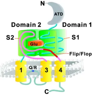
AMPA receptors, like NMDA and kainate receptors, belong to the glutamate ion channel receptor family (Dingledine et al., 1999; Traynelis et al., 2010; Palmer et al., 2005). As ligand-gated ion channels, AMPA receptors are composed of domains that span the membrane to form a pore or channel, and the channel is gated by glutamate. Upon glutamate binding, the receptor rapidly opens its channel pore to allow flow of small cations such as Na+ and K+, which causes an increase in the postsynaptic membrane potential. AMPA receptors mediate the majority of fast excitatory neurotransmission in the CNS, and play crucial roles during neuronal development and synaptic plasticity (Dingledine et al., 1999; Traynelis et al., 2010; Palmer et al., 2005). Each of the AMPA receptor subunits, GluA1-4 (Collingridge et al., 2009), can form homomeric channels by itself or assemble into heteromeric channels (Sobolevsky et al., 2009).
AMPA receptors are subject to RNA alternative splicing (Sommer et al., 1990) and editing (Sommer et al., 1991). RNA splicing and editing are developmentally regulated, and generate additional, functionally different receptors (Dingledine et al., 1999; Palmer et al., 2005; Sommer et al., 1991). All AMPA receptor subunits exist as two splice variants, flip and flop. The alternative splice cassette is found at the C-terminal end of the loop between TMIII and TMIV. Although the change in the receptor subunits is small (only a few amino acids are changed), the effect can be quite dramatic, resulting in altered desensitisation kinetics.
The calcium permeability of the GluA2 subunit is determined by the post-transcriptional editing of the GluA2 mRNA, which changes a single amino-acid in the
TMII region from glutamine (Q) to arginine (R). This is the so called Q/R editing site - GluA2Q is calcium permeable whilst GluA2R is not - positioned at the narrow constriction of the channel in the re-entry loop, exists exclusively in the GluA2 subunit (Figure 1) (Seeburg et al., 1996). Homomeric GluA2Q can form functional channels, which are Ca2+ permeable, whereas GluA2R, the edited R isoform (arginine), cannot form channels alone (Hume et al., 1991; Schiffer et al., 1997). In contrast, a glutamine Q remains at this equivalent position for GluA1, 3 and 4 (Sommer et al., 1991; Seeburg et al., 2003).
Figure 1. Schematic drawing of the topology of an AMPA receptor subunit showing the sequence location of the alternatively spliced flip/flop region (from Pei et al, 2009).
AMPA receptors are tetramers (Sobolevsky et al., 2009; Safferling et al., 2001; Armstrong et al., 2000; Mayer, 2005). AMPA receptors assembled from different subunits exhibit distinct gating, permeation, and rectification properties as well as Ca2+ permeability. GluA2 is a key subunit in controlling AMPA receptor assembly and function (Sans et al., 2003). This is because at the molecular level, GluA2R-containing channels are Ca2+-impermeable, whereas those lacking GluA2R are permeable to Ca2+ (and Zn2+) and exhibit distinctly fast kinetics (Geiger et al., 1995). The GluA2R-containing AMPA
receptors are found in most of the principal neurons in the neocortex, hippocampus, amygdala and cerebellum (Lambolez 1996; Sans et al., 2003).
Excessive AMPA receptor activity has been implicated in various neurological diseases, such as amyotrophic lateral sclerosis (ALS), ischemia and epilepsy (Dingledine et al., 1999; Liu et al., 2007), by a pathogenic mechanism known as excitotoxicity. Several studies demonstrate that excessive activity of Ca2+-permeable AMPA receptors is involved in a wide range of neurological diseases. Thus, blocking excessive AMPA receptor activity would be a promising therapeutic approach for the treatment of these diseases. AMPA receptor antagonists show a broad range of neuroprotection and are better tolerated with lesser side effects as compared with NMDA receptor antagonists.
Various AMPA receptor antagonists have been synthesized; for example, NBQX (6-nitro-7-sulfamoylbenzo[f]quinoxaline-2,3-dione) is a potent, competitive antagonist of AMPA channels, but it also blocks kainate receptors (Honore et al., 1988).
Mechanistically, noncompetitive antagonists are considered better suited for a more selective blockade of AMPA receptors, because they bind to a regulatory site(s) distinct to the agonist site and their actions should not depend on the concentration of an agonist. Noncompetitive antagonists have also the theoretical advantage to counteract excitotoxicity even at high concentration of glutamate and to show less side-effects than competitive antagonists (Parsons et al, 1998). It should be pointed out that all of these small-molecules are typically drug-like and amenable to chemical optimization for oral bioavailability and favourable pharmacokinetic properties.
There are a number of pharmacological agents that affect AMPAR function through interactions outside of the agonist-binding domain (Kew et al., 2005). Perampanel, a selective noncompetitive AMPAR antagonist, has recently gained FDA approval for
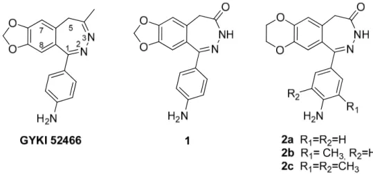
clinical use in the treatment of partial-onset seizures and primary generalized tonic-clonic (PGTC) seizures (Rogawski et al., 2013). Radioligand-binding studies suggest that the blocking site coincides with that of GYKI 52466 (Figure 2), the prototype of 2,3-benzodiazepine noncompetitive AMPAR antagonists (Solyom et al., 2002).
The most promising group of noncompetitive antagonists of AMPA receptors are 2,3-benzodiazepine derivatives, whose prototype GYKI 52466,
1-(4-aminophenyl)-4-methyl-7,8-methylenedioxy-5H-2,3-benzodiazepine, demonstrated significant anticonvulsant and neuroprotective action (Solyom et al, 2002). These antagonists bind at the interface between the S1 and S2 glutamate binding core and channel transmembrane domains, specifically interacting with S1-M1 and S2-M4 linkers, thereby disrupting the transduction of agonist binding into channel opening (Balannik et al., 2005).
More recently, the crystal structures of the rat AMPA-subtype GluA2 receptor in complex with three noncompetitive inhibitors have been reported (Yelshanskaya, et al. 2016). The inhibitors bind to a binding site, completely conserved between rat and human, at the interface between the ion channel and linkers connecting it to the ligand-binding domains. The authors propose that the inhibitors stabilize the AMPAR closed state by acting as wedges between the transmembrane segments, thereby preventing gating rearrangements that are necessary for ion channel opening.
The research group with whom I worked during my PhD has been involved by many years in the synthesis of new 1-(4-aminophenyl)-3,5-dihydro-7,8-methylenedioxy-4H-2,3-benzodiazepin-4-ones (e.g. 1, Figure 2) and in the characterization of their mechanism of action (Grasso et al., 1999; Grasso et al., 2003; Zappalà et al, 2006a). During the development of this research project, the Authors demonstrated that an improvement of AMPAR affinity of 2,3-benzodiazepin-4-ones has been reached by substituting the
benzo-fused dioxole nucleus with the dioxane homologue (i.e. 2, Figure 2) (Zappalà et al, 2006b; Micale et al. 2008). Moreover, the Authors found that the addition of a methyl group at the 3’ position (i.e. 2b) enhanced the antagonist activity, whereas the introduction of another methyl group at the 5’ position (i.e. 2c), was not fruitful for the increase of the potency on GluA2Q receptors (Wang et al., 2015), according to the binding assays and functional tests already performed (Micale et al. 2008).
Figure 2. Model compounds GYKI 52466, 1 and 2a-c.
Furthermore, in collaboration with Dr. Niu, using a laser-pulse photolysis technique, the mechanism of action of compounds 1 and 2 and their binding site on the AMPA receptors were further characterized in the microsecond-to-millisecond time domain (Ritz et al., 2008; Ritz et al., 2011, Qneibi et al., 2012, Wang et al., 2015).
Kinetic studies showed that compound 1 and 2 are noncompetitive antagonist of GluA2Q homomeric receptors. Noteworthy, compound 1 prefers to bind to the open-channel state whereas derivatives 2 show a higher affinity towards the closed-open-channel state of the GluA2Q receptors. By a double-inhibition experiment, it was demonstrated that compound 1 and derivatives 2 do not seem to bind to the same binding site on GluA2Q receptor, and there is no an apparent allosteric interaction between these putative binding sites on the same receptor (Qneibi et al., 2012, Wang et al., 2015).
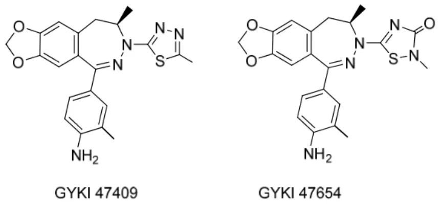
It has been recently reported (Wang et al., 2014) that the introduction of a thiadiazole moiety at the N-3 position of the 2,3-benzodiazepine scaffold yields an enhancement in potency and selectivity on AMPA receptors. The two 2,3-benzodiazepines GYKI 47409 and GYKI 47654 (Figure 3) were found to be far more potent inhibitors of both the closed and open conformations of all four homomeric AMPA receptor channels than the unsubstituted 2,3-benzodiazepine GYKI 52466 as well as N-3 substituted derivatives Talampanel and GYKI 53784. The Authors proposed that the heterocycle at the N-3 position is able to well accommodate into a “side pocket”, generating a strong interaction
with residues surrounding this binding site, as predicted by a four-point pharmacophore model (Rezessy and Solyom, 2004) that suggests a role of the heteroatom of the thiadiazole moiety as H-bond acceptor.
Figure 3. Structure of thiadiazole derivativesGYKI 47409 and GYKI 47654
To better define the structure–activity relationship (SAR) of this class of compounds, we planned to synthesize two groups of analogues of 2,3-benzodiazepines 2a-2c.
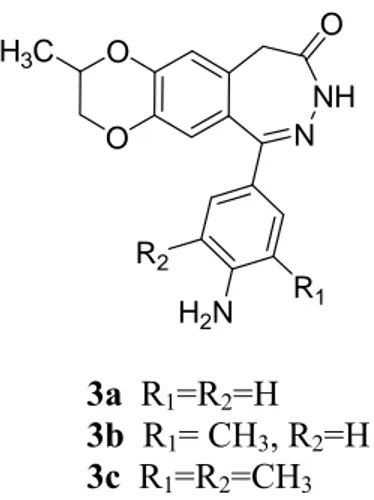
The first group of these compounds (Figure 4) bears a methyl group on 7,8-ethylenedioxy moiety (3a-3c) in order to create additional hydrophobic interactions between the 7,8-ethylenedioxy portion and the receptor site.
O O N NH O H2N R1 R2 H3C 3a R1=R2=H 3b R1= CH3, R2=H 3c R1=R2=CH3
Figure 4. Structure of novel 2,3-benzodiazepines noncompetitive AMPAR antagonists 3a-3c
The second group of 2,3-benzodiazepines has been designed starting from the above-mentioned thiadiazole derivatives GYKI 47409 and GYKI 47654. We decided to insert at the N-3 position a more flexible heterocycle that could better fit into the binding pocket and presumably maintain the same capability to interact with the receptor site via hydrogen bond as the thiadiazole nucleus does. In particular, I synthesized derivatives 4a-4c in which a 3-bromoisoxazolin-5-yl has been linked to N-3 and derivatives 5a-5c in which the same heterocycle was linked to N-3 by means of a methylene spacer (Figure 5). The choice of the 3-bromoisoxazolin-5-yl is in agreement with literature data (Solyom et al, 2007), in which the substituent at N-3 position could be represented by a substituted or unsubstituted 5-or 6-membered, aromatic, saturated or partially saturated heterocyclic ring containing at least two heteroatoms, with one of them being a nitrogen atom.
Figure 5. Structure of GYKI compounds and novel 2,3-benzodiazepines noncompetitive AMPAR antagonists 4a-4c and 5a-5c.
The synthesized compounds have been characterized for their inhibitory properties by a set of functional assays. Using human embryonic kidney cells, i.e. HEK-293 cells, to transiently express homomeric receptor channels, individually, and using whole-cell recording to monitor the receptor function, we have characterized the potency and selectivity of these compounds towards AMPARs.
1B
1.2 Evaluation of novel 2,3-benzodiazepin-4-one noncompetitive AMPAR
antagonist on leukemia Jurkat T cell growth, cell cycle and apoptosis
Over the past years several lines of evidences implicated glutamate in the development and proliferation of different types of cancers inside and outside of the central nervous system (Prickett et al. 2012; The et al. 2012). Beside to its excitatory role in the CNS, glutamate is involved in others cellular and biochemical functions such as proliferation, differentiation and survival of the neural cells (Luján et al., 2005). A number of findings revealed that the inhibition of AMPA receptor activity was able to inhibit migration and to induce apoptosis in human glioblastoma cells (Walczak et al, 2014), and to decrease cell growth in different non-neuronal cancer cell lines (Rzeski et al., 2001). Noteworthy was the evidence that different non-neuronal tumoral cell lines, such as human leukemia Jurkat T cell line, expressed AMPA receptor subunits GluA2-GluA4 (Stepulak et al., 2009), and glutamate might facilitate the spread and growth of leukemia T cells through interactions with GluA3 subunit AMPA receptor (Ganor et al., 2009). Despite these interesting and intriguing results, a deeper molecular and pharmacological characterization of putative AMPA antagonist has not yet been performed.
Recently, Stepulak et al. (2011) has shown that the AMPA antagonist GYKI 52466 reduced the viability of laryngeal cancer cell lines.
With the aim to elucidate the potential mechanism in cell cycle regulation elicited by 2,3-benzodiazepine derivatives, compound 2c, based on primary screening on six different tumor cell lines, has been selected as the most active 2,3-benzodiazepine-4-one derivatives. The ability of compound 2c to modulate the cell cycle distribution and the molecular determinants involved in the cell cycle check points, together with its potential ability to modulated apoptotic pathways in human Jurkat T cell line has been evaluated.
2B
1.3 Development of subtype-specific agonists for NMDA receptor glycine
binding sites
The NMDA receptors are heterologous complexes consisting of several subunits assembled to form tetrameric arrangements. So far, seven different subunits have been identified, each with a specific modulatory influence on the receptor: one GluN1subunit, four GluN2 subunit (GluN2A-D) and two subunits GluN3 (GluN3A-B) (Paoletti, et al., 2007).
The most common NMDAR architecture consists of the coassembly of two GluN1 subunits and two GluN2 subunits organized as a dimer of dimers with a GluN1-GluN2-GluN1-GluN2 pattern with an overall axis of two-fold symmetry within the extracellular domains and with the ion channel domain exhibiting a four-fold symmetry.
NMDARs differ from other ligand-gated ion channels as they present the peculiarity to require two distinct ligands for their activation. Glu and Asp are the endogenous agonists for the receptor and bind to residues located in the GluN2 subunits that are the major determinants of the pharmacological and biophysical properties of these receptors. Glycine or D-serine acts as an essential coagonist and binds to residues located in the GluN1 and GluN3 subunits, increasing the frequency of channel opening channel (Yao et al, 2006: Madry et al, 2008).
The involvement of NMDARs in many neurological disorders and the crucial role of Glu as the major excitatory neurotransmitter in the mammalian CNS, triggered intense research plans aimed at the synthesis of new drug candidates characterized by a selective agonistic or antagonistic activity at these receptors.
During the past several decades, a great number of iGluR ligands have been developed, but few of them are specific for a single subtype. For the glycine site in the GluN1 subunit of NMDA receptors, a large number of antagonists exists, but relatively
few full and partial agonists have been reported (Bräuner-Osborne et al. 2000; Chen et al. 2008; Urwyler et al. 2009).
In recent years, a large number of structures of isolated iGluR agonist binding domains (ABDs) have disclosed important information on the molecular basis for orthosteric ligand recognition, and the mechanisms underlying activation, desensitization, and allosteric modulation (Furukawa and Gouaux, 2003; Inanobe et al., 2005; Yi et al., 2016; Hackos et al., 2016; Jespersen et al, 2014). These studies also show that structural differences exist in the dimer interface between ABDs of GluN1 and the different GluN2A-D subunits (Yi et al., 2016; Hackos et al., 2016).
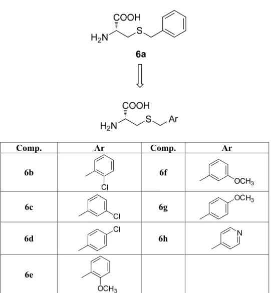
On the basis of these structural differences, agonists capable of differentiating between the glycine binding site of GluN1 in a GluN2 subunit-dependent manner have been recently developed by the research group with whom I worked for a six month period (Maolanon et al. 2017). A series of Ser and Cys analogues have been designed and synthesized and displayed pronounced variation in activity among GluN1/2A-D NMDA receptor subtypes. In particular, S-benzyl substituted L-cysteine (R-form) 6a showed extensive potentiation (169% response relative to glycine) of maximal current at GluN1/2C receptor subtype relative to the current induced by Gly. Thus, this compound may be considered a superagonist at the glycine site of the GluN1/2C receptor. These levels of superagonistic activity are unprecedented among all NMDA receptor agonists described to date (Bräuner-Osborne et al. 2000; Chen et al. 2008; Dravid et al., 2010). Although non-natural R-isomers generally are more active than S-isomers, it is notable that the cysteine analogue 6a is the active enantiomer since this L-form has R absolute configuration.
In order to further evaluate the ability of L-Cys derivatives to differentiate between the glycine binding site of GluN1 in a GluN2 subunit-dependent manner, and to assess the effect of substituents with different electronic characteristics on the agonist properties of
the lead compound 6a, I designed and synthesized new L-cysteine derivatives S-substituted 6b-6g in which a chlorine atom or a methoxy group has been introduced in different positions of the benzyl moiety and compound 6h in which the aromatic ring has been replaced by a 4-pyridyl nucleus. (Figure 6).
Comp. Ar Comp. Ar 6b 6f OCH3 6c 6g 6d 6h N 6e
Figure 6. Structure of the superagonist 6a and of the designed analogues 6b-6h The synthesized compounds have been characterized by two-electrode voltage-clamp (TEVC) electrophysiology using Xenopus oocytes expressing recombinant NMDA receptor subtypes. Concentration–response data for the compounds have been generated in the continuous presence of a saturating concentration of Glu (100–300 μM) at the four GluN1/GluN2A-D NMDA receptor subtypes.
Chapter 2. Glutamate receptors
Currently,(S)-glutamic acid (Glu) is considered the main excitatory neurotransmitter in the mammalian central nervous system (CNS) and is implicated in key processes of brain development such as learning and memory (Riedel et al, 2003), and in several neuropathological conditions (Coyle et al., 2002).
An excessive glutamatergic stimulation can induce neuronal citotoxicity through at least two mechanisms:
• osmotic damage caused by the excessive influx of Na+ in the cell through the ionic channels;
• damage induced by the alteration of the homeostasis of Ca2+.
The effects of glutamate are mediated by its interaction with different receptor subtypes that, according to the mechanism of signal transduction, were grouped into two broad classes: ionotropic receptors (iGluRs) and metabotropic receptors (mGluRs).
The first mediate fast neurotransmission through the depolarization of the membrane potential directly by the opening of the pore-channel that increase the transmembrane flow of mono- and divalentcations.
The iGluRs are divided, on the basis of the name of the main exogenous agonists, in NMDA receptors (N-methyl-D-aspartic acid), AMPA [(RS)-2-amino-3-(3-hydroxy-5-methylisoxazol-4-yl)propionic acid] and KA (kainic acid)receptors (Traynelis et al., 2010). The second family of receptors mediateslow modulatory responses and acts indirectly since the activation of the receptors by the ligand is transmitted from a protein G to the effector that is responsible for the increase in the intracellular second messengers.
Eight different subtypes of mGluRs have so far been identified. They are classified into three subgroups based on homologies in the peptide sequence, signal transduction mechanisms and pharmacology. The mGlu of group I (mGluR1, mGluR5) act by activating the phospholipase C; group II ( mGluR2, mGluR3) and group III (mGluR4,6,7,8) acting through inhibition of adenylcyclase activity (Mayer, 2006; Traynelis et al., 2010).
3B
2.1. Glutamate ionotropic receptors (iGluR)
Glutamate ionotropic receptors are a family of tetrameric receptors channel permeable to Na+, K+, and Ca2+, whose opening and mediated by the interaction with the orthosteric ligand. Although they may be also located in the presynaptic terminal, they have localization predominantly post-synaptic, where they are involved in the fast excitatory neurotransmission and various forms of synaptic plasticity. The glutamate released from the presynaptic terminal interact with their receptors evoking an excitatory current post-synaptic (EPSC) whose profile shows a biphasic pattern. After an initial massive influx of cations (a few ms) mediated by the sudden activation of AMPAR, these receptors desensitize and simultaneously activate the NMDAR argue that the slow component of the EPSC. After 50-60ms, NMDAR the close and the charge flow terminates.
The AMPA receptors are generally co-expressed with the NMDA receptors at the level of the glutamatergic synapses where jointly contribute to the processes of synaptic plasticity that are involved in the phenomena of learning and memory, excitotoxicity and neuroprotection. These receptors typically differ in their kinetics of the response to the presynaptic release of glutamate. The AMPA receptors mediate fast postsynaptic answers even to potential very negative or in the absence of action potentials. Rapid desensitization
of these receptors is responsible of the EPSCs; the NMDA receptors, instead, are characterized by a slower kineticand a weak or nodesensitization.
From the structural point of view the receptor channel is a tetramer, composed of identical subunits (homotetramer) or different (heterotetramer), that are assembled in two subsequent steps. Initially two subunits interact to form a dimer, and then two dimers are organized delimiting the central pore and giving rise to the mature receptor.A recent study aimed to clarify the mechanism assembly, has highlighted that the heterotetramers assemble from heterodimers (Riou et al., 2012).
The different subunits have a shared modular structure that consists of:
a large N-terminal domain (NTD extracellular) that in addition to being involved in the assembly and in the modulation of different subtypes, has the binding site for the allosteric modulators;
an extracellular domain of binding to the agonist (ABD) also known as the S1-S2 domain;
a transmembrane domain (TMD) composed by three transmembraneα-helices (M1, M3 e M4) and by a short loop (M2) which constitutes the filter of ionic selectivity; this re-entrant loop is a peculiar structural feature that distinguishes iGluR subunits from other neurotransmitter-gated ion channels.
a C-terminal domain (CTD) of variable size that has several phosphorylation sites and is involved in the modulation, in traffic and in the localization of the receptor (Madden, 2002).
The recent crystallization of subtype homomeric GluA2 of AMPAR has represented a turning point for the resolution of the structure of the ionotropic receptors, in particular the data obtained show that the region of the pore has a much more compact structure and
symmetrical with respect to the extracellular domains. Moreover, the interactions between the NTD of different subunits are relatively small compared to what previously hypothesized (Sobolevsky et al., 2009).
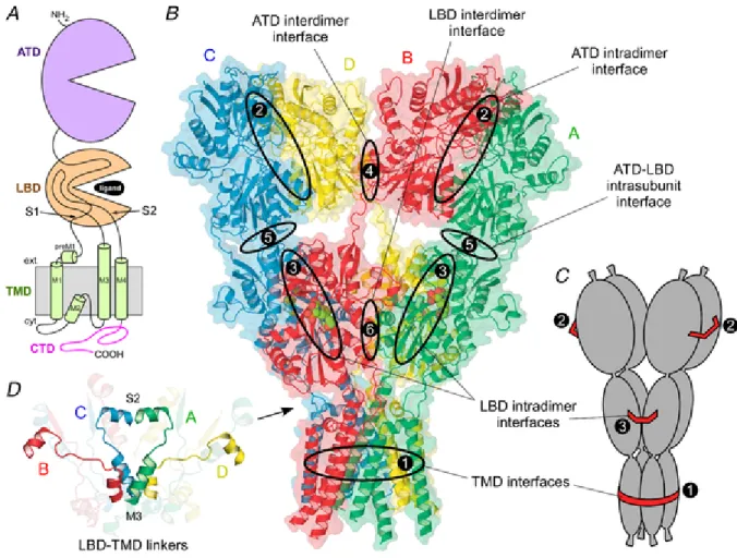
Figure 7. Structure of iGluR. A, topology of iGluR subunit. B, structure of AMPA subtype rat GluA2 receptor in the closed antagonist bound state (3KG2). Strong and weak interfaces are shown as large and small ovals, respectively. C, model of the assembly of four three-compartmental sausages with strong interactions holding together (1) all four bottom compartments, (2) left and right pairs of the top compartments and (3) front and back pairs of the middle compartments. D, LBD–TMD linkers – the iGluR gating transmission domain – include S1–M1, M3–S2 and S2–M4, of which S1–M1 and S2–M4 are shown transparent. The M3–S2 linkers, the central element of the gating machinery, have different conformations and secondary structures for the two diagonal pairs
The large structural and functional diversity of the different receptor subtypes is supported both by the large number of subunits, and from translational and post-transcriptional modifications. For example the pre-mRNA of some subunits may undergo a process of nucleotide editing that produces the incorporation of an arginine (R) in the mature receptor while the genomic DNA in that site code for a glutamine (Q). The point at which occurs the nucleotide editing takes the name of Q/R site and appears to be involved in regulation of neuronal development and in ionic selectivity of receptors containing the subunit GluA2 (Seeburg et al., 1998).
From a pharmacological point of view, in the absence of ligand the channel is closed and the receptor is in a rest state (R), following the binding of the agonist the receptor passes in an activated state (A) and the channel opens allowing the ion flow. After an interval of 1-100 ms in function of the receptor subtype, the channel closes while the binding site is still busy, preventing the binding with another molecule of agonist. In this condition the receptor is in a desensitized state (D) (Talukder et al., 2010)
4B
2.2 AMPA receptors
AMPA receptors are the most abundant ionotropic glutamate receptors in the mammalian brain.
The AMPAR are a heterogeneous family of homo- or heterotetrameric assembly of four different subunits of about 100 kDa identified as GluA1-2-3-4. As previously mentioned, these receptors show a rapid kinetics of activation and desensitization that justifies the fast component of the glutamatergic EPSP.
The pharmacological characteristics of the AMPAR differ greatly in function of the subtypes taken into consideration and the post-transcriptional modifications of the different
subunits. For example, the length of CTD of some subunits is determined by variations in the splicing of the primary transcript. It has been demonstrated that the length of the CTD influence the turnover of AMPAR, in detail the longer CTD reduces the receptor turnover. This mechanism may explain some forms of LTP, mediated by the expression in the synapses of receptors composed of subunits with the longer CTD (Seeburg et al., 1998).
It is well known that as a result of alternative splicing of exons 14 and 15, the isoforms "Flip" and "flop" of the subunits of AMPAR are respectively expressed. The incorporation in the tetramer of variant "Flip" confers a quicker kinetics of activation of the receptor and a slower desensitization (Coleman et al., 2006). During the development, the "Flip" isoform is more expressed with respect to the isoform "flop", while in the adult, the expression levels of the two splice variants are relatively similar. This observation has been confirmed by experimental evidence that suggest the involvement of receptors that contain subunits "Flip" in the growth and maturation of neocortical neurons during the prenatal period.
A further level of heterogeneity of AMPAR is related to the nucleotide editing of primary transcripts of the subunit GluA2.The deamination of an adenosine to inosine, catalyzed by adenosine deaminase, leads to the expression of an arginine (R) in M2 reentrant loop of mature receptor, while the genomic DNA in that position code for a glutamine (Q). The AMPAR incorporating the edited subunits are impermeable to Ca2+ and consequently not mediate the metabolic responses related to the homeostasis of Ca2+
itself. . However, since nearly all GluA2 subunits are edited and the majority of AMPA receptors contain the GluA2 subunit, most AMPA receptors do not flux calcium. However, GluA2-lacking AMPA receptors are common in interneurons and some cortical neurons where their rapid kinetics allows particularly fast synaptic signaling and their calcium permeability mediates novel forms of synaptic plasticity (Isaac et al., 2007).
12B
2.2.1 AMPA receptor subunits
It is now well established that there are four AMPA receptor subunits designated GluA1–GluA4 (formerly GluR1–GluR4), each encoded by a separate gene (Lodge, 2009). AMPA subunit shared the modular structure as the other iGluR subunits.
The large extracellular ATD is involved in receptor assembly, trafficking and modulation. A ligand-binding domain LBD serves as the recognition site for agonists (including the natural agonist glutamate) and also represents the binding site for competitive antagonists. The transmembrane domain forms the ion channel and consists of three membrane-spanning hydrophobic domains and one intramembranous reentrant loop.
A short cytoplasmic carboxy-terminal domain is involved in targeting the receptor to synapses. The peptide segments connecting the ligand-binding domain to the transmembrane domain transmit conformational changes elicited by agonist binding to the transmembrane ion channel domain, allowing agonist binding to gate the channel to the open state; these segments can be considered the “transducing domain” (Szenasi et al., 2008). This region of the channel is critical to binding of noncompetitive antagonists, which prevent channel gating (Balannik et al., 2005).
Each subunit consists of approximately 900 amino acids and exhibits 65–75% sequence homology to other subunits. All AMPA receptors are tetrameric combinations of the four subunits. While homomeric receptors are functional, native AMPA receptors are believed to be heteromers. For example, in hippocampal pyramidal cells of mature rats, the most common subunit configurations are GluA1/GluA2 and GluA2/GluA3 (Wenthold et al., 1996).
The near-full-length crystal structure of GluA2 (Fig. 7 and fig. 8B) revealed a number of interesting features about AMPARs. For example, the ATD and LBD each
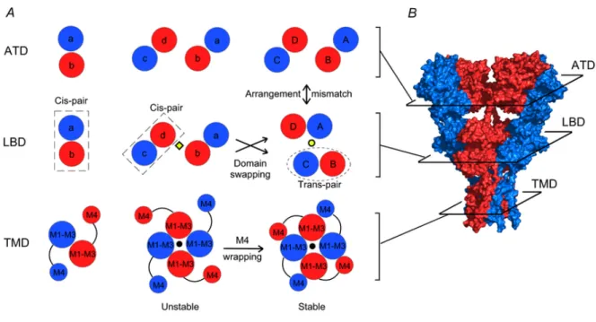
possess a 2-fold rotational symmetry, where subunits of like conformation are positioned opposite to each other (Fig. 8A right panels) (Sobolevsky et al. 2009). In contrast, the TMD displays 4-fold symmetry. Further, there is a mismatch in subunit arrangement between the ATD and LBD. In the fully assembled receptor (Fig. 8A, right panels), subunits that are proximal to each other (B and D) at the ATD level are located distally at the LBD level, and vice versa (Gan et al., 2015).
Figure 8. A Model of AMPAR assembly; B, crystal structure of GluA2 receptor (3KG2) (from Gan et al., 2015).
Figure 8A illustrates a hypothetical model of AMPAR assembly highlighting the intrinsic interactions among subunits at levels of the ATD, the LBD, and the TMD. In this model, the ATD facilitates the formation of dimers (left panels). At the dimer stage, the LBDs of the two participating subunits might associate to form an LBD pair (dubbed ‘cis-LBD pair’). Dimers then associate with each other via interactions within the ion channel core to form an unstable intermediate termed a ‘proto-tetramer’ (central panels). Cis-LBD pairs persist into the proto-tetramer stage leading to an arrangement at the level of the LBD
matching that of the ATD. The M4 transmembrane segment of each subunit then wraps around the ion channel core (M1–M3) of an adjacent subunit – a process we term ‘M4 wrapping’ – and stabilizes the tetramer. At some point during this process, the LBDs separate and exchange their interacting partners to form ‘trans-LBD pairs’ leading to the subunit arrangement mismatch between the ATD and the LBD observed in the fully assembled tetrameric complex (Sobolevsky et al. 2009). We refer to this process as ‘domain swapping’. M4 wrapping in the TMD, along with domain swapping, presumably contributes to the overall intrinsic energetics of tetramerization. Additionally extrinsic factors such as ER chaperones and glycosylation could also have important, albeit undefined, modulatory effects on these subunit interactions facilitating tetramerization.
Recently, the crystal structures of the rat AMPA-subtype GluA2 receptor in complex with three noncompetitive inhibitors have been reported (Yelshanskaya, et al. 2016). The inhibitors bind to a novel binding site, completely conserved between rat and human, at the interface between the ion channel and linkers connecting it to the ligand-binding domains. The authors propose that the inhibitors stabilize the AMPA receptor closed state by acting as wedges between the transmembrane segments, thereby preventing gating rearrangements that are necessary for ion channel opening.
AMPA receptors are associated with a variety of transmembrane proteins that function as auxiliary subunits, including TARPs (transmembrane AMPA receptor regulatory proteins), such as stargazin; cornichon proteins (CNIH-2, CNIH-3); and SynDIG1 (synapse differentially induced gene 1) (Diaz, 2010). The auxiliary subunits regulate channel gating and are involved in subunit folding, assembly, surface expression and the clustering of AMPA receptors at synapses. In addition, the subunits modulate the sensitivity of AMPA receptors to pharmacological agents, including antagonists (Cokić and Stein, 2008).
Binding studies with 3H-AMPA showed that this class of receptors is widely distributed in the CNS and that the expression levels vary in function of the brain regions analyzed. The highest expression levels were observed in the hippocampus, in the cerebral cortex and in the cerebellum. In particular, the subtype prevalent in hippocampus is the heterotetramerGluA1/GluA2, whereas the GluA3receptors are expressed at high levels in the encephalic nuclei. The GluA4 subunit is especially expressed in the early stages of neuronal development and is replaced by the GluA1 in SNC adult, thus suggesting a role in the development of SNC. As regards the cerebral cortex, high levels of expression of the subtypes GluA1 and GluA2/3 were reportedin GABAergic interneurons and in excitatory interneurons.
13B
2.2.2 Post-translational modifications of AMPA receptors
Properties and function of AMPARs may also be modulated by post-translational modifications such as glycosylation, palmitoylation and phosphorylation.
Glycosylation is a protective modification that can occur at 4-6 different sites located in the extracellular domains of each AMPAR subunit. This N-glycosylation may facilitate the maturation of AMPARs and protect them from proteolytic degradation (Jiang et al., 2006).
Palmitoylation is a reversible fatty acetylation that regulates protein trafficking and cellular localization. All AMPAR subunits can be palmitoylated on two cysteine residues in their transmembrane domain TM2 and in their intracellular C-terminal region. The first palmitoylation in TM2 leads to an accumulation of AMPAR in the Golgi apparatus, resulting in a decreased expression of the receptor in the cell surface. On the other hand, palmitoylation at the C-terminal domain contributes to receptor internalization by
disrupting its interaction with the 4.1N protein, known to stabilize AMPAR expression on the surface (Hayashi et al., 2005).
Four and two phosphorylation sites have been reported for the GluA1 and GluA2 subunits, respectively (Lee et al., 2010), all residing in the intracellular C-terminal. Phosphorylation of these Ser e Tyr residues is involved in the mechanisms of synaptic plasticity: hippocampal NMDA-dependent Long-Term Potentiation (LTP) and Depression (LTD).
5B
2.3 NMDA receptors
Expressed in ubiquitous manner in the CNS, NMDA receptors mediate fast gluammatergic neurotransmission in CNS and play important roles in neuronal functions such as the processing of information, learning and memory formation, synaptic plasticity and neuronal development. The NMDA receptors also play an important role in nociception: many antagonists of the receptor have been shown capable of effectively attenuate the pain in both acute and chronic (Bräuner-Osborne et al., 2000; Petrenko et al, 2003).
An abnormal activation of NMDA receptors may lead to an increase in the levels of Ca2+ intracellular up to cytotoxic levels, thus promoting the neuronal death (excitotoxicity). The permeability to calcium represents a peculiarity for NMDA receptors with respect to the other two classes of ionotropic receptors, AMPA and KA, that show instead a predominant permeability to Na+ and K+ ions (Liu et al., 2007). Excitotoxicity mediated by NMDA receptors was observed in many conditions both acute, as ischemia or trauma,and chronic as in many neurodegenerative diseases including Parkinson's, Alzheimer's, Huntington's disease and amyotrophic lateral sclerosis. A hypofunction of
NMDA receptors is involved in the pathophysiology of schizophrenia (Lynch et al., 2002; Vrajováet al., 2010)
The architecture of the NMDA receptors allows them to exist in multiple isoforms, each with specific characteristics of molecular composition, expression spatial and temporal, intracellular localization, functional properties, pharmacological and kinetics (Laube et al. 1998).
The NMDA receptors are heterologous complexes consisting of several subunits assembled to form tetrameric arrangements. So far, seven different subunits have been identified, each with a specific modulatory influence on the receptor: one GluN1subunit, four GluN2 subunit (GluN2A-D) and two subunits GluN3 (GluN3A-B) (Paoletti, et al., 2007).
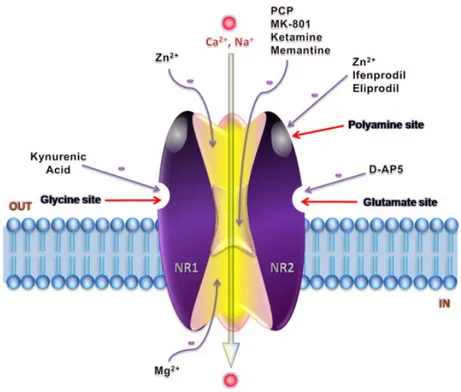
Eight subunits GluN1 are generated by alternative splicing of a single gene, while the GluN2 and GluN3 subunits are encoded by six different genes. The subunits GluN1 and GluN2 are essential for the functions of the receptor and therefore are always present in the complex. A splice variant of the subunit GluN1 combines with at least one subunit GluN2A-D and less frequently with a modulatory subunit GluN3A-B. The most common architecture of the NMDA receptor consists in a co-assembly of two GluN1 subunits and two GluN2 subunits organized as a dimer of dimers with an arrangement GluN1-GluN2-GluN2 disposed around the ion channel (Fig. 9). In the receptor subtypes that express a GluN3modulatory subunit, the most common co-assembly consists in a ternary GluN1-GluN2-GluN3tetramericcomplex. (Paoletti, et al.,2007; Cull-Candy et al., 2001; Schorge et al., 2003).
NMDA receptors differ from other ligand-gated ion channels because they have the peculiarity of requiring two separate ligands for its activation. Glutamate is the endogenous
agonist and binds to residues located in GluN2 subunit that are the main determinants of the pharmacological properties and biophysical of these receptors. The glycine acts as a co-agonist and binds to residues of the GluN1 subunits and probably also of the GluN3subunits, increasing the affinity of the receptor for the glutamate and the frequency of opening of the channel (Yao et al, 2006: Madry et al, 2008).
Moreover, GluN2 subunits have: (i) binding sites for allosteric regulators (e.g. extracellular Zn+2); (ii) binding sites for non-competitive antagonists such as feniletanolammine (e.g. ifenprodil)located at the interface with GluN1 subunits; (iii) two or three sites for polyamines (voltage-dependent and voltage-independent); (iv) a site for channel blockers (e.g. phencyclidine) (Fig. 9) (Gielen et al., 2009).
Figure 9. Heterotetramer GluN1/GluN2 NMDA receptor and its modulatory sites (from Ghasemi et al., 2011).
14B
2.3.1 NMDA receptor subunits
The GluN1 and GluN2 subunits share a common basic topology with other iGluRs (AMPA and KA receptors), characterized by four hydrophobic domains (M1-M4) within the central portion of the sequence: M1, M3 and M4 are membrane-spanning segments, whereas M2 domain forms a re-entrant loop in the membrane and lines the pore of the ion channel.
NMDAR subunits also have a cytoplasmic carboxylic terminal domain (CTD), a large extracellular amino-terminal domain (ATD), and an extracellular loop between M3 and M4. The size of CTD is different in the various subunits and contains several sites of interaction with numerous intracellular proteins such as protein kinases and protein phosphatases.
The ATD has a key role in subunit assembly and it also contains the binding sites of the negative allosteric modulators Zn2+ and ifenprodil-like compounds (Paoletti, 2007; Huggins and Grant, 2005). Endogenous Zn2+ binds at the GluN2A subunit with nanomolar
affinity and at the GluN2B subunit with a >100-fold lower affinity, while it does not affect GluN2C and GluN2D subunits (Paoletti et al. 2000; Rachline et al., 2005). Ifenprodil-like compounds bind selectively at the GluN1-GluN2B interface (Karakas, et al. 2011).
The large extracellular loop shared by M3 and M4 domains (S2 region) forms with the extracellular N-terminus (S1 region) the ligand binding domain (LBD). The S1 region is a highly conserved domain involved in the crucial interaction with the γ-carboxylate of the ligand by means of an arginine residue. Conversely, the S2 region is less conserved among different subtypes; this peculiarly allows the generation of subtype selective ligands. The LBD is a clamshell-like motif in which the two lobes (referred to as D1 and
D2 according to the regions that form them) can adopt different conformational states depending on NMDAR subunit and/or the feature of the ligand.
X-ray studies demonstrated that LBD of isolated GluN1 subunits adopt “closed” conformations in complex with full agonists (i.e. glycine or D-serine) and partial agonists (e.g. D-cycloserine) leading to receptor activation, and that the degree of the domain closure is essentially the same (Furukawa and Gouaux, 2003; Inanobe et al., 2005).
Conversely, site-directed mutagenesis studies performed on Glu binding site of GluN2B subunits co-expressed with GluN1 indicated that partial agonists might induce “half-open” conformations of the LBD (Hansen, et al, 2005).
Finally, the LBD adopts open conformations in the “apo” (ligand-free) form and in complexes with antagonists (Furukawa and Gouaux, 2003). The M2 domain of GluN2 and GluN1 subunits contains an asparagine residue in the so-called QRN site. The name of this site arose from the fact that it is occupied by an asparagine (N) residue in NMDARs and by a glutamine (Q) or an arginine (R) in non- NMDARs, which is critical for high-calcium permeability and voltage-dependent magnesium block (O’Leary et al., 2009; Chaffey and Chazot, 2008).
Several studies revealed that in NMDAR subtypes incorporating a GluN3 subunit, the ion channel conductance, Ca2+ permeability and Mg2+ block are markedly reduced. These functional differences are due to the structure of the channel-lining M2 domain that differs from that of other subunits around the QRN site (Sasaki et al., 2002). In particular, in the GluN3 subunits, an Asn residue is replaced with Gly followed by Arg at the N+1 site, generating at this locus a sequence that is Gly-Arg, whereas in GluN1 the sequence is Asn-Ser and Asn-Asn in GluN2. The presence of the protonated Arg residue in GluN3 is
likely responsible for the specific permeation properties and magnesium sensitivity of the GluN3-containing NMDA receptors (Matsuda et al., 2002).
The four GluN2 subunits have different distributions throughout the CNS. GluN2B and GluN2D are already expressed during embryonic stages, while GluN2A and GluN2C expression begins after birth. GluN2A subunit is ubiquitously expressed in the adult brain and it is particularly abundant in hippocampus and cerebellum. GluN2B predominate in the forebrain especially in the cerebral cortex, hippocampus and thalamic regions (Loftis and Janowsky, 2003).
The expression of GluN2C subunits is nearly entirely confined to the cerebellum, mostly in Purkinje and granule cells with almost no expression in the forebrain. The presence of GluN2D subunit in the adult brain is restricted to a few areas such as the globuspallidus, thalamus, subthalamic nuclei, and superior colliculus (Wenzel et al., 1996) Moreover, there is evidence that triheteromeric GluN1/GluN2A/ GluN2B receptors are present in the cortex and hippocampus and GluN1/GluN2A/GluN2C and GluN1/GluN2B/GluN2D in the cerebellum (Cull-Candy and Leszkiewics, 2004). GluN3A and GluN3B subunits also showed distinct expression in different regions of the CNS. GluN3A is particularly expressed in the cortex, midbrain and hippocampus, whereas GluN3B is predominant in somatic motor neurons in the brain, brainstem and spinal cord (Nishi et al., 2001; Eriksson et al, 2002).
15B
2.3.2 NMDA receptor channel activation
The binding of Glu and glycine to their sites on the NMDAR complex determines the opening of a cation permeable pore, responsible for the postsynaptic depolarization. The NMDARs, as well as other members of the iGluR family, when activated, allow the
passage of Na+ ions into the cell and K+ ions out of the cell, to generate a short-lived depolarization called excitatory postsynaptic potential (EPSP).
Unlike the other two subtypes of iGluRs, the NMDARs ion channels also enable intracellular passage of Ca2+ ions, which can produce strong biochemical signals in the postsynaptic cell, affecting numerous intracellular signaling and processing systems (Popescu, 2005). Ca2+ influx through NMDARs can induce the long-term potentiation (LTP), a long-lasting enhancement in signal transmission that is thought to play a critical role in regulating synaptic plasticity, a cellular mechanism that underlies learning and memory. Ca2+ influx is also required for synapse formation, synaptic maintenance and physiological pruning during development (Cooke and Bliss, 2006).
However, an excessive entry of Ca2+ may cause neuronal cell death through activation of a variety of Ca2+- dependent proteolytic enzymes such as calpains and endonucleases. All this mechanism triggers off excitotoxicity that leads to pathologic neurodegeneration (Arundine and Tymianski, 2003).
Another peculiarity of NMDARs with respect to the other iGluRs is their gating kinetics, which determines the time course of synaptic currents. NMDARs show much slower deactivation kinetics based on their subunit composition and a weak or no desensitization. This has been proven for GluN1/GluN2A receptors, which deactivate more rapidly (ms) than those containing GluN2B or GluN2C, which in turn deactivate at least 10 times more rapidly (s) than NR2D containing receptors (Erreger et al, 2004).
The prolonged deactivation time course of NR2D containing receptors has been recently explained by means of crystallographic studies performed on isolated GluN2D LBD in complex with various agonists and by electrophysiological experiments. These studies revealed unique features for this subunit: the Glu-bound form induces
conformational changes in a region located at the backside of the ligand binding site (called ‘hinge loop’) which are not present in other ligand-bound forms. This Glu-induced conformational variability of GluN2D LBD influences both deactivation kinetics and receptor activity (Vance et al, 2011).
16B
2.3.3 NMDA receptor modulatory binding sites
At resting membrane potential, the NMDARs ion channel is blocked by micromolar concentrations of extracellular Mg2+ ions. This voltage-dependent block can be relieved by a membrane depolarization beyond ~40mV and it is considered a protective mechanism by which the NMDARs avoid the massive influx of Ca2+ into neurons and the consequential neuronal damage.
The asparagine residue following the QRN site of the GluN2 subunits seems to be responsible for the high sensitivity towards magnesium blockade, especially in NMDARs that express GluN2A and GluN2B subunits in comparison to those ones containing GluN2C and GluN2D subunits (Cull-Candy et al, 2001).
Beyond extracellular Mg2+, other endogenous and exogenous ions/compounds may modulate the responsivity of the NMDAR complex. The divalent cation Zn2+ is packaged into synaptic vesicles of axons and when it is coreleased with Glu can modulate neuronal excitability mediated by NMDARs through a reversible and voltage-dependent block, depending of its synaptic concentration. At nanomolar concentrations (IC50 = 10-30 nM) ,
Zn2+ exerts a high-affinity voltage-independent inhibition on GluN2A-containing NMDARs while at micromolar concentrations (IC50 = 20-100 μM) a low-affinity
voltage-dependent channel block has been recorded. However, the inhibition is complete only when the voltage-dependent block occurs.
The high-affinity voltage independent binding site of Zn2+ resides within the cleft of the ATD and stabilizes at a closed conformation within the ATD by means of coordination bonds with His residues (Low et al. 2000). The low-affinity voltage-dependent binding site of Zn2+ instead, is located within the re-entrant M2 pore loop (Paoletti et al., 2000).
A similar interaction model has been proposed for the voltage-dependent GluN2B-containing NMDARs where residues His127 and Glu284 of the ATD come into direct contact with the cation Zn2+, and residues Glu47 and Asp265 coordinate water molecules that increase Zn2+ sensitivity (Karakas et al, 2009). Cu2+ may directly interact with the NMDARs recognition site, acting as a non-competitive antagonist, and high concentrations of Cu2+ were shown to reduce specific glutamate binding (Liu and Zhang, 2000).
Polyamines (e.g. extracellular spermine) enable to potentiate or inhibit glutamate-mediated responses of NMDARs. The potentiating effects of polyamines include an increased affinity of the receptor for subsaturating concentrations of glycine, an increase of glutamate-induced currents in the presence of saturating concentrations of glycine and a decrease in NMDAR desensitization. The inhibitory effects of polyamines on NMDARs include a voltage-dependent blockade and a reduced affinity for Glu.
NMDARs are also very sensitive to changes in H+ concentration. They are partially inhibited under physiological conditions and blocked in hypoxic/ischemic conditions, wherein the overproduction and extracellular accumulation of lactic acid cause a decrease of pH. In such pathological conditions H+ ions provide another important protective mechanism against Ca2+-mediated excitotoxicity.
Protein phosphorylation/dephosphorylation represents another mechanism of NMDARs modulation, with the balance between activity of Src family kinases and tyrosine phosphatases representing the major device. Phosphorylation is a posttranslational modification that occurs at the intracellular C-terminal domains of NMDARs and regulates
important functions such as excitability, trafficking and synaptic plasticity (Salter and Kalia, 2004).
Generally, it is a subunit-preferring reaction that involves the esterification of the hydroxyl groups of specific amino acids. For instance, GluN1 subunit is phosphorylated at Ser890 and Ser896 by two distinct PKC (PKCγand PKCα, respectively) modulating the NMDAR function and/or intracellular localization (Sanchez-Pérez and Felipe, 2005), while the concurrent PKC-PKA phosphorylation of two adjacent serine residues (Ser896 and Ser897) promotes subunit trafficking from the endoplasmic reticulum to the surface membrane (Scott et al., 2003). Also, GluN2A and GluN2B subunits contain several serine and tyrosine residues as phosphorylation sites. In regards to GluN2A subunit, phosphorylation of Ser1232 by CDK5 has been related with increased NMDA-evoked currents and excitotoxicity (Li et al 2001, Wang et al., 2003), and the action of a Src kinase on Tyr1387 with a reduced high-affinity voltage-independent zinc inhibition.
In GluN2B subunits, Src activity may influence endocytosis in some conditions (Snyder et al 2005), whereas a casein kinase (CKII) activity is involved in surface expression (Chung et al., 2007). Moreover, the activity of these subunits can be modulated by Srcfamily tyrosine kinases and Ca2+/calmodulin-dependent protein kinase II (CaMKII) promoting LTP (Barria and Malinow, 2005). GluN2C subunits contain two important serine residues for modulatory activity: Ser1230 (phosphorylated by PKC and PKA) that is located near the extreme of the C terminus and, unlike other GluN2 subunits, does not affect trafficking but instead channel sensitivity (Chen et al., 2006); Ser1096 (phosphorylated by PKB/Akt) that is involved in trafficking to the surface membrane (Chen and Roche, 2009). Three serine residues have been identified as possible targets for kinases in GluN2D subunits, but their function is still unknown. Phosphorylation of GluN3A and GluN3B subunits instead, has not been reported yet.
Other post-translation modifications of NMDARs that can affect their localization or activity include: i) N-glycosylation at the ATD and LBD (Standley and Baudry, 2000); ii) palmitoylation at two different clusters of cysteine residues of the C termini of GluN2A and GluN2B subunits that regulates NMDAR trafficking (Hayashi et al. 2009); iii) S-nitrosylation at both GluN1 and GluN2 subunits that reduces agonist-evoked currents (Choi, et al., 2000); iv) extracellular redox modulation involving disulfide bonds of specific cysteine residue within the ATD and/or LBD of GluN1 and GluN2 subunits and affecting Zn2+ high-affinity voltageindependent binding sites (Choi, et al., 2001).
6B
2.4 Synaptic plasticity
The mechanisms through which the various environmental stimuli and physiological alter the functionality of the synapses and modulate the reorganization of the connections in the CNS, are defined mechanisms of synaptic plasticity. On the basis of the time course of these adjustments, the plasticity is defined in the short term or long term. It is known that the neuronal adaptations in the long term are responsible for many forms of conditional learning, memory and various behavioral aspects of dependencies from substances of abuse. The two main forms of synaptic plasticity long term are the LTP (Long-Term Potentiation) and LTD (Long-Term Depression), which induce respectively a strengthening or a decrease of the synaptic transmission after repeated stimuli over- or under-threshold.
Learning and memory as well as other processes involved in all human behaviour are possible due to the ability of the mammalian brain to undergo experience-based adaptations. Such plasticity is exquisitely regulated by highly intricate molecular mechanisms (Fleming and England, 2010; Shepherd and Huganir, 2007) and it occurs at
the level of synapses that become stronger or weaker in response to specific patterns of activity. These changes mediate the efficiency of synaptic transmission and, consequently, the activity of neuronal networks, ultimately representing the cellular correlate of learning and memory.
Despite being the most thoroughly studied forms of synaptic plasticity, the molecular mechanisms mediating LTP and LTD are still unclear. In general, two molecular mechanisms seem to underlie the changes in synaptic strength: either changes in the amount of neurotransmitters released by presynaptic neurons into the synaptic cleft or changes in the number and function of receptors on the postsynaptic neuron that respond to those neurotransmitters (Fleming and England, 2010). This second mechanism has gained particular support in the last decade, even though Lynch and Baudry had already proposed an increase in the number of synaptic GluRs during LTP more than twenty years ago (Lynch and Baudry, 1984). This idea came back to light after electrophysiological experiments suggested the existence of ‘silent synapses’ (Isaac et al., 1995). These synapses, lacking AMPARs but with NMDARs, upon induction of LTP are converted to ‘functional’ synapses by delivery of AMPARs to the synaptic membrane.
AMPARs in the adult hippocampus contain GluA1/2 or GluA2/3 heteromers, but several lines of evidence point to a central role for the GluA1 subunit in hippocampal LTP, since knockout mice for GluA1 subunit were reported to be deficient in LTP (Zamanillo et
al., 1999). Accordingly, studies in organotypic hippocampal cultures transiently expressing
GFP-tagged AMPARs showed a rapid translocation of GluA1-GFP to dendritic spines following LTP (Shi et al., 1999). Moreover, the rapid translocation of this central subunit to the synaptic membrane requires a high-frequency stimulation and is highly dependent on the activation of NMDARs (Shi et al., 1999), which is consistent with what was described earlier, suggesting the activity-dependent insertion of GluA1-containing AMPARs in the
synaptic membrane. Furthermore, it was shown that the re-insertion of GluA1-containing AMPARs into the plasma membrane from recycling endosomes is enhanced in response to LTP-inducing stimuli, contributing not only to enhance synaptic efficacy but also to supply lipid membrane for the extension of dendritic spines during this phenomenon (Park et al., 2004). Thus, these results seem to suggest the need of a stable pool of GluA1- containing AMPARs in close proximity to synaptic sites for the rapid modulation of the synaptic membrane upon LTP induction. Recent data suggest that GluA1 homomers are the first channels to be inserted during LTP, contributing to the early remodelling of synapses that occurs in the initial phases of this phenomenon, with a subsequent switch to GluA2-containing heteromers, thought to contribute to the consolidation of LTP (Plant et al., 2006) although this finding remains controversial (Adesnik and Nicoll, 2007). Also, the changes in synaptic activity, based on the cycling of AMPARs in and out of synapses, are highly dependent on the phosphorylation of receptors and many studies support a critical role for CaMKII- and PKA-dependent phosphorylation of GluA1 at Ser831 and Ser845, respectively, in LTP. Particularly, while phosphorylation of Ser831 by CaMKII seems to be crucial for the induction of LTP (Lee et al., 2000) but not required for the synaptic delivery of receptors (Hayashi et al., 2000), PKA-mediated phosphorylation of Ser845 is necessary, although not sufficient, for this event (Malinow, 2003).
Regarding LTD, many studies show that this phenomenon results from the endocytosis of AMPARs (Beattie et al., 2000). Indeed, the activation of NMDARs or insulin receptors can cause a loss of synaptically expressed AMPARs (Man et al., 2000). Particularly, NMDAR-dependent LTD is known to require a moderate increase in postsynaptic calcium influx and activation of the calcium-dependent phosphatase calcineurin (Beattie et al., 2000). The activation of this phosphatase mediates the regulation of the phosphorylation of AMPAR subunits, which is also important for LTD
expression. Thus, LTD further requires the dephosphorylation of the GluA1 subunit in Ser831 and 845 (Lee et al., 2000). The mechanisms by which these dephosphorylation states of the GluA1 subunit mediate the internalization of AMPARs are still unclear but may involve differential regulation of AMPAR binding partners (Shepherd andHuganir, 2007). Furthermore, the regulated endocytosis of AMPARs is also dependent of the GluA2 subunit. Interaction between the GluA2 subunit and the clathrin adaptor protein AP2 is required to AMPAR internalization, and also, phosphorylation of this subunit mediates the disruption of the stabilizer GluA2-GRIP interaction, resulting in the removal of synaptic AMPARs, by facilitation of the GluA2-PICK1 interaction (Perez et al., 2001).
The LTP and LTD mediated by NMDAR, are induced by specific patterns of synaptic activation (Malenka and Bear, 2004). As regards the LTP, it is necessary the synchronized activities of neurons pre- and postsynaptic. When the release of glutamate from presynaptic terminals and the depolarization of the post-synaptic cell occur at the same time, the maximal activation of NMDAR receptor takes places. In particular, the GluN1/GluN2B subtype seems to have a central role in the LTP (Malinow and Miller, 1986). These receptors allow the massive influx of Ca2+ and activation of intracellular signal which are ultimately responsible for the increase in synaptic functionality (MacDermott et al., 1986). On the contrary, a low frequency stimulation of the postsynaptic terminal does not cause the depolarization of the post-synaptic cell and then the passage of Ca2+ through the NMDAR is very reduced. This stimulus induces in the
neurone a series of metabolic adaptations targeted to reduce the functionality of the synapses.
A fundamental characteristic of the synaptic plasticity phenomena mediated by NMDAR is that they are synapses-specific. In vitro experiments have shown that it is possible to induce LTP in a specific synapses without causing alterations in the synapses
adjacent. However, it has been demonstrated that the intradendritic diffusion of active form of Ras from an activated synapse to the adjacent ones, is able to favor the induction of LTP (Harvey et al., 2008).
Recent studies suggest that the synchronization of the pre- and postsynapsis can generate an potential of action that propagates to the terminals presynaptic, causing a further release of neurotransmitter and then a further depolarization of postsynapsis. If the activation of presynapsis is repeatedly evoked before (5ms) the depolarization of the postsynaptic neuron ("pre-post"), LTP is induced. On the contrary, when the depolarization of the postsynapsis precedes repeatedly the activation of the presynapsis, LTD is induced. For this mechanism of induction of plasticity, the time profiles of activation of the synaptic compartments are therefore fundamental (Dan and Poo, 2006; Caporale and Dan, 2008).
As previously mentioned, the flow of Ca2+ ions through the NMDAR is the main responsible of the LTP and LTD mediated by NMDAR. The increase in the concentration of synaptic Ca2+ activates the CaMKII, that in turn mediates the phosphorylation of several proteins including the AMPAR, increasing the conductance and translocation to the membrane (Malenka and Bear, 2004). The molecular mechanisms that regulate the induction of LTP however, prove to be very complex and involve other protein kinase as the PKA, PKC, tyrosine kinase and MAPK.
The ultrastructural alterations induced during the LTP involve new protein synthesis, which is promoted by the activation of protein kinase (PKA, CaMKIV, PKM-z, ERK), transcription factors (CREB, BDNF) and "immediate early gene" (ARC) (Sacktor, 2008). Many of the described effects have also been observed in the GABAergic inhibitory synapses, suggesting that the NMDAR receptors are involved in the long term modulation of both excitatory and inhibitory neurotransmission in the CNS (Castillo et al., 2011).
Chapter 3. Results and discussion
7B
3.1 Development of novel 2,3-benzodiazepines as noncompetitive
AMPAR antagonists
17B
3.1.1 Synthesis of compounds 3a-c
The synthesis of compounds 3a-3c is showed in Scheme 1. The synthesis was performed starting from the 2-(3,4-dihydroxyphenyl)acetic acid 7, commercially available, which has been initially converted to the methyl ester 8. Compound 8 reacted then with 1,2-dibromopropane, in the presence of potassium carbonate, to afford compound 9. Ketoesters 10a-10c have been prepared by a Friedel-Crafts acylation of the derivative 9 with the appropriate p-nitrobenzoic acid in the presence of phosphorous pentoxide. The subsequent treatment of 10a-10c with hydrazine gave the 2,3-benzodiazepine 11a-11c, which have been converted into the corresponding amino derivatives 3a-3c by reduction at the nitro group with Raney-Ni/ammonium formate.
HO HO HO HO O O CH3 O O O CH3 O O O O NO2 R1 O O N N O O2N R1 a b c d e O O N N O H2N R1 10a R1,R2=H 10b R1=CH3,R2=H 10c R1R2=CH3 7 8 9 H H COOH COOCH3 R2 R2 R2 11a R1,R2=H 11b R1=CH3,R2=H 11c R1R2=CH3 3a R1,R2=H 3b R1=CH3,R2=H 3c R1R2=CH3
Scheme 1: a) MeOH, HCl, r.t. 12h; b) 1,2-dibromopropane, K2CO3, acetone, Δ, 42h; c)
18B
3.1.2 Synthesis of compounds 4a-4c and 5a-5c
The synthetic procedure to achieve compounds 4a-4c and 5a-5c is showed in Schemes 2-4. The synthesis was performed starting from the intermediate 8 which was made to reacted with 1,2-dibromoethane, in the presence of potassium carbonate, to afford compound 12. Compounds 13a-13c have been synthesized, as above described for derivatives 10, through the acylation of compound 12 with the appropriate p-nitrobenzoic acid. The subsequent treatment of derivatives 13a-13c with hydrazine afford the 2,3-benzodiazepin-4-one 14a-14c (Scheme 2).
HO HO O O CH3 O O O CH3 O O O O NO2 R1 O O N N O O2N R1 a b c 13a R1,R2=H 13b R1=CH3,R2=H 13c R1R2=CH3 8 12 H COOCH3 R2 R2 14a R1,R2=H 14b R1=CH3,R2=H 14c R1R2=CH3
Scheme 2. a) BrCH2CH2Br, K2CO3, acetone, Δ, 42h; b) ArCOOH, P2O5, CH2Cl2, r.t. 16h;
c) NH2NH2 H2O, ethanol, Δ, 20h;
To achieve the synthesis of compounds 4a-4c, in which the 3-bromoisoxazoline nucleus is directly bound to the N3 of the 2,3-benzodiazepine, the intermediates 14a-14c were treated with vinyl bromide in the presence of K2CO3 as base, CuI as catalyst and
N,N′-dimethylethylenediamine (DMEDA) as a ligand. The resulting mixture was heated in a microwave reactor at 100°C for 3 h to afford the corresponding products of N-alkylation 15a-15c. The nucleus 3-bromo-isoxazoline was built via 1,3-dipolar cycloaddition between dipolarophiles 15a-15c and bromonitrile oxide, generated in situ by dehydrohalogenation of the stable precursor dibromoformaldoxime (DBF) to affords compounds 16a-16c. Finally, the reduction of the nitro group gave the desired amino derivatives 4a-4c in good yields (Scheme 3).
Scheme 3. a) Vinyl bromide, CuI, K2CO3 DMF, MW, 3h; b) DBF, NaHCO3, EtOAc, r.t.
72h c) AcOH, Zn, 1h, 0°C-rt.
With a similar approach, derivatives 5a-5c, in which the 3-bromo isoxazoline heterocycle is linked to N3 by means of a methylene spacer, were synthesized.
2,3-Benzodiazepines 14a-14c were N-alkylated with allyl bromide, in the presence of NaH, to give the intermediate on which the 3-bromo-isoxazoline nucleus was built via 1,3-dipolar cycloaddition. To obtain the 4’-amino derivatives, a reduction with Zn powder and acetic acid has been performed, which gave the final compounds 5a-5c in good yields.
Scheme 4. a) allyl bromide, NaH, DMF, 0°C-rt; b) DBF, NaHCO3, EtOAc, r.t. 72h c)
19B
3.1.3 Inhibition of the glutamate-evoked current of AMPA receptors
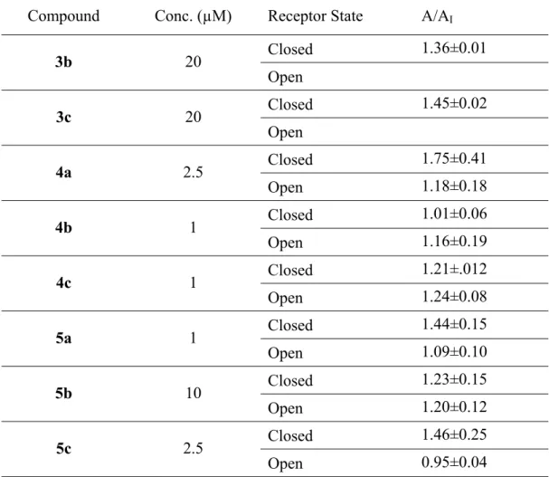
The synthesized compounds 3a-3c, 4a-4c and 5a-5c have been characterized for their antagonistic effects against homomeric GluA2Qflip receptors, transiently expressed in
human embryonic kidney HEK-293 cells. Using whole-cell recording, we measured the glutamate-evoked current amplitude in the absence (control or A) and presence of a compound (AI); a pair of representative whole-cell current response is shown in Figure 10. Specifically, a saturating glutamate concentration (i.e. 3 mM glutamate) was used to measure the potency of a compound for the open form of the channel, since essentially all the channels (~96%) would be in the open state at this concentration. In contrast, a low glutamate concentration (0.1 mM) was used to measure the potency of the compound for the closed-channel form. At a low glutamate concentration, the majority of the receptors would be in the closed-channel state (Wu, et al., 2014). The ratio of the whole-cell current amplitude in the absence and presence of an inhibitor (i.e. A/AI) as a function of inhibitor concentration is reported in Table 1. As in can be seen from the graphic in Figure 11, compounds 5a and 5c showed a higher affinity towards the closed state of the GluA2Qflip
receptor channel, in agreement with Balannik studies (Balannik et al., 2005). It should be noted that compound 4a is a strong inhibitor, judged by the A/AI value, although the t-test showed p=0.051 for the inhibition of the closed- and the open-channel state.
Figure 10. A pair of representative whole-cell traces obtained in 2.5 μM 4a assay on the closed conformation of GluK2 channel.