University of Pisa
Dept. of Psichiatry, Neurobiology, Pharmacology and Biochemistry Science of Drug and Bioactive Substances
Prof.ssa Claudia Martini
Antiapoptotic Strategies in Retinal Degeneration:
a Biochemical and Functional Approach
A Thesis submitted for the degree of Philosophiæ Doctor by:
Ilaria Piano
Under the supervision of:
Prof.ssa Claudia Gargini
Declaration
The work described in this thesis is included in the following papers: MC. Gargini, A. Asta,I. Piano, P. Gasco, C. Musicanti, E. Novelli, E. Strettoi, R. Ghidoni
Inhibition of Ceramide de novo Synthesis in an Animal Model of Retinitis Pigmentosa: Biochemical, Morphological and Functional Effects upon Photoreceptors
CDD Conference 2009 on Neurodegeneration
MC. Gargini, A. Asta,I. Piano, P. Gasco, C. Musicanti, E. Novelli, E. Strettoi, R. Ghidoni
Inhibition of Ceramide de novo Synthesis in an Animal Model of Retinitis Pigmentosa: Biochemical, Morphological and Functional Effects upon Photoreceptors
CDD Conference 2009 on Neurodegeneration
I. Piano, E. Novelli, G. Sala, P. Gasco, C. Gargini, E. Strettoi, R. Ghidoni The Role of sphingolipids in retinal degeneration
FEBS Advanced Course, Lipid Signalling and Disease-2009
E. Strettoi, R. Ghidoni, G. Sala, E. Novelli, I. Piano, P. Gasco, C. Gargini Inhibition of Ceramide de-novo sinthesis in animal models of Retinitis Pigmentosa: Rescue effects upon photoreceptors
Retinal International Meeting, Stresa 26-27 June 2010, Italy
I. Piano, E. Novelli, G. Sala, P. Gasco, C. Gargini, E. Strettoi, R. Ghidoni Sphingolipids in Retinal Degenerations
Sphingolipid Club, Glasgow 30 June-2 July 2010
E. Strettoi, C. Gargini, E. Novelli, G. Sala, I. Piano, P. Gasco, R. Ghidoni Inhibition of ceramide biosynthesis preserves photoreceptor structure and function in a mouse model of retinitis pigmentosa.
Abstract
The sphingolipid ceramide exerts a pro-apoptotic role in variety of cellular and organ systems and increased of de-novo synthesis of ceramide are associated with initiation of cell death. In Retinitis Pigmentosa (RP) photoreceptor death occurs by apoptosis but the individual pathways of this process are unknown. We employed an animal model of RP, the rd10 mutant mouse, to assess the role of ceramide in inherited photoreceptor degeneration. We used Myriocin, a known inhibitor of serine palmitoyltransferase (SPT, the rate-limiting enzyme of ceramide biosynthesis) which was either injected intravitreally in a single dose or administered daily to rd10 mice as eye drops of Solid Lipid Nanoparti-cles (SLNs). Control mice were given intravitreal injections of vehicle alone or unloaded lipid particles, respectively. We found that retinal ceramide levels in rd10 mice double from P14 to P30, the time interval of maximum photoreceptor death in this strain. Intraocular treatment with Myriocin decreases the number of pycnotic photoreceptors in rd10 mice by approximately 50%. Electroretino-gram (ERG) recordings were obtained from animals of various ages chronically treated with Myriocin-SLNs. ERG a-waves persist after P30 in treated mice while these responses are virtually extinct in control littermates. Retinal sec-tions from ERG recorded animals were examined at a confocal microscope to estimate photoreceptor survival. Morphometric analysis of retinas from rd10 mice aged P24 (peak of rod apoptosis) up to P30 showed prolonged survival of photoreceptors in treated animals. This study demonstrates in a mammalian model of RP that it is possible to decrease the rate of apoptotic death of pho-toreceptors in vivo by lowering retinal ceramide levels through inhibition of the de-novo biosynthesis of this molecule. Non-invasive, chronic administrations of nanoparticles loaded with SPT inhibitors are effective in prolonging survival and light responsiveness of photoreceptors.
Contents
Table of contents ii
List of figures iii
1 Introduction 1
1.1 Retina . . . 1
1.1.1 Morphological structure of the retina . . . 1
1.1.2 Retina of the Mouse vs Human . . . 2
1.2 Retinal Degenerations (RDs) . . . 3
1.2.1 Retinitis Pigmentosa: Genetics and Molecular Mechanisms 4 1.2.2 RHO Mutations . . . 5
1.2.3 Mutations in photoreceptor structural proteins . . . 6
1.2.4 Abnormalities in retinal transcriptional factors . . . 7
1.2.5 Rod-phosphodiesterase (PDE) gene mutations . . . 7
1.2.6 The rd10 mutant mouse . . . 9
1.2.7 Secondary cell death of Cone-photoreceptors . . . 10
1.3 RP experimental therapeutic strategies . . . 10
1.3.1 Transplants . . . 10
1.3.2 Implants: Subretinal and Epiretinal prostheses . . . 11
1.3.3 Neuroprotective factors . . . 12
1.3.4 Gene therapy . . . 13
1.3.5 Antioxidant agents . . . 13
1.4 Sphingolipids: Role in RP . . . 14
1.4.1 How the Ceramide works? . . . 15
1.4.2 De Novo ceramide biosynthesis enzyme and its Inhibitor . 16 2 Materials and Methods 21 2.1 Animals . . . 21
2.2 Drug Delivery . . . 21
2.2.1 Acute Treatment: Intravitreal Injections . . . 21
2.2.2 Chronic Treatment . . . 21
2.2.3 Solid Lipid Nanoparticles . . . 22
2.3 Immunohistochemistry . . . 22
2.3.1 Slice preparation . . . 22
2.3.2 Immunoreaction . . . 22
2.4 Western Blot Analysis . . . 23
2.5 Biochemical quantification of retinal ceramide . . . 23
2.6.1 Animal preparation . . . 24 2.6.2 Light stimulation . . . 24 2.6.3 ERG protocols . . . 25
3 Results 26
3.1 Retinal content of Ceramide . . . 26 3.2 Pharmacological effects on Retinal Ceramide levels and rod-photoreceptors
survival . . . 27 3.2.1 Acute Treatment . . . 27 3.2.2 Chronic Treatment . . . 31 3.3 Morphological and functional evaluation of the retina in the later
stages of RP in rd10 mice . . . 36 3.3.1 Time-course of cone-photoreceptors function in rd10 mice 39 3.3.2 Myriocin effects on cone-photoreceptors survival . . . 41 3.3.3 Myriocin effects on inner retina . . . 45
4 Discussion 47
4.1 Effects of Myriocin treatment in the early stage of RP . . . 47 4.2 Effects of Myriocin treatment in the later stage of RP . . . 49
List of Figures
1.1 Stratification and principal classes of retinal neurons . . . 3
1.2 Tunnel vision is a typical sympotom of RP . . . 4
1.3 Fundus views of the retina of a normal human subject (left) and of a RP patient (right) . . . 4 1.4 Phototransduction cascade . . . 5 1.5 Photoreceptors structure . . . 6 1.6 rd1 fundus . . . 8 1.7 rd10 retina . . . 17 1.8 Implants . . . 18 1.9 Sphingolipids biosynthesis . . . 18
1.10 Ceramide role in mitochondria . . . 19
1.11 De Novo formation of the bioactive sphingolipids . . . 19
1.12 SPT role . . . 20
3.1 content of ceramide . . . 26
3.2 Levels of Ceramide in rd10 mice . . . 27
3.3 Pycnotic Photoreceptors . . . 28
3.4 ERG after acute treatment . . . 29
3.5 Lack of functional effects of acute treatment of myriocin . . . 30
3.6 Lack of functional effects of chronic treatment of myriocin in WT 32 3.7 Morphologic and functional effect of chronic treatment with myri-ocin in rd10 mice . . . 33
3.8 Effect of chronic treatment with myriocin in rd10 mice . . . 34
3.9 Average of photoreceptor rows . . . 35
3.10 WT and rd10 comparison (1) . . . 37
3.11 WT and rd10 comparison (2) . . . 38
3.12 Cone amplitude in rd10 control mice . . . 40
3.13 Cone sensibility after Myriocin-SLNs treatment . . . 41
3.14 Istogram of cones b-wave after Myriocin-SLNs treatment . . . 42
3.15 WB analysis from rd10 P40 and P60 mice . . . 42
3.16 Confocal microscopy for inner retina . . . 43
3.17 Confocal microscopy for inner retina . . . 44
3.18 Confocal microscopy for inner retina . . . 44
3.19 Cone OPs . . . 45
Chapter 1
Introduction
1.1
Retina
Vision is very important in humans because the eye is dedicated to capture light reflected by the scene surronding the subject, to encode it into electrical signals and efficiently transfer them to the brain. A cross-sectional view of the eye shows three different layers:
1. The external layer, formed by the sclera and cornea
2. The intermediate layer, divided into two parts: anterior composed by iris and ciliary body and posterior
3. The internal layer that represents the sensory part of the eye, the retina.
1.1.1
Morphological structure of the retina
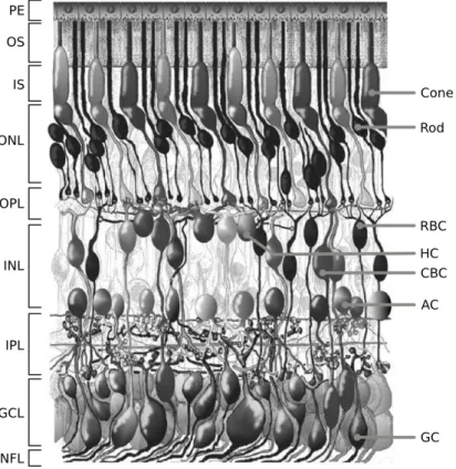
Surrounding the fovea there is a circular area of approximately 6mm in diameter called central retina while beyond this is peripheral retina stretching to the ora serrata that represents the center of the optic nerve. A redial section of the retina shows that the ganglion cells (the output neurons of the retina) lie innermost in the retina closest to the lens and front of the eye, and the photoreceptors (rods and cones) lie outermost in the retina against the pigment epithelium and choroid. A vertical section of the retina is as follows:
• Pigmented Epithelium (PE): placed at the periphery of the retina, is the pigmented cell layer just outside the neurosensory retina that nourishes retinal visual cells, and is firmly attached to the underlying choroid and overlying retinal visual cells. the PE is involved in the phagocytosis of the outer segment of photoreceptors cells and it is also involved in the vitamin A cycle where it isomerizes all trans retinol to 11-cis retinal. PE also serves as the limiting transport factor that maintains the retinal environment by supplying small molecules such as amino acid, ascorbic acid and D-glucose while remaining a tight barrier to choroidal blood borne substances. The ionic enviroment is maintained by a particular exchange system.
• Photoreceptors Layers: contain both Outer and Inner Segment (OS/IS) of the photoreceptors. The OS is the most distal part of photoreceptors and
in this location a multi-stage processe known as phototrasduction begins with light-induced isomerization of a pigment (rhodopsin or cone-opsin). The whole photodrasductive machinery is contained in here. The IS is connected to the OS by a thin cilium, it is the portion of the photoreceptors where most of the biosynthetic processes take place.
• Outer Nuclear Layer (ONL): Contain all the photoreceptors nuclei and the terminal processes of Muller glia.
• Outer Plexiform Layer (OPL): Layer where are synaptical contact between photoreceptors and bipolar cells (second-order neurons).
• Inner Nuclear Layer (INL): Contain the cellular bodies of the all second-order neurons (rod- and cone-bipolar cells, amacrine cells, horizontal cells) and of the non-neural Muller glia.
• Inner Plexiform Layer (IPL): In this layer there are the synaptical contact between second-order neurons and ganglion cells.
• Ganglion Cell Layer (GCL): Contain both ganglion cell and displaced amacrine cellular bodies. Ganglion cells represent the third-order neu-rons, the only neurons generating both spontaneus and light-evoked spike potentials.
• Optic Nerve Fiber Layer (NFL): It’s the output of the axon of the ganglion cells to the brain. Packed together they form the optic nerve and at the level of optic disc assumes the usual myelination.
1.1.2
Retina of the Mouse vs Human
In the mouse retina we can recognise the same layer of the human retina. The mouse retina is rod-dominated (rods 4.5milion versus cones 125.000) and at variance with human retina there aren’t three type of cones but only two: green and blu cones. Similarly to those of humans, photoreceptors of the rodents, rods and cones, absorb photons and convert them into electrical signals. Dim light signals have sufficient energy to excite the rods, while cones are less sensitive and to be excited require much brighter lights. Therefore rod photoreceptor cells operate in the twilight while cone visual cells are active at daylight and subserve color vision. Photoreceptor cells may be connected to the neighbor-ing cells by electrical synapses (gap junctions) and establish chemical synapses with bipolar and horizontal cells. Bipolar cells are subdivided into two main classes: rod and cone bipolar, each bipolar cells make contact with the corre-sponding photoreceptor populations. Cone bipolar cells are furhter divided in two subtype: ON- and OFF-bipolar cells, that respectively depolarize and hy-perpolarize in response to the illumination of their receptive field centers. All rod-bipolar cells are of subtype ON and establish direct connection with ON ganglion cells (RGC) through amacrine cells. Both ON- and OFF cone-bipolar cells are connected directly to respectively ON or OFF RGCs.
Figure 1.1: Morphological representation of the retina in a vertical section of the adult human eye. Retinal layers on the left. Principal neuronal classes on the right. RBC: rod bipolar cell, HC: horizontal cell, CBC: cone bipolar cell, AC: amacrine cell, GC: ganglion cell
1.2
Retinal Degenerations (RDs)
A very high number of genetic mutations affect the eye and a large number of mutations affect photoreceptors or pigment epithelium and may cause retinal degeneration (RDs). Retinitis Pigmentosa (RP) comprises a group of hetero-geneus genetic disorders that cause severe loss of vision in as many as 1.5 milion individuals worldwide (review: [68], [44],[81]). The mutation mainly affects the rod photoreceptors which mediate the scotopic vision (dim light conditions). When rods start to die, in response to genetic abnormality, symptoms appear and the night vision of patients is reduced and the visual field is constricted (tunnel vision). RP usually follows a two-stage process in which first the rod-photoreceptors degenerate, then also cone-rod-photoreceptors start to die resulting in the loss of central vision. At this stage of RP also blood vassel distrophy and intraretinal brown pigment accumulation occur. It is unusual for patients with RP to became totally blind as most of them retain some useful vision well into old ages. Type of RP and related diseases include, among others, Usher syn-drome, Leber’s congenital amaurosis, rod-cone disease, Bardet-Biedl syndrome and Refsum disease.
Figure 1.2: The progressive die of the rod-photoreceptors lead to loss of periph-eral vision.
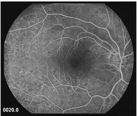
Figure 1.3: A typical brown pigmentation is present in the pathological condi-tion. Pigment deposits, named bone spicules for their shape, are responsiblefor the name of the disease. In these image is also visible a clear attenuation of retinal blood vassels.
1.2.1
Retinitis Pigmentosa: Genetics and Molecular
Mech-anisms
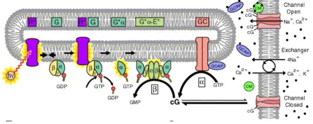
There are three principal types of RP: autosominal dominant (ad), autosominal recessive (ar) and X-linked. RP causing mutations have been identified in more than 35 genes. Genetic mutations (as deletions, insertions, or substitutions) triggering potoreceptors degeneration and then death by apoptosis, often affect the retinoid acid cycle or the phototransduction cascade. Phototransduction starts with a light-induced, conformational change of 11-cis-retinal to all-trans-retinal. This in turn activates opsin molecules, causing sequential activation of transducine and cyclic guanosine monophosphate (cGMP)-phosphodiesterase-6 (PDE6) and subsequent decreases in intracellular cGMP concentration. High cGMP levels maintain cyclic nucleotide-gated (CNG) cation channels in the open state, allowing the influx of cations including sodium and calcium. Reduc-tion of cGMP levels closes CNG channels causing cell hyperpolarizaReduc-tion. This signal is then transmitted to second order neurons in the INL Fig.1.4 ([68]). There are mainly two mutations affecting the phototransductive process: the first concerns, the Rhodopsin gene (RHO), this is the best studied mutation and is responsible for varius forms of autosominal dominant RP (adRP); the second, that affects the phosphodiesterase gene (Pdeb), leads to autosominal recessive RP (arRP) and this is the defect carried by the mouse models used in the present study (explained in more detail later).
Figure 1.4: Pathway of the visual transduction cascade.
1.2.2
RHO Mutations
Rhodopsin is a prototypic member of the G-protein-coupled receptor family and its molecular structure has been known for some time ([79],[80]). The visual pig-ment molecule consists of a protein component (a 348 amino acid sequence orga-nized into three distinct domains: cytoplasmic, transmembrane and intradiscal), and chromophore group (11-cis-retinal a light absorbing polyen structure where phototransduction initiates, fig.1.4) The most common mutation in RHO gene is the change of proline to histidine at codon 23 (P23H), this mutation leads to a phenotype classified as class B1 with a typical features that includes slower progression and better visual function compared to other forms of adRP. Be-sides P23H, there are several other types of mutations in the RHO gene that are classified as follows:
• Class I: mutations basically occur in the C-terminus of the protein, this part of the protein is important for its translocation to the plasma mem-brane and could form a functional chromophore with 11-cis-retinal. • Class II: the most common adRP mutations, were characterized by
muta-tions in the intradiscal membranes and cytoplasmatic domain of rhodopsin, which resulting in misfolding of the protein, defined by the inability to form a functional chromophore with 11-cis-retinal.
• Class III: corresponds to mutations that affect endocytosis (R135L muta-tion).
• Class IV: these mutations affect the rhodopsin stability and post - trans-lational modification, such as T4R mutation that causes adRP in dogs. • Class V: have no abvius initial folding defect but show an increased
acti-vation rate of for transducin.
• Class VI: this mutant appears to fold correctly bat leads to the constitutive activation of opsin in the absence of chromophore and in dark.
Dominance in adRP patients could be due to loss-of-function, gain-of-function or dominant-negative mutations, or any of the above in combination.
Several animal models of rhodopsassociated RP have been generated and in-clude the rhodopsin knockout mouse lacking any functional rhodopsin and thus rapidly undergoes retinal degeneration, and dominant transgenic models such as the P23H mouse ([77]). These animals are an extremely valuable resource in the study of rhodopsin-induced degeneration mechanisms.
1.2.3
Mutations in photoreceptor structural proteins
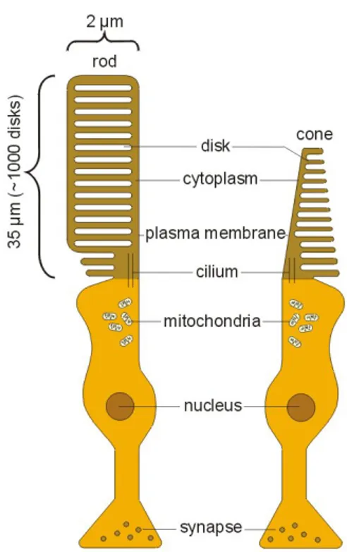
Several of the known RP genes encode proteins that have pivotal roles in the formation and maintenance of the highly complex and delicate cellular struc-ture of the rod (Fig.1.5). Peripherina 2 also known as retinal degeneration slow
Figure 1.5: This figure show a schematic representation of the complex photore-ceptor strcture.
(RDS), a transmembrane glycoprotein, is one of the earliest proteins to be asso-ciated with RP. Mutations in RDS lead to several different retinal phenotypes including arRP, cone-rod dystrophy and adRP
(http://www.sph.uth.tmc.edu/Retnet/home.htm).
RDS estabilishes complexes that have an integral role in the morphogenesis and structure of the stacked discs in both rod and cone OS (see fig.1.5). RDS mu-tations lead to RP by two different mechanisms. First, alterations prevent the protein from forming tetramers, inhibiting its transportation from the transla-tion site in the IS to the OS; second, the mutant protein is incorporated into the nascent disc but causes them to be inherently unstable and RP develops be-cause of the disorganization that ensues in the OS ([26],[42], [57]). Mutations in the RP1 gene are among the most common causes of adRP and are responsible
for 6-10 per cent of cases. The role of the RP1 protein might be central to the morphogenesis of the photoreceptors OS ([107]).
1.2.4
Abnormalities in retinal transcriptional factors
The process that occurs in development, differentiation and maintenance of the photoreceptors are under the control of several transcription factors, such as cone-rod homeobox-cointaining (CRX) and neural retina leucine zipper (NRL) genes. CRX is essential for photoreceptor development and when mutated is associated with the early onset retinal degeneration laber congenital amaurosis (LCA) and adRP; NRL is pivotal in the determination and development of the photoreceptors and also in their function and maintenance in the adult, in patients with NRL mutations, it appears that the detrimental effects might be a result of increased expression of rhodopsin and other photoreceptor gene ([17]).
1.2.5
Rod-phosphodiesterase (PDE) gene mutations
Several forms of the arRP are caused by mutations carried in the PDE gene. Cyclic GMP-phosphodiesterase is composed of four subunits, two with catalytic activity, α and β, and two identical gamma subunits with inhibitory activity. This enzyme is fundamentally important for phototrasduction in photoreceptors ([25]).
cGMP phosphodiesterase type 6 (PDE6) is the specific enzyme in rod-photoreceptor. PDE6 is activated in response to light stimulation of the receptor in the outer segments of rod (Fig.1.4). A visual cascade of biochemical reactions is initiated when rhodopsin is activated by the incident light. Activated rhodopsin stim-ulates the exchange of GDP for GTP and activates the G-protein, transducin. Transducin activates PDE6 by displacing the inhibitory γ-subunits from the active site of the enzyme, thereby greatly stimulating cGMP hydrolysis. PDE6 rapidly reduces the cytoplasmic cGMP concentration, which causes closure of cGMP-gated channels and produces a transient hyperpolarization of the rod plasma membrane leading to a reduction in glutamate release at the synapse ([40]).
In 1924 Keeler and collaborators discovered for the first time the mutations in the PDE gene in rodless mice (r) ([43]), this were a nonsense mutation in the PDE gene coding for the beta-subunit of cGMP phosphodiesterase gene.This spontaneus mutation was later rediscovered in the retinal degeneration mouse (rd1 or rd)([9],[84], [85], [86]) and subsequently in humans with arRP and adRP form of night blindness ([66], [65], [28]).
Mouse model of retinal degeneration has been investigated for many years in the hope of understanding the causes of photoreceptors cell death. The first retinal degeneration observed is rd1. This mutation has been found among several com-mon laboratory strain (such as C3H and CBA/J) and mice homozygous for the rd1 mutation have an early onset severe retinal degeneration due to a murine viral insert (MLV) and a second nonsense mutation in exon 7 of the Pde6b gene enconding the beta subunit of cGMP-PDE. Mice with the rd1 mutation can be easily typed by phenotype based on vassel attenuation and pigment patches in the fundus (Fig.1.6) ([14]).
Biochemical studies comparing retinas from wt and rd1 mice have shown that the lack of cGMP-PDE activity causes a dramatic increase in cytoplasmatic
Figure 1.6: This image show the appearance of the ocular fundus in the rd1 mice retina.
cGMP concentration, this, in turn, leads to a permanent opening of the cGMP gated cation channels on the photoreceptor membrane with an increase of the extracellular ions influx, particularly Ca2+. It has been suggested that this increase in intracellular Ca2+ causes a metabolic overload of the cells, eventu-ally leading to cell death by apoptosis ([16]). Excessive Ca2+ influx has long been regarded as a major factor in photoreceptor degeneration and strong ac-tivation of Ca2+-dependent enzymes has been observed in rd1 photoreceptors.
Certainly, Ca2+ is seen as a trigger of apoptosis through the inactivation of
mitochondria and subsequent activation of apoptotic machinery. On the other hand, Ca2+ influx causes ATP depletion by activating Ca2+ extrusion
mecha-nisms such as Ca2+-ATPaese or the Na-ATPases required to drive N a+/Ca2+
exchangers, while at the same time inhibiting mitochondrial ATP synthesis. A possibility is that the observed rise in retinal Ca2+ levels is only secondary to
the degeneration process and it is not even clear whether it occurs in photore-ceptors or in other retinal cell types. Furthermore, fluctuations in cytosolic Ca2+ may be caused not only by Ca2+ influx from extracellular sources bat
also by intracellular release from the mitochondria, photoreceptor discs or the endoplasmatic reticulum (ER). Further, other mechanisms can lead to cell death such as oxdative stress and activation of the proapoptotic protein. In summary Ca2+, the energetic status, and ER stress are different factors that appear to be interconnected in cell death: a primary rise in intracellular Ca2+ triggered by
ER stress could be followed by mitochondrial dysfunction, an energetic collapse, and a secondary wave of extracellular Ca2+ ([90]).
In addition to rd1 mouse model, other naturally occuring mouse mutants that manifest degeneration of photoreceptors in the retina with preservation of all other retinal cell types have been found such as:
• Purkinje cell degeneration (pcd ) • nervous (nr )
• retinal degeneration slow (rds) • motor neuron degeneration (mnd )
• vitiligo (vit )
• neuronal ceroid lipofuscinosis (nclf ) • cone photoreceptor function loss (cpfl1 )
• and several types of retinal degeneration (rd3, rd4, rd5, rd6, rd7, rd8, rd9 and rd10 ) ([14]).
1.2.6
The rd10 mutant mouse
Retinal degeneration 10 (rd10 ) mouse is a viable model of human retinitis pig-mentosa and this strain is used in the present study. Genetic analysis shows that these mice carry an autosomal recessive mutation that maps into chromosome (Chr) 5. Sequence analysis shows that the retinal degeneration is caused by a missense point mutation in 13th exon in the Pde6b gene of the beta-subunit of
the rod cGMP phosphodiesterase (beta-PDE). The mutation changes codon 560 from CGC to TGC resulting into an arginine to a cysteine. The exon 13 mis-sense mutation is the first known occurence of a remutation in the Pde6b gene in mice and provide a good model for studying the pathogenesis of arRP in human. It also provides a better model than rd1 for experimental pharmaceutical-based therapy for RP because of its later onset, slowly progressing and less severe retinal degeneration compared to that affecting the rd1 ([14]).
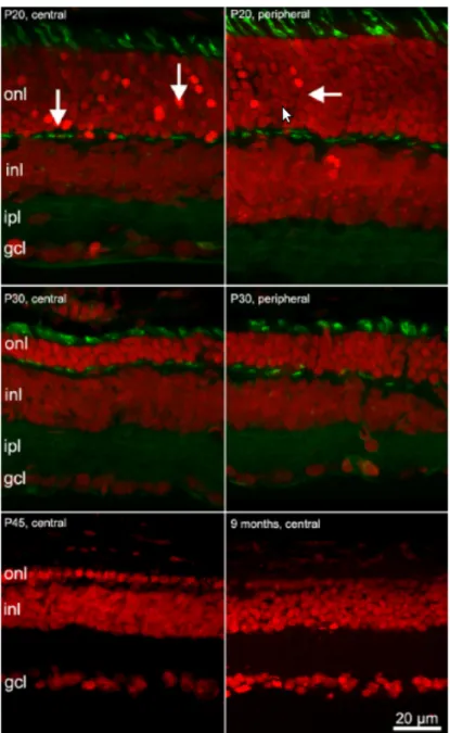
In the rd10 mice the number of photoreceptor rows is 12-14 at postnatal day 10 (P10) and 11-12 rows at P20. From P20 to P25 there is a substantial re-duction in the number of photoreceptor rows and at P30 cones are shortened outer and inner segmented and a deformed shape. At P45, only a single row of photoreceptors is present (Fig.1.7). Degeneration of photoreceptors in rd10 retinas follows a center-to-periphery gradient: the decrease in the number of ONL rows becomes apparent in the central retina first, whereas degeneration in the periphery is delayed ([29]). Atrophic retinal vessels are found at four weeks of age, consistent with retinal degeneration ([15], [29]). After extensive photoreceptor death, dendrites of the bipolar cells exhibit a retraction. Survival of rod bipolar and horizontal cells drops right after the peak of photoreceptor degeneration and decreases slowly over the following months. After that, also the INL disorganizes and typical rosettes appear.
The morphological changes have a physiological correlate. Infact elettroretino-gram (ERG) analysis shows differences between the flash ERG of wt and rd10 mice related with severe damage affecting the distal rod pathway. The loss of functioning rods that occurs in the rd10 mutants between P20 and P30 largely contributes to the drastic reduction in the normalized maximal a-wave that is directly proportional to the suppression of the dark current by light ([10]). Note that alterations in the shape and relative amplitude of the b-wave are de-tected as early as P18 when no morphological modification occur in inner retina. Changes in the efficiency of the synaptic communication of rod-to-rod bipolar cells could explain the alterations found in the ERG by using sinusoidal stimuli. In fact the ERG mesurements obtained by using sinusoidal modulation of the mean luminance confirm the results of the flash ERG, and provide an indication that, even at earliest stage of the degenerative process, bipolar neurons show signs of damage ([29]).
1.2.7
Secondary cell death of Cone-photoreceptors
Photoreceptor degeneration results in vision loss in diseases like RP and age-related macular degeneration. In these diseases, the main cause of clinically significant vision loss is cone cell degeneration rather than rod cell death. In fact most mutations responsible for RP in humans and animal models affect rod-photoreceptor-specific genes, rod apoptosis is often followed by secondary cone degeneration ([71]). Several possible causes have been indicated to explain mutation-indipendent secondary cell death of cone-photoreceptors. These in-clude loss of structural support and/or loss of trophic support, i.e, Rod-derived Cone Viability Factor (RdCVF) ([89]) when rod negatively affect the cells are degenerating and death. In fact, intraocular injections of this factor may to rescue cone photoreceptors ([112]).
Another hypothesis concerning the changes in oxigen consumption levels as a function of rod degeneration: depletion of the photoreceptor population (mainly rods) by any cause would reduce consumption of oxygen flowing from the choroidal circulation. Because this flow of oxygen is unregulated, photoreceptor depletion will cause a chronic increase in oxygen tension in the outer retina, and this increase is toxic to surviving photoreceptor (both rods and cones) ([18]). The vulnerability of photoreceptors to hyperoxia has also been confirmed, and evidence has been reported that rod loss results in oxidative stress to cones ([94]).
Two other hypothesis have recently been developed concerning the secondary death of cones: rods may release toxic substances that negatively affect the neighboring cells and a chronic activation of microglial cells has been described in animal models of RP. It is possible that an immune response might enhance degenerative processes and could in part be responsible for the secondary loss of cone-photoreceptors which are not affected by the primary genetic mutation ([113]).
1.3
RP experimental therapeutic strategies
1.3.1
Transplants
The possibility of retinal transplantation represents a hope for the restoration of vision through cell-replacement therapy. However, getting the transplanted cells to establish the right connections within the retina has been a major problem by far. It appears that an early developmental stage of the retina of acceptors represents a key feature of a successful transplant. Despite decades of exper-imental attempts, transplants have yet to produce better vision in mammals with retinal degeneration because the transplanted cells do not wire up prop-erly. The first successful transplant of a mammalian retina dates back to 1959, when Royo and Quay transplanted fetal rat retinas into the eyes of adults of the same strain. Transplants appear to develop many characteristics of a normal retina ([93]; [115]) and grow to form a second retinal layer underneath the host tissue. Further, transplant of microaggregates containing clumps of few retinal neurons from newborn mice, develop most characteristics of rod photoreceptors including the expression of the rhodopsin and the outer segments ([32], [33]). Unfortunately, both these type of transplantion remain isolated without inter-acting or integrating effectively with the host retinal neurons. On the contrary,
integration with the host retinal neurons is obtained when transplanting retinal stem cells derived from fetal or newborn mice or rats, or from human fetuses. In fact, when these cells are transplanted into both normal and degenerating retinas migrate into all retina layers and develop morphological characteristics of several retinal cell types, mainly photoreceptors ([13], [19], [46]). There is also evidence to show that the best donor cells should be from 3 to 5 days old, approximately two or three days after the peak of rod photoreceptor develop-ment in the mouse retina. This suggests that, newly born rods, rather than the stem cells, are the best candidates for cellular transplantation ([61]).
On a different rationale, promising results for RP treatment have been obtained, still using a cell-based approach ([78]). These study have shown that injections of hematopoietic stem cells (lineage-negative called Lin-HSCs) into the eye of mouse models of RP (rd1 and rd10 mice) result into a dramatic rescue of both blood vessels and photoreceptors, mainly cones. These mice show an improve-ment of the ERG at an age when usually it is completely extinct and the rescue effects are long lasting.
1.3.2
Implants: Subretinal and Epiretinal prostheses
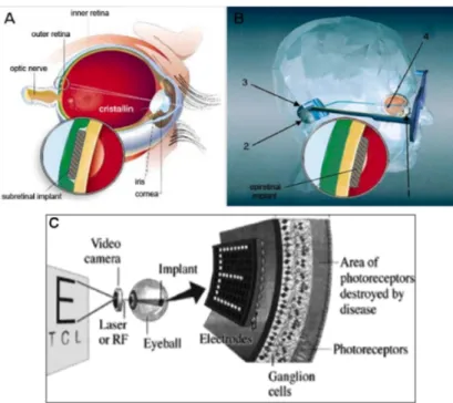
The development of artificialimplants inserted in substitution of an entire part of the pathological retina represents an active line of investigation to treat both RP and other retinal diseases, such as macular degeneration. Advances in mi-crotechnology have facilitated the development of a variety of prostheses that can be connected to the brain or implanted in the eye; these implants have a greater potential for visual restoration as they are well tolerated and remain viable for several years; on the on the hand, retinal prostheses are only effective where the visual pathway distal to the retinal implant is still functional. Two types of prostheses have been developed: subretinal and epiretinal (Fig.1.8). Subretinal prostheses contain microphotodiodes attached to micro-electrodes. These implants, such as the articial silicon retina (5000 micro-electrodes), are placed in the subretinal space between the outer retina and retinal pigment ep-ithelium (Fig.1.8). The photodiodes are stimulated by light pass- ing trough the retina, and the resulting electric current excites adjacent retinal sensory neurons. These implants do not require an external electrical source as incident light is sufficient for stimulation ([116]). Preclinical evaluation shows that sev-eral species of the animal have a tolerance to the implants up to 30 months, and histological examination does not show significant changes in the morphology of the retina. In six patients with RP, who received a subretinal implants, vi-sual perception but also appreciation of brightness, contrast of color, movement, shape and vi- sual eld are improved. Some patients showed also an improvement in visual acuity. Two major advantages of subretinal prostheses are the utiliza-tion of existing forces between the neural retina and retinal pigment epithelium to maintain their position and the potential of a high spatial resolution, as they are posi- tioned close to retinal nerve cells and can stimulate neurons by means of low electrical currents. The main disadvantages of subretinal implants include im- paired nourishment of the inner retina due to the creation of a mechanical barrier between the outer retina and the choroid and the occurrence of trauma to the retina during the implanting. These prostheses also show poor dissipa-tion of heat and therefore could damage the retina. The mechanism of acdissipa-tion of these implants may be by direct stimulation of retinal neurons. A specific
neurotrophic eect elicited locally onto photoreceptors by means of the surgical procedure cannot be excluded: a study of subretinal artificial silicon implants in rats showed a temporary protective eect on the retina, resulting in increased generation of photoreceptors.
Epiretinal prostheses are composed of an array of electrodes implanted on the surface of the retina between the vitreous and inner limiting membrane. The implants receive electrical signals from a camera positioned outside the body (Fig.1.8); the camera transmits light signals to a microchip within the camera. After that, the microchip decodes the signals and relays it, by wireless trans-mission, to a microchip in the epiretinal implant that in turn stimulates the RGCs ([116]). Clinical trials in humans have reported simple visual perception as phosphenes; these implants have survived up to two years.
Respect to subretinal implants, an advantage of epiretinal devices is that the camera can process signals before they reach the implant: this allows optimiza-tion of the signal quality that leads to improved visual percepoptimiza-tion. Epiretinal implants can also use the heat dissipating properties of the vitreous and are therefore less likely than subretinal implants to damage the retina. The super-cial location of the epiretinal devices reduces risk of trauma during both im-plantation and replacement. The principal disadvantage of epiretinal implants, compared to subretinal devices, is the complicated microtechnology and surgi-cal techniques for a secure xation of the implant on the retina; furthermore, the epiretinal implants requires a higher electrical current than the subretinal implants, and these devices are being developed to generate their own currents on stimulation ([50]).
1.3.3
Neuroprotective factors
Most mutations responsible for the RP phenotype selectively affect rod photore-ceptors, however cones undergo degeneration secondary to the loss of rods, often leading to total blindeness. How to save the cones has become a major research challenge because preserving 5 % of all cones may be sufficient to maintain a useful vision, and 50 % of cone functionality would ensure a normal visual acuity. Treatment with neuroprotective factors, such as brain-derived (BDNF), ciliary (CNTF), or glial cell line-derived (GDNF) neurotrophic factor, has been found to protect partially against photoreceptors degeneration in several an-imal models ([24], [51], [97]). Further, neuroprotective factors could work in-dipendently of the genetic mutation that cause RP, and therefore could bypass the very high genetic heterogeneity of this disease. CNTF, initially identified as a factor in chick embryo extract that supported viability of embryonic chick ciliary neurons ([2], [105]) and later purified to homogeneity, exhibits multiple biological effects in the retina ([55], [102]). CNTF promotes the survival and axonal regeneration of retinal ganglion cells ([108], [41], [69]) and acts as neu-ronal differentiation factor, promotes the differentiation of cone photoreceptors expressing green opsin in the developing chick retina ([27], [45], [111]), enhances the expression of bipolar neuron markers in rat retinal cultures and inhibits rod photoreceptor cell differentiation ([6], [23], [72], [92]), and promotes Muller glia genesis from the postnatal retinal progenitor pool ([34]). It has been reported that CNTF regulates the photodrsduction machinery of rod photoreceptor, and the regulation was mediated through Muller cells ([109], [110]).
function stimulating regeneration of COS. In the early stages of degeneration, cone without COS are not only alive but are capable of regenerating their outer segments (woithout COS, cones lose their light-sensig funcion), indicating that at this time the degeneration process is reversible. They found that the delay between loss of COS and cell death is likely in the time frame of about two months in their animal models. Considering the rapid rod degeneration (about 10 days) in these animals, the secondary cone degeneration was relatively slow process, which not only provides an opportunity to study the patology of sec-ondary cone degeneration, but also serves a model for preclinical assessment of potential treatments. Further, this finding araises the possibility that a similar window may exist in patients with RP, in order to allow therapeutic intervention to save cones.
The importance of rod-cone interactions for cone survival was suggested subse-quent to the identification of the first causal mutations in RP ([22], [83]). The rod dependence of cone survival is also supported by the experiments in vitro ([38]) and by the studies of transgenic mice and mutant zebrafish ([39], [30]), suggesting cell-to-cell interactions in photoreceptor degeneration. RdCVF was isolated for its effect on cone survival using embryonic chick cone cultures ([60]). The effect of the injections of RdCVF protein from old mice 6 to 8 months indi-cates that this factor induces cone survival directly but does not act indirectly by stimulating rod survival, as most rods have already degenerated at the time of treatment. This trophic effect appears indipendent from the mechanisms of rod degenaration and thus of the causal mutation when the mutated gene is only expressed in rods. RdCVF induces also a functional rescue, thus validating the potential of this trophic factor for therapeutic applications, and in adressing the secondary degeneration of cones, RdCVF administration may be used to tar-get a pathological mechanisms common to most forms of the disease. A recent study on the P23H rat suggests that RdCVF administration may be efficient in the treatment of autosomal dominant RP due to rhodopsin mutations, which account for 30-40% of cases of human autosominal dominant RP ([112]).
1.3.4
Gene therapy
Cone cell bodies remain present longer than rods in both humans and animals, but these light-insensitive cells can be reactivated for a significant time win-dow after the loss of photosensitivity. Another recent study ([11]) shows that a microbial gene introduced to surviving cone cell bodies reactivated retinal ON and OFF patways and the retinal circuitry for lateral inhibition and direc-tional selective responses. The reactivated cones enabled RD mice to perform visually-guided behaviors. The tested time window of intervention suggests that persisting cone cell bodies (about 25%) was enough to induce ganglion cell activity, even during later stages of degeneration.
1.3.5
Antioxidant agents
Another possible explanation for the slowly progressive death of cones after the death of rods, in RP, is oxidative damage. The demostration that oxidtive dam-age contributes to cone cell death in RP has important clinical implications. It provides a therapeutic target that may apply to all RP patients. The enormous genetic heterogeneity among the diseases that constitute RP is a problem for
the development of treatments that deal with primary genetic defects. If, re-gardless of the genetic defect that leads to rod-cell death, the same treatment can be used to prolong cone survival and function with an enormous impact. As long as cones survive, useful vision is possible and, although it would be ideal to salvage rods as well, it is not necessary, because patients can carry on relatively normal lives with only cone function.
In varius animal models, three agents, α-tocopherol, ascorbic acid, and α-lipoic acid,have been shown to reduce one or more types of oxidative stress ([12], [98], [73], [49], [82]), and these agents may have additive effects when used together ([100]). Another potent antioxidant that is adept at reducing oxidative damage in mitochondria, MnTBAP, does not cross the blood-brain or blood-retinal bar-riers under normal circumstances ([67]), but because the blood-retinal barrier in compromised in RP, MnTBAP was also added the regimen. Daily systemic injections of the mixture starting at P18 in rd1 mouse, the time of onset of cone cell death, reduced staining for acrolein (marker of oxidative damage) in cones at P35 to undetectable levels, indicating that the treatment reduced oxidative damage in cones.
Further, an additional support for the hypothesis that oxidative stress is an im-portant contributor to cone cell death in RP, is shown in Komeima et al. ([48]), they found that compared to paired vehicle controls, daily administration of ei-ther α-tocopherol or α-lipoic acid as monoei-therapy also reduced in a significant increase in cone cell number at P35 in rd1 mice.
Combining this approach with other strategies designed to bolster components of the endogenous defense system directed against oxidative stress, as well as enhancing levels of survival factors, may be needed to provide manigful benefit with RP.
1.4
Sphingolipids: Role in RP
Complex sphingolipids are composed of bioactive backbones (sphingoid bases and ceramides) that can cause cell death when the amounts are elevated by turnover of complex sphingolipids, disruption of normal sphingolipids metabolism or induction of sphingolipid biosynthesis de novo.
Sphingolipid biosynthesis produces several bioactive intermediates formed from complex sphingolipid turnover: ceramides, sphngoid bases ( sphingosine and sphinganine) and sphingoid base 1-phosphates ([36]). The principal enzymes of sphingolipid turnover are sphingomyelinase and ceramidases but there are at least six key enzymes that partecipate in the de novo pathway that form/remove these compounds: serine palmitoyltransferase (SPT), which catalyzes the first reaction of the pathway; ceramide synthase, the enzyme responsible for trapping sphinganine as dihydroceramides; dihydroceramide desaturase, which converts dihydroceramides to ceramides; and enzymes such as sphinganine kinase, sph-ingomyelin synthase, glucosylceramide synthase, etc. which remove or modify these intermediates ([56]).
Ceramide, a sphingolipid metabolic precursor, is a bioactive lipid that has been proposed to be an endogenous mediator of apoptosis; ceramide is actively in-volved in the triggering of apoptosis in many cell systems, including neurons. Increased intracellular levels of ceramide have been shown to occur during hy-poxia, trophic factor removal, treatment with chemotherapeutic agents, heat,
UV radiation, and other stress signals, such as oxidative stress ([31]). Ceramide levels depend on the activity of several enzymes that participate in its synthesis and catabolism. The increase in ceramide in response to apoptotic stimuli may arise from hydrolysis of sphingomyelin due to stimulation of sphingomyelinases, through de novo biosynthesis, or from the combined stimulation of both path-way ([8]). In turn, several biochemical reactions lead to the disappearance of ceramide: its hydrolysis catalyzed by ceramidases, sphingomyelin resynthesis, or ceramide glucosylation.
Only recently, it was demonstrated for the first time that the sphingolipid path-way is involved in photoreceptor apoptosis in Drosophila ([1]), and a mutation in a novel ceramide kinase gene has been recently established as a cause of an autosomal recessive form of RP, suggesting a direct link between sphingolipid-mediated apoptosis and retinal degeneration ([104]).
1.4.1
How the Ceramide works?
The de novo synthesis of ceramide occurs in the endoplasmatic reticulum (ER) and in mitochondria ([62], [96]). In recent years, ceramide has become increas-ingly appreciated as a bioactive lipid that modulates apoptotic/necrotic cellular processes that are integrated at the mitochondrial level, while sphingosine-1-phosphate has been shown to be protective/anti-apoptotic. In fact, it has been proposed that it is the ratio of sphingosine-1-phosphate to ceramide/sphingosine that determines the fate of a cell ([20], [76]);
There are a number of observations that support a proapoptotic role for ce-ramide in apoptosis:
• First, ceramide generation is a common cellular response of a variety of cell types following exposure to apoptosis-inducing agents ([75]);
• Second, the effective doses of these agents required to induce ceramide generation closely matches the dose required to induce apoptosis ([47]); • Third, elevations in cellular ceramide addition in response to these agents
occurs prior to the execution phase of apoptosis ([35], [7]);
• Fourth, exogenous addition of cell-permeable ceramide analogues induces apoptosis in a variety of cell lines ([75]);
• Fifth, ceramide-induced apoptosis is very specific, as a naturally occurring ceramide precursor dihydroceramide does not induce apoptosis ([75]); • Sixth, apoptosis can be inhibited upon blockage of ceramide generation
and cells that are incapable of generating ceramide are often incapable of undergoing apoptosis ([3], [88]).
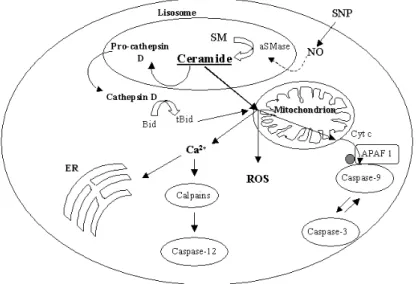
Furthermore, ceramides have been reported to have numerous effects on mito-chondria, including enhanced generation of reactive oxygen species, alteration of calcium homeostasis of mitochondria and ER, ATP depletion, collapse in the inner mitochondrial membrane potential, inhibition and/or activation of the ac-tivities of various components of the mitochondrial electron transport chain and release of intermembrane space proteins. Further, it has been suggested that ceramide conveys death signals to mitochondria by two principal mechanisms:
1. ceramide leads to a selective permeabilization of the outer mitochondrial membrane for proapoptotic proteins as a process independent of inner membrane functions;
2. the second mechanisms presumes that loss of the permeability barrier of the inner mitochondrial membrane is a prerequisite for the subsequent outer membrane permebilization.
Participation of the inner mitochondrial membrane in the release of proapop-totic preoteins involves the obligatory opening of the Permeability Transition Pore (PTP) in the inner membrane. PTP opening occurs as a result of direct in-teraction of ceramide with the pore in the presence of Ca2+or that PTP opening follows mitochondrial Ca2+ overload. Support for this mechanism arises from experiments with many cell types in which ceramide-induced cell death is sup-pressed by PTP inhibitors, such as cyclosporina A and bongkrekic acid ([99], [54]). Models of mitochondrial Ca2+ overload as a consequence of
ceramide-induced Ca2+ release from ER and the direct activation of PTP by ceramides
where largely based on data obtained using artificial C2-ceramide ([74]).
1.4.2
De Novo ceramide biosynthesis enzyme and its
In-hibitor
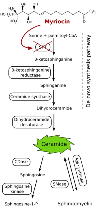
The first demostration of toxicity owing to alteration of de novo ceramide biosynthesis was the discovery that fumonisins inhibit ceramide synthase ([63]). Further, it is possible that many factors that perturb intermediary metabolism affect sphingolipid biosynthesis and thereby alter the amounts of sphinganine, ceramide, etc. as shown in Fig.1.11. It has been shown that some aspects of the toxicity of palmitate for cells in culture is due to sphingolipid biosynthesis and the overexpression of Serine palmitoyltransferase (SPT) can also induce apop-tosis ([95]). SPT is a pyridoxal 5’-phosphate-dependent enzyme that condenses serine with palmitoyl-CoA to produce 3-ketosphinganine (Fig.1.12). For mam-mals and yeast, two gene products (SPT1 and SPT2) are necessary for activity and appear to be associated physically.
Because SPT is a key enzyme for the regulation of sphingolipid levels in cells, the specific inhibitors of SPT are useful for understanding the physiological roles of sphingolipids. Several natural and synthetic inhibitors of SPT have been reported. ISP-1 (Myriocin) from a culture of fungus Isalia sinclairii is a widely used and highly selective SPT inhibitor. The structure of myriocin resembles that of sphingosine (Fig. 1.12). Myriocin inhibits ceramide synthesis in mammalian cells with IC50 values in the nanomolar range, while a
micro-molar concentration of myriocin is necessary for the inhibition of sphingolipid synthesis in S. cerevisiae ([70], [103]).
Figure 1.7: This image shows the progressive degeneration of photoreceptors. Red signal is obtained with ethidium nuclear staining; green signal represent the inner segment of cones and the synaptic endings in the OPL, obtained with PNA.
Figure 1.8: Schematic representations of subretinal (A) and epiretinal electronic implants (B): 1, camcorder mounted in a glass frame; 2, wireless transmitter; 3, extraocular junction box; 4, intraocular implants. In (C) is shown a simpli-fied scheme of electronic epiretinal implants. From http://current.ucsc.edu/04-05/10-18/retina.asp.
Figure 1.9: Biosynthetic and catabolic reactions for formation and removal of the major bioactive backbones of sphingolipids.
Figure 1.10: Schematic representation of the events that ceramide leads in cells for the activation of the mitochondrial apoptotic pathway.
Figure 1.11: SPT1 and SPT2 are the two components of SPT. The sites of action of commonly used inhibitors (myriocin and fumonisin) are also shown. The subcellular locations of these reactions are indicated only for a general context; there are likely to be other sites where some of these reactions occurs. GlcCer, glucosyl-ceramide; Pal-CoA, palmitoyl-CoA.
Figure 1.12: Schematic representation of the de-novo biosynthesis of ceramide and role of SPT. The chemical structure of Myriocin, an inhibitor of SPT is
Chapter 2
Materials and Methods
2.1
Animals
All the experimental procedures involving animals were carried out according to the Italian and European guidelines for animal care (d.l. 116/92; 86/609/CE). Animals were maintained under a 12:12h light:dark cycle. Species and strains involved in this work include: rd10 mice (Jackson Laboratories strain B6.CXB1-Pde6brd10/J) ([14]) and wild-type mice (Jackson Laboratories stain C57BL/6J).
2.2
Drug Delivery
The animals were treated with Myriocin, a known inhibitor of serine-palmitoil-transferase (SPT) which is the rate-limiting factor in Ceramide de-novo synthe-sis.
2.2.1
Acute Treatment: Intravitreal Injections
The acute treatment was obtained by means of intravitreal injections of Myri-ocin. Rd10 mice, aged P19, were anesthetized by intraperitoneal Avertin (0.5 g/ml 2,2,2-tribromoethanol in ter-amylic alcohol; 20 µl/g body weight). Using a glass micropipette driven by an oil Hamilton syringe, 500 nl of the solution of Myriocin (188M, in DMSO) was injected in the vitreous body of the right eye under a dissecting microscope, while vehicle alone was injected in the left eye. Local antibiotics were administered to prevent infections.
2.2.2
Chronic Treatment
We performed the chronic treatment by using eye drops containing Solid Lipid Nanoparticles (SLNs). The animals were divided in two groups. The first one was treated with SLNs loaded with Myriocin, the second one was treated with SLNs unloaded.
The administration of the eye drops started at P14 (the day when the animal first opens the eyes) and ended at the different ages of the animals (P21, P24, P27, P30, P35, P45, etc..). Eye drops were administered to each animal twice a
day. Each administration consisted in 0.5µl for the range P14-21, and in 0.75µl after P21.
2.2.3
Solid Lipid Nanoparticles
Solid Lipid Nanoparticles were pure lipid particles with a diameter of 40-200 nm containing a fatty acid core wrapped in a layer of phospholipids. For fea-sibility testing, SLNs were loaded with the fluorescent dyes coumarin (Across Organics), Nile red (Sigma-Aldrich), or N-(7-nitro-2-1,3-benzox-adiazol-4-yl)-1,2-dipalmitoyl-sn-glycero-3-phosphoethanolamine (NBD-DPPE; NOF Corpo-ration) at a concentration of 0.01-0.02% (wt/wt). For experimental testing, SLNs were loaded with 0.15% (wt/wt) myriocin (Sigma-Aldrich). Briefly, an oil phase composed of melted stearic acid and Epikuron 200 (96% phosphatidil-choline; Cargill) and a water phase consisting of taurocholic acid sodium salt (Prodotti Chimici Alimentari) and water were combined in a warm microemul-sion. Butylated hydroxyanisole and D,L-α-tocopherol were included to prevent oxidation.
The microemulsion was dispersed in cold water, resulting in the solidification of nanodrops which were then washed by tangential filtration (Amicon 8010 stirred ultrafiltration cell; Millipore). SLNs preparations were sterilized by fil-tration through a 0.2 µm filter before use.
2.3
Immunohistochemistry
2.3.1
Slice preparation
Adult mice (age ∼ 2 months) were anesthetized with an i.p. injection of Aver-tine (15 mg/kg, Sigma-Aldrich). Eyes were enucleated and fixed by immersion in 4% paraformaldehyde for 15 min, eyecup was dissected and maintained in paraformaldehyde for 1 hour; washed in 0.1M phosphate-buffered saline (PBS, pH 7.4) and cryoprotected in scalar dilution (10, 20, 30%) of sucrose. Eyecups were then included in Tissue Tek Optimal Cutting Temperature (OCT) com-pound (Miles incorporated, Elkhart NL) and sectioned at −20◦C into a cryo-stat. Serial sections of 18µm in thickness were collected on glass slips coated with gelatin (Fluka Biochemika).
2.3.2
Immunoreaction
The number of apoptotic photoreceptors for retina was assessed by morpholog-ical methods on retinas from rd10 mice injected with Myriocin at P19 and har-vested at P21 (17 animals). Eyes were enucleated and fixed in 4% paraformald-heyde in 0.1M phosphate buffer. Retinas were isolated and stained with 2µM ethidium homodimer, a fluorescent DNA-binding molecule. Whole mount reti-nal samples were inspected with a Leica TCS-SP confocal microscope. The outer nuclear layer, containing the nuclei of photoreceptors, was sampled at 500µm intervals along the 4 retinal meridians. Photoreceptor pycnotic nuclei, condensed as a consequence of apoptosis, were counted in each scanned field and the total number of pycnotic photoreceptors was calculated for each retina with the aid of an image analyser. After the functional analysis of mice treated with Myriocin-SLNs or unloaded-SLNs, eyes were removed, fixed for 1h in 4%
paraformaldehyde in 0.1M PBS (pH 7.4), crioprotected by infiltration with 30% sucrose in the same buffer and frozen at −20◦C on a cryostat stage (Leica). Vertical sections of 14 µm were cut, collected on glass microacope slides, and stained with ethidium homodimer 2 in order to estimate the number of sur-viving photoreceptors by counting the rows of nuclei in the outer nuclear layer on high-resulution images of vertical sections obtained from both central and peripheral retinal areas ([101]).
Photoreceptor morphology and retinal histology were studied by immunocyto-chemistry and confocal microscopy on retinal sections ([29]). Primary antibodies used were: anti-blu/red-green cone-opsins (rbbit polyclonal, 1:1000; Chemicon), anti-PSD95 (rabbit polyclonal, 1:500; Millipore), anti-PKC (mouse monoclonal, 1:100; Sigma Aldrich), anti-Goα (mouse monoclonal, 1:500; Sigma Aldrich). All of the primary antbodies were revealed with secondary antibodies anti-mouse or anti-rabbit conjugated with Alexa Fluo 488 or 568.
2.4
Western Blot Analysis
Cone-opsin proteins were assessed by semi-quantitative western blot, from both rd10 treated and untreated mice retinas, at different ages. 60 g of protein from each sample was electrophoresed on a 12% SDS-polyacrylamide gel. Pro-teins were transferred to PVDF (Immobilon-P Transfer membrane 0,45 µm, Millipore) membrane using transfer buffer (25mM Tris-HCl pH=8.3, 192mM glycine, 20 methanol). The protein blot was blocked by exposure to 3% non-fat dried milk and 0.1% NP-40 (Igepal, Sigma) in 20 mM Tris-HCl, 500mM NaCl (TBS) pH=8 at room temperature for 45 minutes. After the blocking procedure, the membrane was incubated overnight at 4◦C with primary antibodies: rab-bit polyclonal anti-blu cone-opsin or anti-R/G cone-opsin (1:200, Santa Cruz) diluited in blocking buffer. The reactions were revealed with HRP-conjugated secondary antibodies (anti-rabbit IgG, 1:10000, Chemicon), incubated for 2 hours at room temperature. Bands were visualized using a chemiluminescence kit (ECL western blotting detections agents, Amersham Biosciences, UK) and quantified by optical density. On the same blots, cone-opsins content, were normalized to the amounts of β-actin. The PVDF membrane was incubated in stripping buffer (glycine 0,1M pH 2.5) for 5 minutes. Then the membrane was washed in TBS for 6x10 minutes at room temperature. The protein blot was exposed to the blocking medium at room temperature for 45 minutes. Af-ter, the membrane was incubated overnight at 4C with the mouse monoclonal anti-βactin (1:2000, Sigma) in blocking buffer. The reaction were visualized with HRP-conjugated secondary antibodies (anti-mouse, 1:10000) for 2 hours at room temperature.
2.5
Biochemical quantification of retinal ceramide
Basic ceramide (CER) values were assessed biochemically on retinal extracts from rd10 and wt mice aged P14, P22 and P30 (n=6 each group). Animals were anesthetized as above, their eyes quickly removed and the retinas iso-lated in oxygenated ACSF medium, then frozen on dry ice for lipid extraction. Briefly, 30 nmol of lipids, quantified as inorganic phosphate, were incubated in
the presence of 20 µl β-octylglucoside/dioleoylphosphstidilglycerol micelles, 2 mM dithiothreitol, 6 µg of Escherichia Coli DGK (Calbiochem), 1 mM ATP, 1.3 Ci of [y32 -P] ATP (3 Ci/mol) in a final volume of 100 l at 25C for 45 min-utes. Radioactive lipids were separated, along with reference lipid standards, by thin layer chromatography (TLC), with chloroform/methanol/acetic acid/water (10/4/3/2/1, by vol). Endogenous CER content was determined by the DGK assay ([91]). Radioactive CER phosphate spots were visualized by autoradiogra-phy, scraped and counted by liquid scintillation CER values of different animals were averaged and referred to total phospholipids. CER was quantified also from retinas of mice after intraocular injections of Myriocin as described above. Left and right retinas of each animal (n=16) were isolated at P21 and processed separately for CER biochemical assay.
2.6
Electroretinogram
2.6.1
Animal preparation
Animals were dark adapted overnight before the experimental session, mice were anesthetized with an i.p. injection of ketamine (2.5µg/g body weight) and xylazine (0.3µl/g body weight). A single injection was sufficient to maintain the subject deeply anesthetized for the whole experimental session (3 to 4 hours), as verified by the absence of corneal reflexes. Pupils were dilated with drops of 1% tropicamide (rats) or 1% atropine (mouse) (both from Sigma-Aldrich), while constant body temperature of ∼ 37◦C was maintained by an electric thermal blanket placed beneath the animal. A gold, ring-shaped recording electrode was placed over each cornea, a thin layer of methylcellulose (Lacrinorm, Farmigea Pisa) allowed the cornea to remain moist throughout the experiment. A needle electrode inserted in the rear portion of the neck was used for ground signalling in mice. ERG signals were amplified by a 1000x factor and filtered in the frequency range of 0.3 - 500 Hz by a PC board amplifier (LACE elettronica, Roma Italy), digitized at 12.8 kHz by a 16-bit DAQ board (model PCI-MIO-16E4) driven by a custom made LabView 6.1 software (both from National Instruments, Austin TX).
2.6.2
Light stimulation
Light stimulation protocols were generated by a 16-bit PC DAQ interface (PCI-MIO-16E4, National Instruments, Austin TX), voltage-encoded signals were converted into light intensity variations by a device developed in our laboratory, described in [21]. Light stimuli were delivered into a Ganzfeld sphere of 30 cm in diameter, with internal surface coated with highly reflective white paint, in order to ensure an uniform illumination over the whole retinal surface.
For the flash stimulation protocol, an electronic flash unit (Sunpak B3600 DX , Tocad Ltd. Tokyo JP) delivered flashes of white light, whose energy decayed with a τ = 1.7 ms and whose scotopic efficacy was estimated following [59]. The estimated retinal illuminance was 5.7 × 10⁵ Φ (Photoisomerization per rod) per
flash. The whole spectrum of light intensities were achieved by placing optical neutral density filters within the light path. The luminance was expressed as a
function of time by the equation(2.1):
L(t) = L0(1 + m · sin (ωt))
= L0(1 + m · sin (2πνt)) (2.1)
where: L0 is the mean luminance, estimated to be ∼ 40 φ; m is the contrast
value, allowed to assume values from 0 to 1, a constant value of 0.85 was used during all the experiments; ν is the frequency of the generated sinusoid, varying from 0.3 Hz to 30 Hz within each trial.
Estimations of light intensity impinging on the rodent retina were carried out according to [58] using the equations (2.2) (2.3).
F(λ) =
I Ephoton
= I
h · cλ (2.2)
where: F(λ) is the photon density [photons m−2 s−1]; I is the measured
irra-diance [W m−2]; h is the Planck constant (6.6626 × 10⁻³⁴ J sec−1); c is the light’s speed in a vacuum (2.99792 × 10⁸ m s−1) and λ is the wavelength of the
light [m]. Φ ∆t= F(λ)· τ(λ)· ac(λ)· Spupil Sretina (2.3) where: ∆tΦ is the estimated photoisomerization per rod per second delivered to the retina; F(λ) is the photon density; τ(λ) is the transmission of the
pre-photoreceptor ocular media (estimated as 0.7 for both rats and mice); ac(λ) is
the “end-on collecting area” of rod photoreceptors (1.3 µm2for rats, 0.87 µm2 for mice); Spupil is the area of the fully dilated pupil (7.1 µm2for rats, 3.2 µm2
for mice); Sretina is the area of the retinal tissue (55 µm2 for rats, 18 µm2 for
mice).
2.6.3
ERG protocols
Mice were subjected at two different ERG protocols: scotopic and photopic ERG. The responses to the brightest flashes, of the scotopic ERG, include mixed rod and cone components patway, whereas in photopic ERG the response gen-erate only from the cone patway. In scotopic ERG, mice were subjected to six different flash intensities, each repeated five times, with an interstimulus inter-val that ranged from 60s for dim light to 5 min for the brightest flashes. Five ERG traces at each flash luminance were averaged before measurements of a-and b-wave amplitudes. The amplitude of the a-wave was taken as the different between baseline and the lowest value, whereas the amplitude of the b-wave was measured from the trough of the a-wave to the peak of the b-wave.
Isolated cone components were obtained by superimposing test flashes on a background of saturating intensity for rods (30 cd*s/m2).
From the ERG responses obtained by photopic stimulation we analyser the Oscillatory Potentials (OPs) ([52], [114]). OPs are specific components of elec-troretinograms and consist of a series of wavelets embedded in the ascending limb of the ERG b-wave. Analyze the OPs is a method in order to understand the functionality of the inner retina, in fact OPs are generated from the inner retina, presumably amacrine cells and adjacent retinal neuron network ([106]).
Chapter 3
Results
3.1
Retinal content of Ceramide
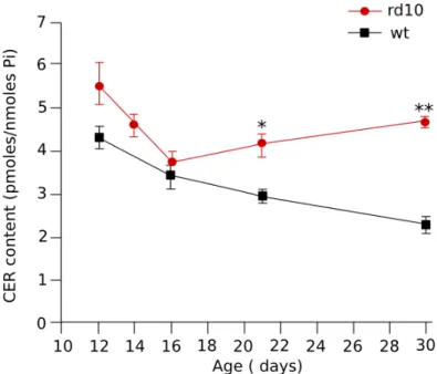
To evaluate if, in the rd10 mouse, there are altered levels of retinal ceramide, retinas were obtained from both rd10 and wt pups between P12 and P30. Total retinal ceramide, normalized to the amount of inorganic phospate (Pi) from total phospholipid, was similar in the two grups until P16; A P21, time of peak of rods degeneration, the levels of ceramide was significantly increased in rd10 retinas respect to wt retinas (3.1). Note that after P16 in rd10 retinas the levels of ceramide increase while in wt retinas the ceramide content starts to decrease.
Figure 3.1: Time course of endogenous ceramide content in retinas of litter-maters during the first month of life: in rd10 retinas a significantly increase of the ceramide levels is in concomitance with the peak of retinal degeneration (* P=0.05 t test;** P=0.001 t test)
3.2
Pharmacological effects on Retinal Ceramide
levels and rod-photoreceptors survival
3.2.1
Acute Treatment
In order to evaluate whether the high levels of ceramide in phatological condi-tions were reduced by a pharmacological treatment, we administered Myriocin, an inhibitor of the biosynthesis of ceramide. Miryocin was administered by intraocular injection on P19 mice followed by ceramide quantification on P21. Myriocin injection into the right vitreous body reduced the retinal ceramide levels with respect to the left eye which was injected with the vehicle alone. The results show that this treatment was effective in bringing ceramide to levels considered normal for P21 mice (3.2).
Figure 3.2: Effect of intraocular injection of Myriocin on retinal ceramide con-tent in rd10 mice. In 14 of 16 animals, Myriocin lowered ceramide concon-tent; Myri-ocin induced a 25.4% reduction in mean ceramide content, from 4.09 pmol/nmol Pi to 3.05 pmol/nmol Pi (SD=0.97) (P=0.011, paired t test)
The protective effect of a single injection of Myriocin on photoreceptors survival was investigated by both counting the number of apoptotic photoreceptors and recording the scotopic ERG, in retinas from treated and untreated-eyes. Iso-lated retinas were stained with a fluorescent nuclear dye to identify pycnotic nuclei (condensed DNA, cell start to die by apoptotic process) and visualized by confocal microscopy. Retinas from treated-eyes had fewer intensely stained nuclei than untreated-eyes (3.3). Qunatitative analysis shows that a single injec-tion of Myriocin was effective in reduced the number of pycnotic photoreceptors (reduced by 52.6%).
The scotopic ERG was recorded from P21 animals after two days after injec-tion the treatment with injecinjec-tion of myriocin (right eye) and vehicle alone (left eye). The scotopic ERG response to flashes of light of different intensities was
measured in dark adapted animals (3.4). Surprisingly, no significant difference could be detected in the ERGs of eyes injected with myriocin (red bar) or with the DMSO vehicle (black bar) (3.5).
Figure 3.3: Fluorescence microscopy of retina whole mount after a single injec-tion of Myriocin. Retinas, from myriocin treated eye (A) and vehicle treated eye (B), were fixed and stained with ethidium homodimer. Quantification of pycnotic nuclei (C), myriocin injection was associated with a reduction in the number of pycnotic photoreceptor nuclei on P21 (** P=0.007, t test)
In order to evaluate if a single injection of myriocin was not sufficient to induce a recovery in retinal fuctional we performed a chronic treatment with myriocin.
Figure 3.4: Example of ERG responses at different intensity of light. The red tracks represent the treated eye with myriocin and the black tacks represent the treated eye with DMSO (vehicle). A single intraocular injection of Myriocin did not have significant protective effects on retinal function, as shown by similar amplitudes and kinetics of a and b-waves in the two experimental conditions
Figure 3.5: Average of eleven treated and untreated eyes. Treatment with a single injection with myriocin didn’t induce significant change in both a-wave and b-wave
3.2.2
Chronic Treatment
The first chronic treatment was performed by instilling, once daily, eye drops of a solution of myriocin (1µl of 3.77 mM solution in DMSO) to the cornea of rd0 mice once daily for 4 days. Microscopic analysis for pycnotic nuclei and ceramide quentification revealed no significant difference between myriocin- and DMSO-treated eyes, suggesting that possibly the drug could not cross the ex-ternal tissue of the eye thus failing to reach the retinal terget.
We therefore turned to a different strategy, based on the use of solid lipid nanoparticles (SLNs), that could enable the drug to diffuse across the ocular tissue. We first investigated whether SLNs pure lipid particles with a diameter of 40-200nm labeled with a fluorescent dye, instilled into the conjuntival sac, could reach the retinal layers ([101]) in wt mice and showed by confocal mi-croscopy the presence of bright aggregates in the outer nuclear layer (ONL) and also between photoreceptors and retinal pigment epithelium ([101]).
SLNs were subsequently loaded with myriocin. The concentration of the drug in different preparations was between 0,4 and 1.0 mM.
First of all, a eye drop solution containing the Myriocin-SLNs was administered to wt mice starting on P14 upto P24. Wt mice included two distinct groups: treated (Myr-SLNs) and control (Vehicle-SLNs) groups. After the treatment sample of retinas from treated mice were collected to evaluate by morphological analysis ([101]), possible adverse effects onthe retinal structure. Other animals from same group were used to evauate the functional performance of the retina by measuring the ERG response (3.6). Subsequently we turned to the treat-ment with myriocin of rd10 mice. The protocol of the daily eye drop treattreat-ment of transgenic mice sterted on P14 to end at different ages (P21, P24, P30, P35) in various animal groups. From P21 to P35, different animals underwent ERG testing of retinal function followed by retinal microscopic analysis (3.7). In both control and myriocin-treated rd10 mice, mean aplitude of the b-waves and a-waves decreased progressively over time in concomitance with retinal de-generation. However, the b-wave amplitude of myriocin treated animals was substantially higher than that recorded from control animals at all time execept P35. Significant diffrences were observed, between the two groups, in the am-plitude of a-wave at P30 and P35 (3.8).
Also, microscopic analysis of vertical section of treated and untreaed retinas, revealed a protective effect of myriocin on the number of photoreceptor rows (3.9); further, the morphology of surviving photoreceptors was preserved as well as dentrites in rod bipolar cell ([101]).