BioS, Biomolecular Sciences Grad school
PhD course in Molecular Biotechnologies
PhD. Thesis
Genes regulated by the transcription factor
Xrx1: a microarray analysis
Advisors Candidate:
Prof.ssa Giuseppina Barsacchi Guido Giudetti
Prof. Massimiliano Andreazzoli
I do not fear computers, I fear the lack of them.
INTRODUCTION... 4
EYE DEVELOPMENT IN VERTEBRATES... 5
Morphogenetic events ... 5
The retina and the optic nerve... 6
Inductive events in the neural plate... 8
The eye field ... 10
Eye field induction and specification ... 11
Proliferation vs. differentiation ... 14
Rx/Xrx1 ... 15
Gene expression in the ciliary marginal zone (CMZ) ... 23
Xenopus laevis as model system ... 25
METHODS ... 27
SOLUTIONS... 28
PROTOCOLS AND TECHNIQUES... 30
Purification of plasmid DNA... 30
Plasmids ... 30
Capped mRNA in vitro synthesis... 31
Antisense labeled probes synthesis ... 31
Xenopus laevis embryos ... 32
Microinjections... 32
Whole-mount in situ hybridization ... 33
In situ hybridization on sections... 34
Bleaching of embryos ... 34
RNA extraction and RT-PCR analysis... 34
Microarray experiments ... 36
Microarray data analysis... 36
RESULTS... 37 AIM OF THE PROJECT... 38 Affymetrix GeneChips®... 38 Experimental design ... 41 Microinjections... 41 Gain-of-function ... 41 Loss-of-function ... 42
RNA extraction, PCR screening ... 44
Microarray hybridization and QC... 48
Microarray hybridization QC... 50
Data normalization... 54
Data annotation ... 57
Gain-of-function, loss-of-function gene lists ... 63
The intersection list... 66
Expression pattern analysis... 69
DISCUSSION ... 82
Experimental setup rationale ... 84
Annotation... 88
List analysis ... 88
The intersection list... 90
FINAL CONSIDERATIONS AND FUTURE DIRECTIONS... 93
Eye development in Vertebrates
Eye development in Vertebrates is a highly complex multi-step process that requires the interaction of different embryonal regions, such as the prosencephalic neuroectoderm, the head ectoderm and neural crest cells. This complexity requires specific inductive signals and precise morphogenetic movements to allow a well coordinated development in space and time.
The formation of a Vertebrate eye is indeed an integral part of head formation, requiring the specification and regionalization of the anterior neural plate through neural induction. The eye will then develop from a specific anterior region of the neural plate, called “eye field”. Eye development will then proceed through the evagination of the optic vesicles and, finally, the cellular differentiation of the lens and retina.
Morphogenetic events
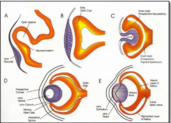
The first morphological evidence of eyes is found at the end of neurulation, when two symmetric evaginations, giving rise to the optic vesicles, extend proximo-distally from the presuntive ventral diencephalon. The optic vesicles are connected to the diencephalon through the optic stalk and send inductive signals to the ectodermal surface of the embryo, promoting the formation of the lens placode (Fig. 1, A-B).The lens actually forms from the head ectoderm, a region that already possesses a lens-forming bias by planar signalling from the presumptive retina and vertical signalling from the underlying endomesoderm (Saha et al., 1989). Thus, the optic vesicles are not necessary for lenses induction but for their correct localization in the head ectoderm. The lens placode then invaginates, detaching from the ectoderm, to give rise to the lens vesicle that interacts with the optic vesicle. In turn, this interaction induces the optic vesicle invagination, starting from the ventral side (Cvekl and Piatigorsky, 1996) (Fig. 1, C). Therefore the optic vesicle becomes a bi-layered optic cup: the proximal side starts producing melanine and will become the retinal pigmented epithelium (RPE), while the distal side will differentiate neurons and will give rise to the neural retina (Fig. 1, D-E).
Fig. 1. Eye morphogenesis. The optic vesicle evaginates from the diencephalon and promotes
the formation of the lens placode (A, B), then invaginates becoming a bi-layered optic cup (C,
D). E: anatomy of a fully developed eye.
The retina and the optic nerve
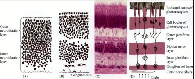
A fully mature neural retina is a stratified structure (Fig. 2, C-D) of alternate nuclear layers, made by the neuron cellular bodies, and plexiform layers, constituted by cellular processes establishing intercellular synaptic connections. Adiacent to the pigmented epithelium, the outer nuclear layer (ONL) is made by the photoreceptor cell bodies; the outer plexiform layer (OPL) separates the ONL and the inner nuclear layer (INL), composed in turn by bipolar, amacrine and horizontal cell bodies; the inner plexiform layer (IPL) precedes the last layer in the retina, the ganglion cell layer (GCL), facing the vitreous body. Within such a complex laminar design, bipolar cells are devoted to establish communication between photoreceptors and ganglion cells, whereas horizontal neurons provide horizonal information transport and integration. The highly intricate retina structure is achieved through a complex system of events, comprising cell proliferation and differentiation, migration and apoptosis.
Cell differentiation in the retina follows a conserved timing in Vertebrates: the first cells to differentiate are the ganglion cells, followed in order by cones, amacrine, horizontal, rods, bipolar, and then Müller glia cells (Young, 1985). A single neuroblast can give rise to all cell types in the retina, either differentiating into three neuron types or into two neuron types and a glial cell (Turner and Cepko, 1987): the differentiation of a specific cell type instead of another thus depends on the cellular environment in which cells become localized and not by their precursors (Cepko et al., 1996; Harris, 1997).
Fig. 2. Retinal development. (A-B) initial separation of neuroblasts. (C) Stratification of the
adult retina. (D) Functional representation of the main synaptic connections in the adult retina.
In several classes of Vertebrates, such as Amphibia and Fishes, the eye keeps growing throughout the lifetime of the animal. In these organisms, a peripheral ring of undifferentiated cells, called ciliary marginal zone (CMZ), keeps generating cell precursors that will differentiate into all the cell subtypes of the neural retina and the retinal pigmented epithelium. In mammals, a homologous structure exists, called pigmented ciliary margin (PCM): this region contains quiescent stem cells that constitute a reservoir for the regeneration of all different cell subtypes in the adult retina (Tropepe et al., 2000).
Recent studies suggest that, alongside the CMZ, another pool of retinal stem cells may exist in the adult retina. In rat, Müller glia cells display neurogenic potential, being able to generate retinal neurons (Das et al., 2006); in zebrafish, Müller glia can de-differentiate and mediate regeneration of injured retinae, thus
suggesting that these cells function as a multipotent retinal stem cells pool that generates retinal neurons by homeostatic and regenerative developmental mechanisms (Bernardos et al., 2007; Fausett and Goldman, 2006).
Inductive events in the neural plate
Although the first morphological hint of eye formation is the evagination of the optic vesicles, the events that trigger eye development start earlier, during gastrulation. Evidence in Xenopus laevis shows that the eye field is specified to some degree already at midgastrula stage (Lupo et al., 2002).
During gastrulation, the endomesoderm interacts with the overlying dorsal ectoderm to induce the neural fate in a broad region, the neural plate (Spemann, 1938). Historically, Nieuwkoop and collaborators proposed an “activation/transformation” model explaining the patterning of the anterior neural plate. During an activation phase the dorsal ectoderm is induced to become prosencephalic neuroectoderm by the underlying mesendoderm; during the following transformation phase, part of the induced tissue receives caudalizing signals by the posterior dorsal mesoderm (Nieuwkoop PD, 1952; Nieuwkoop PD, 1954).
Subsequent studies demonstrated that explanted animal caps from frogs embryos, if dissociated and reaggregated, were somehow ‘activated’ and produced neural tissue in the absence of mesoderm or endoderm (Nieuwkoop, 1963).
The apparent contradiction between the two sets of data is resolved by hypothesizing that activation is exherted by an inhibition of signals that normally inhibit achievement of the neural fate in the ectoderm. Indeed, in Xenopus, secreted molecules, such as noggin, chordin, follistatin, Xnr3, cerberus were found in the dorsal mesendoderm during gastrula and neurula stages (Harland, 2000; Weinstein and Hemmati-Brivanlou, 1999): these molecules act as activators by physically binding BMP4, a TGFβ-like molecule that needs to be repressed in order to convert uncommitted ectoderm into neuroectoderm (Sasai et al., 1994; Zimmerman et al., 1996). Furthermore, the ‘transforming’ activity was identified as residing in secreted molecules such as retinoic acid, wnt, FGF,
BMP, as they are all capable of activating the expression of posterior neural genes in the neuroectoderm (Gamse and Sive, 2001; Munoz-Sanjuan and Brivanlou, 2001; Sasai and De Robertis, 1997). Complex interactions of these secreted molecules, as well as IGF and nodal pathways, with cerberus, chordin, noggin and dickkopf-1 eventually lead to proper regionalization of the anterior neural plate (Houart et al., 2002; Lagutin et al., 2003; Lupo et al., 2002; Pera et al., 2001; Piccolo et al., 1999; Wilson and Houart, 2004).
Initial patterning of the neural plate depends indeed on complex interactions (Wilson and Houart, 2004): for example, in Xenopus laevis a global gradient of wnt proteins and antagonists regulates the antero-posterior positional patterning (Kiecker and Niehrs, 2001). The interaction and integration of different signalling pathways can explain the broad regionalization in the forming neural plate into presumptive prosencephalon, mesencephalon and hindbrain. However, it is important to keep in mind that the further subregionalization of structures such as the prosencephalon is attained by later local signalling that modulates and refines the regional patterning: in this way the expression domains of late induced genes sub-divide the neural plate in discrete territories (Wilson and Houart, 2004).
There is evidence, for example, that points at the isthmus as the local organizer for the midbrain-hindbrain region, and at cells in the anterior neural border as a source of secrete molecular signals, such as FGF-8 and sFRP wnt inhibitors, that promote the expression of telencephalic genes (Echevarria et al., 2003; Houart et al., 1998; Shimamura and Rubenstein, 1997; Tian et al., 2002). wnt activity seems to be important also during this phase: after the broad regionalization imparted by a global Wnt gradient (Kiecker and Niehrs, 2001), a more localized expression of wnt agonists and antagonists could establish and refine an activity gradient for this signalling pathway, thus perfectioning the local patterning (Houart et al., 2002).
Thus, it became clear that not only BMP inhibition si involved in the specification and development of the rostralmost regions of central nervous system: instead, the correct patterning of the anterior neural plate, and by consequence of the eye field, is the result of the interaction and integration of different signalling
pathways (Houart et al., 2002; Lupo et al., 2002; Mukhopadhyay et al., 2001; Pera et al., 2001; Stern, 2001; Wilson and Rubenstein, 2000).
The eye field
At the neurula stage of development in the anterior neural plate, overlapping with the presuntive forebrain, a broad crescent-shaped region is induced and specified, that is equipotentially capable to give rise to eye structures and for this reason is named ‘eye field’. This initially uniform domain becomes divided into two bilateral simmetric eye fields under the influence of the underlying prechordal mesoderm (Li et al., 1997; Pera and Kessel, 1997).
The idea of the generation of two eye fields from one has been controversial over time: Spemann favored the hypotesis that the two eyes are generated by two eye fields (Spemann, 1938), while Adelmann pioneered the notion that two eyes are generated by a single eye anlage (Adelmann, 1929). It is now known that during neurulation a single field is divided along the midline of the embryo into two independent domains, which eventually give rise to the eyes of the embryo, by downregulation of eye-specific markers at the midline and by suppression of the retinal fate (Adelmann, 1936; Eggert et al., 1998; Ekker et al., 1995; Li et al., 1997; Macdonald et al., 1995; Pera and Kessel, 1997).
In zebrafish, the mechanism of eye field separation appears to be different, in that the neural cells of the presuntive ventral diencephalon, initially located posterior to the antero-medial eye field, migrate anteriorly and divide the eye field in two symmetric optic primordia (Moody, 1999; Varga et al., 1999). Mutations such as cyclops, one-eyed-pinhead, schmalspur and squint in genes that are involved in nodal midline signalling, such as ndr2 and sonic hedgehog, lead to the lack of separation of the two domains and to the formation of cyclopic embryos (Chiang et al., 1996; Hatta et al., 1994; Pogoda et al., 2000; Schier et al., 1997; Sirotkin et al., 2000). This underlines the crucial role played by nodal signalling in zebrafish.
Eye field induction and specification
The inductive events responsible for the determination of the eye field are not completely understood yet. By recombining animal cap explants with Spemann’s organizer tissue, Xrx1, a specific marker for the eye field, is induced (Casarosa et al., 1997; Lupo et al., 2002); microinjection of syntethic mRNA of
noggin and chordin induces eye molecular markers expression in explanted
animal caps (Andreazzoli et al., 1999; Lupo et al., 2002). This suggests that BMP inhibition could be sufficient for the initial specification of the eye field. Moreover, it has been demonstrated that the dorsal ectoderm has already received eye field specification signals at the midgastrula stage: explanted dorsal ectoderm, when cultured to later stages, expresses opsin and can produce retinal pigmented tissue (Saha et al., 1992).
The eye field itself is comprised into the larger expression domain of the prosencephalic/mesencephalic marker Xotx2 (Fig. 4): this transcription factor is hypotesized to have a permissive role, as its expression is suppressed in the center of the presumptive eye field, possibly by the Rx protein. This allows the initial expression, as well as the mainteinance of specific transcription factors, bringing to the specification of the eye-field (Bernier et al., 2001; Kenyon et al., 2001; Zuber et al., 2003).
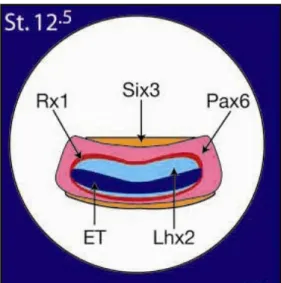
Indeed, molecular evidence indicates that the eye field is specified in the anterior neural plate by the expression of several eye field specific transcription factors (EFTFs), including ET, Xrx1, Pax6, Six3, Lhx2, tll, Optx2 (Chow and Lang, 2001; Zuber et al., 2003), all of them expressed in continuous and overlapping domains (Fig. 3): this further demonstrates that the eye field originates as a single medial domain that is then splitted into two optic primordia (Lupo et al., 2000)
Fig. 3. Expression domains of ET, Rx1, Pax6, Six3, Lhx2, at stage 12.5 (Zuber et al., 2003).
Several of these EFTFs are homologs of Drosophila genes involved in eye development. In Drosophila, eye specification requires the interaction of seven genes: twin of eyeless, eyeless (a Pax6 homolog), eyes absent, sine oculis,
dachsund, eye gone and optix (a Six3 and Optx2 homolog): their expression
pattern, which is overlapping, is regulated by Notch and EGF-R signalling, giving rise to a genic network of protein-protein interactions and feedback regulations (Kumar and Moses, 2001c). Similarly, Vertebrate EFTFs do not seem to interact by means of a linear activation cascade, but are structured into a genic network whose relationships have been recently elucidated (Zuber et al., 2003) (Fig. 4).
Fig. 4. Summary model of eye field induction in the anterior neural plate. Light blue indicates
the neural plate, blue shows the area of Xotx2 expression and dark blue represents the eye field (Zuber et al., 2003). The proposed scheme of interactions among the EFTFs is also presented.
Pax6 is a homeodomain-containing transcription factor expressed in the anterior
neural plate that plays a crucial role in Vertebrate eye formation. Mutations in
Pax6 result in eye malformations knowns as aniridia, Peter’s anomaly, and
cataracts in humans (Glaser et al., 1992; Hanson et al., 1993; Ton et al., 1991) (Fujiwara et al., 1994; Hill et al., 1991) and Small eye syndrome in mice and rats. The Drosophila homologue of Pax6, eyeless, is essential for Drosophila eye formation (Quiring et al., 1994).
Six3 is also expressed in the anterior neural plate (Oliver et al., 1995) and has a
critical role in the formation of the forebrain, as mutations in human SIX3 cause holoprosencephaly (Pasquier et al., 2000; Wallis et al., 1999). Mouse embryos lacking Six3 function lack most of the head structures anterior to the midbrain (Lagutin et al., 2003). Six3 has been shown to play a critical role in anterior neural plate specification and mainteinance, being able to repress wnt, BMP and nodal transcription (Gestri et al., 2005; Inbal et al., 2007; Lagutin et al., 2003). This factor is also crucial to control cell proliferation in the eye field and forebrain, acting through both dependent and transcriptional-independent pathways (Del Bene et al., 2004; Gestri et al., 2005).
Optx2 is expressed in the early precursors of the eye (Jean et al., 1999; Toy
and Sundin, 1999) and its overexpression in Xenopus embryos results in overproliferation of the retinal cells (Toy and Sundin, 1999; Zuber et al., 1999). Targeted elimination of this gene in mice confirmed that it has a role in the proliferation of retinal progenitor cells (Li et al., 2002).
Functional studies in different model systems demonstrated that ET, Xrx1, Pax6,
Six3, Lhx2, tll, and Optx2 are necessary and, in some context, also sufficient for
a correct eye development. Indeed, overexpression of Xrx1, Pax6, Six3, and
Optx2 expands or induces ectopic retinal tissue (Andreazzoli et al., 1999;
Bernier et al., 2000; Chow et al., 1999; Chuang and Raymond, 2001; Loosli et al., 1999; Mathers et al., 1997; Oliver et al., 1996; Zuber et al., 1999). Moreover, the overexpression of each of the same genes activates the expression of the others, while their inactivation reduces the expression of the others, without preventing their initial activation (Andreazzoli et al., 1999; Bernier et al., 2000; Carl et al., 2002; Chow et al., 1999; Chuang and Raymond, 2001; Goudreau et
al., 2002; Lagutin et al., 2001; Lagutin et al., 2003; Loosli et al., 1999; Zhang et al., 2000; Zuber et al., 1999).
Thus, experimental evidence supports a model for a progressive tissutal specification in which the neural induction and regional patterning exherted by
Xotx2 prepare the anterior neural plate to the formation of the eye-field:
subsequently, the EFTFs create a net of mutual feedback interactions that eventually specifies the eye-field for its further development into eyes (Zuber et al., 2003).
Proliferation vs. differentiation
The anterior neural plate is characterized by a prolonged proliferation and retarded differentiation with respect to the posterior neural plate (Papalopulu and Kintner, 1996): this permits the formation of a large encephalic region and a thinner posterior spinal chord. The molecular bases of this differential proliferation timing are largely unknown.
In anamniote Vertebrates such as Zebrafish and Xenopus laevis the neuronal differentiation starts when the neural plate is still planar and opened: as these organisms develop through a swimming tadpole stage, there is the need to rapidly produce a simple but fully functioning nervous system. For this reason, a small group of neuroectodermal cell exits cell cycle and starts differentiating into primary neurons at the end of gastrulation (Hartenstein, 1989; Wilson and Easter, 1992). A second neurogenetic wave will generate the secondary neurons that will substitute the primary ones (Forehand and Farel, 1982).
In Xenopus, the first neurogenetic wave is easily recognizable as three parallel stripes of cells expressing the n-tubulin neural differentiation marker: these stripes have bilateral symmetry and contain, separately and in medio-lateral direction, motor neurons, interneurons and sensory primary neurons (Chitnis et al., 1995).
This process is controlled by the expression of genes of early regional specification such as the Xiro family of genes (de la Calle-Mustienes et al., 2002) that define the zones in which primary neurons are allowed to differentiate or
not. Moreover, the Xiro genes regulate the finer expression of proneural genes such as Xngnr-1 (Ma et al., 1996): the proneural genes allow only one cell to become a neuronal precursor in a single proneural group, by means of lateral inhibition mediated by the neurogenic gene Notch and its ligand, Delta (Chitnis et al., 1995). The proneural genes in fact promote Delta expression in a selected cell, so that the Delta ligand can interact with the nearer cells expressing Notch. The Notch receptor is then cleaved and its freed intracellular domain enters the nucleus and activates the transcription of genes such as the HES family of genes, transcriptional repressors that suppress the neuronal fate in the single cell (Bray, 1998; Chitnis et al., 1995; Ma et al., 1996; Wettstein et al., 1997).
By the end of gastrulation, in Xenopus laevis the proliferating borders of the anterior neural plate are clearly bordered, on the neural plate side, by the expression of neurogenic genes such as Xdelta-1 and Xngnr-1 and of genes involved in cell cycle arrest such as p27Xic-1 and Xgadd45-γ (de la Calle-Mustienes et al., 2002; Hardcastle and Papalopulu, 2000). p27Xic-1 is a cyclin that is highly expressed in cells fated to become neurons, it is necessary to neurogenesis and acts upstream of NeuroD (a specific transcription factor for structural proteins in neural cells: (Lee et al., 1995) and in parallel with Xngnr-1. In the anterior neural plate, transcription factors such as Xsix3, Xoptx2, Xanf-1 and Xbf-1 are involved in delaying differentiation: Xsix3 and Xoptx2 promote proliferative activity leading to retina enlargement (Bernier et al., 2000; Zuber et al., 1999); overexpression of Xanf-1 brings to an enlargement of the neural plate and represses neural differentiation (Ermakova et al., 1999). Xbf-1, a presumptive telencephalic marker, has a role in limiting the neurogenesis at the anterior neural plate border in a concentration-dependent manner (Bourguignon et al., 1998).
Rx/Xrx1
The Rx (retinal homeobox) genes are a small family of homeobox genes that are critical for eye formation. The structure of Rx genes is very conserved and since their discovery they have been described in several vertebrate and
invertebrate species; their number varies among different species, and generally ranges from one to three. Homologues of Rx have been identified in man (RAX), in mouse (mRx) and rat, in chicken (cRax and cRaxL), in teleost fishes (three homologues in zebrafish, Zrx1, Zrx2, Zrx3 and two in medaka, Rx2 and Rx3), in Drosophila (drx), and in Xenopus laevis (Xrx1, Xrx2 and Rx-L) (Casarosa et al., 1997; Eggert et al., 1998; Furukawa et al., 1997; Loosli et al., 2001; Mathers et al., 1997; Ohuchi et al., 1999; Pan et al., 2006; Tucker et al., 2001).
The homeodomains of Rx proteins are extremely well conserved and are, for example, identical between Xenopus, Drosophila and two of the three zebrafish proteins. They belong to the paired-like class of transcription factors: the aminoacidic residue in position 50 of the homeodomain is a glutamine instead of a serine, as in paired class homeobox genes; they possess a HSIEAILG octapeptide and a transactivating OAR domain, such as in other paired-like transcription factors (Furukawa et al., 1997; Simeone et al., 1994) (Fig. 5).
Hd OAR
OCT
Fig. 5. Functional domains of a Rx gene. OCT: octapeptide. Hd: homeodomain. OAR:
trans-activating domain.
In Drosophila, drx is neither expressed in the eye primordia or the eye imaginal discs but it is expressed in the part of the brain called the ellipsoid body and in the clypeolabrum (Eggert et al., 1998; Mathers et al., 1997). drx is necessary to brain development, rather than eye development: drx null mutants possess a normal visual system, while the ellipsoid body is altered (Davis et al., 2003). In chicken, cRax lacks the octapeptide and its sequence is more similar to Xrx1 sequence, whereas cRaxL sequence is highly similar to zebrafish Rx1 and Rx2.
cRax is detectable in the ectoderm anterior to Hensen’s node at stage 4. During
neurulation, cRax and cRaxL expression domains overlap in the anterior neural ectoderm region corresponding to the presumptive prosencephalon and retina (Ohuchi et al., 1999). cRax is expressed, similarly to mice, in the anterior neural folds, in the prospective retina, and in the ventral forebrain (Ohuchi et al., 1999).
cRaxL is expressed in the anterior neural ectoderm somewhat later than cRax.
During the cellular differentiation of the retina, cRaxL is expressed in the initial stages of photoreceptor differentiation, while cRax is not expressed in photoreceptor cells (Chen and Cepko, 2002).
A review of Rx expression patterns in different species reveals that the most conserved aspect of vertebrate Rx expression is its early transcription in the anterior neural plate, followed by the expression in the eyes, ventral forebrain and the pineal gland. This pattern of expression is conserved in the two
Xenopus Rx genes, in medaka Rx3 and in the mouse Rx (Casarosa et al.,
1997; Loosli et al., 2001; Mathers et al., 1997).
In zebrafish, the three Rx homologues, Zrx1, Zrx2 and Zrx3 are all expressed in the anterior neural plate during the neurulation an all are transcribed in the optic vesicles, but have a different modulation during later development. Zrx1 and
Zrx2 share a similar expression pattern in the retina while Zrx3 is transcribed in
the ventral prosencephalon (Chuang et al., 1999; Chuang and Raymond, 2001). This suggests that at a certain evolution stage there was duplication and divergence of the gene functions; their combined expression pattern spans the same expression domain of Rx homologues in Xenopus and mouse (Mathers et al., 1997). During the cellular differentiation of the retina, Zrx1 and Zrx2 are expressed in the adult cone cells, but not in the rod cells (Chuang et al., 1999).
Zrx3 is expressed in the inner nuclear layer of the adult retina.
In medaka, Rx3 is first expressed at late gastrulation and by early neurula stages this gene is strongly expressed in a single field of the developing forebrain. By late neurula stages there is strong retinal expression in addition to the forebrain, but this expression site is progressively lost as the embryo proceeds through somitogenesis, leaving intense expression only in the ventral diencephalon. Adult fishes show Rx3 expression in the inner nuclear layer of the retina as well as the hypothalamus (Deschet et al., 1999). Medaka Rx2 expression begins several hours later than Rx3 in the developing optic vesicle
and then is maintained in the neuroretina, but not in the hypothalamus (Loosli et al., 1998).
A conditional el (eyeless) mutant shows no evagination of optic vesicles. The
eyeless mutant is a consequence of an intronic insertion in the Rx3 locus: the
mutant phenotype demonstrates that this gene is required for the correct migration of retinal progenitor cells and for the determination, evagination and proliferation of the optic vesicles (Loosli et al., 2001; Rembold et al., 2006). Rx3 sequence is more similar to Xrx2 sequence than Xrx1 (Zuber et al., 2003). The
Rx3 mutation neither interfere with the expression of Rx2 nor with the eye field
splitting into the two optic primordia: this suggest that morphogenesis and patterning could be actually separated (Winkler et al., 2000). Rx2 is exclusively expressed during and after gastrulation, in the presumptive and then differentiated retinal tissue (Loosli et al., 2001; Mathers et al., 1997): this suggests that Rx2 itself or a still unidentified Rx homologue acts as a functional homologue of Xenopus Xrx1 (Zuber et al., 2003).
As in Xenopus, the murine Mrx is first activated in the anterior neural plate at stage E7.5, in a region that will give rise to the eyes, the pineal gland, and the diencephalon. At stage E10.5, expression of Mrx is confined to the developing retina and the ventral forebrain. There is a uniform expression in the entire neuroretina of E15.5 embryos. At later stages there is a progressive reduction of Mrx expression in the retina, which initiates in the ganglion cells and proceeds in accordance with the loss of proliferative activity in the retinal cell layers.
Expression in the eye at stage P6.5 is restricted to photoreceptors and the inner nuclear layer; at stage P13.5 no Rx expression is detected (Mathers et al.,
1997). Rx1-/- mutants are anophtalmic and do not develop any early eye
structure such as the optic vesicles or the optic cups (Mathers et al., 1997) (Fig.6), while small eye/small eye homozygous mutants, carrying a mutation in
Pax6, develop anomalous optic vesicles. Rx1-/- mutants show a gradual
phenotype: in the mild phenotype the prosencephalon is present, but optic vesicles are not and the putative eye field region lacks also the expression of
lack completely the prosencephalon and the mesencephalon seems missing to a variable extent (Mathers et al., 1997).
Fig. 6. Mouse Rx1-/- knock-out mutant (Mathers et al., 1997).
The two Xenopus Rx homologues, Xrx1 and Xrx2, share a very similar, if not identical, expression pattern. Xrx1 starts being expressed at low level at developmental stage 11, then strengthens at stage 12.5-13 and keeps being transcribed at a stable level until stage 45, after which it is downregulated (Casarosa et al., 1997). At the end of gastrulation its transcripts can be detected by in situ hybridization demarcating a uniform field of cells in the anterior neural plate. Xrx1 expression is sharply delineated anteriorly from the cells of the cement gland anlage, which in Xenopus is the anteriormost structure (Fig. 7, left). The posterior border of Xrx1 expression is in the proximity of the forebrain/ midbrain boundary. Therefore, it appears that by the end of gastrulation the
Xrx1 early expression domain is primarily localized to the putative forebrain, in a
region which will give rise to the optic vesicles, the neural retina, the diencephalon floor, the optic chiasm and the epiphysis (Eagleson et al., 1995).
Fig. 7. Xrx1 expression in Xenopus laevis. Left: eye-field expression at stage 13. Center:
expression at stage 28: green arrow indicates the prospective pineal gland. Right: Xrx1 expression in the CMZ; red arrow indicates the CMZ.
During neurulation the retina remains the primary site of Xrx1 expression. It is notable that it is expressed only in regions of neural derivation (neural retina and retinal pigmented epithelium) and not in ectodermal deriving tissues such as the lens and the cornea. The pineal gland (epiphysis), and the ventral hypothalamus also express this gene (Fig. 7, center).
Sections of neurula stage embryos show that initially the entire retinal neuroepithelium expresses Xrx1 with a slight accumulation in the ventro-nasal region, but by the time the optic cup is formed, the Xrx1 RNA expression domain is restricted to the cells of the retinal ciliary margin (Fig. 7, right). This is a very important finding as it had been shown that the retinal ciliary margin contains the multipotent retinal stem cells that continually generate the entire repertoire of retinal cell types throughout Xenopus life (Holt et al., 1988; Stiemke and Hollyfield, 1995; Wetts and Fraser, 1988; Wetts et al., 1989). Later in development, Xrx1 is reactivated in the photoreceptor cells (Perron et al., 1998) and keeps being expressed in the CMZ during metamorphosis (Casarosa et al., 2005).
Functional analysis shows that Xrx1 has a relevant role during the specification as well as in cell proliferation control and neurogesis in the anterior neural plate (Andreazzoli et al., 1999; Andreazzoli et al., 2003).
Recently an Rx-like (Rx-L) gene has been identified in Xenopus laevis (Pan et al., 2006). This gene shares homology with Xrx1 at the homeo-, OAR, and Rx domains, but lacks an octapeptide motif. Rx-L is expressed in the developing retina beginning in the early tailbud stage, much after the onset of expression of
Xrx1. In the maturing retina, Rx-L expression is restricted primarily to the
developing photoreceptor layer and the ciliary marginal zone. In a promoter activity assay, Rx-L functions as a stronger transcriptional activator than Xrx1. Antisense morpholino-mediated knockdown of Rx-L expression resulted in a decrease in rhodopsin and red cone opsin expression levels in Xenopus retinas. Injection of the Rx-L antisense morpholino oligonucleotide also resulted in a decrease in the length of both rod and cone outer segments.
These results suggest that Rx-L functions to regulate rod and cone development by activating photoreceptor-specific gene expression, thus having
a substantially different function in retina development with respect to other genes belonging to the Rx family of genes; as Rx-L is a stronger transcriptional activator than Xrx1, its function may be to boost, rather than initiate, promoter activity (Pan et al., 2006).
The effects of overexpression of Xrx1 were examined by injection of Xrx1 synthetic mRNA into dorsal animal blastomeres of 8-cell Xenopus embryos: results are the overproliferation of the neuroretina and ectopic retinal pigment epithelium that invades the optic stalk (Fig. 8, left). In some embryos ectopic retinal tissue as well as anterior neural tube duplication, was observed (Fig. 8, center and right) (Andreazzoli et al., 1999; Mathers et al., 1997).
Fig. 8. Xrx1 overexpression phenotypes. Left: arrow indicates ectopic pigmented retinal
epithelium. Center:. TN1: neural tube. TN2: ectopic secondary neural tube. Right: section of fully developed eye. R1, EP1, R2, EP2: retina (R1,2) and retinal pigmented epithelium (EP1,2) of a double eye structure. Phenotypes are observed on the embryo injected side.
Similar results were obtained in zebrafish by Chuang and Raymond (Chuang and Raymond, 2001).
Xrx1 overexpression ectopically activates Six3 and Pax6 at later stages of
neurulation and downregulates Xotx2 at early neurula stage: this suggests that
Xrx1 could act as a mediator for the early Xotx2 repression in the eye territory
during eye field specification (Andreazzoli et al., 1999).
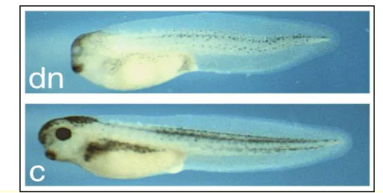
The function of Xrx1 can be inhibited by injecting at 4-cells stage either a dominant negative Xrx1 construct (Xrx1-EnR) or Xrx1-specific morpholinos. Both loss-of-function strategies lead to a variable reduction or loss of eyes and anterior head structures at the level of the telencephalon and ventral diencephalon (Fig. 9). These findings are consistent with the phenotype
observed in Rx-/- mice (Andreazzoli et al., 1999; Andreazzoli et al., 2003; Mathers et al., 1997). At earlier stages of development, Xrx1 functional knock-down leads to the knock-down-regulation of Pax6, Xotx2, XBF-1 and Six3: this suggests that the lack of entire anatomic portions of the head could depend on the impairment of early specification events. In fact, the lack of Xrx1 function does not allow the formation of structures deriving from the neuroectodermal region in which it is normally expressed.
Fig. 9. Xrx1 functional knock-down. C: uninjected embryo. Dn: Phenotype shared by
morpholino- or Xrx1-EnR-injected embryos.
Xrx1 exherts its function by antagonizing the differentiative signals and by
promoting proliferation in a regional specific manner: in fact its expression domain, coinciding with the proliferative region of the anterior neural plate, is defined by the interaction of positive and negative signals. The Xrx1 expression domain is surrounded by cells expressing the Xngnr-1 proneural gene, the neurogenic gene Xdelta-1 and the cell cycle inhibitor p27Xic-1: at this developmental stage these cells, after exiting the cell cycle, are differentiating into neurons. Xrx1 is activated by chordin, noggin, hedgehog and wnt pathways (Andreazzoli et al., 2003; Zuber et al., 1999) and repressed by the activity of
Xngnr-1 and the retinoic acid. If overexpressed, Xrx1 counteracts the
differentiative activity of Xngnr-1, retinoic acid and Xdelta-1. Finally, Xrx1 acts by inducing antineurogenic transcriptional repressors such as Xhairy2 and Zic2, rather than by lateral inhibition (Andreazzoli et al., 2003). In addition, Xrx1 activates transcription of XBF-1. XBF-1, like X-ngnr-1, inhibits p27Xic1 expression and therefore facilitates cell proliferation (Hardcastle and Papalopulu, 2000).
As a result of all these interactions, the Xrx1 expressing cells proliferate, but they do not differentiate.
Analysis of Xrx1 effects on proliferation and on the expression of stem cell or differentiation markers demonstrates that Xrx1 maintains cells in a stem cell state by promoting proliferation and delaying expression of neural identity and differentiation markers (Casarosa et al., 2003; Zaghloul and Moody, 2007a). In summary, Xrx1 is necessary to eye and anterior brain development. There is increasing evidence, mainly from Xenopus studies, that Xrx1 acts as a cell type specific factor that is involved in the proliferation of cells from which the retina and the ventral hypothalamus are derived and could possess a role in regulating the anterior regional specification and neurogenesis. Evidence from medaka and zebrafish suggests that Rx genes might be involved in the
morphogenesis of the optic vesicle. Finally, observations from Rx-/- mice
suggest that, in addition to cell proliferation, Rx genes might have a role in the specification of the retinal progenitors. This is further supported by the recent finding that embryonic stem cells can be specified to form retinal cells by ectopic expression of Rx (Tabata et al., 2004).
Gene expression in the ciliary marginal zone (CMZ)
In Xenopus, four main sub-regions can be identified in the ciliary marginal zone, each well characterized by the differential expression of a subset of genes involved in retinal cell differentiation not only at later stages, but also at early stages of eye development. The temporal order and the relative localization in which these genes are expressed suggest that the molecular events at the level of the CMZ recapitulate in space the events that happen during retinal development in time (Perron et al., 1998).
The four zones in which the CMZ has been subdivided were accordingly named specification zone, proneural and neurogenic zone, cellular determination zone and differentiation zone (Fig. 10).
Fig.10. Model of molecular development in the CMZ. The array of cells from left to right in
this model of the CMZ can also be considered as a temporal sequence of gene expression. The specification zone is the peripheralmost region of the CMZ where the pigmented epithelium folds over the neural retina, characterized by the presence of retinal stem cells that can originate all the cells in the retina. These cells express early eye field specification genes such as Xpax6, Xrx1 and Xsix3. The proneural and neurogenic zone contains proliferating neuroblasts that express the major cell cycle activators, such as cyclin D1, cyclin A2 and cdk2. In this zone the eye field transcription factors are still expressed, as well as the proneural genes homologous to the Drosophila achaete-scute complex (Xash1 and Xash3) and neurogenic genes such as Xnotch-1 and Xdelta-1(Perron et al.,
1998).
In the determination zone, the various retinal cell types are sorted by expression of genes that are homologous to the atonal complex in Drosophila, such as
The differentiation zone is the centralmost one. Cells in this zone are already post-mitotic and do not express Notch, Delta and Xash anymore, but start expressing molecular markers of specific cell types, such as Brn-3 in the ganglion cell layer (Hirsch and Harris, 1997).
The spatial ordering of gene expression, from peripheral to central, reflects a developmental sequence, suggesting a developmental cascade and recapitulates the order of gene expression in the rapidly developing embryonic retinal primordium. The succession of gene expression, so clearly delineated in space (Fig. 10), allows us to see the steps of molecular development arranged in a single linear dimension, and thus provides clear models of which genes are upstream of others. The retinal CMZ of lower vertebrates thus can be considered a powerful system to study the genetic pathway of neurogenesis in vertebrates (Perron et al., 1998).
.
Xenopus laevis as model system
Over the years, the anuran amphibian Xenopus laevis has become a powerful vertebrate model system for experimental embryology and developmental biology. The advantages of using this freshwater African clawed frog as an experimental model stem from the possibility to easily obtain embryos at different stages. The females, in fact can be induced to ovulate at any time of the year by means of a simple hormonal boost. Usually, 1000 to 1500 eggs are produced each time, which are easily fertilized in vitro using testis homogenates. The embryos can easily grow in a Petri dish in simple saline solutions; moreover, they are large and can be microinjected and micromanipulated with no difficulty. In addition, development is rapid: it takes about three days starting from fertilization for an embryo to reach the tadpole stage (stage 42), at which organogenesis is completed.
During the past several years, many new techniques have been devised or adapted for Xenopus, such as in situ hybridization or immunocytochemistry, which allow to visualize gene or protein expression domains in the whole embryo or on sections. Analysis of gene function can be performed by means of two complementary approaches, by gain-of-function and loss-of-function
experiments. Inducible gene expression systems or stage-specific transfection of constructs, by means of lipofection technique, allow to control timing of gene expression in gain-of-function assays, whereas dominant negative proteins (for example dominant negative ligands or transcription factors), or the recently developed antisense morpholino oligo technology, proved to be useful tools to inactivate gene function.
Moreover, the large size of the embryos, the ability of the explanted tissues to survive without requirement for added nutrients and the availability of detailed fate maps make Xenopus a very interesting model system for studies of lineage commitment or induction.