Cerebrospinal Fluid from Patients with
Subarachnoid Haemorrhage and Vasospasm
Enhances Endothelin Contraction in Rat
Cerebral Arteries
Barbara Assenzio1, Erica L. Martin1, Edgaras Stankevicius2, Federica Civiletti1, Marco Fontanella3, Riccardo Boccaletti3, Maurizio Berardino1, AnnaTeresa Mazzeo1,
Alessandro Ducati3, Ulf Simonsen4, Luciana Mascia1*
1 Department of Anesthesia and Intensive Care, Azienda Ospedaliera Città della salute e della scienza di Torino, University of Turin, Turin, Italy, 2 Institute of Physiology and Pharmacology, Medical Academy, Lithuanian University of Health Sciences, Kaunas, Lithuania, 3 Division of Neurosurgery, Department of Neuroscience, Azienda Ospedaliera Città della salute e della scienza di Torino, University of Turin, Turin, Italy, 4 Department of Biomedicine, Pulmonary and Cardiovascular Pharmacology, University of Aarhus, Aarhus, Denmark
Abstract
Introduction
Previous studies have suggested that cerebrospinal fluid from patients with subarachnoid hemorrhage (SAH) leads to pronounced vasoconstriction in isolated arteries. We hypothe-sized that only cerebrospinal fluid from SAH patients with vasospasm would produce an en-hanced contractile response to endothelin-1 in rat cerebral arteries, involving both
endothelin ETAand ETBreceptors.
Methods
Intact rat basilar arteries were incubated for 24 hours with cerebrospinal fluid from 1) SAH patients with vasospasm, 2) SAH patients without vasospasm, and 3) control patients. Arte-rial segments with and without endothelium were mounted in myographs and concentra-tion-response curves for endothelin-1 were constructed in the absence and presence of selective and combined ETAand ETBreceptor antagonists. Endothelin concentrations in
culture medium and receptor expression were measured.
Results
Compared to the other groups, the following was observed in arteries exposed to cerebro-spinal fluid from patients with vasospasm: 1) larger contractions at lower endothelin concen-trations (p<0.05); 2) the increased endothelin contraction was absent in arteries without endothelium; 3) higher levels of endothelin secretion in the culture medium (p<0.05); 4) there was expression of ETAreceptors and new expression of ETBreceptors was apparent;
OPEN ACCESS
Citation: Assenzio B, Martin EL, Stankevicius E, Civiletti F, Fontanella M, Boccaletti R, et al. (2015) Cerebrospinal Fluid from Patients with Subarachnoid Haemorrhage and Vasospasm Enhances Endothelin Contraction in Rat Cerebral Arteries. PLoS ONE 10 (1): e0116456. doi:10.1371/journal.pone.0116456 Academic Editor: Christopher Torrens, University of Southampton, UNITED KINGDOM
Received: May 21, 2014 Accepted: December 10, 2014 Published: January 28, 2015
Copyright: © 2015 Assenzio et al. This is an open access article distributed under the terms of the
Creative Commons Attribution License, which permits unrestricted use, distribution, and reproduction in any medium, provided the original author and source are credited.
Data Availability Statement: All relevant data are within the paper.
Funding: This study was supported by grant CIPE LM2004 from the Comitato Interministeriale per al Programmazione Economica and grant LM2007 from the Ministero della Salute, Programma Ricerca Finalizzata. The funders had no role in study design, data collection and analysis, decision to publish, or preparation of the manuscript.
Competing Interests: The authors have declared that no competing interests exist.
5) reduction in the enhanced response to endothelin after ETBblockade in the low range
and after ETAblockade in the high range of endothelin concentrations; 6) after combined
ETAand ETBblockade a complete inhibition of endothelin contraction was observed.
Conclusions
Our experimental findings showed that in intact rat basilar arteries exposed to cerebrospinal fluid from patients with vasospasm endothelin contraction was enhanced in an endotheli-um-dependent manner and was blocked by combined ETAand ETBreceptor antagonism.
Therefore we suggest that combined blockade of both receptors may play a role in counter-acting vasospasm in patients with SAH.
Introduction
Cerebral vasospasm is one of the most serious complications following aneurysmal subarach-noid hemorrhage (SAH) and is independently associated with poor outcome [1]. Endothelin-1 (ET-1) is thought to be of major importance for cerebral vasospasm [2,3]
In experimental models of SAH, increased sensitivity of cerebral arteries to ET-1 have been invariably reported [4]; increased levels of ETAreceptor mRNA have been inconsistently
demonstrated [5] [6,7]while increased levels of ETBmRNA have been unequivocally reported
[8–10] [11]. So far studies on endothelin-1 receptor regulation did not differentiate between the two conditions of SAH with and without vasospasm [6,9,10,12]. Moreover, andin vitro studies on the cerebral microvasculature have often been used to explain events observed in conductive vessels[13]. Furthermore, although the cerebral endothelium is unique in terms of growth and responsiveness to various vasoactive agonists, results obtained in non-cerebral en-dothelial cells have been extrapolated to the brain vasculature[14,15].
The efficacy of selective receptor antagonists have been tested in experimental models of ce-rebral vasospasm [5,16,17]. Several randomized clinical trials tested the efficacy of ETA
selec-tive blockade and found a reduction in angiographic vasospasm but no improvement in measureable functional outcomes [18–22]. These results pose several questions: 1) the possibil-ity that early brain injury occurring just after the hemorrhage may contribute to the poor out-come; 2) a functional interaction between ETAand ETBreceptors named“cross talk” may play
a role in the pathogenesis of SAH-induced vasospasm.
To overcome the above-mentioned methodological criticisms and to elucidate the function-al interaction between the two receptors, we evfunction-aluated the upregulation of the endothelin sys-tem of the targeted vessels by vasospasm, incubating intact and denuded conductive cerebral vessels with CSF from SAH patients with a conclusive diagnosis of vasospasm obtained by angiography.
We hypothesized that CSF from SAH patients who developed vasospasm would produce an en-hanced contractile response in intact rat cerebral arteries involving both ETAand ETBreceptors.
Materials and Methods
Patient Selection
Ethical approval
The present study was conducted using CSF from patients with aneurysmal SAH confirmed by CT scan and angiography admitted to ICU. CSF had been collected for an observational study
in SAH patients at risk of developing vasospasm (NCT01686763). The institutional review board (Comitato Etico Interaziendale A.O.U. San Giovanni Battista di Torino A.O. C.T.O./ Maria Adelaide di Torino) approved the study. If the patient was unable to give consent at study entry, consent was delayed, and the family was informed of the study. Written permis-sion for using collected data was thus obtained from the patient (if competent) or from the family (in case of death or if the patient remained incompetent).
All the patients fulfilled the following inclusion criteria: 1) angiographic proof of aneurysm; 2) admission within 24 hours of the SAH; and 3) presence of an intraventricular catheter placed either after admission or at the time of surgery. Patients were graded clinically according to the World Federation of Neurological Surgeons (WFNS) scale and classified according to the CT distribution of blood as described by the Fisher scale.
All patients underwent neurosurgical intervention or endovascular procedure to secure the aneurysm within 2 days of admission. Neurological status was evaluated daily using the Glas-gow Coma Scale (GCS), transcranial Doppler of the middle cerebral arteries was performed daily until day 14 and patients were treated according to the guidelines [1].
On day 7 post-SAH, a second angiogram was routinely performed and the patients were then classified as having: 1) angiographic vasospasm (AV) if the angiogram showed 25% or more reduction from baseline caliber without any clinical deterioration; 2) clinical vasospasm (CV) if a reduction in the angiographic caliber >50% of the middle cerebral artery (MCA) was accompanied by controlateral weakness to or global neurological deterioration (two points re-duction in GCS) occurring later than day 3 for anterior or diffuse vasospasm; 3) patients with-out vasospasm (NV) if there was reduction lower than 25% in the angiographic caliber [23] [24].
To test the hypothesis of the present study samples of CSF from patients with the most se-vere vasospasm (clinical vasospasm CV) and from those without vasospasm (no vasospasm NV) were included. As negative control a group of patients with normal pressure hydrocepha-lus (NPI) and normal GCS undergoing lumbar drainage for the diagnosis of NPI was included [25]. In SAH patients CSF was collected daily until day 7, with the samples collected on days 4–5 used for the present study. CSF was drawn into tubes containing EDTA from the intraven-tricular catheter and collected by venintraven-tricular drainage from hydrocephalus patients. Tubes were kept and transported in ice. All CSF samples were centrifuged at 1700g for 15 min at 4°C to remove intact cells and cellular debris, following which the supernatant was frozen at−80°C.
Functional study of the
Ex Vivo Model of SAH-induced Vasospasm
Isolated rat basilar arteries were obtained from male Wistar rats (350–400g) (Charles River Laboratories, Calco, Italy). Rats were group-housed, had free access to standard chow and water in a controlled facility providing a 12-hour light/dark cycle, and were kept according to institutional animal welfare guidelines and legislation, submitted and approved by the local Animal Ethics Committee (Comitato di Bioetica dell’Ateneo dell’Università degli Studi di To-rino) and all efforts were made to minimize suffering. Rats were sacrificed by carbon dioxide overdose followed by cervical dislocation and the cerebral basilar artery was isolated using a stereomicroscope and immersed in ice-cold physiological salt solution (PSS) (CaCl22H2O 2.5mmol/L, NaCl 119 mmol/L, KCl 4.69 mmol/L, glucose 5.5 mmol/L, MgSO47H2O 1.17nmol/L,
NaHCO325 mmol/L, KH2PO41.18 mmol/L and ethylenediaminetetraacetic (EDTA) 0.03
mmol/L). Segments of approximately 2 mm in length were incubated with 5% human CSF (v/ v) obtained from ventricular drainage of the patients included in the study. Incubation lasted 24 hours at 37°C with 5% CO2in O2in Dulbecco’s modified Eagle’s medium (DMEM). The
Following incubation with human CSF, isolated rat basilar arteries were mounted on 40μm tungsten wires in a small vessel myograph (D.M.T., Aarhus, Denmark) for isometric force re-cording [26]. Measurements were recorded using the ICU-Lab software program (KleisTEK Advanced Electronic Systems; Bari, Italy). The vessels were allowed to equilibrate for about 30 min in PSS, which was continuously aerated with oxygen enriched with 5% CO2to maintain a
pH of 7.4. The relation between resting wall tension and internal circumference was deter-mined, and the internal circumference, L100, corresponding to a transmural pressure of 100
mmHg for a relaxed vessel in situ was calculated. The vessels were set to the internal circumfer-ence L1, given by L1= 0.9L100. The effective internal lumen diameter was determined asl1= L1/
π, and was 306±41 mm (mean±SD, N = 106) and 303±38 μm (mean±SD, N = 20) in vessels with and without endothelium respectively.
After a 30 min period to stabilize vessel tone, the contractile capacity was determined by ex-posing the vessels to an isotonic solution containing 123 mmol/l of K+(KCl 123.70 mmol/L, MgSO47H2O 1.17 nmol/L, KH2PO41.18 mmol/L, CaCl22H2O 2.5 mmol/L, NaHCO325
mmol/L, EDTA 0.03 mmol/L, glucose 5.5 mmol/L), obtained by equimolar change of NaCl for KCl in PSS. Only vessels responding by contraction corresponding to a pressure above 13.3 kPa to potassium were included in the study. The presence of an intact endothelium was checked by contracting the vessel using serotonin (5-HT, 10−5mol/L, Sigma-Aldrich, Milan, Italy) and subsequently exposing the segments to histamine (10−5mol/L, Sigma-Aldrich, Milan, Italy) to stimulate endothelium-dependent relaxation. Confirmation of the presence of endothelium was determined by a relaxation>50% (E+). In a separate set of experiments the endothelial cell layer was mechanically removed by gentle rubbing the intimal surface with a human hair (E−). After endothelial cells removal the contractility of the vessels was tested by adding serotonin.
We did not find any difference in serotonin response between groups with or without endo-thelial cells. The response to 5-HT was 1.59±0.56 (n = 6) and 1.43±0.48 mN/mm (n = 8), re-spectively, in vessels with and without endothelium exposed to CSF from the Control group, and 1.59±0.56 (n = 6) and 1.34±0.44 mN/mm (n = 6) in vessels with and without endothelium exposed to CSF from the Non-Vasospasm group, and 1.79±0.50 (n = 6) and 1.74±0.43 mN/ mm (n = 6) in vessels with and without endothelium exposed to CSF from the
Vasospasm group.
In a separate set of vessels, following exposure of the isolated basilar arteries to 5% human CSF, vessels were incubated with specific antagonists to 1) ETA(BQ-123, 1×10⁻⁵mol/L,
Sigma-Aldrich, Milan, Italy), 2) ETB(BQ-788, 1×10⁻⁶mol/L, Sigma-Aldrich S.r.l, Milan, Italy)
3) both ETAand ETB(BQ-123 and BQ-788) 4) vehicle control for 30 min prior to performing
concentration-response curves by cumulative application of ET-1.
Enzyme-linked Immunosorbent Assay (ELISA)
ET-1 concentrations in CSF and in culture medium obtained before and after the incubation of the arteries were determined by ELISA (Human ET-1 (1–21), with sensitivity <0.05 pg/ml; Biomedica Diagnostics Inc, Vienna, Austria).
Immunofluorescence
After the 24-hour incubation in CSF, basilar arteries were embedded in Tissue-Tek (Sakura, Zoeterwoude, NL) and frozen in liquid nitrogen. Cross-sections of the embedded vessels were then cut into 20μm sections. The first antibodies used were rabbit anti-rat ETA(AB3260,
Chemicon International, Vimodrone, Italy), diluted 1:200 or irrelevant isotype contol antibody. All dilutions were performed in PBS with 5% bovine serum albumin (Sigma-Aldrich., Milan, Italy). The second antibodies used were goat anti-rabbit Alexafluor 488 conjugated (A11070, Invitrogen, San Giuliano Milanese, Italy), diluted 1:500 in PBS with 5% bovine serum albumin. The antibodies were detected at the appropriate wavelength using confocal microscopy (Leica TCS SP2, Mannheim, Germany). As control, only secondary antibodies were used. All samples were scanned in the same parameter setting throughout each series of immunoincubation, in-cluding same size of pin hole, gain level, black level, and laser power. The quantification analy-sis of immunoreactivity was carried out on single confocal image using Leica confocal software. As first step, background fluorescence was estimated by analysing the distribution of the pixel intensities in the image areas that did not contain any immunoreactive objects (the background threshold). The background was subtracted by setting the baseline of pixel intensities to the background value; autofluorescence was then subtracted. In the next step, an arbitrarily out-lined polygon, which covered the vessel-occupied and imaged area, was chosen. In the polygon area, the relative area of immunolabelled pixels with an intensity value above the background was calculated. The fluorescence in five different areas on each artery was measured and the mean value for each vessel was used.
Statistical Analysis
Data analysisAll recorded data were analyzed and expressed as tension (mN/mm) developed by the artery, defined as the force induced divided by two times the vessel length because we did not find any differences on potassium induced contraction among groups. In a given experiment, Emax
de-notes the maximal contractile response, and pEC50denotes the negative logarithm of the
con-centration that elicits one-half of the maximal response. For biphasic responses, Emax1and
pEC50(1)were used to describe the high-affinity phase and Emax2and pEC50(2)were used to
de-scribe the low-affinity phase of the concentration-response curves.
Continuous data are presented as mean ± standard error of the mean. Data normally dis-tributed were analyzed by one-way or two-way ANOVA, followed by a Student’s t-test or Tukey-Kramer post-hoc test where appropriate, and data not normally distributed were ana-lyzed by a non-parametric Kruskal-Wallis with Dunn’s post-hoc test. Differences were consid-ered significant when the probability value was<0.05. Statistical analysis was performed using SPSS-statistical software, version 17.0 (SPSS Inc. Chicago, IL).
Results
Inclusion criteria, demographic and clinical data of patients with subarachnoid hemorrhage in-cluded in the present study are reported inTable 1. The control group consisted of six patients (4 females, mean age 54±10) with diagnosed communicating hydrocephalus and normal GCS undergoing lumbar drainage.
Functional Analysis
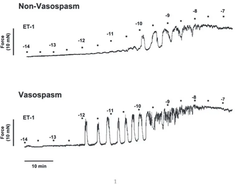
Rat basilar arteries incubated with CSF from Vasospasm patients developed greater contractile forces at lower ET-1 concentrations compared to vessels incubated with Non-Vasospasm CSF, as shown in the representative myograph trace (Fig. 1).
Experiments in arteries with endothelium
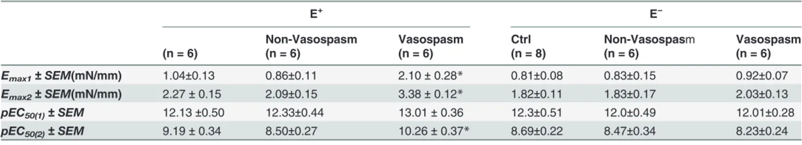
In E+vessels an enhanced concentration-dependent contractile response to ET-1 was present in the Vasospasm group when compared with the Non-Vasospasm and Control groups at ET-1 concentrations between ET-1×ET-10−12.5and 1×10⁻⁷mol/L (p<0.05) (Fig. 2upper panel). Interpo-lation of the concentration-response curves showed a biphasic response in all three groups. Analysis of the Emaxand pEC50for each phase showed that the Emax1, Emax2and pEC50(2)were
significantly higher in the Vasospasm group compared to the Non-Vasospasm or Control groups (Table 2).
Experiments in arteries without endothelium
In E−vessels all the three groups exhibited a similar biphasic response (Table 2). The enhanced contractile response to ET-1 observed in arteries with endothelium and exposed to CSF from patients with vasospasm was absent in arteries without endothelium (Fig. 2lower panel).
ET-1 concentrations in CSF used for vessels incubation were 2.45 ±0.81 pg/ml in Non-Vaso-spasm group and 2.63±1.31 pg/ml in VasoNon-Vaso-spasm group. After dilution, ET-1 concentrations in the culture medium at the beginning of the incubation were undetectable in all groups (data not shown), but ET-1 concentration after 24 hours incubation was significantly higher in the Vasospasm (1.71± 0.4 pg/ml) compared to the Non-Vasospasm(0.82± 0.2 pg/ml) and Control groups (0.91± 0.04 pg/ml) (p<0.05).
Endothelin Receptor Expression in the
Ex Vivo Model of SAH-induced
Vasospasm
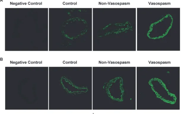
There was a faint autofluorescence in the arterial segments examined by confocal microscopy. When autofluorescence was subtracted ETAreceptors were found to be constitutively
express-ed in the smooth muscle cell layer of arteries exposexpress-ed to CSF from all three patient groups (Fig. 3A). Semiquantitative immunofluorescence for ETBreceptors revealed that the expression
apparently was markedly enhanced in the smooth muscle cell layer comparing arteries
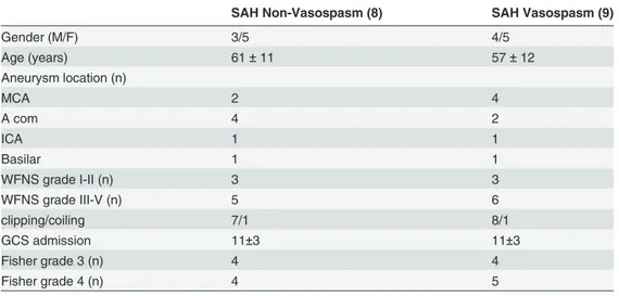
Table 1. Demographic and clinical data of 17 patients studied.
SAH Non-Vasospasm (8) SAH Vasospasm (9)
Gender (M/F) 3/5 4/5 Age (years) 61± 11 57± 12 Aneurysm location (n) MCA 2 4 A com 4 2 ICA 1 1 Basilar 1 1 WFNS grade I-II (n) 3 3 WFNS grade III-V (n) 5 6 clipping/coiling 7/1 8/1 GCS admission 11±3 11±3 Fisher grade 3 (n) 4 4 Fisher grade 4 (n) 4 5
Data expressed as mean± SD or N; MCA = middle cerebral artery, ICA = internal carotid artery, A com = anterior communicating WFNS = World federation of neurological surgeons, GCS = Glasgow Coma Scale doi:10.1371/journal.pone.0116456.t001
incubated with CSF from patients with Vasospasm than in those incubated with CSF from Non-Vasospasm and Control patients (Fig. 3B) (260±9%; p<0.001, results not shown).
Effects of Selective and combined Endothelin Receptor Blockade
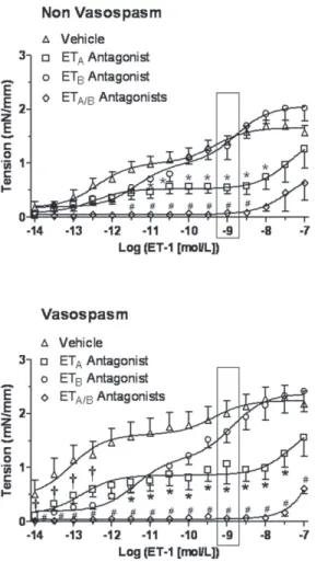
In a separate set of experiments, the ET-1 concentration-response curve was obtained with and without blockade of the individual and combined ETAand ETBreceptors (Table 3). In cerebral
arteries incubated with CSF from Non-Vasospasm group pretreatment with ETAantagonist
(BQ-123) reduced the contractile response for ET-1 concentrations between 1×10⁻¹¹to 1×10⁻⁸ mol/L; pretreatment with ETBantagonist (BQ-788) did not alter ET-1 induced contraction,
while pretreatment with both ETAand ETBantagonists abolished the contractile response for
ET-1 concentrations between 1×10−11.5to 1×10⁻⁸mol/L (Fig. 4, upper panel). In cerebral arter-ies incubated with CSF from the Vasospasm group pretreatment with ETAantagonist
(BQ-123) significantly reduced the contractile response in the high range of ET-1 concentrations
Figure 1. Experimental myograph record of isolated rat basilar arteries incubated with 5% CSF from patients with SAH with and without vasospasm. The contractile force generated during exposure to increasing doses of ET-1 by Log (0.5 mol/L) every 5 min was higher at lower ET-1 concentrationsfor Vasospasm (bottom panel) compared to Non-Vasospasm (top panel).
(between 1×10−11.5to 1×10−7.5mol/L), pretreatment with ETBantagonist reduced the
contrac-tile response in the low range of ET-1 concentrations (between 1×10⁻¹⁴to 1×10⁻¹²mol/L), while pretreatment with both ETAand ETBantagonists completely abolished the ET-1 induced
contraction (Fig. 4, lower panel).
Figure 2. Concentration-response curves elicited by ET-1 in isolated rat basilar arteries with
endothelium (E+) and without enodthelium (E−) following incubation with 5% human CSF from Control (n = 6), Non-Vasospasm (n = 6) and Vasospasm (n = 6) patients (upper panel) and from Control (n = 8), Non-Vasospasm(n = 6) and Vasospasm (n = 6) patients (lower panel). The response to ET-1 was significantly increased in the Vasospasm group compared to Control and Non-Vasospasm groups only in intact vessels. Significance by Kruskal Wallis with Dunn’s post-hoc test where *p<0.05 = Vasospasm versus Controland Non-Vasospasm.
doi:10.1371/journal.pone.0116456.g002
Table 2. Contractile response to ET-1 in rat basilar arteries with (E+) and without (E−) endothelium following incubation with human CSF.
E+ E−
Non-Vasospasm Vasospasm Ctrl Non-Vasospasm Vasospasm
(n = 6) (n = 6) (n = 6) (n = 8) (n = 6) (n = 6)
Emax1± SEM(mN/mm) 1.04±0.13 0.86±0.11 2.10± 0.28* 0.81±0.08 0.83±0.15 0.92±0.07
Emax2± SEM(mN/mm) 2.27± 0.15 2.09±0.15 3.38± 0.12* 1.82±0.11 1.83±0.17 2.03±0.13
pEC50(1)± SEM 12.13±0.50 12.33±0.44 13.01± 0.36 12.3±0.51 12.0±0.49 12.01±0.28
pEC50(2)± SEM 9.19± 0.34 8.50±0.27 10.26± 0.37* 8.69±0.22 8.47±0.34 8.23±0.24
Values are expressed as mean± SEM. Significance was determined by one-way ANOVA with Tukey-Kramer post-hoc where; ctrl = control * p<0.05 = Vasospasm versus Control and Non-Vasospasm.
Discussion
This study for the first time clearly describes the functional response, protein expression, and effects of selective and combined blockade of both ETAand ETBreceptors in two well-defined
conditions: intact conductive cerebral vessels exposed to the biological effects of the hemor-rhagic event (SAH without vasospasm) and vessels exposed to the combined biological effects of SAH and vasospasm. When CSF from patients with SAH and vasospasm was used both re-ceptors were present and mediated the vasoconstriction; therefore combined blockade of both receptors led to the maximal inhibition of the ET-1 induced contraction. The enhanced endothelin contraction was absent in vessels without endothelium suggesting that an endothe-lium-dependent mechanism contribute to the enhanced contractile endothelin-1 response ob-served in arteries conditioned by CSF from SAH patients with vasospasm.
Upregulation of the endothelin system after incubation with CSF from
patients with vasospasm
In most functional studies on isolated vessels the two conditions of SAH, with and without va-sospasm were not differentiated [6,9,10,12]. Only one study with human CSF clearly differenti-ated the two SAH conditions but in that study the role of endothelin-1 was not investigdifferenti-ated [27]. The main novelty of the present study is the selection of human CSF with a definitive di-agnosis of vasospasm established by angiography and integrated with TCD and clinical exami-nation to investigate the endothelin-1 pathway in a model of isolated cerebral vessels.
Interestingly, we found that intact vessels incubated with CSF from SAH patients with vaso-spasm resulted in a greater contractile response to ET-1 demonstrated by the left shift of the
Figure 3. Immunoreactivity of ET Receptors. Representative examples of rat basilar arteries showing immunoreactivity in the smooth muscle cell layer of ETA(panel A) and ETB(panel B) following incubation with human 5% CSF from Control (n = 9), Non-Vasospasm (n = 8) and Vasospasm patients (n = 9).
Negative controls for ETAand ETBantibodies were obtained excluding primary antibodies and in both cases resulted in no specific staining Pictures were
obtained using a × 40 objective. doi:10.1371/journal.pone.0116456.g003
concentration response curve with higher Emax and pEC50(i.e. increased potency and
sensitiv-ity to ET-1) when compared to vessels incubated with CSF from non-vasospasm and control patients. The increased sensitivity to ET-1 induced by organ culture was presumably present in all our different experimental groups and therefore equally influenced all our results. In our study cerebral arteries were incubated with Dulbecco medium containing 5% human CSF. Al-though ET-1 was detectable in all CSF samples of the patients studied, we applied a dilution factor which brought ET-1 concentrations below the detectable range in both groups. There-fore, the amount of ET-1 measured in the culture medium was the result of the stimulation of the arterial segments by CSF from the patients. The choice of CSF from day 4–5 for the present study is explained by the fact that we verified that patients with severe angiographic vasospasm on day 7 (clinical vasospasm) had a significant increase in MCA velocity on TCD on day 4–5 suggesting that molecular mechanisms responsible for vasospasm were already activated at that time. Among the soluble factors derived from the blood, oxidative stress [28], increased coagulation, fibrinolysis cascade, arachidonic acid metabolites and inflammatory mediators [29] may be involved. Recently it has been suggested that SAH induces early activation of the MEK-ERK1/2 pathway in cerebral artery walls, which is associated with upregulation of proin-flammatory cytokines and MMP-9 [30].
The vessels incubated with CSF from patients with SAH and vasospasm had higher secreted ET-1 levels suggesting that soluble factors present in the CSF of this group of patients were able to trigger greater ET-1 secretion compared to CSF from patients without vasospasm. It is a lim-itation that only CSF obtained at day 4–5 were examined, but on the other hand that was where the vasospasm was most pronounced. Moreover, it would be an advantage if secretion and“de novo” production of ET-1 could have been discriminated by measurements of mRNA levels.
Table 3. Contractile response to ET-1 of intact rat basilar arteries following incubation with human CSF and ETAantagonist (BQ-123), ETB
antagonist (BQ-788) and combined ETAand ETBantagonists (BQ-123 and BQ-788).
Biphasic Curves Sigmoidal Curves
Emax1± SEM
(nN/mm)
Emax2± SEM
(nN/mm)
pEC50(1)±SEM pEC50(2)±SEM Emax± SEM
(nN/mm) pEC50± SEM Ctrl Vehicle (n = 7) 0.79±0.26 1.52±1.18 13.13± 0.71 8.67± 0.54 BQ-123 (n = 8) 0.65± 0.07 14.57± 3.40 BQ-788 (n = 7) 2.17± 0.13 9.74± 0.19 BQ-123 + BQ-788 (n = 5) 0.26±0.08 7.67±0.52 NVS Vehicle (n = 8) 1.00± 0.09 1.65± 0.07 12.40± 0.29 9.35± 0.32 BQ-123 (n = 8) 0.53± 0.19† 1.56± 0.51 12.49± 0.62 7.39± 0.53† BQ-788 (n = 8) 1.02± 0.08 2.06± 0.21 11.36± 0.35 8.79± 0.29 BQ-123 + BQ-788 (n = 5) 0.88±0.32 7.42±0.42 VS Vehicle (n = 9) 1.61± 0.32 * 2.29± 0.16* 13.03± 0.54 9.22± 0.64 BQ-123 (n = 9) 0.86± 0.43† 1.91± 0.82 12.81± 0.54 7.29± 0.78† BQ-788 (n = 9) 1.16± 0.10† 2.17± 0.10 11.83± 0.33 9.16± 0.31 BQ-123 + BQ-788 (n = 5) 0.61±0.27 7.31±0.23
Responses were characterized by Emaxand pEC50values (negative logarithm of the molar concentration that produced half-maximum contraction). NVS =
non vasospasm, VS = vasospasm. Values are expressed as mean± SEM. Significance was determined by a one-way ANOVA with a Tukey-Kramer post-hoc analysis between CSF, where* p<0.05 = Vasospasm versus Control and Non-Vasospasm, and by a Student’s t-test between antagonists and vehicle, where†p<0.05 = BQ-123 versus Vehicle.
Involvement of ET
Aand ET
Breceptors after incubation with CSF from
patients with vasospasm
In physiological conditions the biological effects of ET-1 are mediated through activation of the two ET receptors: ETAwhich induces vasoconstriction on smooth muscle cells, whereas the
endothelial ETBmediates vasodilation [31]; consequently endothelium removal leads to the
abolishement of the vasodilator modulation. After SAH a different endothelium dependent ef-fect of ET-1 may be expected and the upregulation of vascular smooth muscle ETBreceptor
may contribute to vasoconstriction [32]. Indeed a functional interaction between ETAand ETB
also named“cross talk” has been recently proposed to explain the limited efficacy of selective ET receptors antagonists [33].
Figure 4. Concentration-response curves for ET-1 after ETA(BQ-123), ETB(BQ-788) or both receptors
blockade on isolated rat basilar arteries following incubation with 5% CSF from Non-Vasospasm (panel A) and Vasospasm (panel B) patients. In the Non-Vasospasm group, the response to ET-1 was significantly reduced by the ETAantagonist (n = 8), was not altered by the ETB(n = 8) antagonist and greatly
reduced by both antagonists (n = 5) compared to Vehicle (n = 8), whereas in the Vasospasm group, the increased sensitivity to ET-1 was significantly reduced by the ETB(n = 9) antagonist in the low range, by the
ETAantagonist (n = 9) in the high range of ET-1 concentrations and was completely abolished by combined
receptors blockade (n = 5) compared to Vehicle (n = 9). Significance at each concentration by a two-way ANOVA followed by a one-way ANOVA with Dunnett’s post-hoc analysis between groups, where *p<0.05 = ETAantagonist versus vehicle,†p<0.05 = ETBantagonist versus vehicle, #p<0.05 = ETAand ETB
antagonists versus vehicle. doi:10.1371/journal.pone.0116456.g004
ETAreceptors have previously been suggested as the main responsible for the pathogenesis
of vasospasm. Thus, after SAH, ETAreceptor expression on smooth muscle cells was
un-changed but blockade of ETAreceptor with clazosentan reduced the contraction to ET-1 [16].
Another two studies in SAH animal models showed upregulation of mRNA expression of the ETAreceptor after SAH [5,6]. In the present study, ETAreceptors were expressed in the smooth
muscle layer of arteries exposed to CSF from all three groups of patients. These findings togeth-er with the increased functional endothelin-1 response in arttogeth-eries exposed to CSF from patients with vasospasm suggest that antagonism of ETAreceptors would be attractive. However, ETB
receptors have also been suggested to play a role in the pathogenesis of vasospasm [10,34,35]. In different models of SAH increased levels of ETBreceptor mRNA with associated enhanced
functional response to ET-1 have been shown [8,11]; in a rat model of SAH, upregulation of ETBreceptor, specific functional response, and mRNA and protein expression were reported
[9]. Selective decrease in ETBreceptor-dependent relaxation [36] and reduced vasospasm after
specific ETBreceptor antagonist [17] have also been reported. In the present study, we found
that the ETBreceptor was not constitutively expressed in smooth muscle cells in control
condi-tions and, more importantly, when using CSF from SAH patientswithout vasospasm but only CSF from SAH patientswith vasospasm induced an apparently marked expression of ETB
re-ceptors in the vascular smooth muscle cell layer.
Finally, we evaluated a) the functional response of intact vessels after selective and com-bined blockade of ETAand ETBreceptors and b) the functional response of intact and denuded
isolated vessels to CSF from SAH patients with and without vasospasm. In the non vasospasm group, only selective ETAblockade induced a dose-dependent right shift of the
concentration-response curve. This is explained by the fact that ETAis constitutively expressed in control
con-ditions; the hemorrhagic event on its own does not cause upregulation of ETBreceptors so that
ETBblockade had no effect. In the vasospasm group, both receptor types were upregulated.
ETBexpression on smooth muscle cells was associated with increased tone and consequently
antagonism of both receptors induced a right shift of the ET-1 concentration-response curves: the effect of ETBblockade was significant at low ET-1 concentrations (high affinity phase)
whereas the effect of ETAblockade was significant at high ET-1 concentrations (low affinity
phase). After blocking both receptors the increased tone was abolished throughout the ET-1 concentration range suggesting the involvement of both receptors in the enhanced contractile response to ET-1. Furthermore the contractile response elicited by incubation with CSF from patients with vasospasm was abolished in vessels without endothelium suggesting that the con-dition of vasospasm after SAH induced an alteration in endothelial function contributing to the enhanced tone. These results may be explained by 1) ET-1 secreted by endothelial cells in the culture medium contributes to vasoconstriction [37]; 2) the existence of ETAreceptor in
the spastic vessels [7]; 3) the dual“vasodilator-vasoconstrictor” role of ETBreceptors as
de-scribed in pulmonary vessels [38,39]; 4) the heterodimerization of ET receptors described in pulmonary hypertension [38]. The latter hypothesis may explain why combination of selective ETAand ETBantagonists resulted in near maximal inhibition of ET-1
induced vasoconstriction.
Conclusion and Perspectives
What additional information does the presenttranslational study provide after several clinical trials [18–20,22,40] with endothelin receptor antagonists which showed reduction in angio-graphic vasospasm but no improvement in measureable functional outcomes?
In a recent metanalysis Vergouwen et al [41] suggested that endothelin receptor antagonists might be effective in selective subgroups of patients. In this perspective our data indeed suggest
that only a well-defined subgroup of patients who develop severe vasospasm induced expres-sion of both receptors mediating vasoconstriction and therefore specific targeting ETA
recep-tors was not sufficient to maximally inhibit vasospasm. The limited efficacy of selective ET receptor antagonists may be due to the functional interaction between ETAand ETBreceptors
named“cross talk”.
The“cross talk” between the two receptors can be functionally interpreted as a cooperative inhibition by combined selective ET receptor blockade of the ET-1 induced contraction [33,42]. In line with this hypothesis in the present study, only blockade of both receptors in-duced an optimal reduction in the contractile response to ET-1.
In conclusion we studied the functional response, protein expression, and the effects of se-lective and combined blockade of both receptors in two well-defined conditions: intact conduc-tive cerebral vessels exposed to the biological effects of the hemorrhagic event (SAH without vasospasm) and vessels exposed to the combined biological effects of SAH and vasospasm. When CSF from patients with SAH and vasospasm was used both receptors were upregulated and mediated vasoconstriction; therefore combined blockade of both receptors led to a maxi-mal inhibition of ET-1-induced contraction. This enhanced ET-1 contraction was abolished in vessels without endothelium suggesting that the condition of vasospasm after SAH induced an alteration of endothelial function contributing to the enhanced tone. Further studies are re-quired to demonstrate the exact nature of the functional interaction or“cross talk” between the two receptors [33].
Acknowledgments
The authors thank Prof. Alessandro Vercelli as animal facility’s supevisor, Dr Federico Sizzano as expert in confocal miscroscopy, Dr Davide Flamini for his help in interpolation analysis, Fla-vio Cristofani as and Valeria Puntorieri from Departement of Anatomy, University of Turin, Italy for technical assistance, animal care support and constructive discussion.
Author Contributions
Conceived and designed the experiments: BA MF AD US LM. Performed the experiments: BA ELM FC. Analyzed the data: BA ELM ES FC. Contributed reagents/materials/analysis tools: MF MB RB. Wrote the paper: BA ELM ES ATM US LM.
References
1. Bederson JB, Connolly ES Jr, Batjer HH, Dacey RG, Dion JE, et al. (2009) Guidelines for the manage-ment of aneurysmal subarachnoid hemorrhage: a statemanage-ment for healthcare professionals from a special writing group of the Stroke Council, American Heart Association. Stroke 40: 994–1025.
2. Mascia L, Fedorko L, Stewart DJ, Mohamed F, terBrugge K, et al. (2001) Temporal relationship be-tween endothelin-1 concentrations and cerebral vasospasm in patients with aneurysmal subarachnoid hemorrhage. Stroke 32: 1185–1190. PMID:11340231
3. Masaoka H, Suzuki R, Hirata Y, Emori T, Marumo F, et al. (1989) Raised plasma endothelin in aneurys-mal subarachnoid haemorrhage. Lancet 2: 1402.
4. Xie A, Aihara Y, Bouryi VA, Nikitina E, Jahromi BS, et al. (2007) Novel mechanism of endothelin-1-in-duced vasospasm after subarachnoid hemorrhage. J Cereb Blood Flow Metab 27: 1692–1701. PMID:
17392694
5. Itoh S, Sasaki T, Asai A, Kuchino Y (1994) Prevention of delayed vasospasm by an endothelin ETA re-ceptor antagonist, BQ-123: change of ETA rere-ceptor mRNA expression in a canine subarachnoid hem-orrhage model. J Neurosurg 81: 759–764. PMID:7931624
6. Kikkawa Y, Matsuo S, Kameda K, Hirano M, Nakamizo A, et al. (2012) Mechanisms underlying potenti-ation of endothelin-1-induced myofilament Ca(2+) sensitizpotenti-ation after subarachnoid hemorrhage. J Cereb Blood Flow Metab 32: 341–352. doi:10.1038/jcbfm.2011.132PMID:21952110
7. Vatter H, Konczalla J, Weidauer S, Preibisch C, Zimmermann M, et al. (2007) Effect of delayed cerebral vasospasm on cerebrovascular endothelin A receptor expression and function. J Neurosurg 107: 121– 127. PMID:17639881
8. Hansen-Schwartz J (2004) Receptor changes in cerebral arteries after subarachnoid haemorrhage. Acta Neurol Scand 109: 33–44. PMID:14653848
9. Beg SS, Hansen-Schwartz JA, Vikman PJ, Xu CB, Edvinsson LI (2007) Protein kinase C inhibition pre-vents upregulation of vascular ET(B) and 5-HT(1B) receptors and reverses cerebral blood flow reduc-tion after subarachnoid haemorrhage in rats. J Cereb Blood Flow Metab 27: 21–32. PMID:16736053
10. Hansen-Schwartz J, Hoel NL, Zhou M, Xu CB, Svendgaard NA, et al. (2003) Subarachnoid hemor-rhage enhances endothelin receptor expression and function in rat cerebral arteries. Neurosurgery 52: 1188–1194; 1194–1185. PMID:12699564
11. Hino A, Tokuyama Y, Kobayashi M, Yano M, Weir B, et al. (1996) Increased expression of endothelin B receptor mRNA following subarachnoid hemorrhage in monkeys. J Cereb Blood Flow Metab 16: 688– 697. PMID:8964809
12. Konczalla J, Vatter H, Weidauer S, Raabe A, Seifert V (2006) Alteration of the cerebrovascular function of endothelin B receptor after subarachnoidal hemorrhage in the rat. Exp Biol Med (Maywood) 231: 1064–1068. PMID:16741050
13. Dietrich HH, Dacey RG Jr (2000) Molecular keys to the problems of cerebral vasospasm. Neurosurgery 46: 517–530. PMID:10719847
14. Ogihara K, Barnanke DH, Zubkov AY, Parent AD, Zhang JH (2000) Effect of endothelin receptor antag-onists on non-muscle matrix compaction in a cell culture vasospasm model. Neurol Res 22: 209–214. PMID:10763512
15. Pluta RM, Boock RJ, Afshar JK, Clouse K, Bacic M, et al. (1997) Source and cause of endothelin-1 re-lease into cerebrospinal fluid after subarachnoid hemorrhage. J Neurosurg 87: 287–293. PMID:
9254095
16. Vatter H, Zimmermann M, Tesanovic V, Raabe A, Schilling L, et al. (2005) Cerebrovascular characteri-zation of clazosentan, the first nonpeptide endothelin receptor antagonist clinically effective for the treatment of cerebral vasospasm. Part I: inhibitory effect on endothelin(A) receptor-mediated contrac-tion. J Neurosurg 102: 1101–1107. PMID:16028770
17. Zuccarello M, Boccaletti R, Romano A, Rapoport RM (1998) Endothelin B receptor antagonists attenu-ate subarachnoid hemorrhage-induced cerebral vasospasm. Stroke 29: 1924–1929. PMID:9731620
18. Macdonald RL, Higashida RT, Keller E, Mayer SA, Molyneux A, et al. (2012) Randomized Trial of Cla-zosentan in Patients With Aneurysmal Subarachnoid Hemorrhage Undergoing Endovascular Coiling. Stroke.
19. Shaw MD, Vermeulen M, Murray GD, Pickard JD, Bell BA, et al. (2000) Efficacy and safety of the endothelin, receptor antagonist TAK-044 in treating subarachnoid hemorrhage: a report by the Steering Committee on behalf of the UK/Netherlands/Eire TAK-044 Subarachnoid Haemorrhage Study Group. J Neurosurg 93: 992–997. PMID:11117873
20. Vajkoczy P, Meyer B, Weidauer S, Raabe A, Thome C, et al. (2005) Clazosentan (AXV-034343), a se-lective endothelin A receptor antagonist, in the prevention of cerebral vasospasm following severe an-eurysmal subarachnoid hemorrhage: results of a randomized, double-blind, placebo-controlled, multicenter phase IIa study. J Neurosurg 103: 9–17. PMID:16121967
21. Macdonald RL, Higashida RT, Keller E, Mayer SA, Molyneux A, et al. (2011) Clazosentan, an endothe-lin receptor antagonist, in patients with aneurysmal subarachnoid haemorrhage undergoing surgical clipping: a randomised, double-blind, placebo-controlled phase 3 trial (CONSCIOUS-2). Lancet Neurol 10: 618–625. doi:10.1016/S1474-4422(11)70108-9PMID:21640651
22. Macdonald RL, Kassell NF, Mayer S, Ruefenacht D, Schmiedek P, et al. (2008) Clazosentan to over-come neurological ischemia and infarction occurring after subarachnoid hemorrhage (CONSCIOUS-1): randomized, double-blind, placebo-controlled phase 2 dose-finding trial. Stroke 39: 3015–3021. doi:
10.1161/STROKEAHA.108.519942PMID:18688013
23. Fontanella M, Valfre W, Benech F, Carlino C, Garbossa D, et al. (2008) Vasospasm after SAH due to aneurysm rupture of the anterior circle of Willis: value of TCD monitoring. Neurol Res 30: 256–261. PMID:17767811
24. Mascia L, Fedorko L, terBrugge K, Filippini C, Pizzio M, et al. (2003) The accuracy of transcranial Dopp-ler to detect vasospasm in patients with aneurysmal subarachnoid hemorrhage. Intensive Care Med 29: 1088–1094. PMID:12774157
25. Walchenbach R, Geiger E, Thomeer RT, Vanneste JA (2002) The value of temporary external lumbar CSF drainage in predicting the outcome of shunting on normal pressure hydrocephalus. J Neurol Neu-rosurg Psychiatry 72: 503–506. PMID:11909911
26. Mulvany MJ, Halpern W (1977) Contractile properties of small arterial resistance vessels in spontane-ously hypertensive and normotensive rats. Circ Res 41: 19–26. PMID:862138
27. Okwuasaba FK, Weir BK, Cook DA, Krueger CA (1981) Effects of various intracranial fluids on smooth muscle. Neurosurgery 9: 402–406. PMID:7301086
28. Nishizawa S, Laher I (2005) Signaling mechanisms in cerebral vasospasm. Trends Cardiovasc Med 15: 24–34.
29. Dumont AS, Dumont RJ, Chow MM, Lin CL, Calisaneller T, et al. (2003) Cerebral vasospasm after sub-arachnoid hemorrhage: putative role of inflammation. Neurosurgery 53: 123–133; discussion 133–125. PMID:12823881
30. Maddahi A, Povlsen GK, Edvinsson L (2012) Regulation of enhanced cerebrovascular expression of proinflammatory mediators in experimental subarachnoid hemorrhage via the mitogen-activated pro-tein kinase kinase/extracellular signal-regulated kinase pathway. J Neuroinflammation 9: 274. doi:10. 1186/1742-2094-9-274PMID:23259581
31. de Nucci G, Thomas R, D’Orleans-Juste P, Antunes E, Walder C, et al. (1988) Pressor effects of circu-lating endothelin are limited by its removal in the pulmonary circulation and by the release of prostacy-clin and endothelium-derived relaxing factor. Proc Natl Acad Sci U S A 85: 9797–9800. PMID:
3059352
32. Clozel M, Gray GA, Breu V, Loffler BM, Osterwalder R (1992) The endothelin ETB receptor mediates both vasodilation and vasoconstriction in vivo. Biochem Biophys Res Commun 186: 867–873. PMID:
1323294
33. Rapoport RM, Zuccarello M (2011) Endothelin(A)-endothelin(B) receptor cross-talk and endothelin re-ceptor binding. J Pharm Pharmacol 63: 1373–1377. PMID:21988418
34. Henriksson M, Stenman E, Vikman P, Edvinsson L (2007) MEK1/2 inhibition attenuates vascular ETA and ETB receptor alterations after cerebral ischaemia. Exp Brain Res 178: 470–476. PMID:17091294
35. Henriksson M, Stenman E, Edvinsson L (2003) Intracellular pathways involved in upregulation of vas-cular endothelin type B receptors in cerebral arteries of the rat. Stroke 34: 1479–1483. PMID:
12750545
36. Vatter H, Zimmermann M, Tesanovic V, Raabe A, Seifert V, et al. (2005) Cerebrovascular characteriza-tion of clazosentan, the first nonpeptide endothelin receptor antagonist shown to be clinically effective for the treatment of cerebral vasospasm. Part II: effect on endothelin(B) receptor-mediated relaxation. J Neurosurg 102: 1108–1114. PMID:16028771
37. Vanhoutte PM, Tang EH (2008) Endothelium-dependent contractions: when a good guy turns bad! J Physiol 586: 5295–5304. doi:10.1113/jphysiol.2008.161430PMID:18818246
38. Sauvageau S, Thorin E, Caron A, Dupuis J (2007) Endothelin-1-induced pulmonary vasoreactivity is regulated by ET(A) and ET(B) receptor interactions. J Vasc Res 44: 375–381. PMID:17495482
39. Davie N, Haleen SJ, Upton PD, Polak JM, Yacoub MH, et al. (2002) ET(A) and ET(B) receptors modu-late the proliferation of human pulmonary artery smooth muscle cells. Am J Respir Crit Care Med 165: 398–405. PMID:11818328
40. Macdonald RL, Pluta RM, Zhang JH (2007) Cerebral vasospasm after subarachnoid hemorrhage: the emerging revolution. Nat Clin Pract Neurol 3: 256–263. PMID:17479073
41. Vergouwen MD, Algra A, Rinkel GJ (2012) Endothelin receptor antagonists for aneurysmal subarach-noid hemorrhage: a systematic review and meta-analysis update. Stroke 43: 2671–2676. PMID:
22871682
42. De Mey JG, Compeer MG, Lemkens P, Meens MJ (2011) ETA-receptor antagonists or allosteric modu-lators? Trends Pharmacol Sci 32: 345–351.