1Research Centre of Systemic Auto-inflammatory Diseases, Behçet’s Disease and Rheumatology-Ophthalmology Collaborative Uveitis Centre, Department of Medical Sciences, Surgery and Neuro-Sciences, University of Siena;
2Department of Ophthalmology,
Humanitas Clinical and Research Centre, Rozzano (Milan);
3Institute of Paediatrics, Università Cattolica Sacro Cuore, Fondazione Poli-clinico Universitario “A. Gemelli”, Rome; 4Ophthalmology Unit of the Department of Medicine, Surgery and Neuroscience, University of Siena;
5Department of Paediatrics, University of Padova, Italy. Jurgen Sota*, MD Antonio Vitale*, MD Claudia Fabiani, MD Bruno Frediani, MD Donato Rigante, MD Gian Marco Tosi, MD Maria E. Zannin, MD Luca Cantarini, MD, PhD
*J. Sota and A. Vitale have contributed equally to this paper.
Please address correspondence to: Luca Cantarini, MD, PhD, Research Centre of Systemic Auto-inflammatory Diseases, Behçet’s Disease and Rheumatology-Ophthalmology Collaborative Uveitis Centre, Department of Medical Sciences, Surgery and Neurosciences, University of Siena, Rheumatology Unit, Policlinico “Le Scotte”, viale Bracci 1, 53100 Siena, Italy. E-mail: [email protected] Received on November 27, 2017; accepted on December 7, 2017.
Clin Exp Rheumatol 2018; 36 (Suppl. 110): S44-S53.
© Copyright CliniCaland
ExpErimEntal rhEumatology 2018. Key words: autoinflammatory diseases, uveitis, retinal vasculitis, retinitis pigmentosa, scleritis
Funding and competing interests on page S-52.
ABSTRACT
Monogenic autoinflammatory diseases (AIDs) are rare entities characterised by improper activation of the innate im-mune system. This in turn determines recurrent episodes of systemic inflam-mation characterised by fever, which is variously combined with a wide range of inflammatory manifestations involv-ing the skin, joints, serous membranes, gastrointestinal tract, and central nerv-ous system. As shown by research ef-forts conducted during the last decade, the eye is not exempt from the systemic inflammatory process and may be in-volved in almost all of the most frequent AIDs, with several distinct peculiarities. Ocular affections may severely impact patients’ quality of life due to orbital pain, impairment of visual acuity, and/ or long-term, sight-threatening compli-cations. Consequently, in the context of a multidisciplinary team, ophthalmolo-gists should be aware of ocular mani-festations related to these disorders as they may have a dominant diagnostic weight in patients with a challenging presentation as well as a salient role in therapeutic choice in sight-threat-ening situations. This review describes a variety of aspects of ophthalmologic involvement in AIDs, looking at both well-recognised eye manifestations as well as rarely reported ocular presen-tations, with a particular focus on the recent literature.
Introduction
Autoinflammatory diseases (AIDs) embrace an expanding group of rare disorders characterised by seemingly unprovoked episodes of self-limited inflammatory attacks with no infec-tious agents, autoreactive T cells or au-toantibodies observed. The pathogen-esis of AIDs is hallmarked by genetic mutations of proteins involved in the modulation of the innate immunity and
leading to the up-regulation of proin-flammatory cytokines, especially inter-leukin (IL)-1β and tumour necrosis fac-tor (TNF)-α (1, 2). The most common monogenic autoinflammatory disorders include familial Mediterranean fever (FMF), TNF receptor-associated peri-odic syndrome (TRAPS), mevalonate kinase deficiency (MKD), idiopathic granulomatous diseases, and cryopyrin-associated periodic syndrome (CAPS) (Table I). CAPS includes three differ-ent clinical phenotypes: familial cold autoinflammatory syndrome (FCAS), Muckle-Wells syndrome (MWS), and chronic infantile neurologic cutane-ous and articular (CINCA) syndrome. These show increasing severity, with FCAS being the least severe and CIN-CA the most severe. From a clinical point of view, AIDs occur most often during childhood, however, the delayed onset of symptoms during adulthood has commonly been reported (3, 4). All AIDs are characterised by recurrent fever episodes and increased inflamma-tory markers variously combined with skin rash and inflammatory episodes affecting the joints, serous membranes, gastrointestinal tract, eyes, and cen-tral nervous system (5). Conversely, a complete wellbeing with normal acute phase reactants characterise inter-crit-ical periods (6). However, AIDs can sometimes acquire a chronic course, especially in cases related to low-pen-etrance mutations, which often lead to adult-onset manifestations and incom-plete or atypical phenotypes (7). Taken together, the rarity of AIDs and their relatively low knowledge among physi-cians along with the broad spectrum of possible clinical presentations and the need for a careful and correct genotype-phenotype correlation, may complicate the differential diagnosis in patients with suspected AIDs thus causing a de-lay in diagnosis (8-10).
literature review and update
J. Sota
1, A. Vitale
1, C. Fabiani
2, B. Frediani
1, D. Rigante
3, G.M. Tosi
4,
Ocular involvement in AIDs may have distinct peculiarities (Table II). Figure 1 illustrates some of the most representa-tive eye lesions of monogenic AIDs. Ocular affections may severely impact the quality of life due to orbital pain, impairment of visual acuity and/or long-term, sight-threatening complica-tions. Hence, an increased awareness of AIDs is warranted among ophthalmolo-gists in order to achieve an early diag-nosis and optimal ocular management. The last decade has seen a number of interesting findings published on ocular involvement in AIDs (11-20). Herein, we review the current knowledge on this issue and provide a broad overview on the possible presentations of ocular affections in these rare diseases. Familial Mediterranean fever FMF is an autosomal recessive autoin-flammatory disorder caused by muta-tions of the MEFV gene, encoding the pyrin protein. This immunoregulatory molecule, also known as marenostrin, is involved in the regulation of inflam-mation, cytokine production, and apop-tosis. The cardinal phenotype of FMF
is represented by recurrent episodes of fever generally lasting 48–72 hours, inflammation of one or more serosal membranes, a non-destructive arthritis usually involving one large joint of the lower limbs, and skin manifestations mostly in the form of erysipelas-like rash (21). Over time, this basilar clinical phenotype of FMF has been expanded with other clinical features including ocular involvement. The earliest pa-per on ophthalmologic affections was published in 1959 by Michaelson and colleagues, who described dotted le-sions consistent with colloid bodies on routine funduscopic examination in 13 out of 23 patients; slit-lamp microscopy subsequently localised the lesions in the Bruch’s lamina (22). Another patient displayed an ocular fundus with a solid-looking mulberry-like mass thought to originate from an adjacent colloid body (22). In 2014, a Turkish working group investigated retinal and choroidal thick-ness (CT) in 30 FMF children by meas-uring each subject’s right eye at the fo-vea and horizontal nasal and temporal quadrants at 500 mm intervals to 1500 mm from the foveal using
spectral-do-main optic coherence Tomography (SD-OCT) (23). Although no significant dif-ferences were found between FMF pa-tients and a control group, the authors highlighted the necessity to perform ocular evaluation during FMF attacks. In this regard, Gundogan et al. carried out a prospective case-controlled clini-cal study evaluating CT during acute attacks in 50 patients suffering from FMF (14). This study identified a sig-nificantly thicker choroid in FMF pa-tients compared with healthy controls. Moreover, CT was positively corre-lated with inflammatory biomarkers, and especially C-reactive protein. The systemic inflammatory process during acute attacks and the following increase of vascular permeability, choroidal ves-sels enlargement and exudation may be responsible for the increase in CT with more impact during acute attacks rather than during symptom-free intervals (14). However, other authors have sug-gested the possible occurrence of con-founding factors behind the increase of CT including the high body temperature during attacks and possible toxic conse-quences of treatment on choroidal tis-Table I. Genetic and clinical features of the main autoinflammatory diseases.
AID Gene/Locus Inheritance Protein Fundamental clinical clues
FMF MEFV/16p13.3 AR Pyrin Fever, serositis, arthralgia or arthritis generally affecting large joints, erysipela-like rash, amyloidosis in untreated patients
TRAPS TNFRSF1A/12p13 AD Tumour necrosis factor Fever, migrating erythematous skin rash, muscle pain in the form of receptor 1 fasciitis, conjunctivitis, periorbital oedema, arthralgia or arthritis, serosal
involvement, amyloidosis
MKD MVK/12q24 AR Mevalonate kinase Fever, polymorphous rash, arthralgia, abdominal pain, diarrhoea, lymph node enlargement, splenomegaly, aphtosis
MA MVK/12q24 AR Mevalonate kinase Psychomotor retardation and growth delay, progressive cerebellar ataxia, dysmorphisms, vision deficits, MKD symptoms
FCAS NLRP3/1q44 AD Cryopyrin Fever, cold-induced urticarial rash, conjunctivitis, arthralgia
MWS NLRP3/1q44 AD Cryopyrin Fever, urticarial rash, conjunctivitis, episcleritis, arthralgia, sensorineural deafness, amyloidosis
CINCA NLRP3/1q44 AD Cryopyrin Fever, urticarial skin rash, uveitis, papilledema, deforming arthritis mainly involving large joints, chronic aseptic meningopathy, sensorineural hearing loss, amyloidosis
Blau syndrome NOD2/16q12.1-13 AD Nucleotide-binding Arthritis, skin rash, granulomatous inflammatory ocular involvement oligomerisation domain 2
AD: autosomal dominant; AID: autoinflammatory disease; AR: autosomal recessive; CINCA: chronic infantile neurologic cutaneous and articular syn-drome; FCAS: familial cold autoinflammatory synsyn-drome; FMF: familial Mediterranean fever; MA: mevalonic aciduria; MEFV: MEditerranean FeVer; MKD: mevalonate kinase deficiency; MVK: mevalonate kinase; MWS: Muckle-Wells syndrome; NLRP3: NACHT, LRR and PYD domains-containing protein 3; NOD2: nucleotide-binding oligomerisation domain 2; TNFRSF1A: tumour necrosis factor receptor super family 1A ; TRAPS: tumour necrosis factor-associated periodic syndrome.
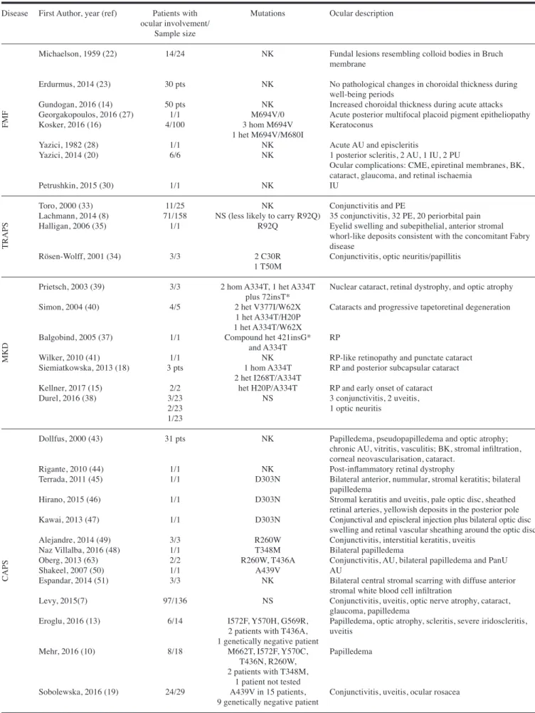
Table II. Current knowledge on ocular involvement in monogenic autoinflammatory diseases.
Disease First Author, year (ref) Patients with Mutations Ocular description ocular involvement/
Sample size
Michaelson, 1959 (22) 14/24 NK Fundal lesions resembling colloid bodies in Bruch membrane
Erdurmus, 2014 (23) 30 pts NK No pathological changes in choroidal thickness during well-being periods
Gundogan, 2016 (14) 50 pts NK Increased choroidal thickness during acute attacks Georgakopoulos, 2016 (27) 1/1 M694V/0 Acute posterior multifocal placoid pigment epitheliopathy Kosker, 2016 (16) 4/100 3 hom M694V Keratoconus
1 het M694V/M680I
Yazici, 1982 (28) 1/1 NK Acute AU and episcleritis
Yazici, 2014 (20) 6/6 NK 1 posterior scleritis, 2 AU, 1 IU, 2 PU
Ocular complications: CME, epiretinal membranes, BK, cataract, glaucoma, and retinal ischaemia
Petrushkin, 2015 (30) 1/1 NK IU
Toro, 2000 (33) 11/25 NK Conjunctivitis and PE
Lachmann, 2014 (8) 71/158 NS (less likely to carry R92Q) 35 conjunctivitis, 32 PE, 20 periorbital pain Halligan, 2006 (35) 1/1 R92Q Eyelid swelling and subepithelial, anterior stromal
whorl-like deposits consistent with the concomitant Fabry
disease
Rösen-Wolff, 2001 (34) 3/3 2 C30R Conjunctivitis, optic neuritis/papillitis 1 T50M
Prietsch, 2003 (39) 3/3 2 hom A334T, 1 het A334T Nuclear cataract, retinal dystrophy, and optic atrophy plus 72insT*
Simon, 2004 (40) 4/5 2 het V377I/W62X Cataracts and progressive tapetoretinal degeneration 1 het A334T/H20P
1 het A334T/W62X
Balgobind, 2005 (37) 1/1 Compound het 421insG* RP and A334T
Wilker, 2010 (41) 1/1 NK RP-like retinopathy and punctate cataract Siemiatkowska, 2013 (18) 3 pts 1 hom A334T RP and posterior subcapsular cataract
2 het I268T/A334T
Kellner, 2017 (15) 2/2 het H20P/A334T RP and early onset of cataract Durel, 2016 (38) 3/23 NS 3 conjunctivitis, 2 uveitis,
2/23 1 optic neuritis
1/23
Dollfus, 2000 (43) 31 pts NK Papilledema, pseudopapilledema and optic atrophy; chronic AU, vitritis, vasculitis; BK, stromal infiltration, corneal neovascularisation, cataract.
Rigante, 2010 (44) 1/1 NK Post-inflammatory retinal dystrophy
Terrada, 2011 (45) 1/1 D303N Bilateral anterior, nummular, stromal keratitis; bilateral papilledema
Hirano, 2015 (46) 1/1 D303N Stromal keratitis and uveitis, pale optic disc, sheathed retinal arteries, yellowish deposits in the posterior pole Kawai, 2013 (47) 1/1 D303N Conjunctival and episcleral injection plus bilateral optic disc
swelling and retinal vascular sheathing around the optic disc Alejandre, 2014 (49) 3/3 R260W Conjunctivitis, interstitial keratitis, uveitis
Naz Villalba, 2016 (48) 1/1 T348M Bilateral papilledema
Oberg, 2013 (63) 2/2 R260W, T436A Conjunctivitis, AU, bilateral papilledema and PanU Shakeel, 2007 (50) 1/1 A439V AU
Espandar, 2014 (51) 3/3 NK Bilateral central stromal scarring with diffuse anterior stromal white blood cell infiltration
Levy, 2015(7) 97/136 NS Conjunctivitis, uveitis, optic nerve atrophy, cataract, glaucoma, papilledema
Eroglu, 2016 (13) 6/14 I572F, Y570H, G569R, Papilledema, optic atrophy, scleritis, severe iridoscleritis, 2 patients with T436A, uveitis
1 genetically negative patient
Mehr, 2016 (10) 8/18 M662T, I572F, Y570C, Papilledema T436N, R260W,
2 patients with T348M, 1 patient not tested
Sobolewska, 2016 (19) 24/29 A439V in 15 patients, Conjunctivitis, uveitis, ocular rosacea 9 genetically negative patient
FMF
TRAPS
MKD
sues (24, 25). In particular, a disruption of microtubules within retinal nerve fibre layer axons was manifested as a reduced birefringence on scanning laser polarimetry without abnormalities on retinal nerve fibre layer thickness meas-ured by OCT (26).
An interesting case report presented the association of FMF with acute poste-rior multifocal placoid pigment epithe-liopathy (APMPPE) in a patient with bilateral blurred vision and scotomata. As APMPPE was believed to be caused by an occlusive choroidal vasculitis, and given the increased frequency of vasculitis in FMF patients, the authors presumed there was a link between APMPPE and FMF (27).
FMF is also considered to be a predis-posing factor for the development of keratoconus (KC), especially in patients carrying homozygous mutations of the MEFV gene. A possible interaction be-tween FMF and KC was identified in 2016 by Kosker et al. who showed the
prevalence of KC was higher in FMF patients than in a control group; 4 out of 100 FMF patients were affected by KC, whereas none of the 300 patients in the control group were affected (16). Furthermore, when compared with the highest prevalence reported in the lit-erature, FMF was shown to be a pre-disposing factor for the development of KC, especially in patients carrying homozygous mutations (16).
With reference to uveal tract inflam-mation, Yazici and Pazarli described a patient with FMF who developed acute anterior uveitis in the left eye and epis-cleritis ten months later (28). Because of the two different forms of eye involve-ment, the authors were tempted to spec-ulate a possible relationship between ocular inflammation and FMF. Follow-ing this, a case series on FMF patients with ocular involvement described 2 cases with anterior uveitis, 2 with poste-rior uveitis, 1 with intermediate uveitis, and 1 case with posterior scleritis (20).
Cystoid macular oedema, epiretinal membranes, band keratopathy, cataract, branch retinal vein occlusion, neovas-cularisation, and glaucoma were also reported as ocular complications. It is noteworthy that 3 out of 6 patients pre-sented a relapsing course, in line with the recurrent nature of FMF (20). How-ever, 2 patients were also diagnosed with Behçet’s disease (BD), as FMF and BD can coexist (29). A second case of intermediate uveitis was reported by Petrushkin et al. who described a female patient with FMF complaining from sudden floaters in the right eye (30). In any case, a careful diagnostic approach is warranted in patients with FMF and uveitis in order to rule out concomitant systemic disorders that may clinically overlap with FMF (20, 21).
TNF receptor-associated periodic syndrome
TRAPS, the most common among the autosomal dominant AIDs, is caused Table II. continued
Disease First Author, year (ref) Patients with Mutations Ocular description ocular involvement/
Sample size
Arvesen, 2017 (53) 1/1 R334Q Granulomatous uveitis
Milman, 2009 (58) 4/4 T605N AU, bilateral PanU, chorioretinitis, scleritis, corneal opacities, retinal disruption
Jimenez-Martinez, 2011 (56) 1/1 M513R BK and PanU
Raiji, 2011 (59) 1/1 D382E Numerous small, white, corneal subepithelial ovoid opacities and conjunctival nodules and later a fulminant PanU
Zeybek, 2015 (60) 1/1 P507S Granulomatous AU
Kim, 2016 (57) 2/2 H480R AU, multiple mild subepithelial opacities on both eyes along with focal posterior synechia
Jain, 2016 (55) 1/1 E667K Right eye: non-granulomatous AU with vitreous haemorrhage
Left eye: chronic AU, multiple iris granuloma, and complicated cataract
Achille, 2016 (62) 1/1 het P268S/SNP5 Bilateral granulomatous PanU Carreño, 2015 (11) 9/9 6 het R334Q/wt 5 PanU, 3 AU, 1 IU
1 het Q809K/wt Optic nerve and retinal features: pale optic disc in 6 eyes, 1 het H520Y/wt indistinct margins in 6 eyes, sheathed optic disc vessels in 4 1 compound het E383D*/D390V eyes, nodular excrescences in the peripapillary area in 13
eyes, hypopigmentation in 6 eyes and mixed hypo- and hyperpigmentation in 7 eyes
Ebrahimiadib, 2016 (12) 6/6 E600A Bilateral non-granulomatous PanU, bilateral retinal vasculitis, papillitis and scleritis
Rosé, 2015 (17) 25/31 NS AU, IU, 64% active vitreous inflammation, 18% active chorioretinitis, 5% active retinal vasculopathy. Complications: 64% synechiae, 55% cataract, 36% IOP, 23% BK, 14% macular oedema, 9% retinal detachment. AU: anterior uveitis; BK: band keratopathy; BS: Blau syndrome; CAPS: cryopyrin associated periodic fever; CME: cystoid macular oedema; FMF: familial Mediterranean fever; het: heterozygous; hom: homozygous; IOP: increased intraocular pressure; IU: intermediate uveitis; KC: keratoconus; MKD: meva-lonate kinase deficiency; NK: not known; NS: not specified for patients with ocular involvement; PanU: panuveitis; PE: periorbital oedema; PU: posterior uveitis; pts: patients; RP: retinitis pigmentosa; TRAPS: tumour necrosis factor receptor-associated periodic syndrome; wt: wild type.
*not found in Infevers (http://fmf.igh.cnrs.fr/ISSAID/infevers/).
by mutations in the TNFRSF1A gene, which encodes for the 55 kDa type-1 re-ceptor of TNF-α. From a clinical point of view, this syndrome is characterised by recurrent inflammatory episodes lasting several weeks with a various combination of the following symp-toms: fever, migrant erythematous rash, myalgia, lymphadenopathy, artic-ular symptoms mainly represented by arthralgia, pleuritic and abdominal pain that may sometimes lead to exploratory laparotomy (31, 32). Ophthalmologic features have contributed to further ex-pand the heterogeneous phenotype of TRAPS. Periorbital oedema and con-junctivitis are the most common ocular manifestations. In this regard, Toro et al. described conjunctivitis and/or peri-orbital oedema in 11 out of 50 TRAPS patients in a study conducted on 16 families (33). Accordingly, based on data from the Eurofever/EUROTRAPS international registry, the largest case series currently published on TRAPS
patients identified periorbital oedema, periorbital pain, and conjunctivitis in 20%, 13%, and 22% of 158 subjects, respectively (8). Moreover, periorbital oedema tended to be significantly more common in paediatric patients as well as in adults with a disease onset during childhood. With regard to genotype-phenotype correlations, eye manifesta-tions were less frequently encountered in patients carrying the R92Q low-pen-etrance mutation (8).
A sporadic case of optic neuritis/papil-litis has also been reported in a German patient harbouring the C30R mutation who also developed fever, arthritis, skin rash myocarditis, and occasional diar-rhoea (34).
An interesting association between TRAPS and Fabry disease was re-ported by Halligan and colleagues in which a 9-year-old boy presenting with symptoms consistent with TRAPS was found to carry the R92Q mutation (35). Among others, eyelid swelling
was identified at physical examination, while an ophthalmologic examination revealed subepithelial, anterior stromal whorl-like deposits, which is a charac-teristic finding of Fabry disease. The authors suggested that the two genetic diseases may interact, potentially wors-ening the clinical phenotype (35). Mevalonate kinase deficiency The second most important autoin-flammatory disorder with recessive inheritance is represented by MKD, determined by mutations in the MVK gene. The MVK gene encodes for me-valonate kinase, a crucial enzyme for the nonsteroidal isoprenoids biosyn-thesis, which in turn determines protein prenylation. The described metabolic defect triggers innate immunity hyper-responsiveness ultimately leading to re-current multi-systemic inflammatory at-tacks classically characterised by fever, painful adenopathy, arthralgia, rash, and abdominal pain that may sometimes Fig. 1. Representative eye lesions in the monogenic autoinflammatory disorders.
(A) Swelling and peripapillary retinal oedema with haemorrhagic papillitis. (B) Retinal vasculitis. (C-D) Diffuse microvascular choriocapillaris vasculitis
showing leakage of fluorescein during fluorescein retinal angiography. (E-F) Diffuse anterior necrotising scleritis resulting in scleromalacia. (G) with scleral thinning. (H) Diffuse anterior non-necrotising scleritis. (I) Granulomatous anterior uveitis. (J) Posterior irido-capsular synechiae. (K-L) Hypopyon in the anterior chamber. From Bascherini et al. 2015 (5).
require surgical examinations for pos-sible acute abdomen. Currently, more than 200 sequence variants of the MVK gene have been associated with MKD, with most of them being single-nucle-otide polymorphisms that lead to mis-sense mutations. To date, the disease is considered as a phenotypic continuum of increasing severity according to the degree of residual enzymatic activity. It ranges from the milder form, hyper IgD syndrome (HIDS), to the most severe variant known as mevalonic aciduria (MA) (Table I). In its severest vari-ant, the clinical phenotype may also be expressed by facial dimorphisms and central nervous system involvement including seizures, ataxia, myopathies, and psychomotor retardation (36). Ophthalmologic features have contrib-uted to MKD clinical heterogeneity as its clinical spectrum continues to ex-pand (15, 18, 37-41). In a study by Pri-etsch et al., which investigated 3 MA patients with retinal dystrophy (RD), cataract accompanied RD in 2 siblings while optic atrophy was a supplementa-ry finding in the third patient (39). Ocu-lar involvement, along with ataxia and short stature, became predominant in patients surviving infancy, suggesting an age-dependent phenotype expres-sion. For all these reasons, Prietsch et al. suggested ocular electrophysiology as an integral part of diagnostic evalua-tion in order to detect retinal disorders in patients with MA (39). Simon et al. also reported progressive blindness due to tapetoretinal degeneration and cata-ract in 4 patients with MKD (40). As supported by several reports in the medical literature (15, 18, 37), retinitis pigmentosa (RP) is the most notewor-thy ocular disease connected to MKD, and particularly to MA. Night blindness with funduscopic and full-field electro-retinogram findings suggestive of RP was reported in a patient with MKD (37). In addition, RP-like retinopathy connected with MA was diagnosed on the basis of Goldmann visual field constrictions and full-field electroreti-nogram after a patient complained of nyctalopia and decreased peripheral vision; ophthalmologic evaluation also revealed findings fitting with punctate cataracts (41).
MKD can even be misdiagnosed as nonsyndromic RP, which might rep-resent the prep-resenting feature in such patients. In this regard, a proband of Dutch origin with RP was found to carry MVK mutations in a study by Siemiat-kowska and colleagues; the MVK gene was subsequently tested in the patient’s family and in a large cohort of patients with nonsyndromic, genetically un-solved RP (18). This study identified 3 subjects from 2 families with nonsyn-dromic RP who were found to have a mutation in the MVK gene. A detailed medical history revealed a mild form of MKD despite a significantly lowered mevalonate kinase activity, leading to misdiagnosis even among expert clini-cians (18).
Recently, 2 brothers of German extrac-tion with a compound heterozygous MVK mutation (H20P/A334T) were described in the first paper reporting an association of MKD and RP with early cataract development (15). As for the cases illustrated by Siemiatkowska and colleagues (18), a relatively mild phe-notype was reported despite the severe effects of the A334T mutation, further suggesting the probable influence of additional genetic and environmental factors on disease manifestation. On this basis, the currently proposed pathogenetic mechanism behind the non-random association between MKD and RP resides in an impaired isopre-nylation, which probably interferes with macromolecules that are essential for the function of photoreceptors (37). It is noteworthy that most of the MKD patients with concomitant RP harbour the A334T (c.1000G>A) in exon 10 of the MKV gene (15, 18, 37, 39, 40), thus suggesting a potential genotype-phe-notype correlation. Therefore, ophthal-mological examination and instrumen-tal ocular evaluation should be recom-mended for all patients with MKD. The identification of cataract represents an interesting finding, which deserves attention in MKD patients (15, 39-41). A possible explanation for cataract could be a direct toxic effect of meva-lonic acid rather than an osmotically-induced opacification. Indeed, young rat lenses cultured for up to 4 days in a medium containing mevalonic acid
exhibited lens opacification and nu-clear cataract within 1–2 days (42). In particular, water and sodium accumu-lated in the lenses, which lost soluble gamma crystallin proteins and potas-sium. Meanwhile, lenses lost the capac-ity to concentrate a potassium analogue (86Rb), possibly due to a slow poisoning
of the cation pump, an effect on mem-brane integrity or both (42).
Other ocular manifestations, including uveitis and optic neuritis, have been reported. An observational multicentre study aimed at classifying clinical and biological features of MKD in adult-hood reported 3 patients with conjuncti-vitis, 2 of them with uveitis, and 1 case of optic neuritis (38).
Cryopyrin-associated periodic syndromes
CAPS represents a group of monogenic AIDs caused by de-novo or dominantly inherited gain-of-function missense mutations of the NLRP3 gene. CAPS encompasses three overlapping syn-dromes sharing general anchor points and presenting an increasing disease severity that ranges from FCAS, the milder form, to CINCA, the most se-vere variant (13).
An increasing number of studies have explored the inflammatory eye involve-ment in patients with CAPS. In this regard, a relevant study on 136 CAPS patients has been recently published with the aim to retrospectively analyse demographic, genetic, and clinical data, and to investigate genotype-phenotype correlations (7). Ophthalmologic in-volvement was described in 71% of the patients. Specifically, 9 patients presented uveitis and 87 had conjunc-tivitis; in 16 cases, ocular manifesta-tions were deemed severe and reported as impaired vision (n=8), optic nerve atrophy (n=6), cataract (n=4), and glaucoma (n=2). Although not statisti-cally explored, severe ophthalmologic manifestations were more frequent in carriers of rare variants. Papilledema, reported as neurologic manifestation, was identified in 29 patients (7). With respect to the clinical features of CINCA, a nonpruritic urticarial skin rash, generally occurring before the 6th month of life, is usually the
present-ing sign. The urticarial rash is often followed by articular manifestations responsible for joint and bone deformi-ties over the long-term. Additional features may expand the protean spec-trum with neurological abnormalities, including, chronic aseptic meningitis and sensory organ involvement with progressive sensorineural hearing loss and eye disease (43). Several types of ocular involvement have also been de-scribed in the CINCA syndrome. An international collaborative descriptive case-report study carried out on 31 pa-tients found optic disc changes as the most common ocular finding in CINCA (43). Indeed, 84% of enrolled subjects presented with optic disc abnormalities including fluorescein angiogram-doc-umented optic disc oedema in 13 out of 31 cases, papilledema in 7 out of 31 patients, and moderate to severe diffuse optic atrophy in 9 out of 31 cases. The involvement of the posterior segment was less frequent, with macular oede-ma found in 4 patients, retinal vasculitis in 3 patients, focal choroiditis in 1 pa-tient, and vitritis in 4 patients. Anterior pole abnormalities were also described: 13% of patients exhibited dry eye and 42% displayed corneal involvement in the form of band keratopathy, stromal infiltration, corneal vascularisation, and cataract (43). On this basis, ophthal-mologists should be familiar with the complex ocular profile of CINCA and constitute an integral part in the man-agement team. Notably, complicated sight-threatening events, such as post-inflammatory retinal dystrophy, may severely impair visual acuity (44). Corneal infiltrates were first reported by Terrada et al. in a patient with CINCA in which slit lamp biomicroscopy re-vealed bilateral anterior nummular stro-mal keratitis, and a fundus examination was evocative of bilateral papilledema (45). This last finding justified a perma-nent visual loss due to optic nerve fibre alteration despite a dramatic ophthal-mologic improvement. Corneal infiltra-tion has also been reported by Hirano et al. who described stromal keratitis and uveitis in a female patient with CIN-CA (46). Of note, both patients were found to harbour the D303N mutation (45, 46). This mutation was also found
in a CAPS patient with conjunctival and episcleral injection in both eyes, and inflammatory cells in the anterior chamber; the patient also had bilateral optic disc swelling and retinal vascular sheathing around the optical discs (47). A focus on ocular manifestations was carried out in a two-centred descriptive study, which described clinical and ge-netic features in a large cohort of Turk-ish paediatric patients with CAPS (13). Among the initial clinical findings, eye affections were identified in 6 out of 14 children. Three of these patients, clas-sified as suffering from severe CAPS, exhibited papilledema and optic atro-phy; 2 patients showed uveitis and the remaining patient had a severe irido-scleritis. One of the patients with uvei-tis had no mutations (13).
A recent study, which described epi-demiological, clinical, and treatment characteristics of 18 Australian patients affected by CAPS, identified papillede-ma in 7 out of 8 CINCA patients and in 1 out of 8 patients diagnosed with MWS; this resolved after biologic treat-ment in 75% of cases (10). Similarly, bilateral papilledema was identified in a MWS patient, which improved after 3 months’ treatment (48). Ophthalmo-logical examination on 3 family mem-bers with MWS identified reticulated mid-stromal changes without corneal opacification in the proband and bilat-eral central corneal opacification in her mother and younger sister; an analysis of their personal history showed that they had also suffered from episcleritis, keratitis, and uveitis (49).
The first association of MWS with an-terior uveitis was described in 2007 when Shakeel and Gouws reported a patient carrying the A439V mutation who complained of photophobia and blurred vision with keratic precipi-tates and hypopyon (50). The A439V mutation was also identified in a ret-rospective, observational cohort study specifically focused on ocular symp-toms in 37 members of a 5-generation family (19). Eye involvement, repre-sented by conjunctivitis followed by anterior uveitis, was the second most common finding after musculoskeletal affections; ocular rosacea was also de-scribed in 8 patients. Ophthalmologic
manifestations were positively corre-lated with headache and skin rash and were significantly more frequent in ge-netically positive subjects than in their genetically negative relatives.
A severe form of ocular involvement may even occur in patients with FCAS, the mildest form of CAPS. An associa-tion between FCAS and keratitis was reported in 2014, with bilateral corneal scars and leukocyte infiltration on slit lamp examination described in 3 family members with FCAS (51).
Ocular involvement in MWS and FCAS can be as severe as that usually attrib-uted to CINCA (48, 51). Indeed, careful ocular assessment may reveal serious issues, including interstitial keratitis and uveitis, which may necessitate the use of biologic agents to avoid ocular complications (48).
Blau syndrome
Both the familial and sporadic forms of the dominantly inherited granuloma-tous AIDs, defined as Blau syndrome (BS) and early-onset Sarcoidosis (EOS) respectively, are caused by gain-of-function mutations in the nucleotide oligomerisation domain (NOD)-2 gene, mainly identified in the NATCH region (Table I) (3, 52). Typically, outbreaks occur during childhood and are charac-terised by non-caseating inflammatory granulomatous structures in the skin, joints, and uveal regions (3). Blurred vision, ocular pain, and photophobia are the most common ophthalmologi-cal complaints (53). BS carries a severe ocular morbidity (17) and is associ-ated with a poor visual outcome (54). Patients harbouring the R334W and R334Q mutations seem to be particu-larly prone to developing panuveitis and have a worse visual prognosis (54). However, novel mutations reported in several case reports and small case series of patients with BS have also highlighted an important impairment of visual function (12, 53, 55-60). The first report of the T605N mutation in the NOD2 gene was described in 4 mem-bers of a Norwegian family with BS, who showed a wide range of eye involvement comprising bilateral panuveitis, anterior uveitis, scleritis, retinal disruption, and a case of corneal opacity (58).
Immunological features of BS at aque-ous humour, vitreaque-ous and blood levels were initially analysed in 2011 in a BS patient with non-pruritic cutaneous rash and polyarthritis, who subsequently de-veloped bilateral band keratopathy and panuveitis (56). The particular ocu-lar milieu enriched in IL-6 and IL-8, along with a differential expression of chemokine receptors on T cells, were found to be related to the novel M513R mutation in the NOD2 gene.
Numerous small and white corneal subepithelial opacities and conjuncti-val nodules were observed in a 2.5-year old female, previously misdiagnosed as an atypical juvenile idiopathic arthritis, who presented with a complicated oph-thalmologic history (59). The patient later developed a fulminant refractory panuveitis. Based on the clinical pres-entation, genetic testing was performed which confirmed the diagnosis of BS and identified the D382E mutation, which had been reported only once pre-viously in the literature (59).
Similarly, Zeybek et al. illustrated the case of a 5-year old male initially diag-nosed with juvenile idiopathic arthritis and found thereafter to have BS in the light of granulomatous anterior uveitis revealed at an ophthalmologic exami-nation; genetic testing determined the identification of a novel P507S muta-tion in the fourth exon of NOD2 gene (60).
The mutational spectrum of BS has re-cently been expanded with further novel mutations identified. Specifically, 2 sib-lings who developed erythematous skin rashes and uveitis were found to carry the novel H480R mutation in the NOD2 gene (57). Interestingly, although uvei-tis is generally the last BS manifesta-tion to emerge (54, 56, 57, 59, 60), one of the siblings displayed an early onset ocular involvement. In addition, the NOD2 common variant, P268S/SNP5, was identified as potentially associated with chronic uveitis in a study of 25 Italian patients suffering from inflam-matory eye involvement compared with 25 healthy controls (61).
The first international, prospective co-hort study of BS was a 3-year, multi-centre, observational study designed to report baseline articular, functional,
and ocular findings in 31 BS patients (17). A total of 25 patients (81%) had ocular involvement and all patients pre-sented with anterior uveitis during their disease course. Posterior and interme-diate uveitis were recorded in 72% and 52% of these patients, respectively, and active vitreous inflammation was iden-tified in 64% of the cohort. Synechiae and cataract complications were identi-fied in more than half the patients with anterior uveitis; ocular hypertension and band keratopathy were recorded in 36% and 23% of patients, respectively. Complications of posterior uveitis in-cluded macular oedema, optic atrophy, and retinal detachment (17).
Unlike skin rash, which is usually the earliest feature of BS, eye disease rarely constitutes the presenting symptom and is usually the last of the triad to emerge (54, 56, 57, 59, 60). However in select-ed cases, uveitis may precselect-ede other dis-ease manifestations (55) or even repre-sent the first sign of BS, which may be later diagnosed via genetic testing. As a case in point, genetic and molecular testing in a family with 7 members af-fected with refractory uveitis revealed a novel mutation converting glutamate to alanine in amino acid 600 (E600A) and an increased basal activity of NOD2, re-spectively (12). In 6 out of the 7 symp-tomatic family members, uveitis was the first disease manifestation and all of them suffered from panuveitis, although with a non-granulomatous type. It was postulated that in this subset of BS with adult onset and uveitis dominancy, the E600A mutation was responsible for a higher penetrance of uveitis and an ear-ly onset of ocular involvement (12). To-gether, these data suggest that atypical or incomplete forms of BS/EOS should be taken into account as possible diag-noses when facing chronic granuloma-tous uveitis (62).
Furthermore, other ocular structures, such as the optic nerve and retinal tis-sues, may be affected in BS. In a study which specifically examined optic nerve and retina in 17 eyes from 9 BS patients, indistinct disc margins were identified in 6 eyes, and optic nerve head pallor in 6 eyes; 4 eyes exhibited sheathed optic disc vessels (11). Hypo-pigmentation and a mixed hyper- and
hypo-pigmentation accompanied by nodular excrescences in the peripap-illary area were also noted in 6 and 7 eyes, respectively. The nodular aspect was related to the granulomatous nature of the inflammatory process and was also identified in patients with ante-rior uveitis. It was concluded that optic nerve abnormalities can be more often associated with BS than previously rec-ognised and screening for mutations in the NOD2 gene, in cases of characteris-tic retinal and opcharacteris-tic disc changes, was recommended (11). Ocular inflamma-tion and joint involvement represent the main clue that guide therapy, as early treatment is required to prevent articu-lar sequelae and visual loss (12, 54, 59). Conclusions
Considering their protean clinical spec-trum, AIDs are rare entities that may present to a wide variety of care profes-sionals, including ophthalmologists. On the basis of current knowledge, ocular involvement in AIDs should not be dis-regarded and ophthalmologists should be aware of ocular manifestations re-lated to these disorders. Indeed, dif-ferential diagnosis may be challenging even among expert clinicians (9) and recognition of peculiar features may represent the turning point. In addition, ocular examination can reveal a seri-ous involvement, such as uveitis and interstitial keratitis, even in the con-text of mild AIDs phenotypes (48, 51). In these cases, a prompt and careful multisystem workup including ocular examination can considerably improve the patients’ prognosis and quality of life. To this extent, the ophthalmologist may have a dominant diagnostic weight and a salient role on therapeutic choice, thus bringing considerable benefits to disease evolution. However, as almost all available studies are small case se-ries or present a retrospective design, many critical issues remain unsolved and further research on this topic is re-quired to clarify unmet needs for strong evidence-based conclusions.
Acknowledgments
The authors acknowledge Dr Melanie Gatt for Health Publishing & Services Srl, for her English language assistance.
Funding
This paper is part of a supplemental is-sue supported by an unrestricted grant from Novartis Farma, Italy through a service agreement with Health Publish-ing & Services Srl. Health PublishPublish-ing & Services Srl provide editorial assis-tance. Article Processing Charges were also funded by Novartis Farma, Italy. Competing interests
L. Cantarini has received consultancies and speaker’s fees from Novartis and SOBI. The the other authors declare no competing interests.
References
1. RIGANTE D, LOPALCO G, VITALE A et al.:
Untangling the web of systemic autoinflam-matory diseases. Mediators Inflamm 2014; 2014: 948154.
2. RIGANTE D, VITALE A, LUCHERINI OM, CANTARINI L: The hereditary autoinflam-matory disorders uncovered. Autoimmun Rev 2014; 13: 892-900.
3. CANTARINI L, IACOPONI F, LUCHERINI OM
et al.: Validation of a diagnostic score for the
diagnosis of autoinflammatory diseases in adults. Int J Immunopathol Pharmacol 2011; 24: 695-702.
4. SAYARLIOGLU M, CEFLE A, INANC M et al.:
Characteristics of patients with adult-onset familial Mediterranean fever in Turkey: analysis of 401 cases. Int J Clin Pract 2005; 59: 202-5.
5. BASCHERINI V, GRANATO C, LOPALCO G et al.: The protean ocular involvement in
mo-nogenic autoinflammatory diseases: state of the art. Clin Rheumatol 2015; 34: 1171-80. 6. FEDERICI S, CAORSI R, GATTORNO M: The
autoinflammatory diseases. Swiss Med Wkly 2012; 142: w13602.
7. LEVY R, GERARD L, KUEMMERLE-DE-SCHNER J et al.: Phenotypic and genotypic
characteristics of cryopyrin-associated pe-riodic syndrome: a series of 136 patients from the Eurofever Registry. Ann Rheum Dis 2015; 74: 2043-9.
8. LACHMANN HJ, PAPA R, GERHOLD K et al.:
The phenotype of TNF receptor-associated autoinflammatory syndrome (TRAPS) at presentation: a series of 158 cases from the Eurofever/EUROTRAPS international regis-try. Ann Rheum Dis 2014; 73: 2160-7. 9. LIDAR M, GIAT E: An Up-to-date Approach
to a Patient with a Suspected Autoinflamma-tory Disease. Rambam Maimonides Med J 2017; 8: e0002.
10. MEHR S, ALLEN R, BOROS C et al.:
Cryopy-rin-associated periodic syndrome in Austral-ian children and adults: Epidemiological, clinical and treatment characteristics. J
Pae-diatr Child Health 2016; 52: 889-95.
11. CARRENO E, GULY CM, CHILOV M et al.:
Op-tic nerve and retinal features in uveitis asso-ciated with juvenile systemic granulomatous disease (Blau syndrome). Acta Ophthalmol 2015; 93: 253-7.
12. EBRAHIMIADIB N, SAMRA KA, DOMINA AM
et al.: A Novel NOD2-associated Mutation
and Variant Blau Syndrome: Phenotype and Molecular Analysis. Ocul Immunol Inflamm 2016; 15: 1-8.
13. EROGLU FK, KASAPCOPUR O, BESBAS N et
al.: Genetic and clinical features of
cryopy-rin-associated periodic syndromes in Turk-ish children. Clin Exp Rheumatol 2016; 34 (Suppl. 102): S115-S20.
14. GUNDOGAN FC, AKAY F, UZUN S, OZGE G,
TOYRAN S, GENC H: Choroidal Thickness Changes in the Acute Attack Period in Pa-tients with Familial Mediterranean Fever.
Ophthalmologica 2016; 235: 72-7.
15. KELLNER U, STOHR H, WEINITZ S, FARMAND
G, WEBER BHF: Mevalonate kinase deficiency associated with ataxia and retinitis pigmento-sa in two brothers with MVK gene mutations.
Ophthalmic Genet 2017; 38: 340-4.
16. KOSKER M, ARSLAN N, ALP MY et al.:
As-sociation Between Keratoconus and Famil-ial Mediterranean Fever in Turkey. Cornea 2016; 35: 77-80.
17. ROSE CD, PANS S, CASTEELS I et al.: Blau
syndrome: cross-sectional data from a mul-ticentre study of clinical, radiological and functional outcomes. Rheumatology (Ox-ford) 2015; 54: 1008-16.
18. SIEMIATKOWSKA AM, van den BORN LI, VAN HAGEN PM et al.: Mutations in the
me-valonate kinase (MVK) gene cause nonsyn-dromic retinitis pigmentosa. Ophthalmology 2013; 120: 2697-705.
19. SOBOLEWSKA B, ANGERMAIR E, DEUTER
C, DOYCHEVA D, KUEMMERLE-DESCHNER
J, ZIERHUT M: NLRP3 A439V Mutation in a
Large family with cryopyrin-associated peri-odic syndrome: description of ophthalmolog-ic symptoms in correlation with other organ symptoms. J Rheumatol 2016; 43: 1101-6.
20. YAZICI A, OZDAL P, YUKSEKKAYA P, ELGIN
U, TEKE MY, SARI E: Ophthalmic manifesta-tions in familial Mediterranean fever: a case series of 6 patients. Eur J Ophthalmol 2014; 24: 593-8.
21. PETRUSHKIN H, STANFORD M, FORTUNE F,
JAWAD AS: Clinical Review: Familial Medi-terranean Fever-An Overview of Pathogen-esis, Symptoms, Ocular Manifestations, and Treatment. Ocul Immunol Inflamm 2016; 24: 422-30.
22. MICHAELSON I, ELIAKIM M, EHRENFELD
EN, RACHMILEWITZ M: Fundal changes
re-sembling colloid bodies in recurrent poly-serositis (periodic disease). AMA Arch
Oph-thalmol 1959; 62: 1-4.
23. ERDURMUS M, BEKDAS M, DEMIRCIOGLU
F, SOYDAN A, GOKSUGUR SB, KISMET E: Retinal and choroidal thickness in children with familial Mediterranean fever. Ocul
Im-munol Inflamm 2014; 22: 444-8.
24. KAYA A, AKSOY Y, SEVINC MK, DINER O: Temperature Control Function of the Cho-roid May Be the Reason for the Increase in Choroidal Thickness during the Acute Phase of Familial Mediterranean Fever.
Ophthal-mologica 2016; 235: 123.
25. KOSKER M, BICER T, CELIKAY O, GURDAL
C: Choroidal Changes in Patients with Famil-ial Mediterranean Fever. Ophthalmologica 2016; 235: 184.
26. FORTUNE B, WANG L, CULL G, CIOFFI GA: Intravitreal colchicine causes decreased RNFL birefringence without altering RNFL thickness. Invest Ophthalmol Vis Sci 2008; 49: 255-61.
27. GEORGAKOPOULOS CD, ANTONOPOULOS I,
MAKRI OE, VASILAKIS P, LIOSSIS SN, ANDO-NOPOULOS AP: Acute posterior multifocal placoid pigment epitheliopathy in a patient with familial Mediterranean fever. Clin Exp
Optom 2016; 99: 385-7.
28. YAZICI H, PAZARLI H: Eye involvement in a patient with familial Mediterranean fever. J
Rheumatol 1982; 9: 644.
29. WATAD A, TIOSANO S, YAHAV D et al.:
Be-hçet’s disease and familial Mediterranean fever: Two sides of the same coin or just an association? A cross-sectional study. Eur J
Intern Med 2017; 39: 75-8.
30. PETRUSHKIN HJ, KARAGIANNIS DA, BIRD A, JAWAD AS: Intermediate uveitis associated with familial Mediterranean fever. Clin Exp
Rheumatol 2015; 33 (Suppl. 94): S170.
31. KIRRESH A, EVERITT A, KON OM, DASGUPTA
R, PICKERING MC, LACHMANN HJ: Trapped
without a diagnosis: Tumour necrosis fac-tor recepfac-tor-associated periodic syndrome (TRAPS). Pract Neurol 2016; 16: 304-7. 32. RIGANTE D, LOPALCO G, VITALE A et al.:
Key facts and hot spots on tumor necrosis factor receptor-associated periodic syn-drome. Clin Rheumatol 2014; 33: 1197-207.
33. TORO JR, AKSENTIJEVICH I, HULL K, DEAN
J, KASTNER DL: Tumor necrosis factor re-ceptor-associated periodic syndrome: a nov-el syndrome with cutaneous manifestations.
Arch Dermatol 2000; 136: 1487-94.
34. ROSEN-WOLFF A, KRETH HW, HOFMANN
S et al.: Periodic fever (TRAPS) caused
by mutations in the TNFalpha receptor 1 (TNFRSF1A) gene of three German patients.
Eur J Haematol 2001; 67: 105-9.
35. HALLIGAN C, HEESE BA, MELLOR G,
MICHELS VV, REED A: A boy with fever and whorl keratopathy. J Rheumatol 2006; 33: 1210-1.
36. FAVIER LA, SCHULERT GS: Mevalonate ki-nase deficiency: current perspectives. Appl
Clin Genet 2016; 9: 101-10.
37. BALGOBIND B, WITTEBOL-POST D,
FREN-KEL J: Retinitis pigmentosa in mevalonate kinase deficiency. J Inherit Metab Dis 2005; 28: 1143-5.
38. DUREL CA, AOUBA A, BIENVENU B et al.:
Observational Study of a French and Belgian multicenter cohort of 23 patients diagnosed in adulthood with mevalonate kinase deficiency.
Medicine (Baltimore) 2016; 95: e3027.
39. PRIETSCH V, MAYATEPEK E, KRASTEL H et al.: Mevalonate kinase deficiency: enlarging
the clinical and biochemical spectrum.
Pedi-atrics 2003; 111: 258-61.
40. SIMON A, KREMER HP, WEVERS RA et al.:
Mevalonate kinase deficiency: Evidence for a phenotypic continuum. Neurology 2004; 62: 994-7.
41. WILKER SC, DAGNELIE G, GOLDBERG MF: Retinitis pigmentosa and punctate cataracts in mevalonic aciduria. Retin Cases Brief Rep 2010; 4: 34-6.
42. CENEDELLA RJ, SEXTON PS: Probing cata-ractogenesis associated with mevalonic
aci-duria. Curr Eye Res 1998; 17: 153-8. 43. DOLLFUS H, HAFNER R, HOFMANN HM et
al.: Chronic infantile neurological cutaneous
and articular/neonatal onset multisystem in-flammatory disease syndrome: ocular mani-festations in a recently recognized chronic inflammatory disease of childhood. Arch
Ophthalmol 2000; 118: 1386-92.
44. RIGANTE D, STABILE A, MINNELLA A et al.: Post-inflammatory retinal dystrophy in
CINCA syndrome. Rheumatol Int 2010; 30: 389-93.
45. TERRADA C, NEVEN B, BODDAERT N et al.:
Ocular modifications in a young girl with cryopyrin-associated periodic syndromes re-sponding to interleukin-1 receptor antagonist anakinra. J Ophthalmic Inflamm Infect 2011; 1: 133-6.
46. HIRANO M, SEGUCHI J, YAMAMURA M et al.:
Successful resolution of stromal keratitis and uveitis using canakinumab in a patient with chronic infantile neurologic, cutaneous, and articular syndrome: a case study. J
Ophthal-mic Inflamm Infect 2015; 5: 34.
47. KAWAI M, YOSHIKAWA T, NISHIKOMORI R,
HEIKE T, TAKAHASHI K: Obvious optic disc swelling in a patient with cryopyrin-associ-ated periodic syndrome. Clin Ophthalmol 2013; 7: 1581-5.
48. NAZ VILLALBA E, GOMEZ dela FUENTE E, CARO GUTIERREZ D et al.: Muckle-Wells
syn-drome: a case report with an NLRP3 T348M mutation. Pediatr Dermatol 2016; 33: e311-4.
49. ALEJANDRE N, RUIZ-PALACIOS A,
GARCIA-APARICIO AM et al.: Description of a new
family with cryopyrin-associated periodic syndrome: risk of visual loss in patients bearing the R260W mutation. Rheumatology (Oxford) 2014; 53: 1095-9.
50. SHAKEEL A, GOUWS P: Muckle-Wells syn-drome: another cause of acute anterior uvei-tis. Eye (Lond) 2007; 21: 849-50.
51. ESPANDAR L, BOEHLKE CS, KELLY MP: First report of keratitis in familial cold autoinflam-matory syndrome. Can J Ophthalmol 2014; 49: 304-6.
52. YAO Q: Nucleotide-binding oligomerization domain containing 2: structure, function, and diseases. Semin Arthritis Rheum 2013; 43: 125-30.
53. ARVESEN KB, HERLIN T, LARSEN DA et al.:
Diagnosis and Treatment of Blau Syndrome/ Early-onset Sarcoidosis, an Autoinflamma-tory Granulomatous Disease, in an Infant.
Acta Derm Venereol 2017; 97: 126-7.
54. PILLAI P, SOBRIN L: Blau syndrome-associ-ated uveitis and the NOD2 gene. Semin
Oph-thalmol 2013; 28: 327-32.
55. JAIN L, GUPTA N, REDDY MM et al.: A Novel
Mutation in Helical Domain 2 of NOD2 in Sporadic Blau Syndrome. Ocul Immunol
In-flamm 2016: 1-3.
56. JIMENEZ-MARTINEZ MC, CRUZ F,
GROMAN-LUPA S, ZENTENO JC: Immunophenotyping in peripheral blood mononuclear cells, aque-ous humour and vitreaque-ous in a Blau syndrome
patient caused by a novel NOD2 mutation.
Int J Immunogenet 2011; 38: 233-42.
57. KIM W, PARK E, AHN YH et al.: A familial
case of Blau syndrome caused by a novel NOD2 genetic mutation. Korean J Pediatr 2016; 59: S5-S9.
58. MILMAN N, URSIN K, RODEVAND E, NIEL-SEN FC, HANNIEL-SEN TV: A novel mutation in the NOD2 gene associated with Blau syndrome: a Norwegian family with four affected mem-bers. Scand J Rheumatol 2009; 38: 190-7. 59. RAIJI VR, MILLER MM, JUNG LK: Uveitis in
Blau syndrome from a de novo mutation of the NOD2/CARD15 gene. J AAPOS 2011; 15: 205-7.
60. ZEYBEK C, BASBOZKURT G, GUL D,
DEMIR-KAYA E, GOK F: A new mutation in blau syn-drome. Case Rep Rheumatol 2015; 2015: 463959.
61. MARRANI E, CIMAZ R, LUCHERINI OM et al.: The common NOD2/CARD15 variant
P268S in patients with non-infectious uvei-tis: a cohort study. Pediatr Rheumatol Online
J 2015; 13: 38.
62. ACHILLE M, ILARIA P, TERESA G et al.:
Successful treatment with adalimumab for severe multifocal choroiditis and panuveitis in presumed (early-onset) ocular sarcoidosis.
Int Ophthalmol 2016; 36: 129-35.
63. OBERG TJ, VITALE AT, HOFFMAN RO,
BOHN-SACK JF, WARNER JE: Cryopyrin-associated periodic syndromes and the eye. Ocul