UNIVERSITÀ DI PISA
FACOLTÀ DI INGEGNERIA
Corso di Laurea Specialistica in Ingegneria Biomedica
Tesi di Laurea Specialistica
Novel design and concepts for biopsy in navigated
bronchoscopy
RELATORI Arianna Menciassi Thomas Langø CANDIDATA Marta Scali Anno Accademico 2011-2012Summary
Introduction ... iv
1 Lung and bronchoscopy ... 1
1.1 Lung ... 1 1.2 Lung cancer ... 2 1.3 Bronchoscopy ... 4 1.3.1 Flexible Bronchoscopy ... 5 1.3.2 Ultrathin Bronchoscopy ... 6 1.3.3 Procedure ... 7 1.4 Biopsy Tools ... 8 1.4.1 Biopsy forceps ... 10 1.4.2 Brush ... 13 1.4.3 Needle ... 14
1.4.4 Foreign body retrieval devices ... 15
1.5 Diagnostic yield ... 16
1.6 Endobronchial ultrasound (EBUS) ... 18
1.7 Navigation systems ... 19
1.7.1 Tracking device ... 21
1.8 Navigation in bronchoscopy ... 24
1.8.1 Electromagnetic navigated bronchoscopy ... 24
1.8.2 Virtual bronchoscopy navigation ... 28
2 Biopsy sampling of lung tissue ... 32
2.1 Biopsy procedure ... 32
2.1.1 Bronchoscopic biopsy without navigation system ... 32
2.1.2 Bronchoscopic biopsy with navigation system ... 33
2.1.3 Steerable and trackable approach with navigation and new biopsy tool ... 36
2.2 Biopsy needle ... 36
2.2.1 Tru-Cut biopsy ... 38
2.2.2 Patents of biopsy needles ... 41
3 New biopsy tool and new procedure ... 47
3.2 Iterative design for new tool ... 47
3.3 Anchor idea ... 51
3.4 Alternative 2-lumen catheter ... 53
3.5 Vesta catheters ... 55
4 First test of principle for anchoring a flexible tip guidewire in tissue ... 58
4.1 Guidewire ... 58
4.2 Tissue ... 60
4.3 Test procedure ... 60
5 Discussion ... 62
Introduction
Bronchoscopy is the main investigative procedure in lung medicine, used for inspection, targeted lavage, cell and lung tissue sampling; usually performed in dedicated pulmonary suites. Bronchoscopy is a minimally invasive intervention and there is a low risk of complications. If CT-images show suspicious lesions, there may be a need to take a sample of tissue for a definitive diagnosis with a biopsy tools, such as biopsy forceps, cytology brushes and transbronchial needles. The success rate of biopsy procedures performed with bronchoscopy is low, i.e. they do not provide a decisive diagnosis and there is often a need for repetitive biopsies. The goal of many ongoing projects is to increase the diagnostic success rate of bronchoscopy and, hence, reduce the need for repeated interventions for the patients.
The aims of this thesis are:
1. Analysis of the problems and limitations of conventional bronchoscopy; 2. Review of lung biopsy tools and techniques in bronchoscopy;
3. Conceptual design for novel biopsy tool;
4. Feasibility of a new concept for anchoring a guide wire in tissue for better in-situ lesion analysis in bronchoscopy (e.g. biopsy)
To achieve these purposes expert pulmonologists have been questioned about the main characteristics of the bronchoscopy procedure and biopsy tool. The medical sentences and the state of the art led to a novel design of biopsy tool and biopsy procedure.
The first chapter describes the state of the bronchoscopy and the instrumentation related to it. After a sketch of the anatomy of the lung and the lung cancer, the bronchoscope and the bronchoscopic procedure has been described. Then there is an overview of all the different biopsy tools such as biopsy forceps, brushes, needles and foreign body retrieval devices. A paragraph is dedicated to the diagnostic yield of the bronchoscopy. The last section of the chapter analyzes the navigation system, the tracking device and the application of the navigation in the bronchoscopy (navigated bronchoscopy). Biopsy procedure is the topic of the second chapter, in particular with biopsy needle. At the end several patents of different design of biopsy needle are shown. The third chapter describes the features of the novel biopsy tool and the possible design of it. Together with that a new bronchoscopy procedure based on the anchorage of a guidewire has been introduced. Finally there is a paragraph
dedicated to the catheters got from Vesta Company, especially Pebax catheter. The fourth chapter describes the first test to verify the principle of anchoring in tissue. The fifth chapter concludes the work with the discussion and all the possible future works.
Chapter 1
1
Lung and bronchoscopy
1.1
Lung
The lung is the essential respiration organ in many air-breathing animals. In mammals the two lungs are located in the chest cavity near the backbone on either side of the heart. Their principal breathing function is to transport oxygen from the atmosphere into the bloodstream, and to release carbon dioxide from the bloodstream into the atmosphere. Further, the lung plays an important role in filtering (renovation), plus warming up and humidifying, the inhaled air, in addition to a broad spectrum of immunological function in the service of antimicrobial protection. Through the lungs the body are exposed to the enviroment (and potensial cancerogenous substances) to a high extent, about 20 m³ each day. The air enters the mouth or the nose, it travels through the oropharynx, nasopharynx, the larynx and the trachea, which divides into two main airways, bronchi (Figure 1.1). The bronchi subdivide about twenty times within the lung, they end up in the bronchioles, branches with a diameter of 3 mm or less. The air passing through the bronchioles reaches the alveoli, where the gas exchange takes place. The blood from the heart arrives to the lungs deoxygenated and come back to the heart oxygenated. Once the blood reaches the left ventricle it is pumped into the systemic circulation [1; 2; 3].
1.2
Lung cancer
Normal, healthy cells grow and divide to form new cells. When normal cells grow old or become damaged, they die, and new cells take their place. Sometimes, this orderly process goes wrong. New cells form when the body does not need them, and old or damaged cells do not die, as they should. The build-up of extra cells often forms a mass of tissue called a growth or a tumor. Tumor cells can be benign (not cancer) or malignant (cancer). Benign tumor cells are usually not as harmful as malignant tumor cells [4]. The main types of lung cancer are small-cell lung cancer (SCLC), also called oat cell cancer, and non-small-cell lung cancer (NSCLC). SCLC comprises about 20% of lung cancers and is the most aggressive and rapidly growing of all lung cancers. NSCLC are the most common lung cancers, accounting for about 80% of all lung cancers [5]. NSCLC type spreads more slowly than SCLC. The most common cause of lung cancer is long-term exposure to tobacco smoke. Other causes can be: genetic factors, radon gas, asbestos, and air pollution. Today, lung cancer is among the most common forms of cancer, and shows the highest mortality rate of all cancers, and is of high priority to the health authorities all around the world, not at least in a cost-effectiveness perspective regarding the investigation and treatment of this condition. Lung cancer accounts for 13% (1.6 million) of the total cases and 18% (1.4 million) of the deaths in 2008 [6]. In the United States about 15% of all cancers are diagnosed in the lung, accounting to 31% of all cancer deaths in males and 26% in females [7]. For this reason it’s important to have the early diagnosis of the lung lesions. Another reason is of course that the earlier it is found, the smaller the lesion is likely to be and, hence, easier to treat to the best for the patients prognosis and also cost-effective for the society.
The doctors can use a wide range of diagnostic procedures and tests to detect and diagnose lung cancer. The chest X-ray is the most common first diagnostic step when any new symptoms of lung cancer are present, but perhaps most often as an accidental finding on X-ray or CT of the chest when patients are investigated for other conditions. This test may reveal suspicious areas in the lungs but is unable to determine if these areas cancerous or not. A computer tomography (CT)-scan is more sensitive and specific than standard chest X-rays in the detection of lung nodules, and for this reason it may be ordered when X-rays show an abnormality or do not show it at all, but there may be a anamnestic and/or clinical
suspicion of lung cancer in a tobacco smoking patient (current or earlier) with COPD (chronic obstructive pulmonary disease), and the older the higher risk of cancer. (Figure 1.2). Other possible radiological tests are magnetic resonance imaging (MRI) and positron emission tomography (PET). MRI is less useful for the lungs due to the air filled organ (not suitable for detection with MRI). The latter often in combination with CT, PET-CT and is more often and widely used, especially looking for metastasis and in a preoperative setting.
Figure 1.2: A)Chest x-ra showing a leftsided lung tumor. B) Coronal CT scan of the lung with the same tumor. C) Sagittal CT scan of the lung with the same tumor. D) Three dimensional
reconstruction of CT images without tumor [8] .
The only certain way to know if lung cancer is present is for a pathologist to check samples of cells or tissue taken from the suspicious lesion. There are many ways to collect samples.
• Sputum cytology: thick fluid (sputum) is coughed up from the lungs by the patient or collected by a pulmonologist, bronchoscopic bronchoalveolar lavage (BAL); • Thoracentesis: The doctor uses a long needle to remove fluid (pleural fluid) from
the chest if present on CT images;
• Bronchoscopy: The doctor uses a thin camera to move into position in the lung and various instruments can be used through a working channel of the bronchoscope to acquire a sample from the suspicious area as seen from the CT or x-ray images, a bronchoscopic biopsy (bronchial or transbronchial biopsy). Endobronchial ultrasound (EBUS) during bronchoscopy may detect central lesions and make collection of pathological material from primary tumors or lymph nodes, otherwise not possible to reach with an ordinary bronchoscope;
• Transthoracic needle aspiration (TTNA): in local anesthesia and guided by CT a small incision (about 1/8-inch) is made in the skin, and the biopsy needle is inserted into the abnormal tissue, tumor, or lung tissue. A small specimen is removed with the needle and sent to the laboratory for analysis;
• Major surgical procedure: thoracoscopy, mediastinoscopy, thoracotomy. When diagnostic material otherwise is not possible to get hold of.
1.3
Bronchoscopy
Bronchoscopy is the most important technique in diagnosing respiratory disorders of airways and lung tissue, regarding the need of cytological and/or tissue specimen. It allows the physicians to look inside the airways by using a long, thin and movable tube equipped with light power, called bronchoscope (videobronchoscope). Today two different techniques of bronchoscopy are available:
Rigid Bronchoscopy: it is performed especially for interventional procedures. This technique requires general anesthesia, whereas the ventilation is maintained by high-jet-frequency ventilation through the rigid tube. Various instruments for diagnostic or therapeutic procedures can be inserted through the rigid bronchoscope [9].
Flexible Bronchoscopy: it is usually performed under local anesthesia and conscious sedation. The flexible bronchoscopy permits to obtain specimens of peripheral lesion for cytological and histological examination. The bronchoscope is longer and thinner than a rigid bronchoscope and is the main choice nowadays for most bronchoscopic procedures. [9].
A bronchoscopy may be performed in order to:
• look for and possibly retrieve a foreign object that were accidentally inhaled into the airways;
• investigate a persistent cough or a cough producing blood;
• obtain a mucus, tissue or liquid samples from the inside the lungs;
• investigate and find the cause of abnormal chest X-ray or CT-scan, both benign and malignant disorders
1.3.1
Flexible Bronchoscopy
Flexible bronchoscopes are generally categorized as video, fiberoptic, or hybrid. The video bronchoscope has charge couple device (CCD) chip at the distal tip that digitally captures the viewing image. The image is transferred electronically through the bronchoscope to the processor, so that the pulmonologist can observe the peripheral areas of the bronchial tree on a monitor. In the fiberoptic bronchoscopes the images are captured by a lens of fixed focal length located at the tip of the bronchoscope and transmitted optically to the proximal head of the instrument via fiberoptic cables. In the hybrid case, a camera is attached to the head of the fiberoptic bronchoscope and the image sent to the processor [10]. The bronchoscope has also a working channel, through which can be passed instruments for cleaning the airways or taking tissue samples. In the latter case biopsy tools are used, such as brushes, forceps, needles or curettes [11].
Typical flexible bronchoscopes have a working (insertion part) length of approximately 600 mm and a total overall length of about 900 mm (Figure 1.3). A diagnostic video bronchoscope has a typical outer diameter of 4.9 – 5.5 mm and a working channel diameter of 2.0 – 2.1 mm. The dimensions of several types of bronchoscope are shown in Table 1.1. Depending on the model, the distal tip can be flexed forward 120° to 210° by pushing a lever located on the head of the bronchoscope (Figure 1.4). It can be extended backward 80° to 160° by pushing the lever the opposite direction. The operator can achieve a 360° view of the airway lumen thanks to the combination between the flexion-extension of the tip and clockwise-counterclockwise rotation of the body of the bronchoscope [10].
Table 1.1: Dimension of some types of bronchoscope [10].
Figure 1.4:a) extension, b) flexion[12].
1.3.2
Ultrathin Bronchoscopy
A bronchoscope with an external diameter of 5 or 6 mm (adult therapeutic bronchoscope) can’t advance more than the third-fourth generation of branching. When the physician wants to reach a peripheral lesion he has to guide the biopsy tools towards the target blindly or guided by fluoroscopy, this is a procedure quite difficult to perform [13]. To aid the doctor an ultrathin bronchoscope can be used, it is small enough to advance further into the peripheral airways than a conventional bronchoscope. M.Oki et al. [14] utilized a thin bronchoscope (outer diameter 3.5 mm) with a 1.7-mm working channel for reaching peripheral pulmonary lesions (Figure 1.5a). A thinner bronchoscope has been used by Timothy D. Soper et al. [15], its outer diameter was 1.6 mm (Figure 1.5b).
a) b)
Figure 1.5: a) standard bronchoscope (left) thin bronchoscope (right), Olympus [14]. b) ultrathin bronchoscope of 1.6 mm diameter next to a conventional bronchoscope [15].
1.3.3
Procedure
In local or general anesthesia the bronchoscope is inserted slowly through patient’s nose or mouth, it passes the vocal cords and it is pushed through the trachea down to the central and peripheral airways. The scope is advanced with guidance assistance from the real-time videoscreen showing the movement and surroundings with high image solution, contrast and color. A large X-ray machine (fluoroscope) may be placed above the patient to send the X-ray pictures to a video monitor (Figure 1.6). During the flexible bronchoscopy the patient is able to breathe on his own and he shouldn’t feel any pain. Rarely infection and bleeding can happen. It’s possible that a tear in the wall of the lung can occur during the biopsy. In this case the air could flow into the membrane around the lung and cause a partial collapse of the lung (pneumothorax). However these complications are rare, so the bronchoscopy can be considered a safe procedure for examining the airways [11] (Figure 1.7).
Figure 1.7: Real Bronchoscopy.
1.4
Biopsy Tools
If CT-images show suspicious lesions, there may be a need to take a sample of tissue for a histopathological diagnosis with a biopsy tools. In particular, the bronchoscopy permits collection of samples from airways and lung tissue by bronchial wash, bronchoalveolar lavage, brushing, endobronchial biopsy (EBB), transbronchial lung biopsy (TBLB), transbronchial needle aspiration (TBNA).
During the bronchial wash and bronchoalveolar lavage the physician injects saline solution through the bronchoscope into the target lung region and then uses suction to retrieve it. By checking the return fluid, one can diagnose bleeding, pneumonia (all kinds of infections), industrial pollutants, inflammatory lung disease and the different kinds of lung cancer. If it is necessary to take samples of the tissue the physician can use several different biopsy tools.
For example during the brushing the physician uses a brush to take microscopic specimens (Figure 1.8).
During the EBB a biopsy forceps (Figure 1.9) are passed through the working channel of the bronchoscope, when they are out from the lumen the forceps are opened and advanced to the target under direct vision. Then the forceps are closed for taking tissue and after that they are withdrawn. The TBLB is performed in the same way but without direct vision of the lesion, the fluoroscopy is used to guide the bronchoscope and visualize the target. The forceps are advanced until resistance is felt.
During the TBNA the doctor uses a needle (Figure 1.10). The bronchoscopist localizes the lesion to be aspirated, the needle is advanced and moved out of the guide sheath. The collection of the sample is done by using a vacuum source applied to the proximal end of a hollow needle. After aspiration, the needle is retracted into the guide sheath and withdrawn through the working channel.
TBNA is different than transbronchial biopsy because your doctor can also diagnose diseases and obtain biopsies from organs or tumors that are outside of the airway. According to the patient’s clinical situation the right biopsy tool is used choosing it among all the different design available.
Figure 1.8: Brush (Endochoice).
Figure 1.9: Forceps (Olympus).
Figure 1.10: Biopsy needle (Conmed)
There are many companies that provide tools for biopsy, such as biopsy forceps, cytology brushes and transbronchial needles. Olympus, Conmed, Cook Medical, Novatech, Boston Scientific and Mediglobe are examples of companies that offer a wide range of tools for
respiratory endoscopy, both single-use and reusable tools. The SuperDimension Company has the family of InReach Tools (SuperTrax), which are the only biopsy tools specifically designed for use with the InReach System, an electromagnetic based navigation system for bronchoscopy.
1.4.1
Biopsy forceps
The biopsy forceps is a surgical instrument that resembles a pair of pliers. They are used as a grasping tool during surgery, for taking samples of tissue. They have different designs according to the interventional situation and target organ structure. The typical biopsy forceps consists of a handle at one end connected to a long, flexible shaft with an enhanced distal flexible segment and, at its distal end, two sharpened cups (the forceps) which oppose upon one another (Figure 1.11).
Figure 1.11: Biopsy tool.
The cups can be round or oval, with or without needle. There can be teeth on the cups, alligator or rat teeth, which improve the grip and aid the anchorage. Usually they are disposable tools but there are some companies that provide also reusable biopsy forceps. They have detachable cups that allow optimal cleaning and enhance instrument life (Table 1.2). The most of biopsy forceps has a twin wire design (Figure 1.12). However, ConMed provide a novel design, the CAM design (Figure 1.13). The CAM design allows having wide jaws span and high capacity jaws (Figure 1.14). The streamlined jaw assembly makes a more smooth passage through the working channel with a better endoscopic view. In the
case of twin wire design there are accessories with protruding components that can cause damage to the biopsy channel of the bronchoscope.
Figure 1.12: Twin wire design [17].
Figure 1.13: CAM design [17].
a) b)
Figure 1.14: a) Jaw span, b) Jaw capacity [17].
Type of biopsy forceps Company Outer diameter (OD) Min.working channel (MWC) Working length Features Oval cups Oval cups with needle Mediglobe [18] Flexibite Single Use 1.8 mm (OD) 2 mm (MWC) 120 cm
Sharp standard cups Large cups opening
Versions with coated metal spiral Rebite Reusable 1.8 mm- 2.3 mm/ 2.0mm- 2.5mm 120 cm Detachable cups Teflon coated inner
pull wire Ground distal spiral
Olympus [19; 20] EndoJaw Single Use 2.0 mm (MWC) 120 cm Ribbed coated sheath to aid insertion. Fenestrated cups for
larger biopsies. Swinging jaw mechanism to aid tangential biopsies. Needle for improved anchorage. Reusable 2.0 mm (MWC) 2.6-2.8 mm(with needle) 105 cm 155 cm
Elongated cups for deeper biopsies. Fenestrated cups for
larger biopsies. Needle for improved anchorage. Rotatable version for accurate positioning. ConMed [17] Precisor Broncho Single Use 1.8 mm (OD) 2.0 mm (MWC) 115 cm
CAM design jaws. Version with coated
shaft enhanced pushability and reduced wear on biopsy channel. Streamlined jaw assembly. Ergonomically designed for comfort. SuperDimension [21] SuperTrax 1.7 mm (OD) 2.0 mm (MWC) 110 cm Shaft reinforced with proprietary sheath. Smooth, bilateral cup edges for clean
bite. Stacked coil design
provides enhanced column strength. CookMedical [22] Captura Single Use 1.8 mm (OD) 2.0 mm (MWC) 110 cm Smooth, variable flexibility sheath. Non-serrated and fenestrated cups. Alligator cups Alligator cups with needle Mediglobe [18] Rebite Reusable 1.8 mm- 2.3 mm (OD) 2.0mm- 2.5mm (MWC) 120 cm Detachable cups. Teflon coated inner
pull wire. Ground distal spiral
Autoclavable.
Olympus [19; 20]
Endojaw
Single Use 2.0 mm (MWC) 115 cm
The same features of oval cups. EndoJaw. Alligator jaws to prevent slippage. SwingJaw Reusable 2.0 mm –with needle 2.8mm (MWC) 105 cm Rotatable for accurate positioning. Elongated cups for
Alligator jaws to prevent slippage. Fenestrated cups for
larger biopsies. ConMed [17] Precisor Broncho Single use 1.8 mm (OD) 2.0 mm (MWC) 115 cm
The same features of oval cups. Precisor Broncho. Round cups Olympus [19; 20] Reusable 2.0 – 2.6 -2.8 mm (MWC) 105 cm
Fenestrated cups for larger biopsies. Minimum tissue damage. Rotatable version for accurate positioning. Version with needle
for improved anchorage. Oval rat tooth Olympus [19; 20] Reusable 2.8 mm (MWC) Mini oval rat tooth 1.2 mm
155 cm 115 cm
Rat tooth to aid anchorage. Elongated cups for
deeper biopsies. Fenestrated cups for
larger biopsies.
Table 1.2: Biopsy forceps
1.4.2
Brush
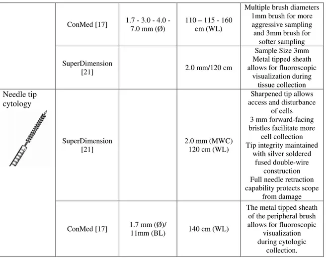
The brushes are used for acquiring cells of the tissue from the region of interest by brushing the tool along the bronchial walls to collect cytological specimens. There are different sizes of the brush according the need. There is also a version with a needle, for aiding the anchorage to the tissue (Table 1.3).
Type of brush Company Brush Ø / brush length Min working cannel (MWC)/ working length (WL) Features Cytology Mediglobe [18] 2.0-2.8 mm (MWC) 120 cm (WL) Teflon tube Brush with ellipsoid tip Ergonomically designed
handle Versions with Extra
Scissor Olympus [19; 20] 1.2 – 2.0- 3.0- 5.0 mm (Ø)/ 10 mm (BL) 2.0-1.0mm (Ø)/ 6.0 mm (BL)_mini brusche 2.0 mm (MWC) 115 mm (WL) 1.2 mm (MWC for mini brushes)
Metal tip for visibility under fluoroscopy Extra fine nylon bristles
and optimal brush hardness Mini brushes for ultrathin channel (1.2
ConMed [17] 1.7 - 3.0 - 4.0 - 7.0 mm (Ø)
110 – 115 - 160 cm (WL)
Multiple brush diameters 1mm brush for more aggressive sampling and 3mm brush for
softer sampling SuperDimension
[21] 2.0 mm/120 cm
Sample Size 3mm Metal tipped sheath allows for fluoroscopic
visualization during tissue collection Needle tip cytology SuperDimension [21] 2.0 mm (MWC) 120 cm (WL)
Sharpened tip allows access and disturbance
of cells 3 mm forward-facing bristles facilitate more
cell collection Tip integrity maintained
with silver soldered fused double-wire
construction Full needle retraction capability protects scope
from damage
ConMed [17] 1.7 mm (Ø)/
11mm (BL) 140 cm (WL)
The metal tipped sheath of the peripheral brush allows for fluoroscopic
visualization during cytologic
collection.
Table 1.3: Brushes.
1.4.3
Needle
The needle is used to collect a tissue sample from a suspicious area of the lung or airway during a TBNA (Table 1.4).
Type of needle Company
Needle Ø / needle length (NL) Min working cannel (MWC)/ working length (WL) Features TBNA Mediglobe [18] 0.8 mm (21 G) (Ø) / 12mm (NL) 2.1mm (MWC)/ 100cm (WL) Coaxial Luer-Lock for Vacuum Syringe and Needle with Side
Hole Removable Safety Clip Lock Mechanism Automatic Needle Retraction by Pressing a Release Button
Olympus (SmoothShot) [19; 20] 0.8mm (21 G) (Ø)/ 13 mm (NL) 2.0 mm (MWC) 115 cm (WC) Smooth passage through the bronchoscope channel
even when the scope is sharply angulated For all tracheal and bronchial regions ConMed (WANG™) [17] 0.9-0.8-0.7mm (20-21-22 G) (Ø) / 13mm (NL)
Smooth hubs and retractable needles to
ensure safe scope passage. Clear PTFE catheters
to allow for direct visualization of
aspirate. Stiffer versions for
central lesions, flexible versions for peripheral lesions, or a combined version for both locations. Locking mechanisms to ensure needle stability when sampling. SuperDimension [21] 2.0 mm (MWC) 130 cm (WL)
Optimal needle length designed specifically for use
within the superDimension® Extended Working Channel (EWC) Catheter delivers unparalleled performance Clear catheter allows
for direct visualization
Table 1.4: Biopsy needle.
1.4.4
Foreign body retrieval devices
In the range of tools there are also devices for removing foreign bodies from inside the airways. Olympus has several types of grasping forceps, baskets and loops (Figure 1.15) that are disposable or reusable. Also Mediglobe provides this type of devices. For example the Polycatch retrieval device (Figure 1.16) with rotatable pouch and the foreign body forceps with rat teeth and detachable cups.
Figure 1.15: Grasping forceps [20]
Figure 1.16: a) Polycatch, b) forceps with rat teeth [18].
1.5
Diagnostic yield
The diagnostic success depends on various factors: skills of the examiner, nodule size and location, as well as on the characteristics of the lesion. The peripheral lung lesions and solitary pulmonary nodules (SPN) are more difficult to evaluate than larger, centrally located lesions. According to the lung cancer diagnosis and treatment guidelines issued by the American College of Chest Physician (ACCP), the diagnostic sensitivity of bronchoscopy for peripheral pulmonary lesions is 63%, whereas that for lesions <2cm is 34% [23] (Table 1.5).
Using an ultrathin bronchoscope facilitates the procedure of reaching the more peripheral targets, thereby increasing the diagnostic yield. M.Oki et al. [14] showed that the diagnostic yield of a thin bronchoscopy (3.5 mm outer diameter) for lesions <2 cm is 57% and ≥2 cm is 73%, values higher than the conventional bronchoscopy.
Naofumi Shinagawa et al. [24] analyzed the relationship between diagnostic sensitivity and the bronchial generation to which an ultrathin bronchoscope is inserted (Figure 1.17). The diagnostic yield is greater than the average diagnostic sensitivity of all lesions (66%) if the ultrathin bronchoscope can be inserted past the fifth bronchial generation.
Figure 1.17: Diagnostic sensitivity [24].
The diagnosis should be achieved by the method that has the most favorable risk/benefit ratio. The pulmonologists choose the appropriate tissue sampling method according to the type of lung cancer (SCLC or NSCLC), the size and location of the tumor, and the presumed stage of the cancer. They can use different tools for taking samples of the tissue or removing foreign object from the airways. The procedures mostly used are: TBLB, TBNA and transthoracic needle aspiration (TTNA).
The flexible bronchoscopy with direct biopsy forceps (TBLB) has a sensitivity of 0.74. A lower sensitivity is shown from washing and brushing techniques, 0.48 and 0.59 respectively. These numbers were obtained from 35 studies of patients with centrally located disease [23]. The same procedures were used for patients with peripheral lesions. The results showed a sensitivity of 0.57 for the transbronchial biopsies, 0.54 for brush biopsy and 0.43 for washings. If TBNA is used the sensitivity reaches 0.65.
In summary, the sensitivity of all biopsy procedures performed with bronchoscopy is low, they do not necessary provide a definitive diagnosis. For this reason it can be necessary to perform a TTNA. This procedure has a higher sensitivity (approximately 0.94), but it increases the risk of complications as well as being an invasive procedure.
The goal of many ongoing projects is to increase the diagnostic yield of procedures with the bronchoscope, because it is a minimally invasive intervention and there is a low risk of complications. This research field is currently very active.
1.6
Endobronchial ultrasound (EBUS)
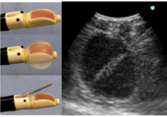
Bronchoscopy using endobronchial ultrasound (EBUS) is a relatively new procedure used in the diagnosis of lung diseases. With the bronchoscope, the operator navigates to the target region of the lungs, and a small ultrasound probe mounted on a video bronchoscope provides an “ultrasound view” of the tissue under the surface.
EBUS is helpful in detecting central nodules or primary tumors located close to the central airways and in mediastinum. The 6 mm diameter scope has a curvilinear ultrasound (US) probe at its distal end which provides a 50 degrees linear continuous B-mode ultrasound image, with color Doppler capability to aid identification of vascular structures (Figure 1.18). Proximal to the US probe, and at 30 degrees to the long axis of the bronchoscope, are a fiber optic lens and a biopsy channel, through which a 22- or 21G biopsy needle can be passed. A disposable latex balloon is placed over the US probe, which is inflated with sterile water to provide a fluid interface between the probe and the tracheal wall.
EBUS also allows detection of peripheral lesions by using an ultra-miniature radial probes (Figure 1.19).
The latest development in the field of EBUS is the use of EBUS-TBNA scopes that provides real time images during needle aspiration procedures [9]. These images help the doctor to reach the target with the biopsy needle and take a sample of the lesion.
Figure 1.19. Radial probe w/balloon and radial image view (360 degrees)
EBUS can increase the diagnostic yield of TBNA to 94%. However, this procedure is limited by the bronchoscope diameter and for this reason it can only be applied in regions of the lungs directly accessible by a bronchoscope [25].
1.7
Navigation systems
Traditional bronchoscopy uses biopsy tools to take samples of the lesions under fluoroscopic guidance, but many pulmonary noduli are not visible with this method. When the physician views a lesion in the CT images, he can decide to perform a bronchoscopy for taking samples of the tissue. To do this procedure he must be able to reach the lesion with the biopsy tool. Generally the physicians don’t know the entire branching structure of the lungs, so when they reach the peripheral areas they can easily get disoriented. Furthermore the lungs move during the procedure due to the breathing and the heartbeat or coughing of the patient, so the intraoperative situation is continually changing.
To solve these problems bronchoscopic navigation systems has been developed which can give the following information [26]:
- Current location of the bronchoscope overlaid onto CT images; - Anatomical structures located behind the bronchial wall;
- Anatomical names of branches currently observed (can be overlaid to create an augmented reality visualization);
- Paths to the desired location where the biopsy should be performed (target visualization).
The navigation system allows to localize and visualize the bronchoscope camera and biopsy tools in relation to preoperative CT images [27], and it guides the pulmonologist to the desired location.
A typical navigation system is composed of [28]:
- Computer workstation: it imports preoperative CT, MRI or other image data. Then typically a point-based registration is performed, that is the system determines the transformation between CT image coordinates and the patient reference coordinates (anatomic landmarks). After that different graphical indications of tool location and anatomy are shown in a video to give some information to the surgeon (Figure 1.20).
- Tracking device: also known as localizer. This device is used to track the positions of instruments relative to patient anatomy via a reference position sensor attached to the patient or the table. The optical tracking system (OT) and the electromagnetic tracking system (EMT) are the two types mostly utilized in medicine. The OT requires to maintain a line-of-sight between the tracking device and the sensor on the instrument to be tracked. The EMT doesn’t have this characteristic, therefore it is able to track instruments such as flexible catheter and needles inside the body (Figure 1.21).
- Associated tools with marker devices: the position and orientation of these tools are continuously measured relative to the navigational tracker.
Figure 1.20: Screenshot of the navigation software providing guidance for TBNA interventions
a) b)
Figure 1.21: Examples of tracking systems: a) Polaris Accedo (optical tracking), b) MiniBird Ascension (electromagnetic tracking).
A navigation system should be robust, precise and accurate. It is precise if it returns the same measurement each time and it is accurate if the mean measurement that the system provides is very close to a reference true value [28]. The robustness is important to be able to track even is there are metallic objects in the vicinity. Some metals cause distortions, but others don’t. The different tracking systems on the market have different pros and cons in this respect.
1.7.1
Tracking device
Intraoperative tracking is the process which uses tracking devices to localize the bronchoscope position and orientation relative to the anatomical environment being navigated [15]. There are three methods available for bronchoscopic tracking:
1. Electromagnetic tracking (EMT); 2. Image-based tracking (IBT); 3. Hybrid approaches.
Electromagnetic tracking
EMT uses the attenuation of oriented electromagnetic signals to determine the absolute position and orientation of a tracker relative to a source, which generates an EM field in which the sensor can be located.
Generally EMT for bronchoscopy includes:
- A small sensor used to track the position and pose of a bronchoscope or biopsy tool relative to an EM field generator [15]. It can be encapsulated in the tip of a flexible catheter pushed through the working channel of a bronchoscope or rigidly attached
to its distal end. It is possible to have more than one sensor to improve the accuracy and robustness of tracking.
- An EM field generator.
- A processing unit to read the different sensors in coordination with the EM field settings.
- A tracking software.
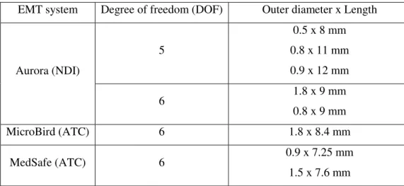
The products commercially available today include the Aurora (Northern Digital Inc., NDI) (Figure 1.22), microBIRD (Ascension Technology Corp., ATC) (Figure 1.23) and MedSafe (ATC) (Figure 1.24). All these systems are able to track miniaturized sensors, which can be integrated into tools for in vivo applications (biopsy tools for bronchoscopy) (Table 1.6).
a) b) c)
Figure 1.22: a) field generator, b) sensor, c) system control unit (Aurora).
a) b)
Figure 1.23: a) Electronic Unit, field generator, b) sensor (MicroBird).
a) b)
EMT system Degree of freedom (DOF) Outer diameter x Length Aurora (NDI) 5 0.5 x 8 mm 0.8 x 11 mm 0.9 x 12 mm 6 1.8 x 9 mm 0.8 x 9 mm MicroBird (ATC) 6 1.8 x 8.4 mm MedSafe (ATC) 6 0.9 x 7.25 mm 1.5 x 7.6 mm
Table 1.6: Table of the miniaturized sensors.
Image-based tracking (IBT)
To track the bronchoscope IBT compares virtual bronchoscopic (VB) images derived from the pre-operative CT image with the real bronchoscopic (RB) images obtained from the camera at the tip of the bronchoscope. Using the video images directly, the system can do the registration between the 3D CT image and the real-time 2D image for calculating the bronchoscope’s 6D position and orientation. During the bronchoscopy 3D image data, 2D CT slices and live video are simultaneously visible on the monitor (Figure 1.25). The IBT doesn’t require additional external hardware, otherwise it requires continuous and unobstructed video input, which can be a challenge to achieve. For this reason it can only be used in regions that are accessible to the bronchoscope [15], and when the view is obstructed from blood or mucus a resynchronization has to be performed.
Hybrid System
A hybrid system uses both EMT and IBT for tracking of the bronchoscope in peripheral airways. An estimate of the position and the pose of the tip is provided by the EMT sensor, then it is improved by CT-video registration.
Timothy D. Soper et al. [15] showed a custom hybrid system for tracking of an ultrathin single-scanning fiber bronchoscope (SFB) in peripheral airways using an EMT system and miniature sensor and IBT of the SFB video with a preoperative CT. They used this method for in vivo navigation within a live pig. Their results showed that the hybrid approach provides a more stable and accurate means of localizing the SFB intraoperatively. The position tracking error was less using hybrid method than using EMT or IBT [15].
1.8
Navigation in bronchoscopy
Four navigation systems are currently available for bronchoscopy: i-LogicTM System (SuperDimension®), SpinDrive® (Veran Medical Technologies), LungPoint® (Broncus), Bf-Navi® (Olympus Medical Systems, Japan). The first two use an EMT system for tracking, while LungPoint® and Bf-Navi® use a virtual navigation system with synchronization between VB and RB.
1.8.1
Electromagnetic navigated bronchoscopy
Electromagnetic-based navigation in bronchoscopy (ENB) enhances standard bronchoscopy by providing a 3D roadmap of the lungs and real-time information about the position of the steerable probe with the EMT system.
ENB requires registration between the coordinate system of the pre-operative CT image and that of EMT system. Today different methods of registration are available: manual marker-based registration [29], automated registration [7] and marker-free registration [27]. Some studies have been done taking respiratory motion into account. Feuerstein et al. [27] use a second cutaneous EMT sensor attached to the patient’s chest which allows to detect the respiratory phase (inspiration or expiration). They reduce the registration error caused by the motion of the body, such as respiratory motion, from 3.5 mm to 2.8 mm [7]. One dilemma with such methods is that the actual movement of the lung is different than the chest wall movement and therefore a modeling step is needed to account for this.
The first animal study with ENB was performed by Becker et al. in 2003 and the same group published the first human study in 2006 [30; 31].
The diagnostic yield of the flexible bronchoscopy (range of 36%-63% for peripheral lesions) can be enhanced by using ENB. The results of different studies (Figure 1.26) show an increase in the diagnostic yield to 69-82% with error around to 6 mm for the registration and to 10 mm for the navigation.
Figure 1.26: Diagnostic yield of ENB [29].
B. Lamprecht et al. demonstrated that the diagnostic yield increases on average from 80% to 87.5% after about 30-40 ENB procedures for each bronchoscopist, even if the diameter of the lesion decreases (Figure 1.27). It means that the diagnostic is independent form the size of the lesion, but depends on the experience of the bronchoscopist [32].
Figure 1.27. Number of performed ENB procedures and corresponding mean diameter of lesion [32].
The use of ENB reduces the risk pneumothorax, which is quite high with TTNA, and it allows to perform biopsy with a good diagnostic rate. The only disadvantage is the cost of the hardware and the software that can be quite expensive.
SUPERDIMENSION
SuperDimension® offers an electromagnetic navigation system (i-Logic InReach system) with all the components and the tools necessary for performing the procedure: a disposable extended working channel (EWC) with a disposable locatable guide (LG) that contains an EM tracking sensor, a catheter with different tools for biopsy and the planning and navigation software (Figure 1.28).
In September 2004 this system was cleared for marketing by the U.S. Food and Drug Administration (FDA). Nowadays is the most used navigation system, it can be summarized in the following four steps [9; 33]:
1. CT images of the patient are obtained with high resolution chest CT. These are reconstructed into a 3D volume and the physician utilizes these images to plan pathways to the target.
2. The plan is loaded into the navigation system computer and the patient is positioned either on a portable tracking system board (the field generator) or the tracking system field generator is placed next to the patient table. The field generator uses a low-frequency AC current to produce a magnetic field. A steerable navigational catheter is inserted through the extended working channel of the bronchoscope (EWC) into the airways. At the tip of this probe there is an electromagnetic sensor that detected real time position and orientation. It is used to register key points of the patient’s anatomy in the magnetic field.
3. Using the plan pathway (step 1) and real-time information (step 2) the operator guides the bronchoscope with the steerable probe toward the lesion, then he steers and advances the probe until it is sufficiently near the target to allow biopsy.
4. Once at the desired location the working channel is locked in position and the probe is removed. Then biopsy tools can be inserted through the working channel for performing the biopsy. Fluoroscopy or radial endobronchial ultrasound can be used during biopsies to locate the real-time position of the lesion.
a)
b) c)
d)
Figure 1.28: a) Bronchoscopy with i-Logic InReach system. b) Planning. c) Procedure.
d) i-Logic components.
VERAN
SpinDrive® is the electromagnetic navigation system proposed by Veran Medical Technologies, it was cleared for marketing in December 2009.
The doctor can plan the bronchoscopy before performing it thanks to SpinDrive® CT Planning Software, then the instruments such as biopsy needles could be used to biopsy. They have miniture EM sensors integrated directly into the tip (Always-on tip Tracked™ instruments). The SpinDrive® system utilizes a software that performs an automatic 4D pathway registration to monitor and compensate for patient respiration and movement (Figure 1.29).
a)
b)
Figure 1.29: a) Bronchoscopy with SpinDrive (Veran). b) View during the procedure: CT image,
3D model, the position of the tip [34].
1.8.2
Virtual bronchoscopy navigation
Virtual bronchoscopy navigation (VBN) is a method in which the virtual bronchoscopic images of the bronchial path to a peripheral lesion are used as a guide to navigate the bronchoscope. The virtual images are automatically produced then they are matched with real images. The time needed for the guidance of bronchoscope is short, because the position of the tip and the direction of its advancement need not be checked by fluoroscopy
or other imaging modalities. The VBN facilitates the guidance of the bronchoscope by indicating the bronchial route to the lesion, this improves the diagnostic yield [35]. This method is mainly a planning tool but it can also be used during bronchoscopy, but manual resynchronization might be needed, since no EM tracking is integrated to accomplish this automatically.
However the VBN presents some limitations. As mentioned, there is the necessity of manually fitting the virtual images to the real ones and there is no method to confirm the arrival of the bronchoscope at the target. One group has been able to automatically match virtual and real images and VBN can be combined with fluoroscopy, CT or EBUS to confirm the arrival [35].
BRONCUS
The LungPoint® virtual bronchoscopic navigation was cleared for marketing in December 2008. It provides a map and visual directions through the lung airways to a target. The LungPoint Planning is included in the system and it helps the doctor to calculate the better path to reach the target before performing the procedure. LungPoint® uses the bronchoscopic video to guide the physician. There is no need for additional hardware or specific catheters (Figure 1.30-Figure 1.31).
Figure 1.30: Multiple views: virtual bronchoscopic animation, the 3D airway tree and CT images [36].
Figure 1.31: Virtual animation synchronized with live movements [36].
NAVI
The Bf-Navi ® (Olympus Medical System, Japan) is a VBN software program that generate a three-dimensional roadmap using chest CT scan data in order to help the doctor find an optimal route to the target. When the route is determinate, the virtual images of the bronchial tree are automatically prepared (Figure 1.32).
Figure 1.32. Virtual images of the route. The circle indicate the direction of the targer [35].
Takashi Ishida et al. used this system to show that diagnostic yield is higher in the VBN-assisted (VBNA) than in the non-VBN-VBN-assisted (NVBNA), 80.8% and 67.4% respectively [37] (Figure 1.33).
Chapter 2
2
Biopsy sampling of lung tissue
2.1
Biopsy procedure
The doctor utilizes the biopsy tools to reach the tissue of interest and take a specimen, which will be examined afterwards in the laboratory. The goal is to discover if the lesion visualized in the CT-images is a malignant tumor or not, improving the possibility of an early diagnosis. The conventional bronchoscope is not able to advance more than to the third or fourth bifurcation (starting from the main division of the trachea), so when the lesion is peripherally located, the physician cannot reach the target with direct visual inspection. The ultrathin bronchoscope allows to arrive closer to the peripheral lesion, but there are lesions that cannot be reached even by a thinner bronchoscope. In these cases the doctor pushes the biopsy tool out of the working channel up to the wall of the lesion. The fluoroscopy helps the doctor to visualize the position of the tool during the procedure when the lesion is not visible from the tip of the bronchoscope. Nowadays it’s possible to use a navigation system in addition to or instead of the fluoroscopy to improve the tracking of the tool.
2.1.1
Bronchoscopic biopsy without navigation system
During conventional bronchoscopic biopsy sampling of a suspicious lesion, the pulmonologist uses a biopsy forceps, brush or needle inserted through the working channel of the bronchoscope and advanced until the wall of the lesion. The scope is used to aim for the lesion based on knowledge about the location from previous CT images (central lesion are bronchoscopic visible, and the bronchoscopist advance the scope directly to the target lesion having the CT images in mind to find the lobar or segment location suggested on the CT images). The biopsy tool is then extended and visually controlled by the video. If the lesion is located outside the bronchus and therefore not visible by the scope, one has to extend the biopsy forceps blindly to the approximate length to the lesion to acquire a cytological sample (most often FNA). If the lesion is not visible from the tip of the
bronchoscope, the fluoroscopy may be used to aid the tracking of the biopsy tool and a tissue biopsy is collected (Figure 2.1).
If the specimen results is negative after the pathologist has inspected it, this can either mean that the lesion is not malignant or that the biopsy sample was not taken from the lesion. This is of course true for the other methods as well.
Figure 2.1: Bronchoscopic biopsy with fluoroscopy [14].
2.1.2
Bronchoscopic biopsy with navigation system
Nowadays there are different types of navigation system available as mentioned in the paragraph 1.8: i-LogicTM System (SuperDimension®), SpinDrive® (Veran Medical Technologies), LungPoint® (Broncus), Bf-Navi® (Olympus Medical Systems, Japan). SuperDimension’s technology consists in a planning and navigation software, a disposable extended working channel (EWC) and a tracked guide wire, which is passed through the EWC into the working channel of the bronchoscope and it is navigated towards the target. The tracked catheter is advanced and steered by manipulating the handle according to the bronchial tree (Figure 2.2). The EWC is locked as soon as it is adjacent to the target, then the guide wire is withdrawn and a biopsy tool can be inserted through the empty lumen.
Figure 2.2: Electromagnetic navigation bronchoscopy setup [38].
The doctor has different simultaneous information that are visible on a screen: axial and sagittal CT view, MIP view, dynamic 3D view, tip view and local view (Figure 2.3).
Figure 2.3: SuperDimension procedure screen showing 6 viewports. The green sphere is the target and the yellow ring represents the catheter handle [39].
Veran, as SuperDimension, presents a software based on an electromagnetic tracking system: SpinDrive. It needs specifics tools, Always-on tip Tracked™ instruments, to see inside the airways and, if necessary, perform biopsy procedures.
During a bronchoscopic biopsy guided by LungPoint®Virtual Bronchoscopic Navigation the doctor can see both the live and virtual views, where the pink circle identifies the target (Figure 2.4). Thanks to a 3D view of the airways (Figure 2.5) he knows where is the target into the bronchial tree and what is the best pathway to reach it (blue line).
Figure 2.4: Bronchoscopic needle biopsy guided by LungPoint navigation [40].
Figure 2.5: Airway tree with target [36].
Due to the small dimension of the biopsy tool the tissue sample could be too small for the examination, so the physician might have to repeat the biopsy procedure for extracting more tissue.
During the biopsy procedure, the physician assumes that the catheter remains close to the target, but this cannot be assumed with certainty. When the guide wire is withdrawn the doctor moves the biopsy tool with the aid of the fluoroscopy or radial endobronchial ultrasound to locate the real-time position of the tip.
2.1.3
Steerable and trackable approach with navigation and
new biopsy tool
In addition to introducing steerability of the catheter with tracking to better reach a target, once the tissue is sampled, the biopsy tool should have a mechanism to prevent the loss of possible tumor tissue during withdrawal of the biopsy tool. For example during the TBNA the doctor pushes the needle inside the lesion for taking the specimen, but when it is retracted the tissue can be lost or contaminated with normal tissue. If the sheath is positioned in contact with the surface of the tissue, the said problem is reduced. It can be useful to have a “closed needle” to easier enter the tool into the lesion, open it once inside the lesion, and then close it again before it is retracted.
A novel design of such a “closed needle” biopsy tools could be useful improving the diagnostic success rate, thereby optimizing the procedure. The biopsy tools must also have the right size and functionality to avoid the risk of complications, like excessive bleeding or pneumothorax. Problems associated with the biopsy procedure are the cause of the current low diagnostic yield, especially for the small peripheral lesion.
This project is developing solutions to improve the situation.
2.2
Biopsy needle
Transbronchial needle aspiration (TBNA) is the only technique that allows the bronchoscopist to sample tissue from beyond the confines of the endobronchial tree (Figure 2.6).
Transbronchial needles are designed to be able to pass through the working channel of a flexible fiberoptic bronchoscope and come in a variety of different styles.
The Wang retractable disposable needle was the prototype and remains the standard for comparison for TBNA. The entire needle apparatus consists of a retractable needle system that is 120 cm long. The inner sheath is tipped with a 21 gauge (diameter), 13 mm (length) needle.
The MW (Millrose/Wang) 122 is a single-lumen 22 gauge needle that is used for obtaining cytology specimens. This needle has an unobstructed lumen for optional suction (Figure 2.7).
The MW 319 is a dual needle. The inner and outer needles have beveled tips. The outer needle is 19 gauge and 15 mm, and the inner needle is a 21 gauge 3 mm. The inner needle serves as a trocar to ease penetration and prevent plugging with bronchial tissue. The inner needle is retracted to expose the outer needle for sampling. This needle is used for obtaining histology specimens. (Figure 2.8, Figure 2.9)
The MW 522 peripheral cytology needle is a single lumen needle and is a 13 mm long, 22 gauge. This needle provides the best flexibility for a peripheral specimen collection.
Figure 2.7. Wang cytology needle [22].
Figure 2.9. Distal end of a dual needle [35].
Common characteristics among these needles include: - A distal retractable beveled needle;
- A middle flexible catheter;
- A proximal control device that manipulates the movement of the needle, the stylet, or both, and a slide port trough which suction can be applied.
To obtain a specimen for cytological examination, the needle is introduced through the working channel with the tip concealed within the metal hub. Once the metal hub is visible from the distal end of the fiberoptic bronchoscope, the needle is advanced and then locked into place. The catheter is retracted until only the tip of the needle is visible. The goal is to penetrate the tracheobronchial wall as perpendicularly as possible.
The needle can be inserted through the bronchial wall with different methods such as Jabbing method, pushing method and cough method.
2.2.1
Tru-Cut biopsy
Transthoracic needle biopsy is usually done by an interventional radiologist. Under sterile conditions, local anesthesia, and imaging guidance, a biopsy needle is passed into the suspected lesion while patients hold their breath (Figure 2.10).
Figure 2.10. Transthoracic biopsy needle.
In transthoracic biopsy the doctor could use a cutting needle.
The needle is composed basically of an outer needle (cannula) and an inner needle (stylet), that is inserted into the hollow core of the cannula. The stylet includes a pointed, tissue penetrating distal end (Figure 2.11).
Figure 2.11: Needle Cook medical [22].
The conventional technique of biopsy with a cutting needle is the TruCut procedure. The needle has a sharp tip and a slot for accommodating the specimen. The slot is covered by a sheath which passes over the needle. At first the doctor pushes the needle into the lesion then slides the sheath, that has sharp edges, for cutting the specimen. After that the needle, the sheath and specimen are withdrawn (Figure 2.12).
Figure 2.12: Trucut needle biopsy [41].
At the first step it’s possible to push into the lesion both the needle and the sheath (sheathed needle technique), then the sheath is withdrawn and slid to cut the specimen (Figure 2.13).
Figure 2.13: Sheated needle technique [41].
This technique is very user dependent, in addition if the slot is not completely covered by the sheath the tissue can be lost during the withdrawing.
At present there is a histological Tru-cut needle available, it is made by Cook Medical (Figure 2.14). It uses a spring-loaded system to push the needle out from the sheath.
The idea to use a cutting needle in bronchoscopy could be evaluate. The needle has to have the right sizes to pass through the working channel of the bronchoscope and enable to take a right size of specimen. In this case an extra sheath, such as an extended working channel, is necessary (Figure 2.15).
Figure 2.15. Bronchoscope with cutting needle.
2.2.2
Patents of biopsy needles
John S. Fisher and Frederick Ahari [42] proposed a biopsy needle composed by (Figure 2.16):
- Outer needle: it is a hollow cylindrical needle with an elongated slot in the side wall and a pointed distal free end.
- Inner needle: it is a hollow tube disposed within the lumen of the hollow needle. It has a helical opening with sharp edges, its axis is oblique to the axis of the elongated slot.
When the needle is into the lesion the tissue is pulled into the slot by the vacuum. The inner tube is then rotated to create a scissors-like shearing action and cut the specimen.
Figure 2.16: Design biopsy needle.
The tool allows to take a sample of tissue without losing it during the way back, because the rotation of the inner needle closes the hole in the outer needle’s wall. If the tool is stiff and not too long, like in the transthoracic biopsy, there isn’t any problem to know how much the tip rotates, but for using this tool during a bronchoscopy the stiffness and the sizes have to be different.
A biopsy tool has the typical length of 1.2 m, it has to pass smoothly through the working channel of the bronchoscope, therefore it cannot have a rigid body. The doctor by rotating the handle on the head of the inner tool causes rotate of the tip, because of the characteristics of the tool it’s difficult to know exactly how much the tip has been rotated. In another patent John S. Fisher [43] described different designs of biopsy needle for scraping tissue of cellular size from a lesion. The needle can have two or three sharp edges. The first sharp edge scrapes the tissue during the proximal to distal travel and it is necessary for penetrating the tissue. The second and third sharp edges are provided by a slot formed in the needle and they scrape the tissue when the needle is inserted or retracted respectively. The proximal end of aspiration biopsy needle is connected with a source of negative pressure which creates a vacuum to pull the tissue into the slot.
In a first design the needle has the second edge coincident with the exterior surface of the needle (Figure 2.17).
Figure 2.17:Tip of the needle [43].
In the patent a lot of designs of needle are described, each has different types of edges and slots (Table 2.1).
Drawing Features
The second sharp edge is elevated with respect to the exterior surface of needle.
The second edge is recessed with respect to the exterior surface of needle.
Needle with the third sharp edge.
a)
The third edge is elevated (a) or recessed (b) with respect to the exterior surface of needle.
b)
A needle with two slots and two sharp edges.
A needle with one slot and two sharp edges. (One of the two edges is elevated or recessed with respect to the exterior surface of the needle)
The second sharp edge is pivotally mounted by transversely disposed hinge means. The edge can open for scraping tissue during distal to proximal travel of the needle.
The bending of the edge can be obtained by using different types of springs or hinge, made in nitinol or other suitable material.
Table 2.1. Edges and slots.
Empirical studies show that the cell-collection ability can be enhanced by using edge made of a polymeric material to which the cellules can cling.
The possibility to have an edge bendable would allow the opening and closing of the hole in the needle’s wall. The third edge could be bendable instead of the second edge, thus when the needle gets in the lesion the hole is open and when it’s withdrawn the edge closes the hole with the sample of tissue inside. There will be the loss of the sample during the retraction if the gap is not closed well, this should be avoided in a biopsy procedure.
Figueredo et al. [44] describe a biopsy needle with capture elements into the needle lumen. They are flexible members that allow the access of the tissue into the lumen and prevent the leakage of the sample during the retraction of the needle. These members comprise four retaining members (Figure 2.18), which extend from a needle sidewall and are spaced symmetrically about a center axis of needle lumen. They are coupled to needle by flexural hinge and have a bend of 30°, 45° or 60°.
When the needle is inserted into the lesion, the tissue pushes on surface a and the retaining member moves as shown in the Figure 2.19.
Figure 2.19. Needle’s tip.
When the needle is retracted the sharp edges of the retaining members cut the tissue and their consequent radially inward movement closes the passage, avoiding the exiting of the sample.
A single retaining member can be used as a tissue capture element (Figure 2.20). In this case the passage into the needle is not completely closed during the retraction.
Figure 2.20. Single retaining member.
The tissue capture element could be stationary, for example a ring with a smaller cross-sectional area than lumen of needle. The ring has sharp edges for cutting the tissue and take a sample (Figure 2.21).
Figure 2.21. Needle with ring inside its lumen.
Another type of needle is described, it has a sidewall opening and a distal end cutting edge. There is retaining members with sharp edge for capturing a sample of tissue (Figure 2.22).
Figure 2.22. Needle with cutting edge.
The retaining members allow the tissue to get inside the empty lumen of the needle during the introduction of the needle into the lesion, and keep the sample inside by closing the passage. The closing could be not complete therefore a part of the sample will be lost during the withdrawal. If the tip of the needle is open the lesion’s tissue is taken as well as the normal tissue, this is not a problem as long as the doctor can take big sample thus the pathologist can analyze the nature of the lesion.
Johnson et al. [45] describe the possibility to use a locatable guide of a navigation system with biopsy tools attached to its distal tip. The biopsy needle, described in this patent, has a scalloping blade on a side surface and a hollow interior cavity (Figure 2.23). The needle is advanced into the tissue then rotated for cutting a tissue sample with the blade.
Figure 2.23. Needle with a scalloping blade.
The needle is closed at the end but the slot under the blade remains open also when the tool is retracted, hence there is the risk of loss of the sample.
Chapter 3
3
New biopsy tool and new procedure
3.1
Features of the new biopsy tool
The normal tissue around a lesion might be compressed compared to normal lung tissue. When the standard biopsy tool such as the biopsy forceps is pushed forward towards the lesion, the compressed normal tissue may cause a situation where the sample from the forceps is taken from this normal compressed tissue instead of the lesion. By using a needle or at least something sharp that can get more easily inside the lesion the doctor can increase the success rate of biopsy sampling. It could be possible to use the standard biopsy forceps if, as first step, an anchoring to the lesion is performed. After that a sheath-catheter with two lumens can host in one lumen the thin anchor wire and in the other lumen the biopsy forceps. Once that the catheter is inside the lesion the biopsy tool is advanced to collect a sample of tissue.
The tip should be small enough to avoid the risks of bleeding or pneumothorax and also should be able to take a sufficient quantity of tissue. In addition to these characteristics a good biopsy tool should have a simple and passive mechanism to collect the sample and a design such as to allow an easy retrieval of the sample without contamination of tumor tissue during retraction.
3.2
Iterative design for new tool
Following the characteristics of the retaining members, described in the Figueredo’s patent, a new design of biopsy needle was conceived, made with an inner and an outer part. All the following drawings have been made by using Google Sketchup, a software for 3D modeling.
The outer needle has an empty lumen and sharp edges to help to get into the lesion. At the end part there is a kind of “non-return valve” for maintaining the sample inside the lumen and guide the inner part. This one is a thin tube with a tip in the shape of an umbrella. The sharp end facilitates the entrance of the tool into the lesion and the sharp edges of the tip allow a better collection of the sample (Figure 3.1).
Figure 3.1. New design of biopsy tool – valve closed.
When the biopsy tool is close to the lesion or inside, the doctor pushes forward the inner part. A sample of tissue is collected during the withdrawal of the thin tube thanks to the sharp edges present in the umbrella and at the end of the outer needle. The force of the tissue collected opens the valve getting the other part of the lumen (Figure 3.2). After that the inter tool is retracted and this movement presses the sample in the other direction causing the clamp of the valve.
![Figure 1.3: Bronchoscope [10].](https://thumb-eu.123doks.com/thumbv2/123dokorg/7574414.111927/11.892.232.694.845.1071/figure-bronchoscope.webp)
![Figure 1.14: a) Jaw span, b) Jaw capacity [17].](https://thumb-eu.123doks.com/thumbv2/123dokorg/7574414.111927/17.892.119.781.855.1113/figure-a-jaw-span-b-jaw-capacity.webp)
![Figure 1.16 : a) Polycatch, b) forceps with rat teeth [18].](https://thumb-eu.123doks.com/thumbv2/123dokorg/7574414.111927/22.892.118.834.303.447/figure-polycatch-b-forceps-rat-teeth.webp)
![Figure 1.17: Diagnostic sensitivity [24].](https://thumb-eu.123doks.com/thumbv2/123dokorg/7574414.111927/23.892.166.748.390.688/figure-diagnostic-sensitivity.webp)
![Figure 1.20: Screenshot of the navigation software providing guidance for TBNA interventions [25]](https://thumb-eu.123doks.com/thumbv2/123dokorg/7574414.111927/26.892.335.578.844.1090/figure-screenshot-navigation-software-providing-guidance-tbna-interventions.webp)
![Figure 1.27. Number of performed ENB procedures and corresponding mean diameter of lesion [32].](https://thumb-eu.123doks.com/thumbv2/123dokorg/7574414.111927/31.892.272.642.836.1053/figure-number-performed-enb-procedures-corresponding-diameter-lesion.webp)

