Nanostructured vectors for the transport of
active molecules through biological
membranes for pharmaceutical and
nutraceutical applications
Unione Europea UNIVERSITÀ DEGLI STUDI DI SALERNO
FONDO SOCIALE EUROPEO
Programma Operativo Nazionale 2007/2013
“Ricerca Scientifica, Sviluppo Tecnologico, Alta Formazione” Regioni dell’Obiettivo 1 – Misura III.4
“Formazione superiore ed universitaria”
Department of Industrial Engineering
Corso di dottorato in Ingegneria
Industriale-curriculum Ingegneria Chimica
(XV ciclo)
Nanostructured vectors for the transport of
active molecules through biological membranes
for pharmaceutical and nutraceutical
applications
Supervisor
Ph.D. student
Prof. Anna Angela Barba
Sabrina Bochicchio
Scientific Referees
Prof. Gaetano Lamberti
Prof. Sotiris Missailidis
Prof. Gabriele Grassi
Ph.D. Course Coordinator
Publications
(Inherent the Ph.D. project)
International Journals
Bochicchio S.; Dapas B.; Russo I.; Ciacci C.; Piazza O.; De Smedt S.; Pottie E.; Barba A.A.; Grassi G.; “In vitro and ex vivo delivery of tailored siRNA-nanoliposomes for E2F1 silencing as a potential therapy for colorectal cancer”; IJP - International Journal of Pharmaceutics, 2017, in press Bochicchio S.; Dalmoro A.; Recupido F.; Lamberti G.; Barba A.A.;
“Nanoliposomes production by a protocol based on a simil-microfluidic approach”; accepted as publication in “Lecture Notes in Bioengineering (LNBE)”, Springer Ed., 2017
Bochicchio S.; Dalmoro A.; Barba A.A.; D’Amore M.; Lamberti G.; “New preparative approaches of micro and nano drug delivery carriers”; Current
Drug Delivery, 2017, 14(2), 203-215
Dalmoro A.; Abrami M.; Galzerano B.; Bochicchio S.; Barba A.A.; Grassi M.; Larobina D.; “Injectable chitosan/b-glycerophosphate system for sustained release: gelation study, structural investigation, and erosion tests”;
Current Drug Delivery, 2017, 14(2), 216-223
Piazza O.; Russo I.; Bochicchio S.; Barba A. A.; Lamberti G.; Zeppa P.; Di Crescenzo V.; Carrizzo A.; Vecchione C.; Ciacci C.; “Cyclin D1 Gene Silencing by siRNA in ex vivo human tissues cultures”; Current Drug
Delivery, 2017, 14(2), 246-252
D’Apolito R.; Bochicchio S.; Dalmoro A.; Barba A. A.; Guido S.; Tomaiuolo G.; “Microfluidic investigation of the effect of liposome surface charge on drug delivery in microcirculation”; Current Drug Delivery, 2017, 14(2), 231-238
Bochicchio S.; Barba A.A.; Grassi G.; Lamberti G.; “Vitamin delivery: carriers based on nanoliposomes produced via ultrasonic irradiation”; LWT -
Food Science and Technologies, 2016, 69, 9 -16
Bochicchio S.; Dalmoro A.; Barba A.A.; Grassi G.; Lamberti G.; “Liposomes as siRNA delivery vectors”; Current Drug Metabolism, 2015, 15, 882-892
Barba A.A.; Bochicchio S.; Lamberti G.; Dalmoro A.; "Ultrasonic energy in liposome production: process modelling and size calculation”; Soft Matter, 2014, 10 (15), 2574 – 2581
Technical Journals
Bochicchio S.; Barba A.A.; Lamberti G.; “Nanovettori per il rilascio di farmaci antitumorali a base di acidi nucleici”; ICF- Chemical and Pharmaceutical Industry italian journal, n. 2, 2015
Dalmoro A., Bochicchio S., Lamberti G., d’Amore M., Barba A.A.; “Intensificazione di processo per nuove formulazioni”; AIDIC news, n. 3, 2016
Work submitted:
Bochicchio S.; Sala M.; Spensiero A.; Scala M.C.; Gomez-Monterrey I.M.; Lamberti G.; Barba A.A.; “On the design of tailored liposomes for peptide delivery”
Book Chapters
Bochicchio S.; Lamberti; Barba A.A.; “Phenomenological and formulation aspects in tailored nanoliposomes production” chapter in book Liposomes, InTech Ed., 2016, in press
Barba A.A.; Bochicchio S.; Dalmoro A.; Caccavo D.; Cascone S.; Lamberti G.; “Polymeric and lipid-based systems for controlled drug release: an engineering point of view; submitted as chapter in book to Pharmaceutical Nanotechnology, Volume IX: Sustained and controlled delivery systems, 2017Work in preparation:
Russo I.; Bochicchio S.; Piazza O.; LambertiG.; BarbaA.A.; Carrizzo A.; Vecchione C.; Zeppa P.; Iovino P.; Bucci, C.; Ciacci C.; “Control of inflammation progression in inflammatory bowel disease by suitable systems for cyclin d1 and e2f1 sirna delivery”; 2017
Proceedings
International
Russo I.; Bochicchio S.; Piazza O.; Barba A.A.; Lamberti G.; Carrizzo A.; Zeppa P.; Vecchione C.; Iovino P.; Bucci, C.; Ciacci C.; “Evaluation of efficacy of new vectors for siRNA CYCLIN D1 and E2F1 delivery in control of cancer progression in inflammatory bowel disease” presented to 12th Congress of European Crohn’s and Colitis organization 15-18/02/2017, Barcelona (Spain)
Bochicchio S.; Dalmoro A.; Recupido F.; Lamberti G.; Barba A.A.; “Nanoliposomes production by a protocol based on a simil-microfluidic approach”, presented to WIVACE 2016/BIONAM 2016 workshop, 4-7/10/2016, University of Salerno, Fisciano (SA) (Italy) and in proceedings on “Lecture Notes in Bioengineering (LNBE)”, Springer Ed.
Sala M.; Bochicchio S.; Spensiero A.; Scala M. C.; Gomez-Monterrey I. M.; Dalmoro A.; Lamberti G.; Campiglia P.; Barba A. A.; “Drug delivery system of peptide KRX 29 for treatment of chronic heart failure”, 15th Naples Workshop on Bioactive Peptides, 23-25 June 2016 Naples (Italy) D'Apolito R.; Bochicchio S., Tomaiuolo G.; Guido S.; Dalmoro A; Barba
A.A.; “Liposomes as drug delivery system: effect of superficial charge on margination in blood flow”, 14th European Symposium on Controlled Drug Delivery, 13-15/04/2016 Egmond aan Zee (Holland)
Bochicchio S.; Sala M.; Spensiero A.; Scala M.C.; Gomez-Monterrey I.M.; Campiglia P.; Dalmoro A.; Lamberti G.; Barba A.A.; “Liposomal structures design for peptide delivery in heart failure therapy”, 14th European Symposium on Controlled Drug Delivery, 13-15/04/2016, Egmond aan Zee (Holland)
Bochicchio S.; Dalmoro A.; Cavallaro G.; Craparo F.; Sardo C.; Giammona G.; Lamberti G.; Barba A.A.; “Nanoparticle based PHEA-DETA-PLA copolymer for NABDs delivery”, 14th European Symposium on Controlled Drug Delivery, 13-15/04/2016, Egmond aan Zee (Holland)
Barba A.A.; Bochicchio S.; Lamberti G; Sala M.; Campiglia P.; “Ultrasonic assisted production of nanoliposomes as peptide delivery vectors”, 42nd Annual Meeting & Exposition of the Controlled Release Society CRS 2015, 26-29/07/2015, Edinburgh (Scotland)
Cascone S.; Bochicchio S.; Lamberti G.; Titomanlio G.; Barba A.A.; “In vitro and in silico models in pharmacokinetic studies”, 42nd Annual Meeting & Exposition of the Controlled Release Society CRS 2015, 26-29/07/2015, Edinburgh (Scotland)
Caccavo D.; Cascone S.; Bochicchio S.; Lamberti G.; Dalmoro A.; Barba A.A.; “Hydrogels-based matrices behavior: experimental and modeling description”, 42nd Annual Meeting & Exposition of the Controlled Release Society CRS 2015, 26-29/07/2015, Edinburgh (Scotland)
Bochicchio S.; Dalmoro A.; Cascone S.; Lamberti G.; Barba A.A.; “dsDNA encapsulating in nanoliposomal structures towards gene therapies”, 1st International Congress of the Controlled Release Society- Greek local chapter 2015, 27-28/05/2015, Athens (Greece)
Dalmoro A.; Bochicchio S.; Apicella P.; d’Amore M.; Barba A.A.; “Micro and nano structured vectors for the drug delivery”, 21st International Congress of Chemical and Process Engineering CHISA 2014, 23-27/08/2014, Praga (Czech Republic)
Bochicchio S.; Dalmoro A.; Grassi G.; Lamberti G.; Barba A.A. “Vectors for vitamins delivery: nano liposomes production via ultrasonic irradiation”, 13th European Symposium on Controlled Drug Delivery, 16-18/04/2014, Egmond aan Zee (Holland)
Dalmoro A.; Bochicchio S.; Lamberti G.; d’Amore M.; Barba A.A. “Vectors for vitamins delivery: shell-core microparticles production via ultrasonic atomization and microwave stabilization”, 13th European Symposium on Controlled Drug Delivery, 16-18/04/2014, Egmond aan Zee (Holland)
Barba A.A.; Bochicchio S.; Dalmoro A.; Lamberti G. “Liposomal SUVs preparation by ultrasonic energy: a new approach based on a conventional technique”, 8th World Meeting on Pharmaceutics, Biopharmaceutics and Pharmaceutical Technology, 31/03-03/04/2014, Lisbona (Portugal)
National
Bochicchio S.; Dalmoro A.; Lamberti G.; d’Amore M.; Barba A.A. “Chemical Engineering approaches in the production of dedicated systems for the dosage of active molecules” Microwaves and ultrasound as process intensification tools for drug carriers production, national convention GRICU 2016, 12-14/09/2016, Anacapri (NA), Italy
Barba A.A.; Lamberti G.; Dalmoro A.; Bochicchio S.; d’Amore M. “Nuovi sviluppi della terapia genica: approcci multidisciplinari al dosaggio controllato di siRNA”, National Workshop, 14-15/09/2015, University of Salerno, Fisciano (SA), Italy
d’Amore M.; Dalmoro A.; Bochicchio S.; Lamberti G.; Barba A.A. “Produzione di sistemi di rilascio ottimali per i Nucleic Acid Based Drugs in terapie antitumorali”, Le Giornate del Farmaco, I edizione , 12/04/2013 University of Salerno, Fisciano (SA), Italy
Contents
Publications ... iii
Proceedings ... v
Contents ... I
Figures index ... VII
Tables index ... XI
Abstract ... XV
1. Topic and aims ... 1
1.1 Nanostructured vectors ... 2
1.2 Aims of this thesis ... 2
1.3 Planned activities ... 3
Gantt _____________________________________________ 3
1.4 Outline of the thesis ... 4
2. Introduction ... 7
2.1 Importance of microencapsulation processes ... 8
2.1.1 Critical issues in microencapsulation ________________ 8
2.1.2 Materials of vectors ______________________________ 9
2.2 Lipid carrier systems for therapeutic and
nutraceutical agents delivery: liposomes ... 10
2.2.1 Critical issues in therapeutic and nutraceutical agents
delivery _______________________________________ 13
3. Liposomes production: State of the art ... 17
3.1 Lipid carrier production ... 18
3.1.1 Methods for lipid nanoparticles production __________ 18
3.1.1.1 High Pressure Homogenization 18
3.1.1.2 Microemulsion 19
3.1.1.3 Emulsification-Solvent Evaporation 20
3.1.1.4 Phase Inversion Technique 20
3.1.2 Methods for liposomes production _________________ 21
3.1.2.1 Lipid hydration techniques 21
3.1.2.2 Liposome size reduction 25
3.1.2.3 Removal of non-encapsulated drugs 25
3.1.2.4 Liposomes stabilization 26
3.1.2.5 Recent development in liposomes production techniques 26
4. Novel developed techniques for liposomes production31
4.1 Ultrasound assisted Thin Film Hydration: layout,
principles and phenomenological aspects ... 32
4.1.1 Generalities ___________________________________ 32
4.1.1.1 Principles of the ultrasound process and benefits 32
4.1.2 Experimental section ____________________________ 34
4.1.2.1 Materials 34
4.1.2.2 Layout 34
4.1.2.3 Liposomes preparation 35
4.1.2.4 Modelling 36
4.1.3 Results and discussion ___________________________ 41
4.1.3.2 Part 2: Experimental results description and interpretation 43
4.1.4 Remarks ______________________________________ 48
4.2 Simil-microfluidic liposome preparation method:
layout, principles and phenomenological aspects ... 49
4.2.1 Generalities ___________________________________ 49
4.2.2 Experimental section ____________________________ 50
4.2.2.1 Materials 50
4.2.2.2 Layout 50
4.2.2.3 Liposomes preparation 54
4.2.2.4 Process productivity evaluation 55
4.2.2.5 Scale up of the ultrasound assisted homogenization process 55
4.2.3 Results and Discussion __________________________ 56
4.2.3.1 Simil-microfluidic apparatus 56
4.2.3.2 Unloaded nanoliposomes production: process parameters effect on
dimensional features 59
4.2.3.3 Productivity of the process 61
4.2.3.4 Scale up of the ultrasound assisted homogenization process 62
4.2.4 Remarks ______________________________________ 64
5. Applications ... 65
5.1 Liposomal vectors for vitamins delivery ... 66
5.1.1 Generalities ___________________________________ 66
5.1.2 Materials _____________________________________ 67
5.1.3 Methodology and sperimental set-up _______________ 67
5.1.3.1 Preparation of MLVs and SUVs loaded with vitamin B12 67 5.1.3.2 Preparation of MLVs and SUVs loaded with vitamin α-tocopherol69 5.1.3.3 Preparation of MLVs and SUVs loaded with vitamin ergocalciferol70
5.1.3.4 Physicochemical characterization of vesicles 70
5.1.3.5 Evaluation of vitamins encapsulation and stability test 70
5.1.4.1 MLVs production from lipidic film via ultrasonic irradiation: loading
and characteristics 72
5.1.4.2 SUVs production from lipidic film via ultrasonic irradiation: loading
and characteristics 73
5.1.5 Discussion ____________________________________ 74
5.1.5.1 Thin film hydration technique assisted by ultrasonic irradiation 74
5.1.5.2 Vitamins solubility and SUVs load analysis 76
5.1.6 Remarks ______________________________________ 79
5.2 Nanoliposomes vectors for ferrous sulfate delivery 81
5.2.1 Generalities ___________________________________ 81
5.2.2 Materials _____________________________________ 82
5.2.3 Methodology and sperimental set-up _______________ 82
5.2.3.1 Preparation of iron loaded nanoliposomes 82
5.2.3.2 Physicochemical characterization of vesicles 84
5.2.3.3 Evaluation of iron encapsulation and stability test 84
5.2.4
Results and Discussion _______________________ 86
5.2.4.1 Iron loaded nanoliposomes production 86
5.2.4.2 Iron loaded nanoliposomes characterization 87
5.2.4.3 A comparison between the simil-microfluidic apparatus and the
classical bench scale techniques 92
5.2.5 Remarks ______________________________________ 94
5.3 Nanoliposomes vectors for peptide delivery ... 95
5.3.1 Generalities ___________________________________ 95
5.3.2 Materials _____________________________________ 96
5.3.3 Methodology and sperimental set-up _______________ 96
5.3.3.1 Peptide KRX29 synthesis 96
5.3.3.2 MLVs and SUVs preparation at different charge ratio 97
5.3.3.3 MLVs and SUVs physicochemical characterization 98
5.3.3.4 Determination of encapsulation efficiency, peptide KRX29 recovery
5.3.3.5 MLVs at constant charge ratio but with different peptide concentration:
preparation and characterization 100
5.3.3.6 Stability test 101
5.3.4 Results ______________________________________ 102
5.3.4.1 MLVs and SUVs at different charge ratio 102
5.3.4.2 MLVs at constant charge ratio and different peptide concentration105
5.3.4.3 Stability test 106
5.3.5 Discussion ___________________________________ 107
5.3.5.1 Effect of formulation 108
5.3.5.2 Efficacy of ultrasound assisted technique in KRX29-liposomes
production 113
5.3.6 Remarks _____________________________________ 114
5.4 Nanoliposomes vectors for dsDNA delivery ... 115
5.4.1 Generalities __________________________________ 115
5.4.2 Materials ____________________________________ 115
5.4.3 Methodology and sperimental set-up ______________ 116
5.4.3.1 Phosphate buffer solution preparation 116
5.4.3.2 Tris-buffered saline solution (TBS) preparation 116
5.4.3.3 Duplex-DNA generation 116
5.4.3.4 dsDNA PolyAcrylamide Gel Electrophoresis (PAGE) analysis 117
5.4.3.5 Preparation of cationic SUVs containing dsDNA 117
5.4.3.6 Physicochemical characterization of vesicles 118
5.4.3.7 Evaluation of dsDNA load and stability test 119
5.4.4 Results ______________________________________ 119
5.4.4.1 dsDNA purity and integrity 119
5.4.4.2 Cationic liposomes: loading and characteristics of the tree formulations 120
5.4.5 Discussion ___________________________________ 124
5.4.5.1 Ultrasound assited technique in dsDNA-liposomes production 124
5.4.5.2 Including DOTAP in liposomes formulation 124
5.4.5.3 Effect in changing DOTAP/DNA charge ratio on liposomes final
5.4.6 Remarks _____________________________________ 127
5.5 Nanoliposomes vectors for siRNAs delivery ... 129
5.5.1 Generalities __________________________________ 129
5.5.2 Materials and methods _________________________ 131
5.5.2.1 Unloaded and siRNA loaded vesicles preparation 131 5.5.2.2 Unloaded and siRNA loaded vesicles characterization 133
5.5.2.3 Cell culture condition 134
5.5.2.4 Colon tissue culture condition 134
5.5.2.5 Uptake study in tissue biopsies 135
5.5.2.6 siRNA-SUVs transfection in cells 135
5.5.2.7 siRNA-SUVs transfection in colon tissue 136
5.5.2.8 Evaluation of E2F1 protein expression after transfection in tissue:
Western blot 137
5.5.3 Results and discussion __________________________ 138
5.5.3.1 Unloaded and siRNA loaded vesicles 138
5.5.3.2 Effects of siRNA-nanoliposomes on the vitality of human colon
carcinoma cell lines 143
5.5.3.3 Uptake study in colon tissue cultures 146
5.5.3.4 Evaluation of E2F1 protein expression after transfection in tissue:
Western blot 147
5.5.4 Remarks _____________________________________ 153
6. Concluding Remarks ... 155
Appendix ... 159
Pubblications ... 160
National Journals _______________________________ 166
Book chapter __________________________________ 167
References ... 169
Figures index
Figure 1. Cellular mimetic behaviour of liposomes as delivery systems (van der
Meel et al., 2014) ... 10
Figure 2 Liposomes representation (Deshpande et al., 2013) ... 11
Figure 3. Process schematization of nanoliposomes production through the ultrasound assisited Thin Film Hydration method; the main steps are reported: lipids/organic solvent solution (1) is introduced in a rotary evaporator (D-1) where solvents are evaporated (2) leading to the formation of a dryied lipid film (3) which is then hydrated (4) and stirred (D-2) After this step a suspension containing MLVs is produced and submitted to a duty cycle sonication process (Z-1) thus producing SUVs (5), finally recovered ... 34
Figure 4. The phenomena which take place during liposome formation ... 38
Figure 5. The model calculation flow-sheet ... 40
Figure 6. Output of the calculation model for the simulated test ... 41
Figure 7. Output of the calculation model for real test Run 1 ... 44
Figure 8. Experimental and calculated liposome diameters for Run 1 ... 46
Figure 9. Output of the calculation model for the real test Run 2.a ... 46
Figure 10. Experimental and calculated liposome diameters for Run 2.a ... 47
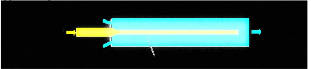
Figure 11. Process schematization of nanoliposomes production through the semicontinuous simil-microfluidic apparatus; the main sections are reported: feeding, pumping, production, homogenization and recovery. From the tanks (D-1, D-2) lipids/ethanol and water solutions are pushed through peristaltic pumps (G-1, G-2) to the production section (I-1) were nanometric vesicles are formed. The hydroalcoholic solution (7) is recovered in a tank (D-3) and subjected to a duty cycle sonication process (Z-1). Finally the suspension (8) is recovered and characterized ... 52
Figure 12. Representation of the liposome formation process by a microfluidic approach ... 58
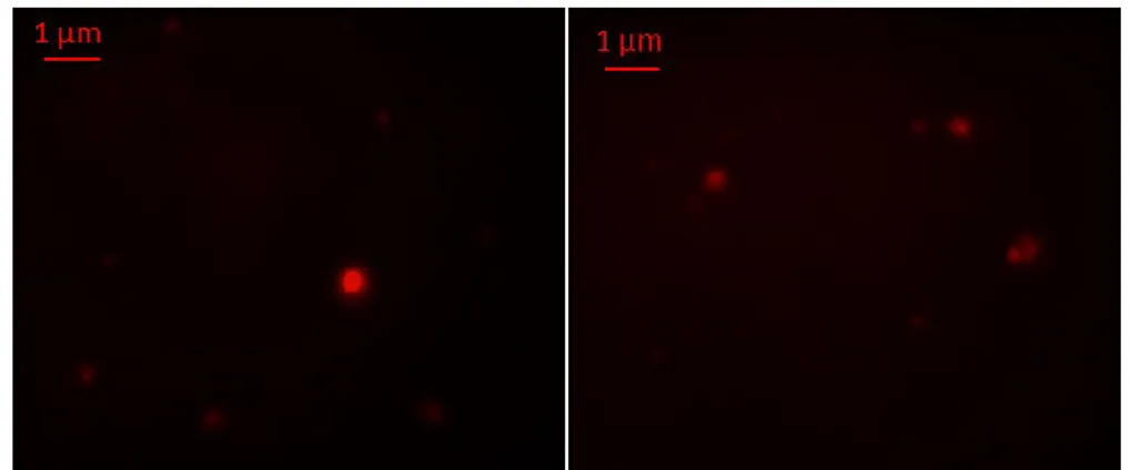
Figure 13. Fluorescence microscopy images of lipid vesicles labelled with Rhodamine B dye and visualized with a 100 X objective ... 59
Figure 14 A) Sonicated and not sonicated liposomes diameter size at different
volumetric flow rates ratio. B) Polydispersity Index (PDI) of sonicated and not sonicated liposomes at different volumetric flow rates ratio. Results are expressed as average of three determinations and reported along with the
standard deviation ... 60
Figure 15 A) Sonicated and not sonicated liposomes diameter size at different
PC concentrations in the hydroalcoholic solution. B) Polydispersity Index (PDI) of sonicated and not sonicated liposomes at different PC concentrations in the hydroalcoholic solution. Results are expressed as average of three
determinations and reported along with the standard deviation ... 61
Figure 16 A) Sonicated and not sonicated liposomes number (Nlip) for unit
volume of solution obtained at different volumetric flow rates ratio. B) Sonicated and not sonicated liposomes number (Nlip) for unit volume of
solution obtained at different PC concentrations in the hydroalcoholic solution .... 61
Figure 17. Linearity between the number of duty cycle sonication rounds
applied and the power supplied to 110 ml nanoliposomes suspension ... 62
Figure 18 A) Liposomes diameter size after different number of duty cycle
sonication rounds. B) Polydispersity Index (PDI) of liposomes after different number of duty cycle sonication rounds. Results are expressed as average of three determinations and reported along with the standard deviation ... 63
Figure 19. Three main units of the simil-microfluidic set-up: a production unit
according to the simil-microfluidic approach, an ultrasound assisted
homogenization and recovery unit, a storage unit ... 63
Figure 20. Ultrasonic irradiation protocol (duty cycle, A) and its effect on
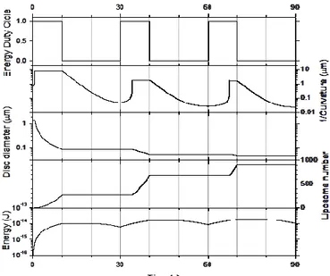
liposomal structures size (from Multilamellar Vesicles –MLVs- to Small Unilamellar Vesicles –SUVs-, B). In Figure A, starting with MLVs, following two irradiation rounds (all irradiation rounds are of 10 seconds and are all followed by a 20-second pause) Large Unilamellar Vesicles-LUVs with micrometric diameter size are obtained as showed by LUVs size distribution in Figure B. In Figure A, starting with LUVs, after three and four more irradiation rounds (50 and 60 seconds), SUVs of nanometric size are obtained as showed by SUVs size distribution in Figure B. ... 68
Figure 21. Optical microscope pictures (in bright field, Obj. 63X) of loaded
MLVs... 72
Figure 22. Vitamins retention in nanoliposomes (SUVs) and released in PBS.
Full symbols: squares, triangles and circles are referred to B12, tocopherol and ergocalciferol, respectively, and represent vitamin retained in the pellet. Open symbols: squares, triangles and circles are referred to B12, tocopherol and ergocalciferol, respectively, and represent the vitamin losses in phosphate
buffer solution ... 74
Figure 23. Fluorescence microscopy images of ferrous sulfate lipid vesicles
Figure 24. Iron retained in nanoliposomes and released in deionized water. The
open symbols are referred to the ferrous sulfate mass retained in the pellet, the full symbols represent the iron losses in deionized water ... 92
Figure 25. Optical microscope pictures (in bright field, Obj. 63 X) of KRX29
loaded liposomes at the different tested charge ratios ... 102
Figure 26. Peptide release profile in Tris-HCl solution at 37°C ... 107 Figure 27. Effect of PG/KRX29 (-/+) charge ratio on recovery efficiency ... 111 Figure 28. Effect of PG/KRX29 (-/+) charge ratio on encapsulation efficiency .... 111 Figure 29. Native PAGE of dsDNA. In the first lane a 100 bp ladder molecular
weight, in the second lane 2 µg of 21 bp dsDNA molecule (simulating "Homo sapiens siRNA probe Luciferase", 12833.4 g/mol). The relative band is well
visible ... 120
Figure 30 Optical microscope pictures (in bright field, Obj. 63 X and 100 X) of
dsDNA loaded Multilamellar Vesicles (MLVs), achieved respectively from the first, second and third formulation ... 121
Figure 31. Fluorescence microscope picture of dsDNA liposome vesicles (Obj
100X). DAPI-labeled dsDNA at left; Rhodamine B-labeled liposomes at right ... 121
Figure 32. dsDNA retention in Small Unilamellar Vesicles (SUVs) and release
in PBS: circles symbols are referred to dsDNA retained in the pellet; squares symbols are referred to dsDNA lost in phosphate buffer solution ... 123
Figure 33. RNA interference mechanism. Double-stranded RNAs (dsRNAs) are
processed by a complex consisting of Dicer and other protein activators and kinase into small interfering RNAs (siRNAs), which are loaded into Argonaute 2 (AGO2) and RNA-induced Silencing Complex (RISC). siRNAs are denatured and become single-strand molecules. The RISC is then active and, using the incorporated single-strand siRNA as template, recognizes the target mRNA to degrade (mRNA cleavage). RISC analyzes mRNAs and identifies as targets only the one with sequences perfect complementary to the 21-22 nucleotides of the siRNA, thus silencing their expression ... 129
Figure 34. Fluorescence microscopy images of merged lipid vesicles labelled
with Rhodamine B dye and visualized with a 100 X objective. A) Multilamellar Vesicles (MLVs) not subjected to the size reduction process; B) Large Vesicles (LVs) after two rounds of duty cycle sonication; C) Small Vesicles (SUVs) after six rounds of duty cycle sonication ... 139
Figure 35. Fluorescence images of siRNA loaded nanometric vesicles labelled
with Rhodamine B (on the left) and siRNA DAPI labelled (on the right)
visualized with a 100 X objective ... 140
Figure 36. UV-image of the agar gel after the electrophoretic assay. Following
the order: M.M., DNA 123 bp; Lane 1, Naked siE2F1-1117; Lane 2, Nanoliposomes-siE2F1-1117; Lane 3, Naked siE2F1-1324; Lane 4,
Figure 37. A) siE2F1-nanoliposome complexes and siE2F1-Lipofectamine®
2000 effects on HT29 cell vitality (*P=0.0001 siE2F1 vs siGL2 nanolipo, Unpaired t test; **P=0.01 siE2F1 vs siGL2 lipo2000, Unpaired t test with Welch correction); B) Un-specific effects on cell of nanoliposomes and Lipofectamine®2000 alone or loaded by the control siGL2; C) Cell counting after siE2F1-nanoliposomes transfection in HT29 cell line; D) E2F1 mRNA levels after siE2F1 –nanoliposomes transfection in HT29 cells (*P=0.0068 siE2F1 vs siGL2 nanolipo, Unpaired t test); E) siE2F1-nanoliposome complexes effects on LoVo109 cell vitality. All data are reported as mean ±
SEM ... 145
Figure 38. Fluorescence microscope images of colon tissue section after 24 h of
incubation with liposomes; 20 X objective. A) cell nuclei were visualized with DAPI, B) unloaded liposomes were Rhodamine B-labelled, C) cell nuclei and labelled liposomes images merge ... 146
Figure 39. E2F1 and GAPDH expressions in donor 1 after siRNA-nanoliposome
transfection at 100 nM. The proteins band quantification is normalized to Actin. The order of the analyzed samples is: Tissue incubated in 1. Only medium; 2. Medium with EC-LPS; Tissue transfected with: 3. siE2F1-1117 nanoliposomes; 4. siE2F1-1117 nanoliposomes in medium with EC-LPS; 5. siE2F1-1324 nanoliposomes; 6. siE2F1-1324 in medium with EC-LPS; 7. Positive control nanoliposomes and 8. Negative control nanoliposomes. Figures A are referred to E2F1 expression levels normalized respect to the not treated (NT) or basal sample. Figures B are referred to E2F1 expression levels normalized respect to the siRNA negative control (CTRL – siRNA) treated sample ... 148
Figure 40. E2F1 and GAPDH expressions in donors 2 (A) and 3 (B) after
siRNA-nanoliposome transfection at 200 nM. The proteins band quantification is normalized to Actin. The order of the analyzed samples is: 1. Tissue fragment incubated in medium; Tissue fragments transfected with: 2. siE2F1-1324 nanoliposomes; 3. Positive control nanoliposomes; and 4. Negative control nanoliposomes. E2F1 expression levels are normalized respect to the basal (not treated) sample and respect to the siRNA negative control (CTRL – siRNA)
treated sample... 150
Figure 41. E2F1 and GAPDH expressions in donors 4 (A) and 5 (B) after
siRNA-nanoliposome transfection at 200 nM and 50 nM. The proteins band quantification is normalized to Actin. The order of the analyzed samples is: 1. Tissue fragment incubated in medium; Tissue fragments transfected with: 2. siE2F1-1324 nanoliposomes; 3. Positive control nanoliposomes; and 4.
Negative control nanoliposomes. E2F1 expression levels are normalized respect to the basal (not treated) sample and respect to the siRNA negative control
Tables index
Table 1. Thesis map ... 5 Table 2 Liposomes classification by size / structure (Coelho et al., 2010, Samad
et al., 2007) ... 12
Table 3. Problems of naked siRNA for clinical uses (David et al., 2012) ... 14 Table 4. Summary of experimental results ... 45 Table 5. Reynolds number relative to the polar phase, the organic phase and the
hydroalcholic solution at the different volumetric flow rates ratio tested ... 57
Table 6. Process parameters adopted in Multiamellar Vesicles (MLVs) and
Small Unilmellar Vesicles (SUVs) preparation, achieved loading and encapsulation efficiency. Results are expressed as average of three
determinations; SD is the standard deviation ... 69
Table 7. Structures size of loaded Multilamellar Vesicles (MLVs) and Small
Unilamellar Vesicles (SUVs). Results are expressed as average of three
determinations; SD is the standard deviation ... 72
Table 8. Large Unilamellar Vesicles (LUVs) and Small Unilamellar Vesicles
(SUVs) mean diameter size and Standard Deviation (SD) after sequential
sonication rounds. Triplicate measurements were performed ... 75
Table 9. Main vitamins properties and Small Unilamellar Vesicles
characterization... 77
Table 10. Sonicated and not sonicated liposomes diameter size, polydispersity
index (PDI) and zeta potential produced at different weight ratio of ferrous sulfate to the total formulation components. Results are expressed as average of three determinations and reported along with the standard deviation (SD) ... 88
Table 11. Theoretical load, effective load and encapsulation efficiency (e.e.) of
sonicated liposomes produced at different weight ratio of ferrous sulfate to the total formulation components. Results are expressed as average of three
determinations and reported along with the standard deviation (SD) ... 91
Table 12. Size, PDI, zeta potential and encapsulation efficiency (e.e.) of ferrous
sulfate loaded nanoliposomes produced adopting the 0.01 w/w Fe/total
Table 13. A comparison in terms of process yield between the use of the
simil-microfluidic set-up and the classical bench scale methods in nanoliposomes production. A volumes of 110 ml (one batch volume), 3000 ml (maximum round bottom flask volume) and 50 ml (maximum sysringe volume) were considered for Simil microfluidic set-up, ultrasound assisted TFI and the EI techniques, respectively with a production time of 2.5 min, 10 min and 24 h ... 94
Table 14. Peptide KRX29 chemico-physical properties ... 97 Table 15. Liposomal formulations for KRX29 encapsulation with different
charge ratio (-/+)... 98
Table 16. Process parameters adopted in Small Unilmellar Vesicles (SUVs)
preparation, starting from Multilamellar Vesicles (MLVs) ... 98
Table 17. Composition of two 1:1 charge ratio formulations with different
amount of peptide KRX29 ... 100
Table 18. Size of unloaded and loaded Multilamellar Vesicles (MLVs) and Small
Unilamellar Vesicles (SUVs) at the different PG/KRX29 studied charge ratios. PDI is the polydispersity index. Results are expressed as average of three
determinations with SD as standard deviation ... 103
Table 19. Zeta potential of unloaded and loaded Multilamellar Vesicles (MLVs)
and Small Unilamellar Vesicles (SUVs) achieved using different PG/KRX29 charge ratios. Results are expressed as average of three determinations; SD is the standard deviation ... 103
Table 20. Encapsulation efficiency and amount of peptide KRX29 accounted for
in MLVs and SUVs loaded with KRX29 at the studied different charge ratios. Results are expressed as average of three determinations; SD is the standard deviation ... 104
Table 21. Theoretical load and effective load of MLVs and SUVs loaded with
KRX29 at the studied different PG/KRX29 charge ratios. Results are expressed as average of three determinations; SD is the standard deviation ... 104
Table 22. Mass balance of KRX29 loaded in MLVs and SUVs at the studied
different charge ratios. Results are expressed as average of three
determinations; SD is the standard deviation ... 105
Table 23. Encapsulation efficiency and amount of peptide KRX29 accounted for
in MLVs loaded with different amounts of KRX29 at the same PG/KRX29 charge ratio (1:1 -/+). Results are expressed as average of three
determinations; SD is the standard deviation ... 105
Table 24. Theoretical load and effective load of MLVs loaded with different
amounts of KRX29 at the same PG/KRX29 charge ratio (1:1 -/+). Results are expressed as average of three determinations; SD is the standard deviation ... 106
Table 25. Mass balance of KRX29 loaded in MLVs at the two studied different
KRX29 concentrations. Results are expressed as average of three
Table 26. Loaded Multilamellar Vesicles (MLVs) and Small Unilamellar
Vesicles (SUVs) dimensional characterization at different DOTAP/dsDNA charge ratio; Results are expressed as average of three determinations; SD is the standard deviation ... 122
Table 27. Zeta potential and encapsulation efficiency of dsDNA-liposomes
complexes formulated starting from different DOTAP/dsDNA charge ratio; Results are expressed as average of three determinations; SD is the standard deviation ... 122
Table 28 Theoretical and effective loads (%) in Small Unilamellar Vesicles
(SUVs) at different DOTAP/dsDNA charge ratio; for the third formulation also Multilamellar Vesicles (MLVs) loads are reported. Results are expressed as
average of three determinations; SD is the standard deviation ... 123
Table 29. Size of produced unloaded Multilamellar Vesicles (MLVs), Large
Unilamellar Vesicles (LUVs) and Small Unilamellar Vesicles (SUVs). Results are expressed as average of three determinations with SD as standard deviation 140
Table 30. Size, PDI and zeta potential of siE2F1-1117 and siE2F1-1324
produced loaded Small Unilamellar Vesicles (SUVs). Results are expressed as average of three determinations with SD as standard deviation ... 141
Abstract
Purpose of the PhD thesis was to develop dedicated lipid nanostructured vectors with tailored features (in terms of size, surface charge, load capability, stimuli responsive ability and stability) through the design of novel production processes expressly developed for nutraceutical and therapeutic agents encapsulation.
The preliminary performed review of the main processes used for liposomes production have underlined that the majority of the conventional and more innovative methods adopted show a number of drawbacks such as few product volumes in output (directly linked to the impossibility in scaling up the process), high energy consumption, long times of production together with the use of toxic solvents and other process drastic conditions. To the light of these literature findings and with the aim to produce nanostructured vectors through more sustainable processes, two novel techniques, sharing the ultrasound technology as process intensification tool used in particles size reduction and homogenization operations, were designed and developed to respond to the needs of a better process performance, improving its efficiency and cutting down energy consumption.
At first, based on the use of ultrasound as alternative energy resource, a solid particles size reduction process was developed and coupled with the bench scale conventional Thin Film Hydration (TFH) method. This technique provides the generation of a lipid film which is formed after solvents evaporation through the use of a rotary evaporator. The dried film is then hydrated, spontaneously producing micrometric vesicles characterized by the presence of several bilayers. Then the method was revisited by adding the ultrasound assisted step developed in order to produce, in a versatile manner, structures with the desired dimension (on micro/nano scale), starting from the micrometric ones.
Four are the main sections composing the set-up to apply this innovative protocol: a feeding section, a solvent evaporation section, a liposomes production/homogenization section and a recovery section. In particular, the homogenization section is composed of a 3 mm sonication tip (operative frequency 20 kHz) which acts on micrometric vesicles sample aliquots.
Subsequently to the realization of the production bench scale apparatus, the phenomenology connected to the vectors constitution was investigated and a dynamic model able to describe the curvature of a lipid bilayer under the effect of ultrasonic energy was then proposed and tested.
In that regard, starting from micrometric vesicles, the ultrasound energy is used to break the lipid bilayer into smaller pieces, then these pieces close themselves in spherical structures producing small vesicles. Moreover the role of several process parameters were also elucidated.
Once established its reliability and due its great potential in reducing time spent, without compromising the integrity of the liposomal systems produced (in terms of structure and load), the ultrasound intensification tool was also used for liposomes homogenization operation during vesicles production through a simil-microfluidic approach.
As a matter of fact, in order to produce higher volumes of lipid vectors, potentially on production scale, directly with nanometric size, a simil-microfluidic apparatus was expressly designed and fabricated, overcoming the limitations of the small output volumes typical of the conventional bench scale techniques.
There are five main sections composing the realized apparatus: a feeding section, a pumping section, a production section, an homogenization section and a recovery section. In particular the homogenization section is composed of a 6 mm sonication tip (operative frequency 20 kHz) directly immersed in the entire hydroalcholic solution containing nanoliposomes.
As previously done, the phenomenological aspects involved in vectors constitution were investigated for this new adopted set-up. In particular, the reproduction of the phenomenology connected to the vesicles formation through a microfluidic approach was achieved by the use of constructive expedients (millimetric diameter of tubes, peristaltic pumps, injection needle). Particularly, nanostructured vectors formation happens at the interfaces between the alcoholic and water phases, when they start to interdiffuse in a direction normal to the liquid flow stream; changes in flow conditions result in size variations of the insertion section of the organic phase reflecting on the vesicles dimensional features.
In that regards, taking into account that size and size distribution are key parameters determining liposomes performance as carrier systems in both pharmaceutical and nutraceutical applications, a control on the produced nanoliposomes dimensional features was demonstrated by tuning the volumetric flow rates and the lipids concentration process parameters. In particular, it was understood that increasing the ratio between the water volumetric flow rate to the lipids-ethanol volumetric flow rate the liposomes dimensional distibution increases; on contrary, ultrasonic energy enhances the homogenization of the hydroalcoholic bulk and, as expected on the bases of previous studies conducted on smaller volumes, its duty cycle application efficaciously promoted a better vesicles dimensional distribution. This result
was also confirmed by working at equal flow rates but at different lipid concentrations. Finally, the developed simil-microfluidic apparatus, working at room conditions and in absence of toxic solvents, makes nanoliposomes production a safe and low cost process, highly productive due to the use of ultrasound which was demonstrated to be a scalable means for process intensification. By using the two developed experimental set-up, several classes of liposomal structures were formulated and produced to respond to specific requests of nutraceutical and pharmaceutical applications. Through the ultrasound assisted tool at first coupled with the conventional THF method and subsequently used as integrant part of the homogenization section of the simil-microfluidic apparatus, different active molecules were successfully encapsulated in lipid nanostructured vectors solving the critical issues linked to their naked administration and transport through biological membranes. In particular, nanoliposomes containing vitamins with different hydrophobicity (α-tocopherol, ergocalciferol, vitamin B12) and ferrous sulfate, with highly interesting features for nutraceutical market, were produced achieving stable loaded nanoliposomes with high encapsulation efficiencies and good dimensional features.
In details, for vitamins-nanoliposomes productions, neuter vesicles with micrometric size, ranging from 2.9 μm to 5.7 μm, were produced, obtaining, after sonication in duty cycle, small vesicles in the average range of 40 nm to 51 nm in size. High encapsulation efficiency (e.e.) was obtained in both micrometric vesicles, with a e.e. % of 72.0 ± 00 % for vitamin B12, 95.0 ± 7.07 % for α-tocopherol and 81.5 ± 2.12 % for ergocalciferol, and small vesicles, with an e.e. % of 56.2 ± 8.51 % for vitamin B12, 76.3 ± 14.02 % for α-tocopherol and 57.5 ± 13.9 % for ergocalciferol (the higher the vitamin hydrophobicity, the higher the encapsulation efficiency). Finally, a comparison between vitamin B12 load achievable with the developed technique and the vitamin load achievable by breaking unloaded preformed liposomes (conventional approach) showed an increase of encapsulation efficiency in small vesicles from 40% to 56.2 %, confirming the effectiveness of the pointed out technique.
Regarding the ferrous sulfate-nanoliposomes, their massive production was possible due to the simil-microfluidic approach with a precise control on particles size and size distribution. In particular, the effect of different weight ratios of iron to the total formulation components (0.06, 0.035, 0.02 and 0.01 iron/total components weight ratio) on the final vesicles encapsulation efficiency was investigated obtaining with the last formulation an high encapsulation efficiency (up to 97%).
In general, ferrous sulfate loaded nanoliposomes, negative charged, with good dimensional features (127-135 nm for not sonicated and 48-76 nm for sonicated liposomes) were successfully produced through the use of the simil-microfluidic method developed, obtaining an elevated process yield if
compared to the classical bench scale techniques (THF and Ethanol Injection).
For pharmaceutical purposes, anionic nanoliposomes containing a new synthetized peptide (KRX29) for a not conventional heart failures therapy and new, cutting edge, nucleic acids based therapeutics agents (NABDs), used in gene therapy, were successfully produced.
Regarding KRX29-nanoliposomes production, micrometric particles of 7.2-11.7 μm were obtained and sized with the use of the developed ultrasound assisted process thus achieving 22 – 35 nm vesicles. The effect of liposomes charge on both peptide encapsulation and recovery efficiencies was at first studied, showing an higher encapsulation efficiency (about 100%) achieved (both in small and large vesicles) by using the higher charge ratio formulation (13:1 (-/+)). Viceversa, the ability to recover the entrapped peptide was obtained for loaded systems (both in small and large vesicles) at the lower charge ratio formulation (1:1 (-/+)). As the charge ratio, also the peptide concentration showed influence on the liposomes encapsulation efficiency.
For NABDs complexes production, at first preliminary experiments in which dsDNA was used to simulate the structure of siRNA molecule were done by testing different dsDNA/DOTAP lipid charge ratio (3:1, 5:1 and 7:1 (+/-)) in order to achieve the higher dsDNA encapsulation efficiency in the smaller carrier possible. DOTAP phospholipid was used due to its positive charge. The performed activities have confirmed the versatility of the ultrasound assisted technique for producing micro (2.2 – 2.9 μm) and nano lipid vectors (28 - 56 nm) encapsulating NABDs. In particular, the charge ratio (+/-) variation from 3:1 to 7:1 (+/-) by changing the amount of positive lipid (DOTAP) used in liposome preparation have allowed to an improved e.e. wich was 64 % and 100 % respectively for small and large vesicles by using the 7:1 (+/-) charge ratio.
Starting from these preliminary tests, siRNAs-nanoliposomes complexes were produced for the inhibition of E2F1 protein expression, studied as a potential way to treat colorectal cancer associated to Inflammatory Bowel Diseases. By the TFH/sonication technique nanoliposomes with 33-38 nm range size and 100% siRNA encapsulation efficiency were obtained. The produced loaded nanoliposomes demonstrated a very low cytotoxicity in cells when compared with the commercial transfection agent Lipofectamine 2000 and an excellent uptake in the cultured human colon mucosa tissues. A remarkable anti-E2F1 expression effect after siE2F1-1324-nanoliposome samples transfection has been demonstrated also in a dynamic human model such the colon tissue microenvironment (i.e. an 80.5% reduction of E2F1 expression respect to the basal tissue was achieved in patient 4), a clear tendency to respond in a patient-dependent way was observed.
All the achieved results highlight the potentiality of the purposely designed nanoliposomes in deliver, in a controlled manner, different active molecules for both pharmaceutical and nutraceutical purposes. The formulative and the chemical engineering approaches adopted in this thesis for nanostructured vectors production respectively enhance the product quality (nanoparticles with tailored features) and make the process more attractive in terms of improved safety and reduced costs.
Chapter One
1. Topic and aims
This chapter underlines the reason why the necessity of nanostructured vectors for the transport of active molecules through biological membranes is of critical importance. Moreover the aims of the thesis and the research organization plan to reach the aims are described.
Keywords
Nanostructured vectors; Advanced Drug Delivery Systems (DDSs); Active molecules; Controlled release; Therapeutic agents
1.1 Nanostructured vectors
Advanced Drug Delivery Systems (DDSs) production is one of the main challenges in the development of effective pharmacological therapies as well as in the growth of new nutraceutical products. Main purposes of an ideal DDSs are: to protect loaded active molecules from degradation in physiological environments; to deliver them in a controlled manner (time period and releasing rate) and towards a specific organ or tissue (targeted drug delivery); to allow the maintenance of the drug level in the body within therapeutic window. Smart features, such as to respond to physiological stimuli and to trigger active molecules release, are also desirable. To achieve all these properties, delivery systems material, size, charge, affinity with entrapped active molecules – biological membranes, play a crucial role. Being characterized by a promising pharmacokinetic and a favorable safety profile, liposomes are ideal carriers of relevant active molecules classes: important micronutrients, such as vitamins and minerals, and powerful and cutting-edge therapeutic agents, such as Nucleic Acid Based Drugs (NABDs) and novel synthetized active ingredients.
1.2 Aims of this thesis
Aims of this research project are: to develop dedicated nanostructured vectors with tailored features (in terms of size, surface charge, load capability, stimuli responsive ability) and the relative suitable characterization procedures; to investigate the mechanisms that regulate vectors constitution and release of the loaded active molecules to propose mathematical descriptions of the main physical phenomena; to study the role of the main process parameters optimizing both production stages and loading/stability – in physiological environments – features of produced nanovectors.
3
1.3 Planned activities
Gantt
Main activity Year 2014 2015 2016 DetailQuadrimester I II III I II III I II III
Literature review Developed vectors for the transport of active
molecules through biological membranes. Nature,
structure and size.
Conventional and emerging technologies to
produce micro-, nano- structured lipid carrier
Bench scale production of lipidic micro-
nano- vectors
Ultrasonic energy in lipid vectors production
(bench scale experimental set-up apparatus) Characterization protocols (morphology, structure,
size)
Lipid bilayers refolding phenomena study,
modeling size prediction
Loaded lipid carriers production by ultrasonic assisted technique (with active model molecules); process parameters optimization
Characterization protocols (morphology, structure,
size, encapsulation efficancy, loading) In vitro testing (stability, realease properties)
Vitamins / Peptides / NABDs loaded vectors
in ex-vivo tissues testing
Vitamins / Peptides / NABDs loaded vectors production (dsDNA as model NABDs, siRNA for specific diseases)
Vitamins / Peptides / NABDs loaded vectors
characterization
Vitamins / Peptides / NABDs loaded vectors
transfection in ex vivo tissues Relevant transport phenomena Relevant formulative and process parameters
tuning
Design and realization of a semi-continuous
experimental set-up
Component selection, plant layout design Set up realization, fluid dynamic investigation,
testing
Tailored (in loading, size and structures) lipid vector production
During the first year, the research activity was focused on the literature review concerning the nanostructured vector for the transport of active molecules through biological membrane and the techniques relatives to their production. At first, based on the use of ultrasound as alternative energy resource, a particles size reduction process was developed and coupled with the bench scale conventional Thin Film Hydration (TFH) method for nanoliposomes production. In particular, the phenomena which take place during liposome formation after sonication were analyzed and a mathematical model for the prediction of the final liposomes size was proposed. Then the activity was focused on the production of nanostructured liposomal vectors, loaded with vitamins, peptides and dsDNA (simulating siRNA structure), through the ultrasound assisted technique developed. In the second year the literature review has been continued and nanostructured vectors able to encapsulate siRNA, for their transfection in cells and ex vivo human tissue, have been produced. The relevant transport phenomena have been analyzed along with the relevant formulative and process parameters tuning. Furthermore, the design of a semi-continuous experimental set-up for a larger scale lipid vectors production, including the component selection and plant layout design, has been started and continued during the third year. In this year the research activity concerns the realization and testing of a simil-microfluidic experimental apparatus for the production of nanostructured vectors with tailored features, in terms of size, charge and load capability was done and the relevant phenomena have been analyzed again along with the formulative and process parameters tuning. Finally, the developed semi-continuous experimental set-up was applied to ferrous sulfate nanostructures vectors production for nutraceutical applications.
1.4 Outline of the thesis
An introduction on the importance of microencapsulation and liposomes as drug delivery systems (Chapter two) is followed by the review of the literature concerning conventional and emerging technologies to produce micro- and nano- structured lipid carrier systems (Chapter three). Then, two developed techniques for liposomes production, the ultrasound-assisted Thin Film Hydration technique and the simil-microfluidic method developed, are shown and their layout, principles and phenomenological aspects are described (Chapter four). Afterwards, all the applications concerning the encapsulation of active molecules for nutraceutical (vitamins and ferrous sulfate) and pharmaceutical (NABDs and peptides) purposes through the ultrasound-assisted Thin Film Hydration technique and the simil-microfluidic apparatus developed are presented (Chapter five). The conclusive part endorses the usefulness of the novel techniques for lipid nanostructured vectors production, based on an ultrasound assisted size reduction/homogenization process used as powerful intensification tool
5
(Chapter six). Finally, the abstracts relative the main pubblications inherent the Ph.D. project are reported (Appendix).Table 1. Thesis map
Chapter 2 Liposomes generalities
Chapter 3 State of art of liposomes production
Chapter 4 Experimental set-up: design and realization Phenomenological aspects
Chapter 5 Applications – each of them is self-standing
Chapter 6 Conclusive remarks about the main findings of this PhD research project
Appendix Abstracts of the main publications inherent the PhD project
Chapter Two
2. Introduction
This chapter outlines the importance of microencapsulation processes with their related critical issues. Moreover the features and the advantages of liposomes as carrier systems for therapeutic and nutraceutical agents delivery are presented along with the necessity of their inclusion in nanostructured vectors.
Keywords
Microencapsulation; Liposome; Controlled Drug Delivery; Pharmaceutical agent; Nutraceutical agent
2.1 Importance of microencapsulation processes
Microencapsulation is a process by which solid, liquid or gaseous active ingredients are packaged within a second material for the purpose of shielding the active ingredient from the surrounding environment (Dubey, 2009). Nanoencapsulation is similar to microencapsulation but at sizes on the nano scale. Microparticles are particles with a diameter of 1-1000 μm, while nanopaticles are <1 μm in size. Micro/nanoencapsulation process has a lot of applications such as in medical, pharmaceutical, cosmetic, food and beverages and agriculture fields. Encapsulation in micro or nanoparticles can be used to protect molecules from the environment, to convert liquid active components into a dry solid system, to mask undesired properties of the active components, to control release of the active components for timed release and/or sustained release (Bansode et al., 2010). In the food sector particles as liposomes have great potential as packaging material and represent an efficient way for controlling nutraceutical delivery and improving and promoting the growth of the nanofood industry (Teixeira et al., 2008). In medical field the delivery of drug at the right time, in the target where it is needed, is essential to realize the full potential of therapeutic molecules (Orive et al., 2005); this can be achieved encapsulating drugs in suitable carriers. Micro and nanoparticles as controlled drug delivery systems can minimize active molecules degradation and loss and, at the same time, reduce side effects and prevent collateral and toxic effects; can increase drug absorbance and bioavailability improving drug biodistribution and penetration in cellular compartment, overcoming the biological membrane barrier.
2.1.1 Critical issues in microencapsulation
There are a large number of microencapsulation processes and the selection of the preferred once is not always a simple task and depends on the active molecules dimension and solubility, the solvents employed, the delivery systems desired size, the process and product economics (Nack, 1970). What is common to the most of the methods certainly is the difficult concerning technical problems such as to control the final properties of the produced carriers in terms of size, stability, drug encapsulation efficiency, all issues becoming more pronounced as particle size is reduced. In particular, bioactive molecules, especially those of new biotechnological production to be used as therapeutic agents, are usually high degradable and sensitive to heat and organic solvents, available only in small quantities and very expensive. Therefore, the stability and the biological activity of the drugs should be not affected during the microencapsulation process, moreover the drug yield and its encapsulation efficiency as well as its stability should be high. Vectors final features and quality should be good and reproducible and
the process should be easy and cheap, useful at an industrial scale. Moreover the process should ensure a residual level of organic solvent lower than the limit value imposed by the European Pharmacopeia (Benita, 2006).
Unfortunately, most of the conventionally used methods to produce active molecules delivery systems are too expensive, not energy-efficient and with any consideration for the source of materials used and manufacturing waste. A process intensification is required in order to achieve a process miniaturization, reduce the capital cost, improve safety and energy efficiency and improve product quality.
To the light of these difficulties affecting the conventionally used techniques, novel microencapsulation processes including simpler scale-up procedures should be developed, which may also increase the scope of commercial products reducing its costs and improving their quality. In that regard the ultrasonication is the key process intensification tool adopted in this thesis for nanostructured vectors production, its principles and benefits will be in dept discussed in 4.1.1.1 paragraph.
2.1.2 Materials of vectors
Micro and nanoparticles, included several reservoirs, such as micro/ nanocapsules, micro/nano-spheres and lipid micro/ nanoparticles, have attracted wide attention of researchers in various areas as they can be structured starting from a variety of materials whose nature can be organic or inorganic. Inorganic structures include magnetic particles, quantum dots, carbon nanotubs and gold particles whereas organic complexes include lipid and polymer conjugated particles (Yildirimer et al., 2011).
Among them, lipidic and polymeric micro and nanoparticles are the most important ones reflecting not only the functions but also the main features of an ideal drug delivery system (Bochicchio et al., 2016b).
In particular, polymeric particles can be prepared from both natural polymers (e.i., chitosan) and synthetic biodegradable and biocompatible polymers (e.i., poly-lactic-co-glycolic acid (PLGA)) (Hadinoto et al., 2013), due to their high structural integrity, stability during storage, and controlled release capability have been widely used as therapeutic delivery vehicles (Peer et al., 2007). Compared to polymeric carriers, the lipid once, which can be prepared from both natural and synthetic phospholipids, have been considered as the more ideal drug delivery vehicles because of their superior biocompatibility and similarity to biological membranes (Figure 1). However, the choice between them is highly dependent on the chemico-physical properties of the active molecules to be encapsulated and on the usage.
Figure 1. Cellular mimetic behaviour of liposomes as delivery systems (van der Meel et al., 2014)
Taking into account that each application requires a particular carrier features in term of size, surface charge, and other specific characteristics such as stimuli responsive ability (i.e. mucoadhesion), the evaluation of the starting material which will constitute the carrier is of fundamental importance.
2.2 Lipid carrier systems for therapeutic and
nutraceutical agents delivery: liposomes
Among the several vectors used or the use of which has been explored/is under investigation, the main role is played by lipid carriers for their advantages of higher degree of biocompatibility and versatility. Lipid-based Drug Delivery Systems (DDSs) are safety and efficacy carriers which can be eaisily tailored to meet different disease conditions, route of administrations, costs, product stability, toxicity and efficacy (Attama et al., 2012). Lipid DDSs can assume disparate structures. Solid Lipid Nanoparticles (SLNs) are lipid-based DDS and represent an evolution of emulsions, the oil of the fat emulsion is replaced by solid lipids giving a solid at room temperature. SLN are stable and can give protection of the incorporated drug from degradation assuring drug controlled release and low cytotoxicity (Weber et al., 2014). These particles can be prepared without the use of organic solvents and a large scale production through high pressure homogenization can be obtained, however the drug loading capacity of conventional SLN is limited by the solubility of drug in the lipid melt and its rapid loss due to the structure of the lipid matrix. To overcome these limits Nanostructured Lipid Carriers (NCLs) have been proposed. These particles are similar to the SLN but are composed of different fatty acids mix, this composition increases the drug loading and prevents drug expulsion (Mukherjee et al., 2009). Lipid Nanocapsules (LNCs) are also lipid particles characterized by a core-shell structure, with a liquid oil core and an external solid lipid layer. These
particles can be produced through the phase inversion temperature (PIT) method (all the production techniques are described in chapter 3).
Finally, there are liposomes, the lipid vesicles object of study of this thesis, whose main features are below discussed.
Liposomes
Liposomes are closed vesicular structures, constituted by one or more phospholipid bilayers, which are formed when membrane lipids are dispersed in an excess of water. Differently from micelle, in the liposomes both the internal and the external regions are aqueous.
According to their lipophilicity, active molecules can be encapsulated in liposomes by different ways: inside the lipid bilayer, in the aqueous volume, or at the interface between the lipid bilayer and the aqueous volume (Figure 2).
Figure 2 Liposomes representation (Deshpande et al., 2013)
In addition liposomes are highly biocompatible and biodegradable carriers known to possess low intrinsic toxicity and immunogenicity (Sawant and Torchilin, 2012). Liposomes are also versatile systems in terms of size and chemical modifications. They can be easily reduced in size and coated with different polymers and their surface can be chemically modified with specific ligands to give active targeting. These characteristics together with their similarity to biological membranes make them vectors of great interest when compared with the more common polymer-based vectors, even if the polymeric vectors can be intelligent carriers due to the stimuli-responsive behaviours (Cascone et al., 2012, Dalmoro et al., 2010, Dalmoro et al., 2012, Barba et al., 2013).
Liposomes can be classified on the basis of their size and their composition (Coelho et al., 2010), as detailed in Table 2.