Neurobiology of Disease
Changes in mGlu5 Receptor-Dependent Synaptic Plasticity
and Coupling to Homer Proteins in the Hippocampus of
Ube3A Hemizygous Mice Modeling Angelman Syndrome
Marco Pignatelli,
1,2* Sonia Piccinin,
1,2* Gemma Molinaro,
3Luisa Di Menna,
3Barbara Riozzi,
3Milena Cannella,
3Marta Motolese,
3Gisella Vetere,
4,5Maria Vincenza Catania,
6,7Giuseppe Battaglia,
3Ferdinando Nicoletti,
1,3Robert Nistico`,
1,4and Valeria Bruno
1,31Department of Physiology and Pharmacology, Sapienza University of Rome, 00185 Rome, Italy,2Pharmacology of Synaptic Plasticity Unit, European Brain
Research Institute, 00143 Rome, Italy,3Istituto di Ricovero e Cura a Carattere Scientifico (IRCCS) Neuromed, 86077 Pozzilli, Italy,4IRCCS Fondazione Santa
Lucia, 00143 Rome, Italy,5Institute of Cell Biology and Neurobiology, National Research Council, 00143 Rome, Italy,6Institute of Neurological Sciences,
National Research Council, 95126 Catania, Italy, and7IRCCS Oasi Maria SS, 94018 Troina, Italy
Angelman syndrome (AS) is caused by the loss of Ube3A, an ubiquitin ligase that commits specific proteins to proteasomal degradation.
How this defect causes autism and other pathological phenotypes associated with AS is unknown. Long-term depression (LTD) of
excitatory synaptic transmission mediated by type 5 metabotropic glutamate (mGlu5) receptors was enhanced in hippocampal slices of
Ube3A
m⫺/p⫹mice, which model AS. No changes were found in NMDA-dependent LTD induced by low-frequency stimulation. mGlu5
receptor-dependent LTD in AS mice was sensitive to the protein synthesis inhibitor anisomycin
, and relied on the same signaling
pathways as in wild-type mice, e.g., the mitogen-activated protein kinase (MAPK) pathway, the phosphatidylinositol-3-kinase (PI3K)/
mammalian target of rapamycine pathway, and protein tyrosine phosphatase. Neither the stimulation of MAPK and PI3K nor the
increase in Arc (activity-regulated cytoskeleton-associated protein) levels in response to mGlu5 receptor activation were abnormal in
hippocampal slices from AS mice compared with wild-type mice. mGlu5 receptor expression and mGlu1/5 receptor-mediated
polyphos-phoinositide hydrolysis were also unchanged in the hippocampus of AS mice. In contrast, AS mice showed a reduced expression of the
short Homer protein isoform Homer 1a, and an increased coupling of mGlu5 receptors to Homer 1b/c proteins in the hippocampus. These
findings support the link between Homer proteins and monogenic autism, and lay the groundwork for the use of mGlu5 receptor
antagonists in AS.
Key words: Angelman syndrome; hippocampus; Homer proteins; LTD; metabotropic glutamate receptors
Introduction
Long-term depression (LTD) of excitatory synaptic transmission
mediated by type 5 metabotropic glutamate (mGlu5) receptors is
amplified in the hippocampus of Fmr1 knock-out mice modeling
fragile X syndrome (FXS;
Huber et al., 2002
;
Bear et al., 2004
), a
genetic disorder associated with autism in
⬃30–35% of affected
children (
Kelleher and Bear, 2008
). Pathological behavioral
phe-notypes of Fmr1 knock-out mice are corrected by germline
ma-nipulations that reduce the expression of mGlu5 receptors
(
Do¨len et al., 2007
) or by treatments with negative allosteric
modulators (NAMs) of mGlu5 receptors (
Bhakar et al., 2012
;
Michalon et al., 2012
), suggesting that exaggerated mGlu5
recep-tor activity contributes to the pathophysiology of FXS. Moving
from these findings, clinical studies are underway to test the
ef-fectiveness of mGlu5 receptor NAMs in the treatment of FXS (for
review, see
Krueger and Bear, 2011
;
Hagerman et al., 2012
).
Aberrant protein synthesis lies at the core of synaptic
modifi-cations associated with FXS (
Feng et al., 1995
), and mGlu5
receptor-dependent LTD in the hippocampus relies on dendritic
protein synthesis (
Huber et al., 2000
; but see also
Moult et al.,
2008
;
Waung and Huber, 2009
). Because an aberrant protein
synthesis is a common motif of autism spectrum disorders
(
Kelleher and Bear, 2008
), there is increasing interest in
examin-ing mGlu5 receptor activity in other models of monogenic
autism.
Auerbach et al. (2011)
found that mice carrying
heterozy-gous loss-of-function mutations of the tuberous sclerosis
complex-2 (Tsc2) showed a reduced mGlu5 receptor-dependent
LTD in the hippocampus. Some of the phenotypes of Tsc2
⫹/⫺mice were corrected by cross-breeding with Fmr1 knock-out
mice or by treatment with a positive allosteric modulator of
mGlu5 receptors. Thus, deviations in either direction in mGlu5
Received May 2, 2013; revised Feb. 11, 2014; accepted Feb. 18, 2014.Author contributions: M.P., S.P., G.M., F.N., R.N., and V.B. designed research; M.P., S.P., G.M., L.D.M., B.R., M.C., M.M., and G.V. performed research; G.M., M.V.C., and G.B. analyzed data; F.N., R.N., and V.B. wrote the paper.
We thank Prof. P. Worley, Department of Neuroscience, Johns Hopkins University School of Medicine, Baltimore, MD, for kindly providing Arc antibody.
*M.P. and S.P. contributed equally to this work. The authors declare no competing financial interests.
Correspondence should be addressed to Valeria Bruno, MD, PhD, Department of Physiology and Pharmacology, Piazzale Aldo Moro 5, 00185 Rome, Italy. E-mail: [email protected].
DOI:10.1523/JNEUROSCI.1846-13.2014
receptor-mediated protein synthesis and synaptic plasticity can
lead to shared pathological phenotypes (
Auerbach et al., 2011
).
Here, we examined mGlu5 receptor-dependent synaptic
plastic-ity in a mouse model of Angelman syndrome (AS), a disorder
char-acterized by developmental delay, epilepsy, hyperactivity, and
autistic features (
Steffenburg et al., 1996
;
Williams, 2005
). AS is
caused by mutations or deletions of the
ma-ternally inherited Ube3A gene, because the
paternal allele of Ube3A is epigenetically
si-lenced in neurons (
Kishino et al., 1997
). A
mouse model of AS has been generated by
knocking out 3 kb of the sequence
ortholo-gous to exon 2 of the human Ube3A gene
(
Jiang et al., 1998
). Ube3A is an E3 ubiquitin
ligase, which provides substrate specificity
to the ubiquitin proteasome system (UPS).
The UPS plays a critical role in the
regula-tion of synaptic plasticity (
Ehlers, 2003
;
Dong et al., 2008
), and Ube3A knock-out
mice display impaired hippocampal
long-term potentiation (
Jiang et al., 1998
,
Weeber
et al., 2003
) and visual cortex plasticity
(
Yashiro et al., 2009
;
Sato and Stryker,
2010
). Here, we report that AS mice show a
selective amplification of mGlu5
receptor-mediated LTD in the hippocampus and
al-terations in mGlu5 receptor coupling to
Homer proteins.
Materials and Methods
Drugs. (RS)-3,5-dihydroxyphenylglycine
(DHPG), 2-methyl-6-(phenylethynyl)-pyri-dine (MPEP), ( E)-2-methyl-6-stryrylpyri-dine(-)-2-oxa-4-aminocyclo[3.1.0]hexane-4, 6-dicarboxylic acid (LY367385), U0126,
D-2-amino-5-phosphonopentanoic acid (D
-AP5), UBE1-41, and anysomicin were ob-tained from Tocris Cookson. Phenylarsine oxide (PAO) and rapamycin were obtained from Sigma-Aldrich.
Animals. Heterozygous Ube3A mice were purchased from The Jackson Laboratory (Jack-son code: 129-Ube3atm1Alb/J) and maintained in a C57BL/6 background. The genotyping was carried by PCR analysis using the following
primers: 5
⬘-GCTCAAGGTTGTATGCCTTG-GTGCT-3⬘ (oIMR1965); 5⬘-AGTTCTCAA
GGTAAGCTGAGCTTGC-3⬘ (oIMR1966);
and 5
⬘-TGCATCGCATTGTCTGAGTAGGT-GTC-3⬘ (oIMR1967; The Jackson Laboratory).
Mice were kept under environmentally controlled conditions (ambient temperature, 22°C; humidity, 40%) on a 12 h light/dark cycle with food and water ad libitum. All experi-ments were performed on mice of either sex. Experiments were performed following the Guidelines for Animal Care and Use of the Na-tional Institutes of Health. All efforts were made to minimize animal suffering and to re-duce the number of animals used.
Electrophysiology. Hippocampal slices were prepared from 4-week-old to 5-week-old
Ube3A maternal deficient mice (Ube3Am⫺/p⫹
“AS” mice) and their wild-type (Ube3Am⫹/p⫹)
littermates, as previously described (Nistico` et
al., 2013). Brains were rapidly dissected out
and parasagittal slices (400m) were prepared
and incubated in artificial CSF (ACSF) containing the following (in mM):
124 NaCl, 3.0 KCl, 1.0 MgCl2, 2.0 CaCl2, 1.25 NaH2PO4, 26 NaHCO3, 10
glucose, saturated with 95% O2, 5% CO2, pH 7.4. The CA3 region was
not removed from the slices. Slices were allowed to recover for 2– 4 h and then placed on a nylon mesh, completely submerged in a small chamber (0.8 ml), and superfused with oxygenated ACSF (30 –31°C) at a constant
A
C
0 100 200 300 400 500 PP rati o 1.0 1.3 1.6 1.9 2.2 2.5 PP interval (msec)E
D
Fiber volley amplitude (mV)
0.1 0.2 0.3 0.4 0.5 fEPSP slope (mV/mS ) 0 0.3 0.6 0.9 1.2 1.5 0 15 30 45 60 75 90 105 20 40 60 80 100 120 Time (min) LFS fEPSP slope (% ) 10 ms 0.5 mV 10 ms 0.5 mV
F
DHPG, 100 μM 10 ms 0.5 mV DHPG, 100 μM 0 10 20 30 40 50 80 Time (min) 20 40 60 80 100 120 fEPSP slope (% ) 60 70 0 10 20 30 40 5 0 Time (min) 60 70 20 40 60 80 100 120 fEPSP slope (% )Ube3A micem+/p+ Ube3A micem-/p+ Ube3A micem+/p+ Ube3A micem-/p+
Ube3A micem+/p+ Ube3A micem-/p+
Ube3A micem+/p+ Ube3A micem-/p+
Ube3A micem+/p+
Ube3A micem-/p+
Ube3A mice + post-DHPGm+/p+
Ube3A mice + post-DHPGm-/p+
G
DHPG MPEP 0 10 20 30 40 5 0 8 0 80 Time (min) 60 70 20 40 60 80 100 120 fEPSP slope (% ) < 105 kDa < 43 kDa Ube3A > β-actin > Ube3A mice m-/p+ Ube3A mice m+/p+B
DHPG, 100 μM DHPG, 100 μM]
*Ube3A micem+/p+ Ube3A micem-/p+
10 ms 0.5 mV
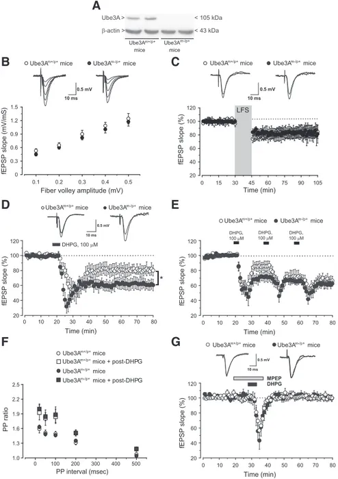
Figure 1. Enhanced mGlu5 receptor-dependent LTD in Ube3Am⫺/p⫹mice. A, Immunoblot analysis of Ube3A in hippocampal
slices from Ube3Am⫹/p⫹(wild-type) mice and Ube3Am⫺/p⫹mice. B, Input– output relation of fEPSPs as a function of
presyn-aptic fiber volley size at the Schaffer collateral/CA1 pyramidal cell synapses. Each plot represents 7– 8 separate recordings for each strain. Superimposed representative fEPSPs evoked in response to increasing stimulus intensity are shown. C, LTD induced by
low-frequency stimulation (LFS; 1 Hz, 15 min) of Schaffer collaterals. The fEPSP slope (mean⫾SEM)isplottedaspercentageofthe
pre-LFS baseline. Insets show fEPSPs from a representative experiment during a baseline interval and 60 min after LTD. D, LTD
induced by bath application of DHPG (100M, 5 min). Values are means⫾SEMofdataobtainedfromslicesof9–12miceforeach
strain. *p⬍ 0.05 (2-tailed unpaired Student’s t test) versus values obtained in slices from wild-type mice. E, Depression of fEPSP
induced by two consecutive applications of DHPG in slices from wild-type and Ube3Am⫺/p⫹mice. Values are means⫾ SEM of
data obtained from eight mice for each strain. F, PPF induced by pairs of stimulation delivered at several interstimulus intervals (20,
50, 100, 200, 500 ms) at baseline and 60 min after DHPG application. Data (means⫾ SEM) are expressed as the ratio between the
second and the first response. G, Synaptic depression induced by DHPG (100M, 5 min) in the presence of MPEP (10M) in slices
flow rate of 2.5–3.0 ml/min. The slope of the field EPSPs (fEPSPs) was recorded from the apical dendrite layer of the CA1 pyramidal cells by
means of saline-filled glass electrodes of⬃2–4 M⍀ resistance.
Stimulat-ing monopolar electrodes were placed in Schaffer collateral/commissural afferents, and stimulation amplitude was adjusted so as to produce one-half of the maximal response. Signals were filtered at 3 kHz and digitized at 10 kHz. After the stabilization of the fEPSP, LTD was induced by low-frequency stimulation (1 Hz for 15 min) or following DHPG
appli-cation (100M, 5 min). In some experiments DHPG was applied in the
presence of the NMDA receptor antagonistD-AP5 (50M), the
protea-some inhibitor UBE1-41 (50M), the protein tyrosine phosphatase
in-hibitor PAO (15M), the mammalian target of rapamycine (mTOR)
inhibitor rapamycin (20 M), the extracellular regulated kinase1/2
(ERK1/2) kinase inhibitor UO126 (20M), or the protein synthesis
in-hibitor anisomycin (20M).D-AP5 was applied 20 min before DHPG
and maintained during the recording session; UBE1-41 (50M, from a
mother solution of 50 mMin dimethyl sulfoxide) was applied to the slices
during the recovery time for 60 min before placement in the recording
chamber (Citri et al., 2009). All the other drugs were applied as indicated
(see figures).
Immunoblotting. Slices prepared as described for electrophysiological
studies were allowed to recover forⱖ3 h. Slices were then incubated with
DHPG (100M) for 5 min and then snap frozen in liquid nitrogen.
Samples were homogenized at 4°C in a lysis buffer composed of Tris-HCl
10 mM, pH 7.4; NaCl, 150 mM; EDTA, 5 mM; Igepal 1%; protease (Santa
Cruz Biotechnology) and phosphatase (Sigma-Aldrich) inhibitor mix-ture. Five microliters of tissue extracts were used for protein
determina-tion. Proteins (30g) were resuspended in SDS-bromophenol blue
reducing buffer with 40 mMDTT and used for protein analysis.
Immu-noblotting was performed with the following primary antibodies: Ube3A (Bethyl Laboratories), mGlu5 receptor (Millipore Biotechnology), p-ERK1/2 (Thr202/Tyr204; Santa Cruz Biotechnology), ERK (Cell Sig-naling Technology), p-Akt (Ser473; Cell SigSig-naling Technology), Akt (Cell Signaling Technology), and Arc (activity-regulated cytoskeleton-associated protein; kindly provided by Prof. P. Worley, Department of Neuroscience, Johns Hopkins University School of Medicine, Baltimore, MD). After incubation in primary antibody overnight at 4°C, immuno-blots were incubated with HRP-conjugated secondary antibodies (Cal-biochem) and developed by ECL (Hybond ECL, GE Healthcare Europe). Measurement of polyphosphoinositide hydrolysis in hippocampal slices. Group I mGlu receptor-stimulated polyphosphoinositide (PI) hydroly-sis was also measured in hippocampal slices obtained from postnatal day
(P) 21–P30 Ube3Am⫺/p⫹mice and their wild-type littermates as
de-scribed previously (Nicoletti et al., 1986). Briefly, hippocampi were sliced
(350⫻ 350m) using a McIlwain tissue chopper. Forty microliters of
gravity-packed slices were then incubated for 60 min in 250l of buffer
containing 1Ci of myo-[3H]inositol. Slices were incubated with LiCl
(10 mMfor 10 min) followed by DHPG (100M). One hour later, the
incubation was stopped by the addition of 900l of
methanol/chloro-form (2:1). After further addition of 300l of chloroform and 600 l of
water, samples were centrifuged at low speed to facilitate phase separation, and the upper aqueous phase was loaded into Dowex 1-X-8 columns for the
separation and quantification of [3H]Inositolmonophosphate (InsP).
Gene expression analysis by real-time PCR. Total RNA was isolated from hippocampi using TRIzol reagent (Invitrogen) according to the manufacturer’s protocol and retrotranscribed into cDNA by using Su-perScript III Reverse Transcriptase (Invitrogen). Real-time PCR was per-formed on the StepOnePlus (Applied Biosystems). PCR was perper-formed by using Power SYBR Green PCR Master Mix Kit (Applied Biosystems) according to the manufacturer’s instructions. Thermal cycler conditions were as follows: 10 min at 95°C, 40 cycles of denaturation (45 s at 95°C), and combined annealing/extension (1 min at 60°C). Sequences of
primers used were as follows: Homer 1a: forward 5⬘-TCTTCAGTC
TCCTTTGACACCA-3⬘ and reverse
5⬘-CATGATTGCTGAATTGAAT-GTG-3⬘; pan-Homer 1: forward 5⬘-TGGACTGGGATTCTCCTCTG-3⬘
and reverse 5⬘-TGTGTCACATCGGGTGTTCT-3⬘; mGlu5 receptor:
for-ward 5⬘-ACGAAGACCAACCGTATTGC-3⬘ and reverse 5⬘-AGACTT
CTCGGATGCTTGGA-3⬘; cyclophilin A: forward 5⬘-TCCAAAGACA
A
20 40 60 80 100 120 fEPSP slope (% ) 0 20 40 100 Time (min) 60 80B
DHPG Ube3A micem+/p+ 40 60 80 100 120 fEPSP slope (% )Ube3A mice + UBE1-41, 50 μMm+/p+
Ube3A micem-/p+
Ube3A mice + UBE1-41, 50 μMm-/p+
0 20 40 100 Time (min) 60 80 20 DHPG
*
LFS
20 40 60 80 100 120 fEPSP slope (% )C
0 10 30 60 Time (min) 40 50 20 70 80 Ube3A micem-/p+Ube3A mice + UBE1-41, 50 μMm-/p+
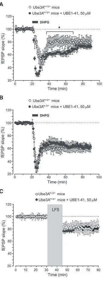
Figure 2. Pharmacological inhibition of proteasomal degradation did not affect synaptic plasticity in AS mice. Slices were preincubated with proteasome inhibitor UBE1-41 for 60 min before recording. A, Amplification of DHPG-induced LTD by UBE1-41 in hippocampal slices from
wild-type mice. Values are means⫾SEMofdataobtainedfromfourmicepergroup.*p⬍0.05
(2-tailed unpaired Student’s t test). B, C, The lack of effect of UBE1-41 on DHPG-induced (B) or
low-frequency stimulation (LFS)-induced (C) LTD in slices from AS mice. Values are means⫾
GCAGAAAACTTTCG-3⬘ and reverse 5⬘-TCTTCTTGCTGGTCTTGC
CATTCC-3⬘.
Concentrations of mRNA were calculated from serially diluted standard curves simultaneously amplified with the samples and nor-malized versus cyclophilin A mRNA levels.
Coimmunoprecipitation. Hippocampi were homogenized at 4°C in a lysis buffer (as above) and 1 mg of total proteins were resuspended in
a coimmunoprecipitation buffer (50 mMTris,
pH 7.4, 120 mMNaCl, 0.5% Nonidet P-40, 1
mMEDTA, 1 mMEGTA). Proteins were
tum-bled overnight at 4°C with 5g of antibody
anti-Homer 1b/c or anti-pan-Homer (Santa Cruz Biotechnology). Protein A agarose bead slurry (GE Healthcare) was added for 2 h, and the beads were then washed with coimmuno-precipitation buffer. Western blotting was per-formed with antibodies against Homer 1b/c, pan-Homer, and mGlu5 receptor (Millipore Biotechnology). Protein extracts from the cere-bral cortex of normal and mGlu5 receptor knock-out mice (stored in our laboratory) were used as positive and negative controls, respectively.
Statistical analysis. Electrophysiological data were normalized to the averaged value of the ini-tial slope of the fEPSP obtained during the 20 min period before the application of the conditioning stimulus or DHPG. Data are expressed as the
means⫾ SEM. Significant differences between
groups were determined using two-tailed un-paired Student’s t test performed on a 10 min average taken 50 min after DHPG application.
Statistical significance was set at p⬍ 0.05. All
ex-periments and the analysis of data were per-formed in a blind manner. For all statistical comparisons, the n used was the number of animals rather than number of slices. For bio-chemical experiments, statistical analysis was performed using two-way ANOVA plus Fisher’s PLSD test or the Student’s t test.
Results
Enhancement of mGlu5
receptor-dependent LTD in the hippocampus of
AS mice
We measured basal synaptic transmission
and activity-dependent synaptic plasticity at
the Schaffer collateral–CA1 synapses in
hip-pocampal slices prepared from Ube3A
ma-ternal deficient mice (Ube3A
m⫺/p⫹AS
mice) and wild-type (Ube3A
m⫹/p⫹)
litter-mates. No Ube3A was detected in
hip-pocampal slices of AS mice, as expected (
Fig.
1
A). AS mice did not show alterations in
basal synaptic transmission (
Fig. 1
B; p
⬎
0.05;
Jiang et al., 1998
), and in LTD induced
by low-frequency stimulation, which is
known to be dependent on NMDA receptor
activation (for review, see
Manabe, 1997
).
Stimulation at 1 Hz for 15 min induced a
similar depression of synaptic transmission
in slices from wild-type and AS mice
(wild-type: 85
⫾ 9%, n ⫽ 7; AS: 82 ⫾ 11%, n ⫽ 8,
p
⬎ 0.05;
Fig. 1
C). In contrast, LTD induced
by bath application of DHPG (100
M, 5
min) was amplified in AS mice (wild-type:
81
⫾ 8%, n ⫽ 9; AS: 61 ⫾ 6%, n ⫽ 12, p ⬍ 0.05;
Fig. 1
D). The
amplification was unaltered in the presence of the NMDA receptor
antagonist,
D-AP5 (50
M; 60
⫾ 7%, n ⫽ 4), excluding any role for
endogenous NMDA receptor activation in the DHPG/LTD
pheno-fEPSP slope (% ) 0 20 40 60 80 100 120 20 40 60 80 100 120 Time (min) fEPSP slope (% ) 0 20 40 60 80 100 120 20 40 60 80 100 120 Time (min) fEPSP slope (% ) DHPG, 100 μM UO126, 20 μM DHPG, 100 μM Rapamycin, 20 nM 0 20 40 60 80 20 40 60 80 100 120 Time (min) DHPG, 100 μM PAO, 15 μM
B
C
D
Ctrl DHPG, 100 μM p-ERK > ERK > < 42 kDa < 44 kDa < 42 kDa < 44 kDa Ctrl DHPG, 100 μME
Ube3A micem+/p+ Ube3A micem-/p+
Ube3A micem+/p+ Ube3A micem-/p+ Phospho-p44 Phospho-p42 Densitometric analysis of p-ERK/ERK Ctrl DHPG, 100 μM Ube3A mice m+/p+Ube3A mice m-/p+Ube3A mice m+/p+Ube3A mice m-/p+ 0 0.2 0.4 0.6 0.8 1.0 1.2 p-Akt > Akt > < 60 kDa < 60 kDa Ctrl DHPG, 100 μM Ctrl DHPG, 100 μM
F
Ube3A micem+/p+ Ube3A micem-/p+ 0 0.2 0.4 0.6 0.8 1.0 Densitometric analysis of p-Akt/Akt Ctrl DHPG, 100 μM Ube3A mice m+/p+ Ube3A mice m-/p+ 10 ms 0.5 mV 10 ms 0.5 mV 10 ms 0.5 mVUbe3A micem+/p+ Ube3A micem-/p+
Ube3A micem+/p+ Ube3A micem-/p+
0 20 40 60 80 Time (min) 20 40 60 80 100 120 fEPSP slope (% ) DHPG, 100 μM Anisomycin, 20 μM Ube3A micem+/p+ Ube3A micem-/p+
10 ms 0.5 mV
A
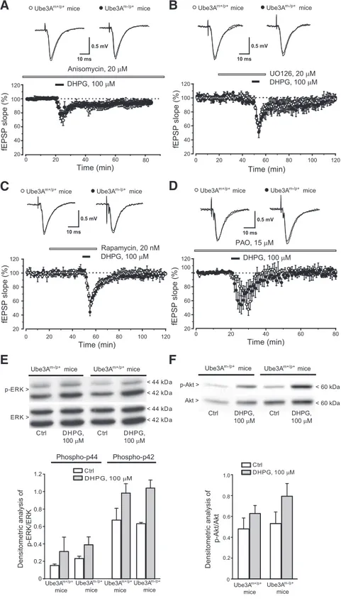
Figure 3. Examination of the intracellular signaling pathways mediating mGlu5 receptor-dependent LTD in hippocampal slices
from wild-type and Ube3Am⫺/p⫹mice. A–D, Depression of fEPSP induced by DHPG in the presence of anisomycin (A), UO126 (B),
rapamycin (C), and PAO (D) in slices from wild-type and Ube3Am⫺/p⫹mice. Values are means⫾SEMofdataobtainedfrom4–5
mice for each strain. E, F, DHPG-stimulated MAPK (E) and PI3K (F ) pathways in slices from the two genotypes. Values are means⫾
SEM from slices obtained from 3– 4 individual mice. Two-way ANOVA analysis of p-ERK and p-Akt data showed a drug effect (F(3,28)⫽ 0.355 and F(3,12)⫽ 0.396, respectively) but not genotype effect or drug–genotype interaction.
type of AS mice. As expected (
Huber et al.,
2001
), two consecutive applications of
DHPG produced maximal depression of
fEPSPs in slices from wild-type mice. In
contrast, only one application of DHPG was
sufficient to achieve saturated levels of LTD
in slices from AS mice, such that maximal
depression did not differ between the two
genotypes (wild-type: 67
⫾ 6%, n ⫽ 8; AS:
62
⫾ 5%, n ⫽ 8, p ⬎ 0.05;
Fig. 1
E).
Paired-pulse facilitation (PPF), a presynaptic form
of short-term synaptic plasticity (
Zucker,
1989
), did not differ between wild-type and
AS mice at multiple interpulse intervals (
Fig.
1
F; p
⬎ 0.05). The increase in PPF induced
by DHPG was also similar between the two
genotypes, indicating no changes in the
pre-synaptic component of group I mGlu-receptor-dependent LTD in
AS mice (
Fig. 1
F; p
⬎ 0.05).
We performed pharmacological studies to dissect the
rela-tive contribution of mGlu1 and mGlu5 receptors in
DHPG-induced LTD in the two genotypes. The mGlu5 receptor NAM
MPEP (10
M) abolished DHPG-induced LTD in both
geno-types (wild-type: 98
⫾ 3%, n ⫽ 7; AS: 98 ⫾ 4%, n ⫽ 8;
Fig. 1
G;
for data with MPEP in normal mice, see
Faas et al., 2002
;
Hou
and Klann, 2004
;
Volk et al., 2006
). In contrast,
DHPG-induced LTD was unaffected by the mGlu1 receptor
antago-nist LY367385 (3
M) in both wild-type and AS mice (data not
shown). Thus, activation of mGlu5 receptors mediated
DHPG-induced LTD in both genotypes.
Knowing that both DHPG-induced and NMDA
receptor-dependent LTD are affected by ubiquitination inhibitors (
Citri et
al., 2009
), we induced LTD in slices preincubated for 1 h with the
proteasome inhibitor UBE1-41 (50
M). DHPG-induced LTD
was amplified by UBE1-41 in slices from wild-type mice during
the first 40 min after DHPG (
Fig. 2
A). In contrast, UBE1-41 did
not affect DHPG-induced LTD in slices from AS mice (
Fig. 2
B),
indicating that the action of the proteasome inhibitor was
oc-cluded by the lack of Ube3A. We also examined NMDA
receptor-dependent LTD induced by low-frequency stimulation in AS
mice, finding no effect of UBE1-41 application (
Fig. 2
C).
Examination of the signaling pathways mediating the
enhanced mGlu5 receptor-dependent LTD in AS mice
We first examined whether DHPG-induced LTD in wild-type
and AS mice under our experimental conditions was sensitive
to the protein synthesis inhibitor anysomicin (20
M). This
treatment abolished DHPG-induced LTD in both genotypes.
(
Fig. 3
A).
Multiple intracellular signaling pathways, including the
mitogen-activated protein kinase (MAPK) pathway, the
phosphatidylinositol-3-kinase (PI3K)/Akt/mTOR pathway, and tyrosine phosphatase
(PTP)-dependent pathways, are involved in
mGlu5-receptor-dependent LTD in the hippocampus (for review, see
Gladding et
al., 2009
;
Collingridge et al., 2010
;
Lu¨scher and Huber, 2010
). We
examined the involvement of these three pathways by inducing
mGlu5 receptor-dependent LTD in the presence of the PTP
in-hibitor PAO (15
M), the ERK1/2 kinase inhibitor U0126 (20
M), or the mTOR inhibitor rapamycin (20 n
M). Treatment of
hippocampal slices with each of these inhibitors had no effect on
basal synaptic transmission but fully blocked DHPG-induced
LTD in both wild-type and AS mice (
Fig. 3
B–D; p
⬎ 0.05). In
addition, all these treatments did not reverse changes in PPF
induced by DHPG (data not shown). These data suggest that
mGlu5 receptor-dependent LTD has the same molecular
require-ments in the two genotypes. In addition, DHPG-induced
phos-phorylation of ERK1/2 and Akt in hippocampal slices did not
differ significantly between wild-type and AS mice (
Fig. 3
E, F ). It
was still possible that Ube3A-target proteins that are regulated by
the MAPK or PI3K/Akt/mTOR pathways in response to mGlu5
receptor activation could be altered in AS mice. We measured the
expression of Arc, the product of an early inducible gene that has
been implicated in mechanisms of mGlu5 receptor-dependent
LTD (
Park et al., 2008
;
Waung et al., 2008
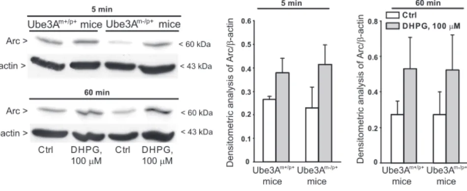
). Basal Arc protein
levels did not change in hippocampal slices from AS mice (
Fig. 4
;
Greer et al., 2010
). A 5 min exposure of hippocampal slices to
DHPG (100
M) increased Arc protein levels to the same extent
in wild-type and AS mice (
Fig. 4
).
Enhanced coupling of mGlu5 receptors with the long
isoforms of Homer proteins in the hippocampus of AS mice
We next examined the possibility that the enhancement of
mGlu5 receptor-dependent LTD could rely on mechanisms that
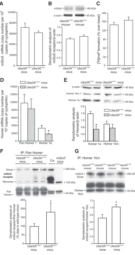
lie upstream in the signal propagation. mGlu5 receptor mRNA
and protein levels were not altered in the hippocampus of AS
mice (
Fig. 5
A, B). We extended the analysis to agonist-stimulated
PI hydrolysis, which represents the canonical signal transduction
pathway activated by mGlu5 receptors (for review, see
Nicoletti
et al., 2011
). DHPG enhanced [
3H]InsP formation to the same
extent in hippocampal slices prepared from wild-type and AS
mice (
Fig. 5
C). Interaction between mGlu5 receptors and Homer
proteins (
Tu et al., 1998
;
Xiao et al., 1998
) has been implicated in
LTD induction (
Ronesi and Huber, 2008
;
Takayasu et al., 2010
;
Ronesi et al., 2012
). Interestingly, mRNA and protein levels of the
short, activity-induced Homer 1a isoform were reduced in the
hippocampus of AS mice, whereas levels of the long, constitutive
Homer 1b/c isoforms were unchanged (
Fig. 5
D, E). We also
measured mGlu5 receptor protein levels in hippocampal
pro-tein immunoprecipitated with either pan-Homer or Homer
1b/c antibodies. mGlu5 receptor levels were significantly
in-creased in pan-Homer immunoprecipitates from AS mice,
compared with wild-type mice (
Fig. 5
F ). A significant increase
in mGlu5 receptor levels was also found in Homer 1b/c
im-munoprecipitates from AS mice (
Fig. 5
G).
Discussion
Drug treatment of AS remains an unmet clinical need, and
pharmacological options to control symptoms of the disease
have been only partially effective. Ube3A has been implicated
0 0.1 0.2 0.3 0.4 0.5 0.6 0 0.2 0.4 0.6 0.8 Arc > β-actin > < 60 kDa < 43 kDa Arc > β-actin > 60 min < 60 kDa < 43 kDa Ctrl DHPG, 100 μM Ctrl DHPG,
100 μM Densitometric analysis of Arc/
β-actin Ctrl DHPG, 100 μM 5 min 60 min Ube3A micem-/p+ 5 min Ube3A mice m+/p+ Ube3A mice m-/p+ Ube3A mice m+/p+ Ube3A mice m-/p+
Densitometric analysis of Arc/
β-actin
Ube3A micem+/p+
Figure 4. Stimulation of Arc expression by DHPG in hippocampal slices from wild-type and Ube3Am⫺/p⫹mice. Representative
immunoblots are shown. Values are means⫾ SEM of data obtained from slices of three mice. Two-way ANOVA showed a drug
in the regulation of activity-dependent
synaptic plasticity (
Jiang et al., 1998
;
Weeber et al., 2003
), but its role in mGlu
receptor-dependent forms of synaptic
plasticity is unexplored. Here, we have
shown that mGlu5 receptor-dependent
LTD was enhanced in the hippocampus
of AS mice, and this was associated with
alterations in mGlu5 receptor coupling
with Homer proteins. mGlu5
receptor-dependent LTD was also enhanced by the
proteasome inhibitor UBE1-41, as
ex-pected (
Citri et al., 2009
), and the action
of UBE1-41 was occluded in AS mice. Thus,
the enhancement of mGlu5
receptor-dependent LTD in AS mice can be
as-cribed to the impairment of the ubiquitin/
proteasome system. Changes in mGlu
receptor-dependent LTD in the
hip-pocampus of AS mice were specific
be-cause LTD induced by low-frequency
stimulation
(e.g.,
NMDA
receptor-dependent LTD) was unaltered. This
con-trasts with the finding of a reduced
NMDA receptor-dependent LTD in the
visual cortex of AS mice (
Yashiro et al.,
2009
). We highlight that DHPG-induced
LTD under our experimental conditions
was insensitive to NMDA receptor
block-ade, as expected.
LTD mediated by group I mGlu
recep-tors at the Schaffer collateral–CA1
syn-apses requires dendritic protein synthesis
(
Waung and Huber, 2009
). In FXS mice,
LTD is enhanced and becomes
indepen-dent of new protein synthesis because of
the lack of FMRP, which normally
re-strains translation of LTD-related
pro-teins (
Huber et al., 2002
;
Hou et al., 2006
;
Nosyreva and Huber, 2006
). In apparent
contrast with these findings, the mGlu5
receptor-dependent LTD in AS mice was
sensitive to the protein synthesis inhibitor
anisomycin to the same extent as in
con-trol mice. Thus, although the defect of
Ube3A is expected to prolong the half-life
of postsynaptic proteins, mGlu
receptor-dependent LTD in AS mice retains its
sen-sitivity to de novo protein synthesis.
Arc, which is the product of an
imme-diate early gene, has been directly related
to mechanisms of LTD mediated by group
I mGlu receptors. Rapid translation of Arc
mediates mGlu1/5 receptor-dependent
LTD in hippocampal neurons through a
persistent increase in the rate of AMPA
receptor endocytosis (
Waung et al., 2008
).
FXS mice show increased Arc levels in
dendrites, and lentiviral-mediated
ex-pression of FMRP in these mice
normal-izes both Arc levels and LTD in the
hippocampus (
Niere et al., 2012
). We
ex-pected to find changes in Arc levels in the
mGlu5 >
β-actin >
< 145 kDa
< 45 kDa
Densitometric analysis of mGlu5 receptor
/β -actin 0 0.2 0.4 0.6 0.8 1.0
mGlu5 mRNA (copy number per 10
copies of cyclophilin) 0 2000 4000 6000 8000 10000 0 30 60 90 120 150 180 [ H]InsP
formation (% over basal)
6 3
A
B
C
Ube3A mice m+/p+Ube3A mice m-/p+ Ube3A mice m+/p+ Ube3A mice m-/p+ Ube3A mice m+/p+Ube3A mice m-/p+ Ube3A mouse m+/p+ Ube3A mouse m-/p+ Homer 1b/c Homer 1a 0 2000 4000 6000 8000 10000 12000 14000Homer mRNA (copy number per
10 copies of cyclophilin)
Pan Homer 1 Homer 1a Densitometric analysis
of Homer /β -actin 2.0 0 6
*
E
D
Ube3A micem+/p+ Ube3A micem-/p+ Ube3A micem+/p+ Ube3A micem-/p+F
Homer > 1b/c IP: Homer 1b/c mGlu5 > DimerDensitometric analysis of mGlu5 receptor/Homer 1b/
c Ube3A mice m+/p+Ube3A mice m-/p+ Ube3A mouse m+/p+ Ube3A mouse < 280 kDa < 45 kDa Ube3A mice m+/p+ Ube3A mice m-/p+
IP: Pan Homer
Pan Homer >
Ctx
Densitometric analysis of mGlu5 receptor/Pan Homer (% data in wild-type mice
) Ube3A mice m+/p+ Ube3A mice m-/p+ < 140 kDa mGlu5 mice -/-Dimer > Monomer > mGlu5 receptor 0 0.2 0.4 0.6 0.8 1.0 1.2
G
m+/p+ Ube3A micem-/p+ < 45 kDa < 49 kDa < 49 kDa β-actin > Homer 1b/c > Homer 1a > 0 50 100 150 200 250*
1.6 1.2 0.8 0.4 < 280 kDa < 45 kDa m-/p+ Ube3A mouse m+/p+ Ube3A mouse m-/p+*
*
Ube3A miceFigure 5. Changes in mGlu5 receptor coupling to Homer proteins in the hippocampus of Ube3Am⫺/p⫹mice. A, B, mGlu5
receptor mRNA and protein levels in the hippocampus of wild-type (A) and Ube3Am⫺/p⫹(B) mice. Values are means⫾ SEM of
4 – 8 mice per group. C, DHPG-stimulated inositol phospholipid hydrolysis in hippocampal slices. Values are means⫾ SEM and
were obtained from slices obtained from five mice (here, slices were pooled and the experiment was performed in triplicate). The experiment was repeated twice with identical results. D, mRNA levels of pan-Homer and Homer 1a. E, Homer 1b/c and Homer 1a
protein levels. Values are means⫾ SEM of four mice per group. *p ⬍ 0.05 versus the respective wild-type (Ube3Am⫹/p⫹) mice
(Student’s t test; t values: D, 4.42; E, 3.915). F, G, Levels of mGlu5 receptors in pan-Homer (F ) and Homer 1b/c (G)
immunopre-cipitates. In F, values (n⫽ 6–7) were calculated from two independent experiments and data are expressed as percentage of
values (means⫾ SEM) obtained in wild-type mice. *p ⬍ 0.05 versus wild-type mice values (Student’s t test; t values, ⫺2.2895).
hippocampus of AS mice because Arc is a substrate for Ube3A
(
Greer et al., 2010
). In contrast, Arc levels did not differ between
wild-type and AS hippocampal slices under the same conditions
used for the induction of LTD. However, we cannot exclude the
possibility that differences in Arc levels between wild-type and AS
mice are present, but anatomically restricted and too small to be
revealed by immunoblot analysis.
Recent data suggest that in addition to triggering protein
deg-radation, ubiquitination can modify protein–protein
interac-tions and protein localization and activity (
Hicke, 2001
;
DiAntonio and Hicke, 2004
;
Chen and Sun, 2009
). We therefore
took steps to find at which level the lack of Ube3A could affect the
propagation of mGlu5 receptor signaling. Using specific
pharma-cological inhibitors, we showed that mGlu5 receptor-dependent
LTD in AS mice relied on the same signaling pathways that
me-diate LTD in wild-type mice, i.e., the PTP, MAPK, and PI3K/
mTOR pathways (
Gladding et al., 2009
;
Collingridge et al., 2010
;
Lu¨scher and Huber, 2010
). Stimulation of at least the MAPK and
PI3K pathways by DHPG was unaltered in AS mice, suggesting
that changes in the activity of these pathways are not responsible
for the enhanced mGlu5 receptor-dependent LTD. mGlu5
recep-tor expression and mGlu5 receprecep-tor-mediated PI hydrolysis were
also unaltered in AS mice.
AS mice differed from wild-type mice in the coupling
mech-anism of mGlu5 receptors to Homer proteins. Long, constitutive
isoforms of Homer proteins (Homer 1b, 1c, 2, and 3)
multim-erize through their C-terminal coiled-coil domains and target
mGlu1a and mGlu5 receptors to the postsynaptic density
through interactions with SHANK (SH3 and multiple ankyrin
repeat domains protein). In addition, long isoforms of Homer
link mGlu1a and mGlu5 receptors to signaling molecules, such as
PIKE (phosphoinositide-3 kinase enhancer), EF2K (the
elonga-tion factor 2 kinase), the inositol-1,4,5-trisphosphate receptors
TRPC1 and TRPC3, N-type calcium channels, and M-type
po-tassium channels (
Brakeman et al., 1997
;
Tu et al., 1998
,
1999
;
Xiao et al., 1998
;
Kammermeier et al., 2000
;
Yuan et al., 2003
;
Kim
et al., 2006
). In contrast, Homer 1a, a short and activity-inducible
form of Homer lacking the coiled-coil domain, acts as a
domi-nant negative isoform by uncoupling mGlu1a or mGlu5
recep-tors from postsynaptic effecrecep-tors (
Kammermeier and Worley,
2007
).
Recent evidence links Homer proteins to mGlu5
receptor-mediated synaptic plasticity and autism. Disruption of mGlu5
interaction with Homer proteins blocks mGlu5
receptor-dependent LTD and protein synthesis in normal mice (
Ronesi et
al., 2012
). In FXS mice, mGlu5 receptors are less associated with
the long Homer isoforms and more associated with Homer 1a
(
Giuffrida et al., 2005
;
Ronesi et al., 2012
). Genetic deletion of
Homer 1a corrects several phenotypes in FXS mice, but not the
enhancement of mGlu5 receptor-dependent LTD in the
hip-pocampus (
Ronesi et al., 2012
). The gene encoding for Homer 1
has been identified as a novel risk gene for nonsyndromic autism.
Rare Homer 1 gene variants that potentially affect protein
func-tion cosegregate closely with autism among children of affected
families (
Kelleher et al., 2012
).
In AS mice, changes in the coupling of mGlu5 receptors to
Homer proteins were opposite to those seen in FXS mice. AS
mice showed reduced Homer 1a mRNA and protein levels,
and increased association of mGlu5 receptors with Homer
proteins in immunoprecipitates. The reduction of Homer 1a
in AS mice was unexpected because Homer 1a is a substrate for
ubiquitination, and proteasome inhibitors are known to
en-hance Homer 1a levels (
Ageta et al., 2001
). Perhaps the lack of
Ube3A enhances the stability of a negative regulator of Homer
1a, which may function at transcriptional or translational
lev-els. Alternatively, in AS mice the Homer 1a phenotype may lay
upstream of the proteasome system, which may help to
ex-plain why Homer 1a levels are not enhanced despite the lack of
Ube3A. It will be interesting to examine whether an enhanced
coupling of mGlu5 receptors to long Homer isoforms has any
influence on the efficiency of the ubiquitine/proteasomal
sys-tem. Whatever the mechanism, the reduction of Homer 1a
suggests that mGlu5 receptors are more efficiently coupled to
postsynaptic effectors in AS mice. This fits nicely with the
enhanced mGlu5 receptor-dependent LTD found in AS mice,
although the precise mechanism that is ultimately responsible
for the amplification of LTD is unknown. It is intriguing that
in FXS and AS mice opposite changes in mGlu5/Homer
cou-pling are associated with an enhanced mGlu5
receptor-dependent LTD. One should take into account the fact that a
reduced association of mGlu5 receptors to long Homer
iso-forms restrains receptor coupling to postsynaptic effectors on
one side (
Kammermeier and Worley, 2007
), but enhances the
agonist-independent “constitutive” activity of mGlu5
recep-tors on the other side (
Ango et al., 2001
). Whether changes in
the constitutive activity of mGlu5 receptors have any role in
the pathological phenotype of AS mice is unknown.
In conclusion, we have described for the first time the
associ-ation of AS with abnormalities in the interaction between mGlu5
receptors and Homer proteins, and an enhancement of mGlu5
receptor-dependent LTD in the hippocampus. These findings lay
the groundwork for the use of mGlu5 receptor antagonists in
models of AS.
References
Ageta H, Kato A, Hatakeyama S, Nakayama K, Isojima Y, Sugiyama H (2001) Regulation of the level of Vesl-1S/Homer-1a proteins by
ubiquitin-proteasome proteolytic systems. J Biol Chem 276:15893–15897.CrossRef
Medline
Ango F, Pre´zeau L, Muller T, Tu JC, Xiao B, Worley PF, Pin JP, Bockaert J, Fagni L (2001) Agonist-independent activation of metabotropic gluta-mate receptors by the intracellular protein Homer. Nature 411:962–965. CrossRef Medline
Auerbach BD, Osterweil EK, Bear MF (2011) Mutations causing syndromic autism define an axis of synaptic pathophysiology. Nature 480:63– 68. CrossRef Medline
Bear MF, Huber KM, Warren ST (2004) The mGluR theory of fragile X
mental retardation. Trends Neurosci 27:370 –377.CrossRef Medline
Bhakar AL, Do¨len G, Bear MF (2012) The pathophysiology of fragile X (and what it teaches us about synapses). Annu Rev Neurosci 35:417– 443. CrossRef Medline
Brakeman PR, Lanahan AA, O’Brien R, Roche K, Barnes CA, Huganir RL, Worley PF (1997) Homer: a protein that selectively binds metabotropic
glutamate receptors. Nature 386:284 –288.CrossRef Medline
Chen ZJ, Sun LJ (2009) Nonproteolytic functions of ubiquitin in cell
signal-ing. Mol Cell 33:275–286.CrossRef Medline
Citri A, Soler-Llavina G, Bhattacharyya S, Malenka RC (2009) N-methyl-D
-aspartate receptor- and metabotropic glutamate receptor-dependent long-term depression are differentially regulated by the
ubiquitin-proteasome system. Eur J Neurosci 30:1443–1450.CrossRef Medline
Collingridge GL, Peineau S, Howland JG, Wang YT (2010) Long-term
de-pression in the CNS. Nat Rev Neurosci 11:459 – 473.CrossRef Medline
DiAntonio A, Hicke L (2004) Ubiquitin-dependent regulation of the
syn-apse. Annu Rev Neurosci 27:223–246.CrossRef Medline
Do¨len G, Osterweil E, Rao BS, Smith GB, Auerbach BD, Chattarji S, Bear MF (2007) Correction of fragile X syndrome in mice. Neuron 56:955–962. CrossRef Medline
Dong C, Upadhya SC, Ding L, Smith TK, Hegde AN (2008) Proteasome inhibition enhances the induction and impairs the maintenance of
late-phase long-term potentiation. Learn Mem 15:335–347.CrossRef Medline
sig-naling via the ubiquitin-proteasome system. Nat Neurosci 6:231–342. CrossRef Medline
Faas GC, Adwanikar H, Gereau RW 4th, Saggau P (2002) Modulation of presynaptic calcium transients by metabotropic glutamate receptor acti-vation: a differential role in acute depression of synaptic transmission and
long-term depression. J Neurosci 22:6885– 6890.Medline
Feng Y, Zhang F, Lokey LK, Chastain JL, Lakkis L, Eberhart D, Warren ST (1995) Translational suppression by trinucleotide repeat expansion at
FMR1. Science 268:731–734.CrossRef Medline
Giuffrida R, Musumeci S, D’Antoni S, Bonaccorso CM, Giuffrida-Stella AM, Oostra BA, Catania MV (2005) A reduced number of metabotropic glu-tamate subtype 5 receptors are associated with constitutive Homer pro-teins in a mouse model of fragile X syndrome. J Neurosci 25:8908 – 89016. CrossRef Medline
Gladding CM, Fitzjohn SM, Molna´r E (2009) Metabotropic glutamate receptor-mediated long-term depression: molecular mechanisms.
Phar-macol Rev 61:395– 412.CrossRef Medline
Greer PL, Hanayama R, Bloodgood BL, Mardinly AR, Lipton DM, Flavell SW, Kim TK, Griffith EC, Waldon Z, Maehr R, Ploegh HL, Chowdhury S, Worley PF, Steen J, Greenberg ME (2010) The Angelman syndrome protein Ube3A regulates synapse development by ubiquitinating arc. Cell
140:704 –716.CrossRef Medline
Hagerman R, Lauterborn J, Au J, Berry-Kravis E (2012) Fragile X syndrome and targeted treatment trials. Results Probl Cell Differ 54:297–335. CrossRef Medline
Hicke L (2001) Protein regulation by monoubiquitin. Nat Rev Mol Cell Biol
2:195–201.CrossRef Medline
Hou L, Klann E (2004) Activation of the phosphoinositide 3-kinase-Akt-mammalian target of rapamycin signaling pathway is required for metabotropic glutamate receptor-dependent long-term depression.
J Neurosci 24:6352– 6361.CrossRef Medline
Hou L, Antion MD, Hu D, Spencer CM, Paylor R, Klann E (2006) Dynamic translational and proteasomal regulation of fragile X mental retardation protein controls mGluR-dependent long-term depression. Neuron 51:
441– 454.CrossRef Medline
Huber KM, Kayser MS, Bear MF (2000) Role for rapid dendritic protein synthesis in hippocampal mGluR-dependent long-term depression.
Sci-ence 288:1254 –1257.CrossRef Medline
Huber KM, Roder JC, Bear MF (2001) Chemical induction of mGluR5- and protein synthesis-dependent long-term depression in hippocampal area
CA1. J Neurophysiol 86:321–325.Medline
Huber KM, Gallagher SM, Warren ST, Bear MF (2002) Altered synaptic plasticity in a mouse model of fragile X mental retardation. Proc Natl
Acad Sci U S A 99:7746 –7750.CrossRef Medline
Jiang YH, Armstrong D, Albrecht U, Atkins CM, Noebels JL, Eichele G, Swe-att JD, Beaudet AL (1998) Mutation of the Angelman ubiquitin ligase in mice causes increased cytoplasmic p53 and deficits of contextual learning
and long-term potentiation. Neuron 21:799 – 811.CrossRef Medline
Kammermeier PJ, Worley PF (2007) Homer 1a uncouples metabotropic glutamate receptor 5 from postsynaptic effectors. Proc Natl Acad Sci
U S A 104:6055– 6060.CrossRef Medline
Kammermeier PJ, Xiao B, Tu JC, Worley PF, Ikeda SR (2000) Homer pro-teins regulate coupling of group I metabotropic glutamate receptors to N-type calcium and M-type potassium channels. J Neurosci 20:7238 –
7245.Medline
Kelleher RJ 3rd, Bear MF (2008) The autistic neuron: troubled translation?
Cell 135:401– 406.CrossRef Medline
Kelleher RJ 3rd, Geigenmu¨ller U, Hovhannisyan H, Trautman E, Pinard R, Rathmell B, Carpenter R, Margulies D (2012) High-throughput se-quencing of mGluR signaling pathway genes reveals enrichment of rare
variants in autism. PLoS One 7:e35003.CrossRef Medline
Kim JY, Zeng W, Kiselyov K, Yuan JP, Dehoff MH, Mikoshiba K, Worley PF, Muallem S (2006) Homer 1 mediates store- and inositol 1,4,5-trisphosphate receptor-dependent translocation and retrieval of TRPC3
to the plasma membrane. J Biol Chem 281:32540 –32549. CrossRef
Medline
Kishino T, Lalande M, Wagstaff J (1997) UBE3A/E6-AP mutations cause
Angelman syndrome. Nat Genet 15:70 –73.CrossRef Medline
Krueger DD, Bear MF (2011) Toward fulfilling the promise of molecular
medicine in fragile X syndrome. Annu Rev Med 62:411– 429.CrossRef
Medline
Lu¨scher C, Huber KM (2010) Group 1 mGluR-dependent synaptic
long-term depression: mechanisms and implications for circuitry and disease.
Neuron 65:445– 459.CrossRef Medline
Manabe T (1997) Two forms of hippocampal long-term depression, the counterpart of long-term potentiation. Rev Neurosci 8:179 –193. CrossRef Medline
Michalon A, Sidorov M, Ballard TM, Ozmen L, Spooren W, Wettstein JG, Jaeschke G, Bear MF, Lindemann L (2012) Chronic pharmacological mGlu5 inhibition corrects fragile X in adult mice. Neuron 74:49 –56. CrossRef Medline
Moult PR, Correˆa SA, Collingridge GL, Fitzjohn SM, Bashir ZI (2008) Co-activation of p38 mitogen-activated protein kinase and protein tyrosine phosphatase underlies metabotropic glutamate receptor-dependent
long-term depression. J Physiol 586:2499 –2510.CrossRef Medline
Nicoletti F, Iadarola MJ, Wroblewski JT, Costa E (1986) Excitatory amino acid recognition sites coupled with inositol phospholipid metabolism:
developmental changes and interaction with␣1-adrenoceptors. Proc Natl
Acad Sci U S A 83:1931–1935.CrossRef Medline
Nicoletti F, Bockaert J, Collingridge GL, Conn PJ, Ferraguti F, Schoepp DD, Wroblewski JT, Pin JP (2011) Metabotropic glutamate receptors: from the workbench to the bedside. Neuropharmacology 60:1017–1041. CrossRef Medline
Niere F, Wilkerson JR, Huber KM (2012) Evidence for a fragile X mental retardation protein-mediated translational switch in metabotropic gluta-mate receptor-triggered Arc translation and long-term depression. J
Neu-rosci 32:5924 –5936.CrossRef Medline
Nistico` R, Mango D, Mandolesi G, Piccinin S, Berretta N, Pignatelli M, Feli-gioni M, Musella A, Gentile A, Mori F, Bernardi G, Nicoletti F, Mercuri NB, Centonze D (2013) Inflammation subverts hippocampal synaptic plasticity in experimental multiple sclerosis. PLoS One 8:e54666. CrossRef Medline
Nosyreva ED, Huber KM (2006) Metabotropic receptor-dependent long-term depression persists in the absence of protein synthesis in the mouse
model of fragile X syndrome. J Neurophysiol 95:3291–3295.CrossRef
Medline
Park S, Park JM, Kim S, Kim JA, Shepherd JD, Smith-Hicks CL, Chowdhury S, Kaufmann W, Kuhl D, Ryazanov AG, Huganir RL, Linden DJ, Worley PF (2008) Elongation factor 2 and fragile X mental retardation protein control the dynamic translation of Arc/Arg3.1 essential for mGluR-LTD.
Neuron 59:70 – 83.CrossRef Medline
Ronesi JA, Huber KM (2008) Homer interactions are necessary for metabo-tropic glutamate receptor-induced long-term depression and
transla-tional activation. J Neurosci 28:543–547.CrossRef Medline
Ronesi JA, Collins KA, Hays SA, Tsai NP, Guo W, Birnbaum SG, Hu JH, Worley PF, Gibson JR, Huber KM (2012) Disrupted Homer scaffolds mediate abnormal mGluR5 function in a mouse model of fragile X
syn-drome. Nat Neurosci 15:431– 440, S1.CrossRef Medline
Sato M, Stryker MP (2010) Genomic imprinting of experience-dependent cortical plasticity by the ubiquitin ligase gene Ube3a. Proc Natl Acad Sci
U S A 107:5611–5616.CrossRef Medline
Steffenburg S, Gillberg CL, Steffenburg U, Kyllerman M (1996) Autism in Angelman syndrome: a population-based study. Pediatr Neurol 14:131–
136.CrossRef Medline
Takayasu Y, Takeuchi K, Kumari R, Bennett MV, Zukin RS, Francesconi A (2010) Caveolin-1 knockout mice exhibit impaired induction of mGluR-dependent long-term depression at CA3-CA1 synapses. Proc Natl Acad
Sci U S A 107:21778 –21783.CrossRef Medline
Tu JC, Xiao B, Yuan JP, Lanahan AA, Leoffert K, Li M, Linden DJ, Worley PF (1998) Homer binds a novel proline-rich motif and links group 1 metabotropic glutamate receptors with IP3 receptors. Neuron 21:717–
726.CrossRef Medline
Tu JC, Xiao B, Naisbitt S, Yuan JP, Petralia RS, Brakeman P, Doan A, Aakalu VK, Lanahan AA, Sheng M, Worley PF (1999) Coupling of mGluR/ Homer and PSD-95 complexes by the Shank family of postsynaptic
den-sity proteins Neuron 23:583–592.CrossRef
Volk LJ, Daly CA, Huber KM (2006) Differential roles for group 1 mGluR subtypes in induction and expression of chemically induced hippocampal long-term depression. J Neurophysiol 95:2427–2438. Medline
Waung MW, Huber KM (2009) Protein translation in synaptic plasticity:
mGluR-LTD, Fragile X. Curr Opin Neurobiol 19:319 –326.CrossRef
Medline
translation of Arc/Arg3.1 selectively mediates mGluR-dependent LTD through persistent increases in AMPAR endocytosis rate. Neuron 59:84 –
97.CrossRef Medline
Weeber EJ, Jiang YH, Elgersma Y, Varga AW, Carrasquillo Y, Brown SE, Christian JM, Mirnikjoo B, Silva A, Beaudet AL, Sweatt JD (2003) De-rangements of hippocampal calcium/calmodulin-dependent protein ki-nase II in a mouse model for Angelman mental retardation syndrome.
J Neurosci 23:2634 –2644.Medline
Williams CA (2005) Neurological aspects of the Angelman syndrome. Brain
Dev 27:88 –94.CrossRef Medline
Xiao B, Tu JC, Petralia RS, Yuan JP, Doan A, Breder CD, Ruggiero A, Lanahan AA, Wenthold RJ, Worley PF (1998) Homer regulates the association of
group 1 metabotropic glutamate receptors with multivalent complexes of
Homer-related, synaptic proteins. Neuron 21:707–716.CrossRef Medline
Yashiro K, Riday TT, Condon KH, Roberts AC, Bernardo DR, Prakash R, Weinberg RJ, Ehlers MD, Philpot BD (2009) Ube3a is required for experience-dependent maturation of the neocortex. Nat Neurosci 12:
777–783.CrossRef Medline
Yuan JP, Kiselyov K, Shin DM, Chen J, Shcheynikov N, Kang SH, Dehoff MH, Schwarz MK, Seeburg PH, Muallem S, Worley PF (2003) Homer binds TRPC family channels and is required for gating of TRPC1 by IP3
recep-tors. Cell 114:777–789.CrossRef Medline
Zucker RS (1989) Short-term synaptic plasticity. Annu Rev Neurosci 12:13–
