PH. D. THESIS
Molecular Biology
DNA-protein interaction dynamics at the Lamin
B2 replication origin
Luca Puzzi
ADVISOR
Prof. Mauro Giacca
2014
of Professor Arturo Falaschi
List of Figures and Tables...vi
List of Abbreviations...viii
List of Publications...x
ABSTRACT......xii
1. INTRODUCTION 1.1 Regulatory mechanisms of eukaryotic DNA replication...2
1.1.1 Eukaryotic DNA replication: an overview...2
1.1.2 Multiple levels of DNA replication regulation within cell-cycle progression...5
1.1.3 From initiation to elongation in eukaryotic DNA replication...8
1.1.4 Additional requirement for DNA replication...19
1.1.5 The still obscure determinants of origin specification...23
1.2 The AP-1 protein family...26
1.2.1 AP-1 complex protein composition and general features...26
1.2.2 Structure of AP-1 proteins...28
1.2.3 AP-1 expression, regulation and activity...32
2. AIM OF THE WORK 2.1 AP-1 and DNA replication...39
3. RESULTS 3.1 Characterization of T98G-HF stable cell clone expressing HA/Flag HOXC13...41
3.2 HOXC13-HF Cp-immunoprecipitation analysis...46
3.3 Analysis of AP-1 binding onto the LaminB2 origin...50
3.4 High resolution analysis of the LaminB2 origin of DNA replication...51
3.4.1 Validation of the high resolution ChIP technology...52
3.4.2 Analysis of the LaminB2 origin of DNA replication by high resolution ChIP...53
3.5 Spatial and temporal analysis of AP-1 proteins binding onto the LaminB2 origin...61
3.6 Effects of the disruption of topological structure on protein binding across the LaminB2 origin region...69
4. DISCUSSION 4.1 c-Fos and c-Jun are involved in the process of DNA replication...74
4.3 c-Fos and c-Jun interaction with LaminB2 origin relies on chromatin topology...75
4.4 A speculative model for origin activation...77
5. MATERIAL AND METHODS 5.1 Cell culture, synchronization and Merbarone treatment...79
5.1.1 Cell synchronization and FACS analysis...80
5.1.2 Merbarone treatment...80
5.2 Antibodies...80
5.3 in vivo topoisomerase I and II mapping...81
5.4 Chromatin immuno precipitation analyses (ChIP)...82
5.4.1 X-ChIP...82
5.4.2 Native chromatin immuno precipitation analyses (NChIP)...83
5.4.3 Sequential chromatin immunoprecipitation analyses (Re-ChIP)...83
5.5 PCR analyses...84
5.5.1 Competitive PCR...84
5.5.2 High resolution LaminB2 PCR...85
REFERENCES
List of Figures and Tables
Figure 1.1: Regulation of DNA replication by origin usage...3
Figure 1.2: Different types of DNA replication origins …...7
Figure 1.3: Model of the regulation of DNA replication...10
Figure 1.4: Schematic pictures of ORC1-5 and Cdc6 proteins from S. cerevisiae...12
Figure 1.5 The many functions of the origin recognition complex...14
Figure 1.6: Proposed features to determine the selection and activation of replication origin...19
Figure 1.7: Model for the progressive restriction of initiation potential during G1 phase...20
Figure 1.8: A/T-rich and C/G-rich islands at DNA replication origins...24
Figure 1.9: Coiled coil protein structure...29
Figure 1.10: The Jun-Fos heterodimer...30
Figure 1.11: Transcriptional and post-translational activation of AP-1...33
Table 1.1: Different AP-1 genes regulation in tumor development and suppression...36
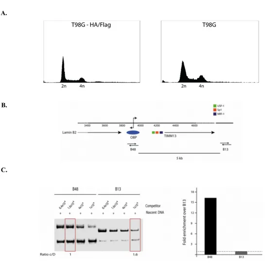
Figure 3.1: Flow cytometry and nascent DNA analyses on the T98G-HA/Flag stable clone...41
Figure 3.2: X-linked HA ChIP analysis on T98G HA/Flag stable clone...43
Figure 3.3: in vivo cleavage analyses of Topoisomerase I and II...45
Figure 3.4: Co-IP partners of HOXC13-HA after X-linked ChIP and Native ChIP...47
Figure 3.5: ChIP analysis for AP-1 proteins on the LaminB2 origin...48
Figure 3.6: Schematic representation of the position of the 12 overlapping PCR segments...50
Figure 3.7: Primer analysis after amplification of a DNA amplicon obtained using the 1L-12U primers or total genomic DNA from starved (G0) T98G cells...52
Figure 3.8: Primer efficiency analyses performed using G0 DNA...53
Figure 3.9: Input chromatin profile in the presence and absence of formaldehyde crosslink...54
Figure 3.10: H2B and acetylated H3(K14) NChIP analysis...55
Figure 3.11: Pre-RC proteins binding profiles across LaminB2 origin...56
Figure 3.12: c-Fos and c-Jun ChIP analysis...57
Figure 3.13: ChIP analysis to detect binding, across the LaminB2 origin, of other potential protein candidates...59
Figure 3.14: Flow cytometry profile and expression levels of AP-1 proteins during the cell cycle...60
Figure 3.15: Western Blot analyses of AP-1 proteins in different phases of the cell cycle...62
Figure 3.16: ChIP analysis for AP-1 and ORC4 proteins through the cell cycle...63
Figure 3.17: Co-IP of ORC4 in AP-1-ChIP samples across the cell cycle...64
Figure 3.18: Development of the Re-ChIP procedure...66
Figure 3.19: AP-1/ORC4 chromatin binding profiles by re-ChIP analyses...67
Figure 3.20: Effect of the topoisomerase II inhibitor merbarone on DNA synthesis...68
Figure 3.21: Effects of merbarone on protein:DNA interactions across the LaminB2 origin region...70
Figure 4.1: Alignment of the “non-canonical sequences” to the 1.1 Kbps region analyzed...76
Figure 4.2: Proposed model...77
Table 5.1:Primers used for high resolution protein binding analysis at the LaminB2 replication origin...86
List of Abbreviations
AP-1 = Activator protein 1 ACS = ARS consensus sequence APC = Anaphase promoting complex ARS = Autonomously replicating sequence ATP = Adenosine triphosphate
BAH = Bromo adjacent homology domain BrdU = Bromo deoxy-Uridine
bps = Base pairs
CDK =Cyclin-dependent-kinase
ChIP = Chromatin immunoprecipitation Co-IP = Co-immunoprecipitation CRE = Cyclic AMP responsive element Ct = Cycle threshold
DDK = Dbf4-dependent-kinase DTT = Dithiothreitol
EDTA = Ethylenediamine tetra-acetic acid HA = Hemaagglutinin protein
MCM = Mini-Chromosome-Maintenance proteins ORC = Origin Recognition Complex
PBS = Phosphate buffered saline PCR = Polymerase chain reaction PI = Propidium iodide
pre-IC = pre-initiative complex pre-RC = pre-replicative complex RC = replicative complex
SSB = single-stranded binding protein ssDNA = single-stranded DNA TAD = transactivation domain
TPA = 12-O-tetradecanoylphorbol 13-acetate TRE = TPA responsive element
WB = Western Blot WCL = whole cell lysate WCE = whole cell extract
List of Publications
Luca Puzzi, Laura Marchetti, Fiorenzo A. Peverali, Giuseppe Biamonti, Mauro Giacca, “DNA-protein interaction dynamics at the Lamin B2 replication origin”, Cell Cycle, 2015, 14 (1): 64-73
Publications not included in this thesis
L. Marchetti, L. Comelli, B. D’Innocenzo, L. Puzzi, S. Luin, D. Arosio, M. Calvello, R. Mendoza-Maldonado, F. Peverali, F. Trovato, S. Riva, G. Biamonti, G. Abdurashidova, F. Beltram, A. Falaschi, “Homeotic proteins participate in the function of human DNA replication origins”, Nucleic Acids Research, 2010 (22): 8105-19
The regulation of human DNA replication operates via a time-defined program of activation and deactivation of approximately 30,000 replication origins distributed along the genome. Due to the complexity of this process, each step requires a sequence of cascade checkpoints and licensing events, most of which are well conserved from yeasts to humans. A multi-protein complex assembles onto each origin causing the local unwinding of the DNA double helix and the start of two oppositely moving replicative forks. Despite the cis-acting elements necessary for origin firing are almost elucidated, the mechanism that governs the selection of a specific DNA sequence as human (and, more generally, metazoan) origin, in the course of G1 phase of the cell cycle, is still poorly understood. The lack of DNA-sequence consensus between replication origins characterized so far, together with the poor binding-specificity displayed by the Origin Recognition Complex, suggest that origin selection might rather be determined by local chromatin structures and/or trans-acting factors. With regard to the latter possibility, it was interesting to find out that a DNA region specifically bound by the AP-1 proteins, is located close to the start site of the human Lamin B2 replication origin.
In the study conducted during this Ph.D. program, the possible role of AP-1 transcription factors in origin specification was explored by investigating the involvement the principal moieties of this protein family, c-Fos and c-Jun, within the replicative complexes in living human cells. The data reported in this thesis provides evidence that both c-Fos and c-Jun interact with the LaminB2 origin of DNA replication and indeed participates in origin function. Participation of these proteins to origin binding is consistent with their interaction with both ORC4 and HOXC13, two members of the replicative complex, and is cell cycle defined, occurring before origin firing. Furthermore the observations point to the existence of specific and
along with origin activation. In this view, AP-1 proteins could contribute to recruit and stabilize the replicative complexes onto the LaminB2 origin, in presence of specific chromatin and topological configurations.
Chapter
1
Introduction
The work reported in this dissertation explores the possible connection between two traditionally separated fields of biology, the regulation of DNA replication and the function of activator protein 1 (AP-1) transcription factors. To provide the conceptual frame of my experimental work, the two following paragraphs will focus on a description of the mechanisms of DNA replication (paragraph 1.1) and of the structure and function of AP-1 proteins (paragraph 1.2). I will try to summarize what appears to be missing for a satisfactory understanding of DNA replication regulation in metazoan organisms, and to what extent the AP-1 proteins could be involved in this process.
1.1 Regulatory mechanisms of eukaryotic DNA
replication
1.1.1 Eukaryotic DNA replication: an overview
DNA is the most important molecule for all living organisms, which has to be maintained intact to allow the survival of the organisms themselves. The importance of this process is demonstrated by the presence of many steps that are finely regulated, involving different proteins both for the process itself, and as controllers. One of the most valuable contributions to our understanding of DNA replication is the replicon model proposed in 1963 by Jacob and Brenner [1], who postulated the existence of two fundamental elements that regulate DNA replication: a cis-acting sequence within the genome, called the “replicator” from which replication starts and a positive trans-acting factor called the ‘‘initiator’’ able to recognize specifically the sequence of the replicator within the genome. In response to appropriate cellular signals, the initiator directs the local unwinding of the replicator sequence and recruits additional factors to initiate the process of DNA replication. Once DNA replication starts, the replication fork proceeds until genome duplication is completed.
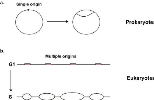
The replicon theory was initially verified by using a bacterial chromosome (Figure 1a). In Escherichia coli, the initiator protein DnaA binds with high affinity to the replication origin oriC, which contains multiple DnaA-binding sites [2]. Eukaryotic genomes are very large and their replication rate is slow, when compared to the prokaryotic replicon model. Nevertheless, the process of DNA replication is made possible by the start of DNA replication at 30.000–50.000 different chromosomal locations, known as origins of DNA replication, that are specifically selected and activated in each cell cycle [3] (Figure 1b). The process of DNA synthesis relies on a spatio-temporal coordinated cycle of activation and deactivation of the origins,
restricted to a relatively narrow window of the eukaryotic cell cycle, namely the S phase. The two main advantages of this mechanism are that the overall time required to duplicate the entire genome is reduced and that the generation of single-stranded DNA (ssDNA) is much more localized and transient, helping preserving genome integrity [4,5]. Actually, the activation of all origins localized in eukaryotic genomes leads to the formation of tandemly arranged replication units, each of which can conceptually be considered as an analog of the bacterial replicon [6]. Studies performed in different organisms have clearly demonstrated that more origins are prepared for replication in G1 than those that are actually used during the S phase. This phenomenon, which is known as origin redundancy, is likely to represent a foolproof mechanism, ensuring that replication restarts through the activation of “dormant origins” when replication forks are arrested [7] (Figure 1.2).
Figure 1.1: Regulation of DNA replication by origin usage. While prokaryotes have a single origin
on a circular chromosome (a) in eukaryotes instead, multiple origins are found on a single, linear chromosome (b). This is useful to achieve a faster replication.
Considering DNA replication as a process, it can be divided into three steps: initiation, elongation and termination. During initiation [8], a specific DNA sequence is selected to be an origin (i.e. the start site) of DNA replication, usually in correspondence to loci of actively transcribed genes and AT-rich sequences, and initiator proteins assemble thereon. This results in the formation of a multi-protein complex which is responsible for the local melting of the DNA duplex, which is necessary for proteins to have access to the template strands. Subsequently, the complex stabilizes the ssDNA that is formed, and two replication forks, thanks to DNA helicases and polymerases, start to replicate the two parental DNA strands in opposite directions. The elongation step [9] is actually the continuation of the unwinding activity by the two fork complexes. It ensures simultaneous replication of both parental DNA strands also outside of the origin sequence. When two replication forks converge, they merge and termination of replicon duplication occurs [10]. Significant differences exist between DNA replication mechanisms in lower and higher eukaryotes. In the former organisms, such as the budding yeast Saccharomices cerevisiae, replication origins are well defined genetic elements containing the conserved and essential autonomously replicating sequence (ARS) consensus sequence (ACS) directly bound by the origin recognition complex (ORC) proteins in an ATP- dependent manner [8] which serves as a platform for the assembly of the pre-replicative complex (Pre-RC). On the contrary, in the fission yeast Schizosaccharomyces pombe, origins are much larger and the ORC complex does not interact with any conserved consensus sequence but binds to AT-rich origin sequences thanks to the AT-hook DNA binding domain of the ORC4 subunit [11], showing an evolution in the origin recognition process in eukaryotes. Conversely, higher eukaryotic organisms display a number of origins. These are at least 100-fold more abundant and, at present, no sequence specific replicators have been found [12]. In spite of these disparities between lower and higher eukaryotes, the proteins that regulate replication are highly conserved in function from yeast to Drosophila, from Xenopus to man, suggesting a common mechanism in the replication function that does not depend on the origin sequence itself [13].
1.1.2 Multiple levels of DNA replication regulation during cell cycle
progression
The complexity of DNA replication in higher eukaryotes implies that any deregulating factor could lead cells to enter apoptosis or progress to tumorigenesis. Because such a strict relationship between replication and tumor proliferation exists, this process has to be strictly controlled by many levels of regulation. The first regulatory step of DNA replication concerns the activation, or initiation of the replication origins. This starts from the end of M phase and the onset of G1 phase [14], when several proteins take part in the pre-RC complex assembly by selecting the DNA sequences that are going to become replication origins and, by binding to these regions, promote the recruitment of other proteins involved in origin activation. During the G1 phase, for all the sequences selected as origins, a timing time of replication initiation is assigned and only a subset of the origins will fires immediately after entry into the S phase (early origins). The remaining ones (middle and late origins) are programmed to fire in an ordered manner after early origins. This results in an organized spatio-temporal activation of replication clusters of different subchromosomal domains at different times during the S phase [15]. In response to genotoxic damage, the DNA damage response pathway prevents entry into S phase by the activation of the G1/S border checkpoint, the components of which are highly conserved in eukaryotes. DNA damage is detected by the ataxia telangiectasia mutated related (ATR) protein which acts as a sensor and leads to the activation of CHK1 protein kinase, which in turn activates effectors (p53 and Cdc25A) that interact with the cell cycle machinery to inhibit cell cycle progression by controlling the association of Cdc45 with chromatin, preventing the transition between G1 to S phase [16, 17].
After origin firing, at the beginning of S phase, the pre-RC is re-organized due to the degradation or modification of several of its members, as a regulatory mechanism to avoid re-replication [18, 19]. Moreover, the temporal separation of pre-RC assembly
from origin activation is actually another key event ensuring that new pre-RC cannot assemble on origins that have already fired [18]. These mechanisms rely on the activity of CDKs (cyclin-dependent kinases), cell-cycle regulated kinases which act on several target proteins [8] controlling the time of replication initiation at specific origins [15, 20]. Because the activity of these kinases remains high from the S phase onset to the end of the following mitosis, re- licensing cannot occur until the beginning of the next subsequent cell cycle [21].
The mechanism that regulates the timing of replication is not completely understood. Originally, it was thought that early replication is a prelude to transcription because transcriptionally active euchromatic regions replicate early and inactive heterochromatic regions late. The molecular relationship between transcription and replication in regulating these temporal programs is unclear, and certainly goes beyond the actual need of DNA-binding proteins to access regions in which chromatin is unfolded [22, 23]. Studies in metazoa have indeed confirmed the recurring correspondence between initiation of DNA replication and transcriptionally active regions [24, 25]. Nevertheless, the timing of origin activation has been reported to correlate with a developmental program rather than with transcription per se [26, 27].
Given the complexity and importance of DNA replication elongation for the maintenance of genome integrity, many different checkpoint pathways are active within the S phase, as demonstrated by studies in the yeast model [28]. These checkpoints encompass the whole phase of DNA synthesis, as well as the switch to G2 phase, and comprise a variety of mechanisms to prevent replication defects, repair damaged replication forks and enable fork reactivation. For their role in the overall control of the cell cycle progression, as well as the control of genomic stability, they are often referred to as cell cycle checkpoints [29]. Very interestingly, the induction of a cell cycle checkpoint often results in the retroactive regulation of the recruitment of key members of the pre-RC to the origin site. For example, in the budding yeast, hydroxyurea treatment not only blocks fork progression from early origins but also prevents the firing of late origins, and this mechanism was shown to depend on
Rad53 and Mec1 [30]. The same conclusion was also obtained following induction of double strand breaks, and the protein involved in this regulation was shown to be ORC2 [31].
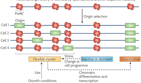
Figure 1.2: Different types of DNA replication origins. The origins that will be activated at the
following S phase are selected during G1 phase and may vary according to several parameters, such as the environmental conditions or cell fate. Four examples of DNA replication origin position in a growing cell population are shown. A cluster of flexible origins contains origins that can be activated differently in different cells; according to physiological or abnormal growth conditions. Inactive (or dormant) origins are not frequently used or not used at all; whereas constitutive origins are fixed and always set at the same position by chromatin or transcriptional constraints, inactive origins can be activated increasing the number of origins per replication cluster. Adapted from [13].
Moreover, a post initiation role was recently proposed for the protein Cdc6 which does not affect replication elongation, nor checkpoint activation in vivo due to the absence of CHK1 activation [32, 33]. In contrast, Cdc6 seems to be crucial for activation of S phase checkpoints in a Xenopus cell-free DNA replication system [34]. Altogether, this information indicate that the same factors involved in DNA replication initiation are also important actors in the regulation of the replication process at different stages during the cell cycle, being the targets of many checkpoint
controls.
1.1.3 From initiation to elongation in eukaryotic DNA replication
DNA replication starts from the stepwise recruitment of the replication machinery to the various origins on the chromosome. The recruitment process is an essential part of the initiation process, to be distinguished from the subsequent replication of the DNA by the replisome (named elongation). As reported in the previous paragraph, initiation is a major step at which DNA replication is regulated: the ordered recruitment of the pre-replication proteins onto the origin is indeed responsible for controlling the process of initiation of DNA replication in terms of both space and time. Furthermore, the subsequent inactivation or removal of some of the protein prevents re-replication during S phase. For these reasons, initiator proteins are crucial in regulating origin activity. The basic mechanism of initiation occurs in several steps that finally lead to bidirectional replication from the origin. These steps can be summarized as follows.
1. Recognition: labeling of the origin by ORC, Cdc6 and Cdt1;
2. Licensing or initiative assembly: loading of the DNA-helicase (MCM complex or minichromosome maintenance complex), to form the pre-RC;
3. Unwinding: activation of the DNA helicase or by protein kinase activity;
4. Elongative assembly: loading of the complete replisome, including DNA polymerase enzymes and SSB (single-stranded DNA binding protein).
The ordered sequence of these four steps allows the switch from initiation to elongation; each of these steps is briefly summarized in figure 1.3
1. Recognition: in this step, the ORC complex recognizes and marks the origins,
which is proposed to occur between the late M and G1 phase [14], and provides an anchorage point for two other proteins entering the complex during the course of G1
phase, Cdc6 and Cdt1. ORC is a six-protein heterocomplex containing ORC1–6 proteins (Figure 1.3) in an equal stoichiometric ratio. It was first isolated in yeast cells due to its specific binding to origin sequences [35]. Although the ORC1–6 proteins are evolutionary conserved in all eukaryotes, the recognition of specific sequences is a property lost in ORC except for the fission yeast Schizosaccharomices pombe, in which a preference for AT-rich sequences exists. This is direct evidence for the absence of a consensus DNA sequence in metazoa and, at the same time, leads to the conclusion that ORC cannot be considered as a true “initiator” protein by itself. The most impressive proof of this concept, and also of the preservation of ORC among eukaryotes, is when recombinant ORC1–6 proteins from human were found to replace the frog ORC1–6 proteins in vitro to initiate DNA replication in a sequence-independent manner [36]. To date, it is not clear which DNA or chromatin structure ORC recognizes. Most likely this is a particular chromatin structure governed by epigenetic determinants and not primary DNA sequence. This possibility is supported by several, recent observations and will further be discussed later.
Most ORC subunits belong to the superfamily of AAA+ ATPases (ATPases Associated with various cellular Activities) and share conserved motifs [37] (Figure 1.4). The ATP-binding activity is required in the process of origin DNA recognition. Indeed, in Saccharomyces cerevisiae, the ORC1 ATPase activity is inhibited until the Cdc6 protein, which is also an AAA+ ATPase, is recruited and activates ORC1 ATPase, thus resulting in the specific recognition of the origin [38]. The role of the ORC6 protein in DNA binding and pre-RC assembly is controversial and represents a sort of enigma. ORC6 is an essential protein for viability in yeast but is not required for DNA binding in vitro. In metazoan cells, complexes with lower amounts of ORC6 than the other ORC1–5 proteins are still active, whilst in Drosophila ORC6 was shown to have intrinsic DNA binding activity and any point mutation in its DNA binding domain negatively affects DNA synthesis [39, 40]. In yeast, ORC is bound to origins throughout the cell cycle and re-replication is avoided by phosphorylation of ORC2 and ORC6 by CDK1.
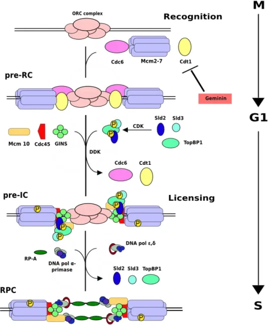
Figure 1.3: Model of the regulation of DNA replication. In eukaryotes, a replication origin is
recognized by ORC at the M/G1 transition. Then, Cdc6 and Cdt1 proteins load the MCM helicase to form the pre-RC complex in G1 phase. Geminin inhibits Cdt1 and consequently pre-RC re-formation. CDK and DDK become active in late G1 and activate the MCM helicase; in addition, CDK inhibits any further licensing. To this end, CDK phosphorylates Sld2 and Sld3 proteins and DDK phosphorylates MCM proteins giving rise to the pre-initiation complex (Pre-IC), Finally loading of primase, polymerase and RPA allows DNA replication to start forming its fundamental unit, the replisome. Modified from S. J. Aves, DNA replication initiation-Methods in Molecular Biology (521) 2009.
However, in other eukaryotes, ORC binding is regulated based on a mechanism known as the “ORC cycle” [41]. This is used to avoid re-replication and consists in the dissociation of ORC1 from the chromatin-bound ORC2–5 complex and its subsequent degradation in cells at the end of G1 phase and beginning of S phase [42]. This process is regulated by CDK1-cyclin A phosphorylation [43]. Of note, recent studies have identified roles for ORC proteins other than the direct control of DNA replication initiation [44] (Figure 1.5). ORC1 has been reported to participate in gene silencing via its BAH domain (Figure 1.4), providing a direct interaction with the silent chromatin protein Sir1 in S. cerevisiae [45], as well as with heterochromatin protein 1 (HP1) in Xenopus, Drosophila [46] and mammals [47]. In both cases, ORC1 helps Sir1 and HP1 to propagate silenced chromatin. Other ORC proteins have been reported to be important for heterochromatin maintenance. ORC2 and ORC3 are associated with constitutive heterochromatin and HP1 in Drosophila; in human cells depletion of these proteins causes HP1 disruption leading to compromised gene silencing, sister chromatid cohesion and centromere function in mitosis [48]. Depletion of ORC1 and ORC5 also results in loss of HP1, but from large heterochromatin foci instead of the centric one where ORC2 and ORC3 are present during mitosis [49]. Studies in both Drosophila and mammalian cells have revealed that ORC6 coordinates cytokinesis with pre-RC formation and chromosome segregation, independent from the rest of the complex [50]. It has been proposed that ORC6 may also participate in positioning of the ORC at the origins of DNA replication, similar to the role of TFIIB in positioning transcription pre-initiation complex at the promoter [51]. Human ORC6 was shown also to localize to kinetochores and reticular-like structures around the cell periphery during mitosis, and to be necessary for the proper progression of this stage of the cell cycle [52]. Human ORC2 also is present at the centrosome during all the cell cycle and, when depleted, mitotic defects and multiple centrosomes arise [48]. Recently, human ORC1 was reported to have a similar role in controlling centrosome copy number [53].
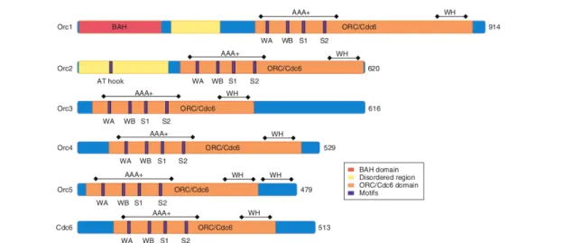
Figure 1.4: Schematic pictures of ORC1-5 and Cdc6 proteins from S. cerevisiae. All ORC1-5 and
Cdc6 proteins contain an AAA+ domain as part of a larger ORC/Cdc6 homology domain (highlighted in orange). Motifs in the AAA+ domain include Walker A (WA), Walker B (WB), Sensor-1 (S1) and Sensor-2 (S2). The winged-helix domain (WH) is involved in DNA binding. ORC1 contains an additional BAH (bromo-adjacent homology) domain (highlighted in pink). ORC1 and ORC2 have disordered regions (yellow); a DNA-binding, AT- hook motif was identified in S. cerevisiae ORC2, and many of these regions have also been found in disordered regions of S. pombe ORC4. The total number of amino acids for each protein is indicated at the right side. Adapted from: [37].
2. Initiative assembly. The next step is to load the heterohexameric DNA helicase
onto the origin (Figure 1.3). This is accomplished by two proteins, Cdc6 and Cdt1, which recruit the mini chromosome maintenance (MCM) helicase to finally achieve the pre-RC assembly onto the origin. “Replication licensing” is a conventional term that is used to describe the process in which origins are “licensed” when the MCM helicase is loaded onto them in the G1 phase of the cell cycle [54]. Cdc6 is also an AAA+ ATPase (see Figure 1.4), which is required to load the MCM helicase onto the complex in the G1 phase, as shown in experiments performed in budding yeast, which also revealed the importance of its ATPase activity to exert this function [55]. In particular, the Cdc6 and ORC ATPases act sequentially, with Cdc6 required initially. In a recently proposed model, Cdc6 and origin chromatin set off a molecular
switch in ORC for pre-RC assembly [38]. Indeed, in S. cerevisiae the ORC1 ATPase activity is inhibited until Cdc6 protein is recruited and activates ORC1 ATPase. This produces a conformational change in the ORC-Cdc6-DNA complex to achieve a ring-like structure with increased specificity for the origin sequence. Origin DNA inhibits ATP hydrolysis by Cdc6 and stabilizes the complex, whereas mutations in the origin sequence can increase Cdc6 ATPase activity, resulting in a less stable Cdc6-DNA complex. This means that ORC binding to the origin is not specific unless Cdc6 is also bound, thus Cdc6 rather than ORC is responsible for origin selection [38]. The structure suggested for Cdc6, which was deduced by comparison with the ORC structure, is similar to the atomic structure of the archaeal homologue, ORC1/Cdc6. ORC1 and Cdc6 proteins are homologues (Figure 1.4), and indeed archaeal species have one protein Orc1/Cdc6 acting both in origin recognition and in MCM helicase loading.
The Cdt1 protein, like Cdc6, is also necessary to load the MCM helicase during G1 phase of the cell cycle of eukaryotes [8]. This protein, which was initially found in fission yeast, is clearly conserved in eukaryotic evolution. As Cdc6 ATPase is required for Cdt1 binding onto the origin in vitro, it has been suggested that a Cdt1-MCM complex is loaded onto the ORC-Cdc6-origin complex during initiation [55] [56]. Cdc6 and Cdt1 then dissociate and, finally, ORC hydrolyzes ATP and this completes the MCM helicase loading reaction [38] [55]. As stated in paragraph 1.1.2, licensing is blocked during S, G2, and M phases of the cell cycle to prevent re-replication. Re-replication is actually avoided by the concurrence of several, redundant mechanisms that block MCM loading during S, G2 and M phases. Pre-RC complexes can be assembled only in the course of G1 phase, but are activated for origin firing only during S phase. A higher level of regulation is catalyzed by CDK, which operates at many redundant levels to avoid licensing in most eukaryotes [57].
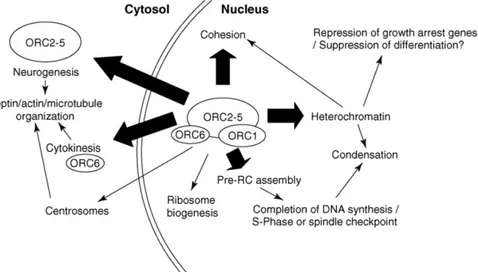
Figure 1.5 The many functions of the origin recognition complex. This diagram describes roles for
ORC proteins that are supported by functional evidence (indicated with a thick arrow), while roles that can be explained indirectly or that are supported primarily by localization or physical association of ORC are indicated with a thin arrow. Adapted from [44].
These include the localization and degradation of several pre-RC components. Besides the already mentioned ORC1 protein in higher eukaryotes, another modified protein is Cdc6: in yeast it is degraded after CDK phosphorylation [58]. Another degree of regulation to block re-replication occurs through a protein known as Geminin (Figure 1.3), which was discovered in frog egg extracts [59] and is only found in metazoans. Geminin binds to and inhibits Cdt1 thus preventing replication licensing by blocking the loading of the MCM helicase [59]. The same role for Geminin is reported to also occur in human cells [19]. The redundancy of these mechanisms in avoiding re-replication has been proposed to provide a key driving force in the evolution of licensing control [60]. Because there is no any unique mechanism effective to inhibit pre-RC components, multiple mechanisms are required for an efficient block of the re-replication. However, as the number of inhibitory mechanisms increases, the relative importance of any single mechanism decreases. During evolution, these regulatory mechanisms may be gained or lost. This could explain the different regulatory mechanism of Cdc6 between yeast and human
cells as well as the appearance of Geminin in metazoa to provide an additional mechanism for preventing re-replication, which may have been important in supporting an increase in genome size with respect to lower eukaryotes.
3. Unwinding and 4. Elongative assembly. These two steps refer to the activation of
helicase activity at the origin and to replisome assembly respectively; due to their close interconnection they are therefore described together.
The MCM complex is believed to be the engine of the replicative helicase. This complex is a hexamer comprising of six related polypeptides (MCM2–7), all of them with AAA+ ATPase activity. They are coded by a family of six paralogous genes, which are conserved from yeast to human. All six members are essential genes as described in a pioneering work in fission yeast in which MCM was as a complex that contained all six subunits in 1:1:1:1:1:1 stoichiometric ratio having a ring-like structure [61].
In G1 phase, pre-RCs with the MCM2–7 helicase bound are present on almost all origins. Indeed, about 90% of all origins that are bound by ORC also contain the MCM complex bound [62]. Nevertheless, MCM is loaded in an inactive state in the pre-RC, when CDK activity is low. The next step is to activate the MCM helicase. This is achieved by the binding of several other proteins to the origin, up to the loading of the replisome (Figures 1.3). The multi-protein complex assembled on the origin at this stage is referred to as the pre-initiation complex (pre-IC), and is required for the activation of the MCM2–7 helicase. Both helicase activation and replisome loading require phosphorylation by two different kinases which are regulated independently of each other, but by similar mechanisms. The kinase activity is established by a protein heterocomplex, thus both kinases are inactive in monomeric form and are activated by the binding of an activating subunit, Cyclin for CDK and Dbf4/Drf1 proteins for DDK [63], respectively. Thus, CDK is Cyclin-dependent kinase (comprising heterodimer of different Cdk and a Cyclin) and DDK is Dbf4-dependent kinase (comprising of Dbf4/Drf1 and Cdc7). In mammals, while there is only one DDK, there are at least four CDKs (Cdk1–4) and four classes of cyclins (A,
B, D, and E) required for cell cycle progression [64]. Thus, the substrate specificity by different Cdk-cyclin complexes drives the cell cycle. In budding yeast, there is only Cdk1 or Cdc28 enzyme, but there are six B-type cyclins (Clb1–6) needed for S and M phases [64]. Cdk1-Clb5 complexes are active in regulating DNA replication and Cdk1-Clb2 for regulating mitosis. The Cdk2 homologue is used in DNA replication. By analogy, Cdk2-cyclin E and Cdk2-cyclin A act as yeast Cdk1-Clb5 for DNA replication, whereas Cdk1-cyclin B act as yeast Cdk1- Clb2 for mitosis [22]. Cell cycle regulation of the unstable subunit ensures cell cycle regulation of the kinase activity. With CDKs, other levels of regulation occur including protein inhibitor binding, phosphorylation by other kinases and cyclin subcellular localization [64]. About DDK, the mechanism is simpler because Dbf4 protein is absent in G1 phase due to its proteosomal degradation by the APC (anaphase promoting complex) and as cells enter S phase, Dbf4 is stabilized and the APC is inactivated by CDK phosphorylation.
Much evidence indicates that the MCM2–7 complex is a target of phosphorylation by DDK, and this occurs in several eukaryotes [65]. Studies performed in yeast have identified phosphorylation sites in the N-terminus of MCM4, MCM2 or MCM6 to be important for formation of the pre-IC and for DNA replication [66]. Pre-RC activation due to phosphorylation of the MCM complex by DDK leads to the loading of additional replication factors, such as MCM10, the GINS complex and Cdc45 giving rise to the pre-initiation complex (Pre-IC).
MCM10, which is not a MCM2–7 homologue [67], is needed for the recruitment of the Cdc45 protein after pre-RC formation and for stabilizing the replisome, a mechanism conserved from yeast to humans [68].
The GINS complex, the name of which is based on the numbers 5, 1, 2, and 3 in Japanese (Go, Ichi, Nii, San), is composed of the Sld5, Psf1, Psf2, and Psf3 proteins, and is needed for replication by functioning interdependently with Cdc45 protein in the loading of the replisome. Most of these proteins are conserved in eukaryotic organisms [69]: only yeast Sld3 does not have any homologue in metazoa, whereas Sld2 and Dbp11 are related to mammalian RecQ4L and TopBP1, respectively; the
GINS complex is highly conserved in yeast [70], Xenopus [71] and human [72]. MCM complex phosphorylation also leads to the loading of Cdc45 protein onto origin chromatin in a mechanism which is conserved from yeast [70] to human [69]. Cdc45 protein is needed for loading of the replisome, including DNA polymerases and RPA, the eukaryotic SSB (Figure 1.3), and moves with the replication fork [16]. An interesting point is: how does phosphorylation of MCM2, MCM4, or MCM6 by DDK activate the helicase and allow the replisome loading? One hypothesis is that DDK phosphoryation leads to a conformational change in the MCM5 protein that activates the helicase and represents a signal for the subsequent binding of the Cdc45 protein [22]. In some cases, Cdc45 has been reported to bind the origin earlier in G1 phase, before MCM activation by DDK. In this case, it is possible that Cdc45 protein may be weakly bound to origin chromatin in the G1 phase, and that it is later stabilized by CDK rather than DDK regulation.
The role of CDK in promoting origin activation has also been thoroughly investigated. In yeast, CDK phosphorylates Sld2 and Sld3 [73], causing them to bind Dpb11 (DNA Polymerase B possible subunit, a subunit of DNA polymerase ε holoenzyme, also called Pol2 or PolB), which in turn serves as an anchor for DNA polymerase, RPA and the GINS complex to reach the replisome.
To summarize, a large number of proteins are needed to load the replisome onto the origin (many of them appear in Figure 1.3). These proteins help to activate the MCM helicase. Indeed, the association of MCM2–7, Cdc45, and GINS constitutes a complex named the CMG complex which, when purified from Drosophila embryos, has helicase activity in vitro [74]. Moreover, these proteins bring the DNA polymerases onto the origin, thereby coupling helicase activation and replisome loading. It is also evident that CDK and DDK regulate similar events independently. The DNA is unwound by the helicase and then replicated by the replisome. It is not clear which is the exact role of MCM10 in this model but its requirement for Cdc45 loading and replisome stability is well established.
1.1.4 Additional requirement for DNA replication
Despite several replication origins have been identified to date, no consensus sequence has been reported to predict their localization in metazoans. In addition, ORC, the protein complex that marks all replication origins and is needed for the sequential assembly of the full replicative complex (RC), exhibits little sequence specificity in higher eukaryotes [75] [76]. Recent data highlight that metazoan origins are modular and hence the binding of ORC might be determined by the combination of different elements encompassing both DNA primary structure (e.g. AT-rich sequences and CpG-islands, promoter regions, dinucleotide repeats, matrix attachment regions) and local DNA topology and epigenetics [77] (Figure 1.6). This latter consideration is strongly emerging in last decade due to different data suggesting that chromatin affects the selection and activation of DNA replication origins. In S.cerevisiae, ORC is important in nucleosome positioning around ARS1 origin [78] which might help to conserve its epigenetic and autonomous status [13]. Binding of bacteria initiator DnaA is dependent upon negatively (-) supercoiled DNA [79, 80] and a similar mechanism is required for origin function in bacteriophage λ [81].
Figure 1.6: Proposed features to determine the selection and activation of replication origin.
Many characteristics have been described at metazoan replication origins which can contribute to the selection of a given origin. AT-rich elements, CpG islands, and DNA regions that can be easily unwound (DNA unwinding elements (DUEs) have been reported. At the DNA level, secondary structures, such as cruciform DNA and the formation of loops and nuclear matrix interactions (matrix attachment region or MAR) have been reported. At the chromatin level, nucleosome-free regions, Dnase-sensitive zones as well as histone acetylation have been noticed, but whether these characteristics direct participate in origin definition or are a consequence of chromatin organization for transcription is difficult to conclude. The presence of a possible link between transcription features and replication origin recognition has been described but evidence remains scarce. Adapted from [77].
In Drosophila (-) supercoiled DNA dramatically increases ORC affinity but not specificity to DNA [82], suggesting a common mechanisms for DNA replication initiation across different species. Again in Drosophila, it was shown that Histone 4 (H4) hyperacetylation foci overlap with ORC2 but not with Double parked protein (Dup – a replication protein present in the S phase also in humans) foci, indicating co-localization with pre-RC but not with moving forks [83]. A driving force for origin selection and activation could also be the chromatin re-organization associated with development and/or with cell cycle progression, particularly with the G1/S transition. These features favors a model [83, 84] in which two stages need to be passed in G1 in order to achieve origin specification: the former is the timing decision point (TDP), where early and late replication domains are established, and the latter is the origin decision point (ODP), which selects only a portion of the sites previously licensed to be used in the next S phase [85] (Figure 1.7).
establishing an origin of DNA replication. A particular, chromatin structure is maintained by a peculiar DNA conformation and topology [86]. If this model holds true, topoisomerases, which are enzymes able to alter the topology of a DNA region, should play a determining role in origin function, as topology-modifying events may be required for the formation of the pre-RC. Indeed, both topoisomerases I and II were found to interact with the Lamin B2 origin, and to be essential for origin firing in close interaction with ORC [82, 87].
Figure 1.7: Model for the progressive restriction of initiation potential during G1 phase. In early
G1 phase, many sites distributed throughout the genome have an equal potential to be used as early replication origins. At the time decision point (TDP), late replicating chromosomal domains become excluded from the pool of potential early replicating origins. At this time, origins within these early replicating domains still have an equal potential for initiation. At the origin decision point (ODP), a subset of these potential origins are chosen for initiation in the upcoming S phase. Adapted from [85].
Together with the DNA topological changes, other elements link replication and transcription. Among these, the involvement of transcription factors (TFs) is the most striking one, due to its strong influence on origin selection itself. Considerable evidence indicating the direct involvement of transcription factors in DNA replication has arisen from the investigation of DNA viruses, including adenovirus, polyomavirus (Py), SV40 and the bovine or human papilloma viruses (BPV and HPV, respectively) [88]. The binding sites of transcription factors are functional elements within or proximal to the replication origins of a variety of viral and eukaryotic
systems [89]. Thus, DNA-bound transcription factors appear to actively recruit initiator proteins to the DNA, indicating their central, albeit auxiliary, role in viral replication initiation [90]. The recruitment of initiators by DNA-bound transcription factors is not limited to virus-encoded initiators. In its latent infection stage, during the replication process, the Epstein Barr virus (EBV) employs the cellular ORC as initiator proteins [88]. Transcription factors aid the recruitment of initiator proteins to origins not only by direct interaction but also by altering local chromatin structure. Chromatin structure generally inhibits DNA replication as well as transcription since it reduces accessibility of proteins to DNA. It has to be underlined that some transcription factors are able to bind their target sites even when these are folded into the nucleosome [91]. In S. cerevisiae various transcription factors positively or negatively regulate the ARS1 replication activity [92]. ARS1 chromatin structure appears to be altered and this change seems to correlate with the activity of the origin; in particular, TFs might regulate the initiation step after ORC binding because ORC can also bind the silent origins [62]. In Drosophila, binding sites for Myb and E2F are located in the chorion gene and are required for its amplification [93]. Flies expressing mutants of Myb and E2F1 or of its partner Dp1 show diminished chorion gene amplification and mislocalization of ORC2 [93]. The autonomously replicating monkey sequence Ors8 contains binding sites for the transcription factor Oct-1 [94]. Interestingly, important transcription factors (and proto-oncogenes) such as c-Myc [95] and the homeotic box family of proteins (HOX) [96, 97] and USF-1[98] were recently found to bind well characterized human replication origin sequences and participate in origin activation, again hinting at transcription events as actors in origin specification.
1.1.5 The still obscure determinants of origin specification
As already mentioned in paragraph 1.1.1, the selection of defined and adequately distributed replication origins seems to represent the safest way to achieve complete genome duplication in eukaryotes. Specific sites named ARS, determined by precise sequence motifs, were found in S. cerevisiae [92]. However, this is not the case for other eukaryotes, in which the sequences directing replication initiation appear to be far less defined. Extreme situations have been reported in Drosophila and Xenopus early embryos, in which replication initiation occurs at random sites along the chromosomes. Strikingly, during embryonic development, in correspondence to remodeling of nuclear structure and chromatin organization, initiation events become restricted to preferred regions [99]. In agreement with the fact that preferred sites of initiation are selected during development, DNA synthesis does not start at random locations in somatic mammalian cells. Also in this context, the mechanism that governs the selection of replication origins in metazoan genomes taking place in the G1 phase of the cell cycle, is still not clear. What makes the understanding of origin specification difficult is mainly the high degree of degeneration of metazoan origin sequences [77]. In human cells, only a few of the overall origins are well characterized and these share no evident sequence similarity [100, 101]. More recently, genome-wide approaches have led to the identification of several origins, but still no consensus sequence has been clearly identified, besides a relative frequency of CpG islands and asymmetric A/T stretches in correspondence to highly active origin sequences, and of the presence of transcription factor binding sites [86, 102] (Figure 1.8). One of the most probable candidates, which could likely contribute to origin specification, is a local chromatin environment ideally suited for the pre-RC assembly. In fact, this transition from sequence-specific to epigenetic specification of replication origins, might have contributed to the plasticity required by a multicellular organism to express a wide variety of genetic programmes from an identical genetic content [13]. In this context, it is clear that not only DNA sequence but also
epigenetic marks have to be correctly transferred to daughter cells. However, chromatin accessibility could not be the only requirement for origin specification, as many specific sequences in a range from 1 to 6 Kb have been described to be capable of maintaining their activity at ectopic positions in the genome [103]. This could result from the combination of several elements, due to the origin modularity (refer to paragraph 1.1.4), such as an open and transcriptionally active chromatin structure, bent DNA structures, close proximity to gene promoters, binding sites for sequence-specific proteins or asymmetric AT-rich stretches. According to this scenario, origins could take advantage of (or “parasitize”) regions that are maintained in an accessible conformation for structural reasons or to facilitate transcription, as suggested by the preference of origins to map near promoters in many cases [86].
This “opportunistic” origin specification would remove the selective pressure to maintain each single origin sequence in the genome for its individual contribution to replication. This model is supported by at least two considerations:
i) eukaryotic origins are present in excess through the genome; not all of them fire in every S phase and many remain silent and are inactivated by replication forks passing by during S phase [104];
ii) open chromatin appears to be the underlying feature that is deterministic for ORC binding as revealed by genome-wide approaches [25].
Thus, origin specification likely relies on other factors; these could include proteins, which display a preference for certain origin features (like sequence or other structural properties) and target the RC proteins onto the origin by direct or indirect protein-protein interactions. So far, several proteins have been identified, that can either specify sites of ORC binding, or in any case have a role in DNA replication initiation. Among these it is worth to mention AIF-C, Trf2, Ku80, EBNA1 and HMG1a proteins [105, 106, 107, 108]. In many of these cases, the proposed proteins were shown to function as “ORC-chaperons” in targeting ORC to chromatin regions, thus contributing to origin formation and to a more specified binding of ORC.
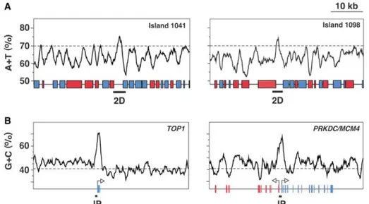
Figure 1.8: A/T-rich and C/G-rich islands at DNA replication origins. A. A/T content through the
1041 and 1098 A/T-rich islands in S. pombe. Red and blue boxes represent genes transcribed towards the left and the right, respectively. Genomic regions labeled 2D represent restriction fragments containing an active ORI. Dashed lines indicate the average intergenic A/T content (70%). B. G/C content across the first two exons of the human TOP1 gene and the bidirectionally transcribed PRKDC and MCM4 genes. Arrows indicate the transcription direction. Red and blue bars represent exons. Black boxes labeled IP represent DNA fragments immuno-precipitated by ChIP analysis with anti-human Orc2p antibodies. Dashed lines indicate the anti-human average genomic G/C content (41%). Scale as in (A). Adapted from [86].
In this context, it was very interesting to find that another family of transcription factors, namely the AP-1 proteins, displays an affinity for origin sequences [102, 109]. A possible role for AP-1 proteins in origin decision would be particularly intriguing, because it could represent a basis for the interplay between DNA replication and transcription, as well as explain the proto-oncogenic properties displayed by these proteins.
In order to better understand to what extent AP-1 proteins could be involved in origin function, some knowledge about their structure and function is required; accordingly, the next paragraph will be focused on this family of transcription factors involved in several aspects of the cell life.
1.2 The AP-1 protein family
1.2.1 AP-1 complex protein composition and general features
Activator protein-1 (AP-1) was first identified as a transcription factor that binds an essential cis-element of the human metallothionein lla (hMTIla) promoter and is required for its optimal basal activity both in vivo and in vitro [110, 111]. Soon after, the AP-1 binding sites were also recognized as taking part in the transcriptional activation of several cellular and viral genes in response to cell treatment with the tumor promoter TPA (12-O-tetradecanoylphorbol 13-acetate), being located at regions thus collectively named TPA-responsive element (TRE) [112].
AP-1 collectively describes a group of diverse nuclear proteins structurally and functionally related, forming dimers and belonging to different sub-families including:
• Jun protein family comprising c-Jun, JunB and JunD;
• Fos protein family composed of c-Fos, FosB, Fra-1 and Fra-2;
• Activating transcription factor (ATF) protein family consisting of ATF-1, ATF-2 (also known as CREB2 or CREBP-2), ATF-3, ATF-4, ATF-5, ATF-6, ATF-7 (also known as ATF-A), B-ATF and CREB1;
• Jun-dimerizing partners (JDP) protein family encompassing JDP-1 and JDP- 2; • Musculo-aponeurotic fibrosarcoma (Maf) protein family comprising c-Maf,
Maf-A, Maf-B, Maf-G, Maf-F, Maf-K and Nrl.
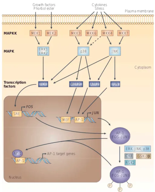
AP-1 activity is induced by many diverse stimuli, both internal and external to the cell, such as growth factors, cytokines, neurotransmitters, polypeptide hormones, cell–matrix interactions, bacterial and viral infections and a variety of physical and chemical stresses [113]. These stimuli activate mitogen activated protein kinase (MAPK) cascades that enhance AP-1 activity through the phosphorylation of distinct
substrates as it will be reviewed in paragraph 1.2.3.
Each of these proteins is differentially expressed and regulated; thus, in every cell type the broad combinatorial possibilities provided by the large number of AP-1 proteins determine the AP-1 dimer binding specificity and affinity and, consequently, the spectrum of regulated genes [114]. A common feature of all these proteins is the presence of an evolutionarily conserved basic DNA-binding domain combined with a leucine zipper, namely the bZIP domain. Dimerization is the fundamental pre-requisite for DNA binding mediated by the basic domain [115], with the composition of the leucine zipper being responsible for dimer specificity and stability. Elucidation on the structure of the AP-1 proteins will be discussed in the next paragraph. Whereas the Jun proteins exist both as homo- and hetero-dimers, the Fos proteins, which cannot homodimerize, form stable heterodimers with Jun and Maf proteins enhancing their DNA-binding activity [116]. Jun-Fos heterodimers bind preferentially the TRE DNA sequence, which is a heptamer consensus sequence (5′-TGA(C/G) TCA-3′), whereas Jun-ATF dimers bind with higher affinity to another consensus sequence known as the cyclic AMP responsive element (CRE) (5′-TGACGTCA-3′) [117]. The Jun-JDP dimer instead, binds with the same affinity to both CRE and TRE sequences. However, the AP-1 binding site exhibits some degree of degeneracy [118]; as a consequence, the sequences to which the AP-1 complex binds may differ upon interaction with structurally unrelated proteins such as NFAT, or proteins from the Ets or Smad families [119] and thus, may also differ in many natural promoters and enhancers of AP-1-regulated genes [117].
The individual Jun and Fos proteins have significantly different transactivation potentials. Whereas c-Jun, c-Fos and FosB are considered strong transactivators, JunB, JunD, Fra-1 and Fra-2 exhibit only weak transactivation potential. In fact, under specific circumstances, the latter might also act as repressors of AP-1 activity as a competitor for binding to AP-1 sites or by forming ‘inactive’ heterodimers with c-Jun, c-Fos or FosB [114].
A wide range of physiological and pathological stimuli regulate AP-1 activity such as cytokines, growth factors, stress signals, infections and oncogenic stimuli [120].
Regulation of the network of AP-1 activity can be achieved at different levels, including changes in AP-1 subunit-encoding gene transcription, control of the stability of their mRNAs, post-translational processing and turnover of pre-existing or newly synthesized AP-1 subunits [117], and specific interactions between AP-1 proteins and other transcription factors and cofactors. This network of molecular regulations will be reviewed in the subsequent paragraphs.
1.2.2 Structure of AP-1 proteins
Dimerization of Jun and Fos proteins occurs between their so called “leucine-zipper” (ZIP) regions via hydrophobic interactions, as originally elucidated used site-directed mutagenesis [121]. The “leucine-zipper” is one of the many structural motifs that characterize proteins able to bind DNA. It consists of an extensive α-helix in which every seventh amino acid a leucine is present. Due to this arrangement, the leucine side chains protrude from one side of the α-helix and form a hydrophobic surface that mediates dimerization [122]. The hydrophobic surfaces of two α-helices wrap around each other as a result of van der Waals interactions, meanwhile closely located hydrophilic amino acids make contact tightening the overall structure in a thermodynamically favorable manner (Figure 1.9). Leucines in the ZIP domain of Jun can be replaced with other hydrophobic residues such as phenylalanines, without any adverse effects on the formation of Jun:Fos heterodimers [123]. In addition, other hydrophobic residues present between the leucines, that together with them form the characteristic 3-4 repeat of α-helices involved in “coiled-coil” interactions [124], are also as important for mediating Jun:Jun and Jun:Fos dimerization [125]. However, hydrophobic interactions alone do not seem to account for the specificity in dimer formation among ZIP proteins. For instance, c-Fos dimerizes with the various Jun proteins but not with GCN4, Myc or another Fos molecule [121, 125]; CREB, which is another ZIP protein, forms homodimers that interact with the cyclic-AMP response
element (CRE) [126] and was not found to interact with either c-Jun, JunB or c-Fos [125]. Surprisingly CREBP-2, which is a protein highly related to CREB, was found to form heterodimers with c-Jun but not with c-Fos and these heterodimers, as well as CREB homodimers, interact with the CRE but not with the TRE sequence [127] [128]. Measurement of the dissociation temperatures, indicates that the increased DNA-binding activity of the Jun:Fos heterodimer is due to its increased thermostability if compared to the Jun:Jun homodimer. Whereas the heterodimer dissociates between 37°C and 42°C, the homodimer dissociates between 25°C and 37°C [125]. The higher thermostability of the heterodimer is responsible for potentiating its DNA-binding activity by increasing the number of molecules present at any given time in the dimeric state. In vitro binding experiments of c-Jun:c-Fos heterodimers with the TRE sequence have shown that addition of an excess of unlabeled binding sites to preformed protein-DNA complexes, resulted in a very rapid disappearance of the preformed DNA complex containing c-Jun homodimers, while the protein-DNA complex formed by c-Jun:c-Fos heterodimers was much more stable [129].
c-Fos, on the other hand, does not dimerize even at 4°C [125] and this can be explained by the presence of electrostatic repulsions between negatively-charged side chains, which are abundant in its ZIP region. Thus, once the ZIP domain of c-Fos is replaced with the one of GCN4 or c-Jun, chimera proteins are capable of homodimerization [130]. While the ZIP region mediates the dimerization of these proteins and hence dictates the specificity of complex formation, the interaction with DNA, instead, occurs via a region found immediately upstream of the “leucine-zipper” (Figure 1.10). This region is known as the “basic region” due to the abundance of positively charged residues [131]. Sequence analyses reveal that the “basic region” is highly conserved among all of the Jun and Fos proteins [131] and that it is also conserved in the various CREB and ATF proteins which interact with a sequence similar to the TRE. Site-directed mutagenesis provides a proof that whereas this region is only responsible for DNA-binding, it is not involved in dimerization.
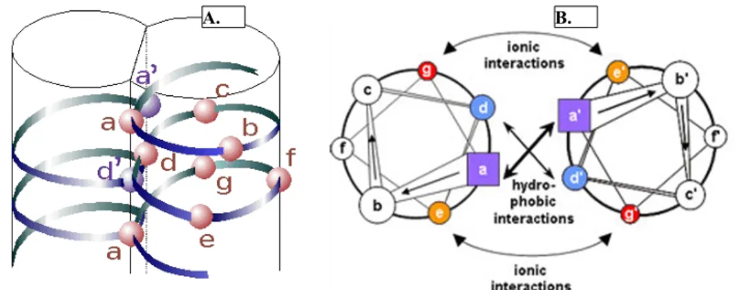
Figure 1.9: Coiled coil protein structure. A coiled-coil protein consists of two identical strands of
amino acid sequences that wrap around each other. The first and fourth position (a and d) are generally apolar or hydrophobic amino acids. When the two strands coil around each other, positions a and d are internalized, stabilizing the structure (A), while positions b, c, e, f, and g are exposed on the surface of the protein. Positions e and g are tighten the structure by ionic interactions (B). Adapted from
Hitchcock-DeGregori – Tropomyosin 2008.
The spacing between the ZIP and the “basic region” is also very critical. In fact, a duplication of five amino acids that are located between the two regions, and therefore alters their phasing, generates a c-Jun protein variant that can still dimerize with wild type c-Jun or c-Fos but is incapable of binding to DNA [132]. Furthermore, the chimeric dimers formed between the variant protein and either c-Jun or c-Fos also fail to bind DNA and inhibit transactivation [125]. In addition to these domains, a region close to the basic domain required for transcriptional activity of dimers, is the transactivation domain (TAD) (Figure 1.10). Within the TAD N-terminus different phosphorylation sites are present, thus phosphorylation of serine 63 and serine 73 residues of c-Jun by the Jun N-terminal kinase (JNK) family of kinases results in a large increase in its ability to interact with the CBP/p300 family of cofactors and, similarly, in the transcriptional activation potential of the protein [133]. The N-terminus of c-Jun also contains a δ-domain, which is the docking site for JNK and mediates ubiquitin-dependent degradation of the protein [134]. The presence of conserved sequences outside the ZIP and basic regions also in the various Fos
proteins [135] suggests that these regions may be involved in the interaction with various components of the transcriptional machinery. In fact, deletion of the c-Fos C-terminus, strongly reduces its ability to cooperate with c-Jun or JunD [136].
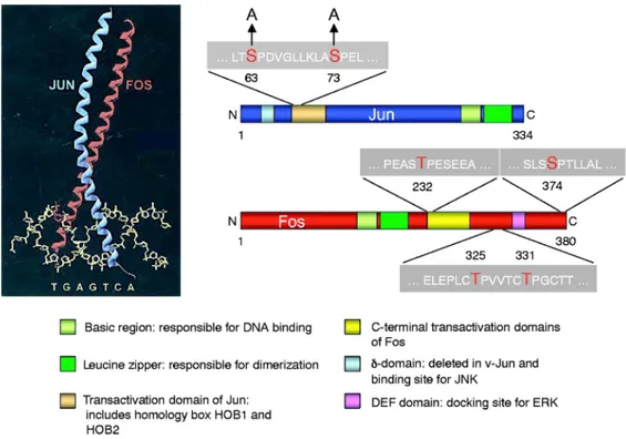
Figure 1.10: The Jun-Fos heterodimer. The c-Jun and c-Fos proteins exhibit several domains,
including the so called bZIP domain or leucine zipper plus the basic domain which is required for their interaction with the AP-1 site (TGAGTCA) forming an X-shaped α-helical structure. In addition, transactivation domains and docking sites for several kinases, such as JNK or ERK, are present. These kinases modulate the activity of both proteins phosphorylating two serine and threonine residues. JNK specifically phosphorylates serine residues within the transactivation domain of c-Jun at position 63 and 73 and thereby regulates its transactivation activity; in fact a Jun mutant in which these residues are mutated to alanine (Jun-AA) generates a protein that cannot be activated by JNKs. ERK phosphorylates threonine residues at positions 325 and 331 and a serine residue at position 374 of c-Fos. Additionally, a c-Fos-related kinase phosphorylates a threonine residue at position 232 of c-Fos . Adapted from [114].
1.2.3 AP-1 expression, regulation and activity
The expression of the AP-1 proteins is critical for the decision of the cell fate and thus is regulated at multiple levels, which include control the transcription of their genes, post-translational modifications and dimer composition (refer to paragraph 1.2.1). It has to be underlined that expression of c-Fos and c-Jun in response to many different stimuli causes protein kinase C (PKC) activation, but also their own transcription is induced by PKC activation [137]. While expression of c-Fos is very rapid and highly transient as well as is its turnover as a protein [138], induction of the c-Jun mRNA in response to stimulation is also transient, but the messenger is significantly more stable. [139]. Other agents, like TNF-α or TGF-β [140] lead to a longer lasting induction of c-Jun while their effects do not modify the c-Fos mRNA stability. Three cis-elements have been found to mediate c-Fos induction. The first one is a CRE sequence proximal to the TATA box occupied by ATF or CREB [141]; the second one is a Sis inducible enhancer (SIE) recognized by STAT proteins [142] and the last one is a serum response element (SRE) recognized by a dimer of serum response factor (SRF) and the ternary complex factors (TCF) [143]. Analyses of the promoter of the human c-Jun gene revealed that, upstream of two TATA-like sequences located 24-30 bps upstream a cluster of transcription initiation sites, there is a sequence recognized by the AP-1 complex itself, suggesting that transcription of c-Jun is subjected to a positive autoregulatory loop [144]. Due to the importance of AP-1 proteins for cell proliferation, presence of negative regulations are required for normal cell function. As already mentioned, the c-Fos transcript is subjected to a rapid turnover due to the presence of an RNAse target, AU-rich sequence in the 3' untranslated region (3' UTR); also c-Fos down-regulates its own gene product in a negative autoregulatory manner [145]. The same AU-rich sequence is also present in the c-Jun transcript, but in this case down-regulation of c-Jun is due to the binding of JunB or JunD homodimers or c-Jun:CREB and c-Jun:ATF-2 heterodimers to the AP-1 binding sites upstream the c-Jun gene [126, 128, 146]. The most common