Meola, Lorenzo Di Venanzio and Antonio Salvetti
Lorenzo Ghiadoni, Stefano Taddei, Agostino Virdis, Isabella Sudano, Virgilio Di Legge, Mario
Essential Hypertension
Endothelial Function and Common Carotid Artery Wall Thickening in Patients With
Print ISSN: 0194-911X. Online ISSN: 1524-4563
Copyright © 1998 American Heart Association, Inc. All rights reserved.
is published by the American Heart Association, 7272 Greenville Avenue, Dallas, TX 75231 Hypertension
doi: 10.1161/01.HYP.32.1.25
1998;32:25-32
Hypertension.
http://hyper.ahajournals.org/content/32/1/25
World Wide Web at:
The online version of this article, along with updated information and services, is located on the
http://hyper.ahajournals.org//subscriptions/
is online at: Hypertension
Information about subscribing to Subscriptions:
http://www.lww.com/reprints
Information about reprints can be found online at: Reprints:
document. Permissions and Rights Question and Answer
this process is available in the
click Request Permissions in the middle column of the Web page under Services. Further information about Office. Once the online version of the published article for which permission is being requested is located,
can be obtained via RightsLink, a service of the Copyright Clearance Center, not the Editorial Hypertension
in
Requests for permissions to reproduce figures, tables, or portions of articles originally published Permissions:
Thickening in Patients With Essential Hypertension
Lorenzo Ghiadoni, Stefano Taddei, Agostino Virdis, Isabella Sudano, Virgilio Di Legge, Mario Meola,
Lorenzo Di Venanzio, Antonio Salvetti
Abstract—Intimal-medial thickening of the carotid wall is considered an early marker of atherosclerosis. Endothelial
function is impaired in the presence of various cardiovascular risk factors that are implicated in the pathogenesis of atherosclerosis. To evaluate the relationship between vascular reactivity and carotid intimal-medial thickening, in 44 (mean6SD age, 45.768.8 years; range, 28 to 60 years; 31 men and 13 women) patients with essential hypertension who had never been treated and whose history of increased blood pressure was no longer than 12 months, we evaluated several parameters: intimal-medial thickening of the common carotid arteries (by B-mode ultrasound); forearm vascular response (by strain-gauge plethysmography) to intrabrachial infusion of acetylcholine (0.15, 0.45, 1.5, 4.5, and 15 mg/100 mL forearm tissue per minute), an endothelium-dependent vasodilator, or sodium nitroprusside (1, 2, and 4 mg/100 mL forearm tissue per minute), an endothelium-independent vasodilator; calculated minimal forearm vascular resistances (the ratio between mean arterial pressure and maximal forearm vasodilation induced by 13 minutes of ischemia and 1 minute of exercise); and left ventricular mass index (on echocardiography profile). Carotid wall intimal-medial thickening showed a significant (P,0.001) inverse correlation with vasodilation to acetylcholine (r520.58) and age (r520.40), whereas no correlation was observed with the response to sodium nitroprusside or with minimal forearm vascular resistances, left ventricular mass index, systolic and diastolic blood pressures, and plasma cholesterol and glucose levels. Moreover, vasodilation to acetylcholine showed no correlation with minimal forearm vascular resistances or left ventricular mass index. Although comparison of different vascular “districts,” such as the forearm microcirculation and carotid artery, does not allow for a conclusive interpretation, the present data indicate that in patients with essential hypertension, carotid wall thickening is associated with reduced endothelium-dependent vasodilation and suggest that endothelial dysfunction might be involved in early arterial structural alterations. (Hypertension. 1998;32:25-32.)
Key Words: endotheliumn carotid arteries n hypertension, essential n acetylcholine n sodium nitroprusside
A
lthough essential hypertension is defined as a genetic disease characterized by consistently elevated BP val-ues, its clinical relevance arises from the associated increased predisposition to cardiovascular morbidity and mortality.1Probably the most important mechanism by which essential hypertension acts as a cardiovascular risk factor is the induction of atherosclerosis.1 However, the pathological
events leading from high BP to atherosclerotic lesions are still to be fully clarified.
It is well documented that functional or morphological alterations of endothelial cells appear to be critical to the evolution, progression, and clinical manifestation of athero-sclerotic vascular disease.2,3In addition, endothelial
dysfunc-tion has been documented in the presence of atherosclerotic lesions,4 and it predicts the development of atherosclerotic
lesions in epicardial vessels of cardiac transplant patients.5
Moreover, endothelial dysfunction has also been documented in the presence of different cardiovascular risk factors in-volved in the pathogenesis of atherosclerosis itself, such as
hypertension,6 –9 aging,9,10 menopause,10
hypercholesterol-emia,11,12diabetes mellitus,13,14and smoking.15
In recent years, ultrasound imaging of the extracranial carotid arteries has been extensively used to detect early arterial wall abnormalities. Moreover, IMT of the carotid wall has been considered an early marker of atherosclerosis16and
a possible index of coronary artery atherosclerosis.17
The aim of the present study was to evaluate the relation between carotid wall IMT with different functional and structural cardiovascular parameters and humoral factors in patients with essential hypertension. Moreover, to better clarify the link between essential hypertension and induction of atherosclerosis, we avoided confounding factors, such as previous pharmacological antihypertensive treatment or the frequently encountered impossibility of ascertaining the du-ration of hypertensive disease. Therefore, in the present study, we selected only those patients who had never been treated and who had a documented history of essential hypertension no longer than 12 months.
Received January 19, 1998; first decision February 2, 1998; revision accepted February 11, 1998. From the I Clinica Medica, University of Pisa, Italy.
Correspondence to Dr Stefano Taddei, I Clinica Medica, University of Pisa, Via Roma 67, 56100 Pisa, Italy. E-mail [email protected] © 1998 American Heart Association, Inc.
Methods
Patients
The study population included 30 normotensive subjects (20 men and 10 women) and 44 patients with essential hypertension (31 men and 13 women). Subjects with marked hypercholesterolemia (total cholesterol.6.2 mmol/L), a heavy smoking habit (.10 cigarettes/ d), diabetes mellitus, cardiac or cerebral ischemic vascular disease, impaired renal function, and other major pathologies were excluded from the study. In accordance with institutional guidelines, the protocol was approved by the ethics committee of the University of Pisa. All patients were aware of the investigational nature of the study and gave their written consent.
Essential hypertension patients were recruited with the collabora-tion of 10 general practicollabora-tioners working in the city of Pisa. The practitioners were asked to enroll patients with recent-onset essential hypertension (no more than 12 months). It is worth noting that patients were enrolled only if they reported a history of periodic BP measurements. This criterion excluded the possibility of detecting hypertension that corresponded to the first BP measurement in a patient’s life, which would have led to difficulty in ascertaining the time of onset of hypertensive disease. In all cases, subjects were characterized by the presence of a positive family history of essential hypertension and a supine arterial BP (after 10 minutes of rest) consistently.140/90 mm Hg, as measured by mercury sphygmo-manometer three times at 1-week intervals. Secondary forms of hypertension were excluded as follows: renovascular disease was excluded in most cases by renal artery color Doppler echography, whereas in 16 of 44 patients, renal angiography was performed; renal parenchymal diseases was ruled out by the normality of serum creatinine levels, urine examination, and renal echotomography; primary aldosteronism was ruled out by the absence of hypokalemia and the normality of plasma renin activity and aldosterone; pheo-chromocytoma was ruled out by the normality of plasma catechol-amine levels and the absence of an adrenal or abdominal mass on echotomography. Mean age was 45.768.8 years (range, 28 to 60 years). BP values were 158.8613.2/101.766.9 mm Hg. As control subjects, 30 healthy normotensive volunteers (mean6SD age, 44.766.6 years; mean6SD BP, 116.863.6/75.464.1 mm Hg) were enrolled.
Drugs
Ach HCl (Farmigea SpA) and SNP (Malesci) were obtained from commercially available sources and freshly diluted to the desired concentration by adding normal saline. SNP was dissolved in glucosate solution and protected from light with aluminum foil. Experimental Procedures
Each subject underwent the following examinations: B-mode ultra-sound imaging of the carotid artery, echocardiographic examination, and FBF studies.
Carotid Artery B-Mode Ultrasound Imaging
High-resolution B-mode ultrasound examination of the carotid ar-teries was performed with a Biosound 2000 IISA with a 7.5-MHz transducer. Sonography and readings were carried out by trained and certified sonographers with regular quality-control checks (Division of Vascular Ultrasound Research, Bowman Gray School of Medicine of Wake Forest University), as previously described by Crouse et
al.18Patients were examined in the supine position, and each carotid wall and segment was examined independently from continuous angles to identify the thickest intimal-medial site. Each scan of the common carotid artery began just above the clavicle, and the transducer was moved cephalad past the bifurcation and along the internal carotid artery. Three segments were identified on each side: the distal 1.0 cm of the common carotid proximal to the bifurcation, the bifurcation itself, and the proximal 1.0 cm of the internal carotid artery. At each of the three segments for both near and far walls in the left and right carotid arteries, the sonographer identified two interfaces: on the near wall, the first interface (interface 2) was the adventitial-medial boundary, and the second (interface 3), the intimal-luminal boundary; on the far wall, the first interface (interface 4) was the luminal-intimal boundary, and the second (interface 5), the medial-adventitial boundary. Thus, the distances between interfaces 2 and 3 and between 4 and 5 define the IMTs on the near and far walls, respectively. When these interfaces had been imaged distinctly, the sonographer reduced the gain and time-gain control settings to as low as possible to decrease artifacts; the video images that included the maximum near- and far-wall IMTs at each of the 12 segments were then recorded. Images were transcribed onto SVHS1⁄2-in. videotape. Frames that identified the maximum near- and far-wall IMTs within each segment were interfaced to a high-resolution monitor, and maximum wall diastolic (minimal carotid diameter) thickness was calculated at each site. Under our experimental conditions, the variability of measurements was 7.960.3%. For this study, the mean of the aggregate of all 12 sites and the maximum of all 12 sites were calculated. Because of the highly significant correlation between these two parameters (r50.83, P,0.0001), the analysis was performed with only the maximum thickness of all 12 sites.
Patients with an IMT,1 mm were considered normal, patients with an IMT between 1 and 1.3 mm were considered to have wall thickening, and patients with an IMT.1.3 mm were considered to have plaque.18,19The operator (L.D.V.) was blinded with respect to the results of other evaluations.
Echocardiographic Evaluation
Echocardiograms (SIM 500, Esaote Biomedica) were performed from parasternal and apical windows with the subjects in the left lateral decubitus position. An ECG tracing with an easily discernible QRS complex was chosen. The dimensions of the left ventricle, septum, and posterior wall were obtained at the beginning of the QRS complex, with the ultrasound beam passing through the left ventricle at the level of the tips of the mitral valve leaflets. Following the recommendations of the Penn Convention, we measured (in centimeters) the diastolic thickness of the left interventricular septum and left ventricular posterior wall, as well as the diastolic dimension of the left ventricular chamber; these measurements were inserted into the mathematical model for the prediction of LVMI according to Devereux et al.20The operator (V.D.L.) was blinded with respect to the results of other evaluations.
FBF Studies
All studies were performed at 8AMafter an overnight fast, with the subjects lying supine in a quiet, air-conditioned room (22°C to 24°C). A polyethylene cannula (21 gauge, Abbot) was inserted into the brachial artery under local anesthesia (2% lidocaine) and con-nected through stopcocks to a pressure transducer (model MS20, Electromedics) for systemic mean BP (1/3 pulse pressure1diastolic pressure) and heart rate (model VSM1, Physiocontrol) monitoring and for intra-arterial infusions. FBF was measured in both forearms (experimental and contralateral forearm) by strain-gauge venous plethysmography (LOOSCO, GL LOOS).21Circulation to the hand was excluded 1 minute before FBF measurement by inflating a pediatric cuff around the wrist at suprasystolic blood pressure. Details concerning the sensitivity and reproducibility of the method as performed in our laboratory have already been published.22
Forearm volume was measured according to the water-displace-ment method, and drug infusion rates were normalized for 1 dL of tissue by adjusting the concentration of the stock solution to the
Selected Abbreviations and Acronyms
Ach5 acetylcholine BP5 blood pressure FBF5 forearm blood flow IMT5 intimal-medial thickening LVMI5 left ventricular mass index MFVR5 minimal forearm vascular resistance
SNP5 sodium nitroprusside
desired infusion rate. Drugs were infused at systemically ineffective rates through separate ports via three-way stopcocks. The operator (A.V.) was blinded with respect to the results of other evaluations. Endothelium-dependent vasodilation was estimated by performing a dose-response curve to intra-arterial Ach (cumulative increase of the infusion rates were 0.15, 0.45, 1.5, 4.5, and 15mg per 100 mL of forearm tissue per minute for 5 minutes at each dose). Endothelium-independent vasodilation was assessed with a dose-response curve to intra-arterial SNP, a direct smooth muscle cell relaxant compound,23 (cumulative increase by 1, 2, and 4mg per 100 mL of forearm tissue per minute for 5 minutes at each dose). These rates were selected to induce vasodilation comparable to that obtained with Ach.
MFVRs were calculated as the ratio between maximal postische-mic FBF increase (after 13 minutes of ischemia plus 1 minute of dynamic hand exercise) and intra-arterial mean arterial pressure, as previously described.24The sequence of the experimental interven-tions was randomized, and a 45-minute recovery period was allowed between the three experimental steps.
Data Analysis
Results are expressed as mean6SD. Differences between means in Table 1 were analyzed by Student’s t test for unpaired data. FBF data were analyzed as absolute values by ANOVA for repeated measures; Scheffe´’s test was applied for multiple comparison testing. Differ-ences were considered statistically significant at P,0.05.
Interactions between carotid wall thickening and forearm vasodi-lation to Ach, SNP (considered as the maximum effect), age, BP, LVMI, MFVR, plasma total cholesterol, and plasma HDL and LDL cholesterol were calculated by both simple correlation and multiple regression analyses.
Results
The baseline systemic demographic, hemodynamic, and hu-moral characteristics for normotensive subjects and essential hypertension patients are summarized in Table 1. No differ-ence was found between the two groups, with the exclusion of BP values. The mean carotid maximal IMT was 1.060.1 mm in normotensive subjects and 1.260.3 in essential hyperten-sion patients (P,0.01 versus normotensives). According to the results of the carotid scanning, hypertensive patients were divided into three groups: (1) n513 patients were found to be normal (ie, an IMT of 0.960.1 mm); (2) n520 patients had IMT thickening (IMT of 1.260.1 mm); and (3) n511 patients had plaque (IMT of 1.660.2 mm). These subgroups of hypertensive subjects presented no differences in the hemo-dynamic and humoral characteristics considered (Table 2).
Ach caused dose-dependent vasodilation, which was shown to be significantly blunted in essential hypertension patients compared with normotensive subjects (FBF rose
TABLE 1. Characteristics of Study Subjects
Parameter Normotensive Subjects (n530) Essential Hypertension Patients (n544) Age, y 44.766.6 45.768.8 Sex, M/F 20/10 31/13 Smoking, yes/no 7/23 10/34
Body mass index, kg/m2 22.461.4 22.961.6
Systolic BP, mm Hg 116.663.6 158.8613.8* Diastolic BP, mm Hg 75.464.1 101.766.9* Heart rate, bpm 73.663.5 72.465.0 Plasma glucose, mmol/L 4.960.3 4.860.4 Plasma total cholesterol, mmol/L 4.860.3 5.260.4 Plasma HDL cholesterol, mmol/L 1.160.2 1.160.2 Plasma LDL cholesterol, mmol/L 3.060.3 3.460.4 Carotid IMT, mm 1.060.1 1.260.3*
MFVR, units 1.461.1 2.660.9*
LVMI, g/m2 94.367.9 110.3621.5*
Data are mean6SD. *P,0.05 or less.
TABLE 2. Demographic, Hemodynamic, and Humoral Characteristics of Essential
Hypertension Patients (n544) Divided Into Three Subgroups According to Carotid Wall Thickening Parameter Normal #1 mm (n513) Thickening 1 to 1.3 mm (n518) Plaque .1.3 mm (n513) Age, y 44.067.7 46.368.1 47.369.9 Sex, M/F 9/4 12/6 10/3
Body mass index, kg/m2 22.160.7 23.460.9 22.660.8
Smoking, yes/no 3/10 4/14 3/10
Systolic BP, mm Hg 15866.8 154.2612.4 158.269.5 Diastolic BP, mm Hg 102.166.3 101.464.7 103.763.7
Heart rate, bpm 66.366.1 65.165.4 68.367.1
Glucose, mmol/L 4.660.4 4.760.2 4.960.4
Total cholesterol, mmol/L 5.460.4 5.260.3 5.160.4 HDL cholesterol, mmol/L 1.160.2 1.060.1 1.160.2 LDL cholesterol, mmol/L 3.563.5 3.360.4 3.360.3
Carotid IMT, mm 0.960.1 1.260.1* 1.660.2*†
MFVR, units 2.960.8 2.660.9 2.760.7
LVMI, g/m2 110.4621.4 105.8621.6 111.6622.8
Data are mean6SD.
from 3.460.5 to 24.864.8 and 3.560.4 to 35.167.1 mL per 100 mL of forearm tissue per minute, respectively; P,0.001) (Figure 1). In contrast, the response to SNP was similar in controls and hypertensive patients (FBF rose from 3.660.4 to 27.963.6 and 3.460.6 to 26.564.3 mL per 100 mL of forearm tissue per minute, respectively) (Figure 1). When the response to Ach was also examined in the three subgroups of hypertensive patients, it was found that endothelium-dependent vasodilation was significantly lower in essential hypertension patients with thickening (FBF rose from 3.460.5 to a maximum of 21.963.1 mL per 100 mL of forearm tissue per minute) and plaque (FBF rose from 3.460.7 to a maximum of 17.963.7 mL per 100 mL of forearm tissue per minute) than in hypertensive
patients with a normal IMT (FBF rose from 3.260.6 to a maximum of 29.665.2 mL per 100 mL of forearm tissue per minute; P,0.05) (Figure 2). However, in the normal IMT subgroup, the vasodilating effect of Ach was still lower than that in normotensive subjects (P,0.05). The response to SNP was still similar in the three hypertensive subgroups (for normal IMT, FBF rose from 3.460.5 to a maximum of 27.864.3; for those with thickening, FBF rose from 3.560.4 to a maximum of 26.463.6; and for those with plaque, FBF rose from 3.660.6 to a maximum of 24.263.5 mL per 100 mL of forearm tissue per minute) (Figure 2). In normotensive subjects, no significant correlation was found between carotid IMT and the response to Ach or SNP (r520.10, P5NS and
r520.08, P5NS, respectively).
Figure 1. FBF increase above basal levels (b) induced by intra-arterial Ach (mg per 100 mL of forearm tissue per minute) (left) and SNP (SNP,mg per 100 mL of fore-arm tissue per minute) (right) in 44 essential hypertension patients (E) and 30 normotensive subjects (F). Data are shown as mean6SD. and reported as absolute values. Asterisks denote significant difference between hyperten-sive patients and normotenhyperten-sive subjects (*P,0.001).
Figure 2. FBF increase above basal levels (b) induced by intra-arterial Ach (mg per 100 mL of forearm tissue per minute) (left) and SNP (mg per 100 mL of forearm tissue per minute) (right) in 44 essential hypertension patients divided into three subgroups: patients with ,1 mm IMT (F), those with IMT of 1 to 1.3 mm (E), and those with IMT.1.3 mm (M). Data are shown as mean6SD and reported as absolute values. Asterisks denote a significant difference between the three sub-groups of hypertensive patients (*P,0.05 or less).
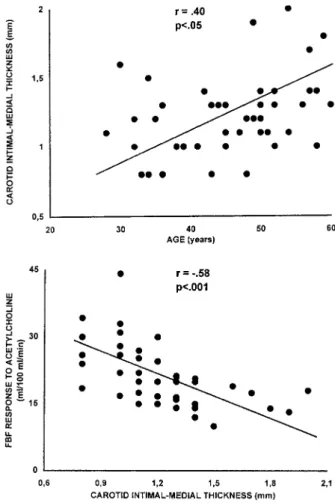
In essential hypertension patients, a significant inverse correlation was found between carotid artery IMT and Ach-induced forearm vasodilation, as evaluated in terms of FBF response to the highest infusion rate of the agonist (15mg per 100 mL of forearm tissue per minute; r520.58, P50.0003) (Figure 3), whereas no correlation was observed with the response to SNP (FBF response to the highest infusion rate (4 mg per 100 mL of forearm tissue per minute, r520.06,
P5NS). Age was positively and significantly correlated with
carotid artery IMT (r50.40, P50.022) (Figure 3) and in-versely and significantly with the maximal response to Ach (r520.51, P50.001).
In addition, no correlation was observed between carotid artery IMT and systolic (r520.05, P5NS) or diastolic (r520.2, P5NS) BP. However, when the data from essential hypertension patients were considered together with data from normotensive subjects, a positive correlation was ob-served between IMT and systolic (r50.39, P,0.001) and diastolic (r50.34, P,0.01) BP. Finally, no correlation was observed between carotid IMT and MFVR (r520.2, P5NS), LVMI (r50.09, P5NS), total cholesterol (r520.08, P5NS), HDL cholesterol (r50.003, P5NS), LDL cholesterol (r520.04, P5NS), or plasma glucose (r520.13, P5NS).
Finally, multivariate regression analysis showed that the correlation between carotid IMT and the other parameters
(r250.51) was influenced most by the maximal response to
Ach (r250.35) and age (r250.15). When the effect of aging
was taken into account in the analysis, the relationship between the response to Ach and carotid IMT remained statistically significant (from P,0.004 to P,0.03).
Discussion
At the present time, B-mode ultrasonography is extensively used to detect early structural changes in carotid arteries because the thickening process in these areas is considered a prognostic marker for the development of atherosclerosis16
and appears to correlate with coronary lesions.17
In the present study, we tested the possible existence of a relationship between early structural changes in carotid arteries and endothelial dysfunction in essential hypertension patients. Given the possibility that regression or induction of cardio-vascular alterations, whether structural or functional, could be caused by long-term pharmacological treatment or different duration of hypertension,25–28the present study was designed
to recruit never-treated essential hypertension patients with a sufficiently accurate determination of disease onset. To pur-sue this aim, we cooperated with general practitioners who were asked to recruit subjects known to be undergoing measurement of BP values at least once every 6 months. Only subjects whose documented report of high BP values was no longer than 1 year were then enrolled.
Following these inclusion criteria, we selected a fairly homogeneous study population represented by never-treated essential hypertension patients with a short duration of the disease. Moreover, apart from high BP, the hypertensive patients studied were characterized by normal glucose and lipid profiles and a moderate prevalence of smoking history. In line with previous evidence,5–9 these patients showed
endothelial dysfunction because vasodilation to Ach but not to SNP was found to be reduced compared with that of normotensive control subjects. However, the interesting find-ing of the present study is that when essential hypertension patients were divided into three subgroups according to the different carotid IMT findings, the response to Ach, while still impaired in patients with a normal IMT compared with normotensive patients, showed a further significant reduction in patients with thickening of the carotid artery and an even greater reduction in patients with plaque. Because the vaso-dilating effect of SNP proved to be similar in the three subgroups of hypertensive patients, the present results indi-cate that IMT of the extracranial carotid arterial wall is associated with blunted endothelium-dependent vasodilation in essential hypertension. This hypothesis is reinforced by the finding that in this study population of essential hypertension patients, carotid IMT showed a negative and significant correlation with maximal response to Ach but not to SNP. In contrast, carotid IMT of the control subjects showed no correlation with vasodilation to Ach.
Finally, in essential hypertension patients, the response to Ach was not correlated with the calculated MFVR or LVMI, which are indexes of arteriolar29
and cardiac28
structural alterations, respectively. Moreover, no correlation was found between IMT of the carotid arteries and LVMI or MFVR. Figure 3. Relationship between carotid wall thickening (x axis)
and age (y axis, top) and FBF response to the highest infusion rate of Ach (15mg per 100 mL of forearm tissue per minute) (y axis, bottom) in 44 essential hypertension patients.
These results suggest that in our study population of never-treated essential hypertension patients with a relatively low cardiovascular risk, there is a possible link between endothelial dysfunction and early structural changes of a large conduit artery, such as the carotid. On the other hand, the lack of correlation between endothelial dysfunction and MFVR or LVMI suggests that such alterations could be determined by different mechanisms. This possibility is in agreement with the evidence that blunted endothelium-depen-dent vasodilation is a “primitive” phenomenon and not secondary to the development of hypertension because this response is present in the normotensive offspring of essential hypertension patients,30shows no significant correlation with
BP values,9,31
and is not reversed by BP normalization.32,33
In contrast, cardiac and microvascular structural alterations seem to be more closely related to BP values.26,28
The lack of correlation between carotid IMT and LVMI is at variance with previous reports.34,35
However, the discrep-ancy can be explained by the fact that in the article by Roman et al,34
the study population included both normotensive and hypertensive patients in the analysis, with a wide range of LVMI values. Moreover, the study of Cuspidi et al35 also
considered patients with concentric remodeling.
Taken together, the present findings suggest that a dys-functional endothelium can be a predisposing factor to the development of atherosclerosis. If this is the case, it is worth noting that the subgroup of essential hypertension patients with a normal carotid IMT was characterized by a response to Ach that was significantly lower than that observed in normotensive controls. It is therefore conceivable that a certain degree of endothelial dysfunction is necessary to observe a detectable association with atherosclerosis. How-ever, another possible explanation for the latter result could lie in the major limitation of the present study, namely, the comparison of two different vascular districts, the forearm and carotid vasculature, with different structures (microcir-culation and large arteries, respectively). The forearm vascu-lature is not usually affected by atherosclerosis; in addition, vascular reactivity in the microcirculation is sometimes dif-ferent from that observed in large arteries.36
It is therefore crucial to avoid conclusive statements, and further studies are needed to provide more detailed confirmation of the associ-ation between endothelial dysfunction and early development of atherosclerosis in the carotid arteries.
The mechanism through which a dysfunctioning endothe-lium could promote atherosclerosis is related to the evidence that endothelial dysfunction is caused by an alteration in the
L-arginine–NO pathway,37 leading to a reduction of NO
bioavailability.38
NO appears to be not only a potent vasodi-lator but also an endogenous inhibitor of platelet aggrega-tion,39vascular smooth muscle cell growth and migration,40,41
leukocyte adhesion,42
and adhesion molecule expression.43
It is clear that an alteration in theL-arginine–NO pathway may
reduce this potentially antiatherosclerotic activity. In addi-tion, a dysfunctional endothelium can also produce prosta-noids such as thromboxane A2, which causes vasoconstriction
and platelet aggregation,44and/or oxygen free radicals, which
can destroy NO45and cause vascular damage.46Of relevance
is the finding that in the aorta and carotid artery of
sponta-neously hypertensive rats, endothelial dysfunction is associ-ated with monocyte/macrophage infiltration,47
suggesting that endothelial activation could constitute an early event in hypertension, leading to both increased monocyte adherence and chemotaxis and abnormal production of endothelium-derived constricting factors. Alternatively, the monocytes/ macrophages might themselves secrete constricting factors or further activate endothelial cells. In conclusion, it may be suggested that in essential hypertension, the mechanisms causing endothelial dysfunction can potentially lead to ath-erosclerosis. This hypothesis seems to be confirmed by the finding that an impairment in the NO system is not an exclusive characteristic of essential hypertension.37,38 It has
also been demonstrated in the presence of the majority of cardiovascular risk factors, such as aging,48
hypercholesterol-emia,12diabetes,13and smoking,15all pathological conditions
characterized by an increased predisposition to the develop-ment of atherosclerosis. As a final speculation, it is possible that the increased wall thickness may impair diffusion of endothelial vasoactive substances to smooth muscle cells, thereby impairing endothelium-dependent vasodilation. How-ever, this hypothesis seems to be indirectly excluded by the results with SNP. This compound acts directly on smooth muscle as NO source cells, and no different effect was observed in normotensive subjects or essential hypertension patients with normal IMT compared with hypertensive pa-tients with plaque.
Another major factor to be considered is the role of aging. The present study confirms previous evidence indicating a positive correlation between IMT of the carotid artery and aging.34,36 There is also a well-documented association
be-tween advancing age and impaired endothelium-dependent vasodilation.9 –11
Thus, aging itself could be the main phenom-enon involved, leading in a parallel manner to both the development of carotid structural alterations and impaired endothelium-dependent vasodilation. An alternative possibil-ity is that the summation of the simultaneous negative effects of aging and hypertension could result in more pronounced impairment of endothelial function, thereby leading to ath-erosclerosis. The consideration that our study population was represented by essential hypertension patients with very low cardiovascular risk points to the possibility that aging could play a key role in explaining the association between carotid artery structural alterations and endothelial dysfunction. However, even after accounting for the role of aging, the inverse relationship between the response to Ach and IMT remained statistically significant.
Finally, in these essential hypertension patients, we found no correlation between carotid IMT and other important atherosclerosis risk factors, such as high BP and unfavorable glucose and lipid profiles, a result that is at variance with several previous observations.36,49,50
A likely explanation could be the fact that, in accordance with the enrollment criteria, the present study population was characterized by the presence of high BP values as the only major cardiovascular risk factor. Thus, glucose and lipid profiles not only were within the normal range but also had a minimum range of variation. Therefore, these experimental conditions are prob-ably inadequate to detect an association between these risk 30 Atherosclerosis and Endothelial Function
factors and carotid IMT. This possibility is reinforced by the evidence that analysis of the relationship between BP values and IMT in the entire study population (normotensives plus hypertensives, characterized by a larger range of BP values) showed a positive correlation between the parameters considered.
In conclusion, the present study indicates an association between carotid artery IMT and impaired endothelium-de-pendent vasodilation. Whether these vascular alterations are casually associated or indicate a causal relationship between endothelial dysfunction and development of atherosclerosis in essential hypertension remains to be established.
References
1. Wilson PWF, Kannel WB. Hypertension, other risk factors and the risk of cardiovascular disease. In: Laragh JH, Brenner BM, eds. Hypertension:
Pathophysiology, Diagnosis and Treatment. New York, NY: Raven Press
Publishers; 1995:99 –114.
2. Ross R. The pathogenesis of atherosclerosis: an update. N Engl J Med. 1986;314:488 –500.
3. Chobanian A. Pathophysiology of atherosclerosis. Am J Cardiol. 1992; 70:3G–7G.
4. Yasue H, Matsuyama K, Matsuyama K, Okamura K, Morikami Y, Ogawa H. Response of angiographically normal human coronary arteries to intracoronary injection of acetylcholine by age and segment: possible role of early coronary atherosclerosis. Circulation. 1990;81:482– 490. 5. Davis SF, Yeung AC, Meredith IT, Charbonneau F, Ganz P, Selwyn P,
Anderson TJ. Early endothelial dysfunction predicts the development of transplant coronary artery disease at 1 year post transplant. Circulation. 1996;93:457– 462.
6. Linder L, Kiowski W, Buhler FR, Luscher TF. Indirect evidence for the release of endothelium-derived relaxing factor in the human forearm circulation in vivo: blunted response in essential hypertension.
Circu-lation. 1990;81:1762–1767.
7. Panza JA, Quyyumi AA, Brush JE Jr, Epstein SE. Abnormal endothelium dependent vascular relaxation in patients with essential hypertension.
N Engl J Med. 1990;323:22–27.
8. Taddei S, Virdis A, Mattei P, Salvetti A. Vasodilation to acetylcholine in primary and secondary forms of human hypertension. Hypertension. 1993;21:929 –933.
9. Taddei S, Virdis A, Mattei P, Ghiadoni L, Gennari A, Basile Fasolo C, Sudano I, Salvetti A. Aging and endothelial function in normotensive subjects and essential hypertensive patients. Circulation. 1995;91: 1981–1987.
10. Taddei S, Virdis A, Ghiadoni L, Mattei P, Sudano I, Bernini G, Pinto S, Salvetti A. Menopause is associated with endothelial dysfunction in women. Hypertension. 1996;28:576 –582.
11. Creager MA, Cooke JP, Mendelsohn ME, Gallagher SJ, Coleman SM, Loscalzo J, Dzau VJ. Impaired vasodilation of forearm resistance vessels in hypercholesterolemic humans. J Clin Invest. 1990;86:228 –234. 12. Casino PR, Kilcoyne CM, Quyyumi AA, Hoeg JM, Panza JA. The role of
nitric-oxide in endothelium-dependent vasodilation of hypercholesterol-emic patients. Circulation. 1993;88:2541–2547.
13. Williams SB, Cusco JA, Roddy M-A, Johnstone MT, Creager MA. Impaired nitric oxide mediated vasodilation in patients with non insulin-dependent diabetes mellitus. J Am Coll Cardiol. 1996;27:567–574. 14. Ting HH, Timini FK, Boles KS, Creager SJ, Ganz P, Creager MA.
Vitamin C improves endothelium-dependent vasodilation in patients with non-insulin-dependent diabetes mellitus. J Clin Invest. 1996;97:22–28. 15. Heitzer T, Yla¨-Herttuala S, Luoma J, Kurz S, Munzel T, Just H,
Olschewski M, Drexler H. Cigarette smoking potentiates endothelial dysfunction of forearm resistance vessels in patients with hypercholes-terolemia: role of oxidized LDL. Circulation. 1996;93:1346 –1353. 16. Poli A, Tremoli E, Colombo A. Ultrasonographic measurement of the
common carotid artery wall thickness in hypercholesterolemic patients: a new model for the quantification and follow up of preclinical atheroscle-rosis in living human subjects. Atheroscleatheroscle-rosis. 1988;70:253–261. 17. Salonen JT, Salonen R. Ultrasonographically assessed carotid
mor-phology and the risk of coronary heart disease. Arterioscler Thromb. 1991;11:1245–1249.
18. Crouse JR III, Craven TE, Hagaman AP, Bond G. Association of coro-nary disease with segment specific intimal-medial thickening of the extracranial carotid artery. Circulation. 1995;92:1141–1147.
19. Borhani NO, Mercuri M, Borhani PA, Buckalew VM, Canossa-Terris M, Carr AA, Kappadoga T, Rocco MV, Schnaper HW, Sowers JR, Bond G. Final outcome results of the multicenter isradipine diuretic atherosclerosis study (MIDAS). JAMA. 1996;276:785–791.
20. Devereux RB, Alonso DR, Lertas EM, Gottlib GJ, Campo E, Sachs I, Reichek N. Echocardiographic assessment of left ventricular mass: com-parison to necropsy findings. Am J Cardiol. 1986;57:450 – 458. 21. Whitney RJ. The measurement of volume changes in human limbs.
J Physiol (Lond). 1953;121:1–27.
22. Pedrinelli R, Taddei S, Graziadei L, Salvetti A. Vascular responses to ouabain and norepinephrine in low and normal renin hypertension.
Hypertension. 1986;8:786 –792.
23. Schultz KD, Schultz K, Schultz G. Sodium nitroprusside and other smooth muscle relaxants increase cyclic GMP levels in rat ductus deferens. Nature. 1977;265:750 –751.
24. Pedrinelli R, Taddei S, Spessot M, Salvetti A. Maximal postischemic forearm vasodilation in human hypertension: a reassessment of the method. J Hypertens. 1987;5(suppl 5):S431–S433.
25. Agabiti-Rosei E, Rizzoni D, Castellano M, Porteri E, Zulli R, Muiesan ML, Bettoni G, Salvetti M, Muiesan P, Giulini SM. Media:lumen ratio in human small resistance arteries is related to forearm minimal vascular resistance. J Hypertens. 1995;13:341–347.
26. Schiffrin LE, Deng LY. Structure and function of resistance arteries of hypertensive patients treated with a b-blocker or a calcium channel antagonist. J Hypertens. 1996;14:1247–1255.
27. Folkow B. The structural factor in hypertension with special emphasis on the altered geometric design of the systemic arteries. In: Laragh JH, Brenner BM, eds. Hypertension: Pathophysiology, Diagnosis and
Treatment. New York, NY: Raven Press Publishers; 1995:481–502.
28. Devereux RB, Pickering TG, Harshfield GA, Kleinert HD, Denby L, Clark L, Pregibon B, Jason M, Kleiner B, Borer JS, Laragh JH. Left ventricular hypertrophy in patients with hypertension: importance of blood pressure response to regularly recurring stress. Circulation. 1983;68:1550 –1558. 29. Rizzoni D, Muiesan ML, Porteri E, Castellano M, Zulli R, Bettoni G,
Salvetti M, Monteduro C, Agabiti-Rosei E. Effects of long-term antihy-pertensive treatment with lisinopril on resistance arteries in hyantihy-pertensive patients with left ventricular hypertrophy. J Hypertens. 1997;15:197–204. 30. Taddei S, Virdis A, Mattei P, Ghiadoni L, Sudano I, Salvetti A. Defective
L-arginine-nitric oxide pathway in offspring of essential hypertensive patients. Circulation. 1996;94:1298 –1303.
31. Panza JA, Quyyumi AA, Callahan TS, Epstein SE. Effect of antihypertensive treatment on endothelium-dependent vascular relaxation in patients with essential hypertension. J Am Coll Cardiol. 1993;21:1145–1151.
32. Creager MA, Roddy MA. Effect of captopril and enalapril on endothelial function in hypertensive patients. Hypertension. 1994;24:499 –505. 33. Kiowski W, Linder L, Nuesch R, Martina B. Effect of cilazapril on
vascular structure and function in essential hypertension. Hypertension. 1996;27(pt 1):371–376.
34. Roman MJ, Saba PS, Pini R, Spitzer M, Pickering TG, Rosen S, Alderman MH, Devereux RB. Parallel cardiac and vascular adaptation in hypertension. Circulation. 1992;86:1909 –1918.
35. Cuspidi C, Lonati L, Sampieri L, Pellizzoli S, Pontiggia G, Leonetti G, Zanchetti A. Left ventricular concentric remodeling and carotid structural changes in essential hypertension. J Hypertens. 1996;14:1441–1446. 36. Gariepy J, Simon A, Massonneau M, Linhart A, Segond P, Levenson J,
Groupe PCVMETRA. Echographic assessment of carotid and femoral arterial structure in man with essential hypertension. Am J Hypertens. 1996;9:126 –136.
37. Panza JA, Casino PR, Kilcoyne CM, Quyyumi AA. Role of endotheli-um-derived nitric oxide in the abnormal endothelium-dependent vascular relaxation of patients with essential hypertension. Circulation. 1993;87: 1468 –1474.
38. Taddei S, Virdis A, Ghiadoni L, Magagna A, Salvetti A. Cyclooxygenase inhibition restores nitric oxide activity in essential hypertension.
Hyper-tension. 1997;29(pt 2):274 –279.
39. Radomski MW, Palmer RMJ, Moncada S. Endogenous nitric oxide inhibits human platelet adhesion to vascular endothelium. Lancet. 1987; 2:1057–1068.
40. Garg UC, Hassid A. Nitric oxide-generating vasodilators and 8-bromo-cyclic guanosine monophosphate inhibit mitogenesis and proliferation of cultured rat vascular smooth muscle cells. J Clin Invest. 1989;83: 1774 –1777.
41. Dubey RK, Ganten D, Lu¨scher TF. Enhanced migration of smooth muscle cells from Ren-2 transgenic rats in response to angiotensin II: inhibition by nitric oxide. Hypertension. 1993;22:412. Abstract.
42. Kubes P, Suzuki M, Granger DN. Nitric oxide: an endogenous modulator of leucocyte adhesion. Proc Natl Acad Sci U S A. 1991;88:4651– 4655. 43. De Caterina R, Libby P, Peng HB, Thannichal VJ, Rajavashisth TB,
Gimbrone MA, Shin WS, Liao JK. Nitric oxide decreases cytokine-in-duced endothelial activation. J Clin Invest. 1995;96:60 – 68.
44. Shirahase H, Fujiwara M, Usui H, Kurahashi K. A possible role of thromboxane A2 in endothelium in maintaining resting tone and
pro-ducing contractile response to acetylcholine and arachidonic acid in canine cerebral arteries. Blood Vessels. 1987;24:117–119.
45. Gryglewski RJ, Palmer RMJ, Moncada S. Superoxide anion is involved in the breakdown of endothelium-derived vascular relaxing factor.
Nature. 1986;320:454 – 456.
46. Alexander RW. Hypertension and the pathogenesis of atherosclerosis.
Hypertension. 1995;25:155–161.
47. Clozel M, Kuhn H, Hefti F, Baumgartner HR. Endothelial dysfunction and subendothelial monocyte macrophages in hypertension: effect of angiotensin converting enzyme inhibition. Hypertension. 1991;18: 132–141.
48. Taddei S, Virdis A, Mattei P, Ghiadoni L, Basile Fasolo C, Sudano I, Salvetti A. Hypertension causes premature aging of endothelial function in humans. Hypertension. 1997;29:736 –743.
49. Folsom AR, Wu KK, Shahar E, Davis CE, for the Atherosclerosis Risk in Communities (ARIC) Study Investigators. Association of homeo-static variables with prevalent cardiovascular disease and asymptom-atic carotid artery atherosclerosis. Arterioscler Thromb. 1993;13: 1829 –1836.
50. Folsom AR, Eckfeldt JH, Weitman S, Ma J, Chambless LE, Barnes RW, Cram KB, Hutchinson RG, for the Atherosclerosis Risk in Communities (ARIC) Study Investigators. Relation of carotid artery wall thickness to diabetes mellitus, fasting glucose and insulin, body size, and physical activity. Stroke. 1994;25:66 –73.