DETECTION OF CARBAMIC AND ORGANOPHOSPHOROUS PESTICIDES IN WATER SAMPLES USING A CHOLINESTERASE BIOSENSOR BASED ON PRUSSIAN BLUE
MODIFIED SCREEN PRINTED ELECTRODE
3.1 Introduction
The determination of pesticides has became increasingly important in recent years because of the widespread of these compounds [1-7].
Cholinesterase-based biosensors are considered as one of the best alternatives to the classical methods (GC, HPLC) for faster and simpler detection of some environmental pollutants [8-11]. The simplicity and low-cost of the equipment also make possible the “in situ” measurement of pesticides.
The quantification of anticholinesterase pesticides is based on the measuring of the decreased enzyme activity after exposure of the enzyme, either in the free form or immobilized, to an inhibitor. This measure can only provide information about the total anticholinesterase toxicity of a given sample without the possibility of selectively detect and quantify different pesticides. The method is thus suitable only as a screening tool providing a rapid response and signalling the existence of contaminated samples. Various amperometric [12-15], potentiometric [16,17,18] and conductimetric biosensors [19] have been developed using this approach and some of them have been applied to test food products. For amperometric detection of cholinesterase activity, both the substrates acetylcholine and acetylthiocholine have been extensively used [20-24]. The latter is preferable because it avoids the use of another enzyme, choline oxidase, which is usually used with acetylcholine.
However, the amperometric measurement of thiocholine, produced by the enzymatically catalysed hydrolysis of acetylthiocholine, has proven to be difficult at classic electrode surfaces due to the high overpotential needed as well as the possible problems of surface passivation [25, 26]. To overcome
these drawbacks, the use of modified electrode surfaces capable of oxidising thiocholine at low applied potentials and without electrode surface passivation has been proposed. The already studied cobalt phthalocyanine mediator remains one of the most used electrocatalysts for thiocholine detection. Hart’s group [27] was the first to show the advantages of such mediator and its possible application for environmental monitoring of pesticides. The best example of the use of cobalt phthalocyanine was when it was mixed with the graphite ink used to construct a screen printed electrode, thus producing a low cost bulk modified cobalt phthalocyanine electrode ready for mass production [27,28]. In addition, the nature of screen printed electrodes and the ease of measurements make these sensors suitable for in situ application. Such electrodes, have also been used as sensors for thiocholine measurement with ferricyanide ions [29]. However, in this case, the mediator was kept free in solution and this posed some practical problems due to the low light stability of the ferricyanide ions.
Recently, with the use of a modified screen printed electrodes (SPE) has been demonstrated the detection of some important thiols at low applied potential (200 mV vs. Ag/AgCl) through the electrocatalysis of thiol oxidation in the presence of Prussian Blue as mediator [30]. High amperometric signals, with a corresponding low detection limit, were obtained for thiocholine. The easy of surface modification together with the high stability of the Prussian Blue layer led us to investigate in details the possibility of applying these sensors for pesticide analysis. In this work we present a comparison between our Prussian Blue modified sensors and cobalt phthalocyanine and ferricyanide sensors. The Prussian Blue modified electrodes were also compared to screen printed electrodes modified with gold film by electrochemical deposition in order to evaluate the advantages and disadvantages of all these systems for thiocholine detection. After this, a practical application of Prussian Blue modified electrodes with immobilised acetylcholinesterase is presented. The detection method is achieved via a two step measurement designated as “medium exchange” method [37]. The first step involves the inhibition, which is performed in a solution obtained with the working buffer and
the sample mixed in a 1:1 ratio. The second step is performed after the washing of the electrode and in a working buffer solution where the enzymatic substrate acetylthiocholine is added and the thiocholine is amperometrically detected. In this way, the enzyme acts as a capture agent for the pesticide, and, because of the irreversibility of the inhibition, the successive enzymatic reaction can be carried out in a clean buffer solution, avoiding the effect of any interfering compound eventually present in real samples, such as SDS (sodium dodecyl sulfate) for example. Real water samples were then analysed with this system demonstrating the suitability of the method.
3.2 Experimental 3.2.1 Apparatus
Amperometric measurements were carried out using a VA 641 amperometric detector (Metrohm, Herisau, Switzerland), connected to an X-t recorder (L250E, Linseis, Selb, Germany).
Cyclic voltammetry (CV) was performed using an Autolab electrochemical system (Eco Chemie, Utrecht, The Netherlands) equipped with PGSTAT-12 and GPES software (Eco Chemie, Utrecht, The Netherlands).
3.2.2 Electrodes
Screen-printed electrodes (SPEs) were home produced with a 245 DEK (Weymouth, England) screen printing machine and using different inks obtained from Acheson (Milan, Italy). Graphite-based ink (Electrodag 421) silver ink (Electrodag 477 SS RFU) and insulating ink (Electrodag 6018 SS) were used, the substrate was a flexible polyester film (Autostat HT5) obtained from Autotype Italia (Milan, Italy). The electrodes were produced in foils of 20. The diameter of the working electrode was 0.2 cm resulting in an apparent geometric area of 0.03 cm2. The silver ink was used to print the reference
0.6 V was applied between the silver ink and an external Ag/AgCl electrode for 20 seconds in a phosphate buffer solution in the presence of 0.1 M KCl [31].
3.2.3 Reagents
All chemicals from commercial sources were of analytical grade. Cobalt phthalocyanine and gold chloride were purchased from Fluka (St. Louis, USA) and potassium ferricyanide from Carlo Erba (Milano). Acetylcholinesterase (AChE) from electric eel, butyrylcholinesterase (BChE) from horse, bovine serum albumine, S-butyrylthiocholine chloride, acetylthiocholine chloride and glutaraldehyde were purchased from Sigma Chemical Company (St. Louis, USA). Cadmium nitrates from Carlo Erba (Milano); cupric and zinc sulfate from Sigma; SDS (sodium dodecyl sulfate) and sodium fluoride from Fluka. Nafion (perfluorinated ion-exchange resin, 5 % v/v solution in lower alcohols/water) was obtained from Aldrich (Steinheim, Germany). Chloropyrifos-methyl(O,O-diemethhyl-O-(3,5,6-trichloro-pyridyl)phosphoro-thioate), aldicarb (2-methyl-2-(methylthio)propionaldehyde-O-methylcarbamoyloxime), carbaryl (1-naphthyl methylcarbamate) were purchased from Riedel-de-Haen (Seelze, Germany) and paraoxon from Sigma Chemical Company (St. Louis, USA).
3.2.4 Thiocholine sensors
3.2.4.1 Preparation of Prussian Blue modified screen printed electrodes
Prior to Prussian Blue modification, screen printed electrodes (SPE) were pre-treated in a 0.05 M phosphate buffer + 0.1 M KCl, pH 7.4 by applying an anodic potential of 1.7 V for 3 min. Prussian Blue modification of SPEs was then accomplished by placing a drop (10 µL total volume) of "precursor solution" onto the working electrode area. This solution is a mixture obtained by adding 5 µL of 0.1 M potassium ferricyanide (K Fe(CN) ) in 10 mM HCl to 5 µL of 0.1 M ferric chloride in 10
mM HCl. The drop was carefully applied exclusively on the working electrode area. The electrodes were shaken gently on an orbital shaker for 10 minutes and then rinsed with a few millilitres of 10 mM HCl. The probes were then left 90 min in the oven at 100°C to obtain a more stable and active layer
of Prussian Blue. The Prussian Blue modified electrodes were stored dry at room temperature in the dark.
3.2.4.2 Preparation of Cobalt Phthalocyanine bulk modified screen printed electrodes
The preparation of cobalt phthalocyanine modified SPEs was obtained by following the procedure by Hart and co-workers [27]. Before the printing step, the graphite ink was carefully mixed with 5% of cobalt phthalocyanine powder in order to make the paste homogenous. After this, the ink was used to print the working electrode of SPE as for the other electrodes (same geometry and surface area as above).
3.2.4.3 Preparation of the SPEs with gold film
SPEs fabricated in our laboratory have been modified with a gold thin film. The first step was a pre-treatment of the working electrode surface in 0.05 M phosphate buffer + 0.1 M KCl, pH 7.4, by applying positive potential of 1.7 V for 3 min.
The second step was the deposition of the gold film. This was accomplished by applying a positive potential 0.3 V vs Ag/AgCl in solution of AuCl3, 1000 ppm, for 6 min. Finally, the third step was the
conditioning of the gold film surface. This was achieved by recording 10 cyclic voltammograms at 0.05 Vs-1, using potential windows between 0.2 V and 0.6 V vs Ag/AgCl.
3.2.5 Thiocholine measurements
Thiocholine was produced enzymatically by AChE using acetylthiocholine as substrate (because thiocholine is not commercially available). For this purpose, 1 mL of 1 M acetylthiocholine solution was prepared in phosphate buffer 0.1 M (pH= 8), and 100 units of AChE were added to this solution. After 1 h, the concentration of thiocholine produced by AChE was estimated spectrophotometrically by Ellman’s method. For this purpose, 900 µL of phosphate buffer solution (0.1 M, pH= 8), 100 µL of 0.1 M DTNB, and 5 µL thiocholine solution (diluted 1:100 in water) were put in a spectrophotometric cells. The absorbance was measured, and the real concentration was evaluated by using the Lambert– Beer law with the known molar extinction coefficient of TNB (ε=13,600 M-1
cm-1) [39]. After 1 h, the
acetylthiocholine hydrolysis is completed, and 1 mL solution of 1 M thiocholine is obtained. The solution is stable for 1 day at 4°C.
In the case of PB modified SPE, thiocholine measurements were performed using amperometric batch analysis in a stirred phosphate buffer solution 0.05 M + 0.1 M KCl, pH 7.4 (10 mL) with an applied potential of + 200 mV vs Ag/AgCl.
Cobalt phthalocyanine modified SPEs were used with the same procedure, but the phosphate buffer solution was pH 8 (10 mL) and the applied potential +100 mV vs Ag/AgCl.
In both cases, when a stable baseline was reached (1 minute), the analyte was added and the response was recorded.
In the case of gold modified SPE, they were immersed in phosphate buffer solution 0.05 M + 0.1 M KCl, pH 7.4 (10 mL) with an applied potential of +850 mV vs Ag/AgCl. When the stable baseline was reached (20 minutes), the analyte was added and the response was recorded.
Thiocholine measurements were also carried out with unmodified SPE in a solution containing 1 mM of ferricyanide ions in a phosphate buffer solution 0.05 M + KCl 0.1M, pH 8 (10 mL) with an applied
potential of +300 mV vs Ag/AgCl. After 1 minute, required for baseline stabilisation, the analyte was added and the response was recorded.
3.2.6 Cholinesterase biosensor based on Prussian Blue modified SPE
Prussian Blue modified SPEs were used as substrate for enzyme immobilisation. Two different biosensors were produced immobilizing, in one case acetylcholinesterase to create an AChE biosensor, and in the other case butyrylcholinesterase to obtain a BChE biosensor.
For this purpose a cross-linking method using two steps was adopted. 2 µL of a glutaraldehyde solution were applied with a syringe exclusively on the working electrode. For the AChE biosensor, a 1% v/v solution of glutaraldhehyde (diluted in water) was used, while for the BChE biosensor a 0.25% v/v solution (diluted in water) was utilised. The solution was then left to evaporate. Then, 2 µL of a mixture of BSA, enzyme and Nafion® were applied on the working electrode. The mixture was
obtained by mixing 25 µL of BSA (5% w/v prepared in water), 25 µL of Nafion (1% v/v diluted in water) and 25 µL of an enzyme stock. In the case of AChE biosensor, acetylcholinesterase was used in a concentration of 15 U/ml. For BChE biosensor, 4 U/ml of butyrylcholinesterase were used.
The total amount of enzyme immobilised onto one single electrode was 0.01 U and 0.0025 U for acetylcholinesterase and butyrylcholinesterase respectively. Once the solution had evaporated, the biosensors were kept in phosphate buffer solution 0.05 M + 0.1 M KCl, pH 7.4 at 4°C.
3.2.7 Acetylthiocholine and butyrylthiocholine measurement with AChE and BChE biosensors
Acetylthiocholine measurements were performed using an amperometric “drop” procedure in phosphate buffer solution (0.05 M + 0.1 M KCl, pH 7.4) with an applied potential of + 200 mV vs Ag/AgCl. The drop (50 µL) of buffer containing different amount of acetylthiocholine was placed onto the AChE biosensor in such a way that the counter and reference electrodes were also covered. After
applying the potential, the signal was recorded continuously and the current value at the steady state was taken. The time needed for the stabilisation of the current was found to be between 2 and 10 minutes depending on substrate concentration. For butyrylthiocholine measurement the same procedure was carried out using a BChE biosensor.
3.2.8 Inhibition measurement using AChE and BChE biosensors
The inhibitory effect of different pesticides on AChE and BChE biosensors was evaluated by determining the decrease in the current obtained for the oxidation of thiocholine that was produced by the enzymes. To do this the cholinesterase biosensor was first incubated in the pesticide solution for a certain period (incubation time) and then rinsed three times with distilled water. After that, the response toward the substrate was measured as described above and the degree of inhibition was calculated as a relative decay of the biosensor response (Equation 3.1).
I%= [(I0-Ii)/I0] × 100 (3.1)
when Io and Ii represent the biosensor response before and after the incubation procedure, respectively.
Four standard pesticides were tested in this work. Aldicarb and carbaryl were tested as carbamate pesticides while paraoxon and chlorpyrifos-methyl were used as example of standard organophosphate pesticides. In the case of chlorpyrifos-methyl, an oxidation step is required in order to obtain the oxo-form which is known to have a greater inhibitory effect towards ChE enzymes than the thiophosphate form [34]. The oxidation was carried out using the procedure described by Ivanov et al. [32]. When real samples were used, before oxidation, a dilution 1:1 with phosphate buffer 0.1 M + 0.2 M KCl, pH 7.4 was required.
To evaluate the possible interference effect due to the presence of detergent in the sample, two different experiments were carried out. The biosensor was incubated in phosphate buffer solutions containing SDS (sodium dodecyl sulfate). In the first experiment the biosensor was rinsed after the incubation time and the residual activity was measured in a new phosphate buffer solution.
In a second experiment, in the other hand, the substrate was added into the same buffer solution containing the interference specie without any washing step after the incubation time.The same procedure was also adopted to evaluate the possible interference effect due to heavy metals and fluoride. In this case copper, fluoride, zinc and cadmium were tested. Concentrations used for both detergent (SDS) and heavy metals are those reported as the maximum admissible value for waste water [43].
3.2.9 Sample collection
Waste water samples were supplied by ACEA (municipal water company) and collected in different days from input to the water softener. Tiber river samples were also collected. All the samples were tested before and after the spiking with pesticide. Each measurement was also performed before and after a filtration step, using a 20 µm filter.
3.3 Result and discussion 3.3.1 Thiocholine sensors
As already stated in the introduction, the measurement of thiocholine is of central importance for pesticide measurement with ChE biosensors in order to avoid the use of a bienzymatic system. In this work we have investigated the response of four different thiocholine sensors. The comparison of their performances in terms of thiocholine signal, stability and response time would allow us to select the most suitable sensor for the successive ChE enzyme immobilisation. All the sensors investigated are
based on the use of SPEs and three of them have already been described in the literature relative to application. The Co-phthalocyanine modified sensor is well known and many examples of its use can be found in literature [24,27,28]. This is also the case for the use of ferricyanide ions as mediator, even if its applications to real samples have not been so numerous [29]. In the case of Prussian Blue modified electrodes, their first application for this purpose was quite recently proposed by our group [30]. Thus for all these sensors, the measuring procedure needs only to be adapted and optimised. A current/potential plot, obtained with a gold film modified electrode in presence of thiocholine (1.5 10-6 M), is shown in fig 3.1. The oxidation current increased rapidly at a potential near +800 mV vs Ag/AgCl, reaching a relative maximum at +850 mV vs Ag/AgCl. This applied potential was then chosen as optimum applied potential for further measurements.
The use of gold electrodes for the amperometric measurement of thiols has also been amply reported, even if it is always hampered by the surface passivation of the electrode [26]. Moreover, gold SPEs are more expensive than those based on graphite with a negative impact on the overall analysis cost. To overcome these problems, a new type of gold based electrode is proposed in this work. A graphite SPE was modified with an electrochemically deposited gold film and its performance in terms of thiocholine measurement was evaluated. Due to the fact that there are no reports of the use of gold film modified electrodes for thiol measurement, a study has also been performed to choose the best applied For a summary of the relative performance, Table 3.1 shows the analytical parameters obtained for amperometric thiocholine detection using the four proposed sensors at their optimised conditions. In the case of SPE utilising hexacyanoferrate ions in solution, a low detection limit was achieved (3 10-7
M) with a high sensitivity and fast response time. However, a major drawback associated with this system is the fact that the mediator has to be used in soluble form. This leads to an overall instability in the system as ferricyanide is light sensitive.
Fig. 3.1: Current/applied potential plot of SPE modified with gold film. Potential range 0.4 to 1 V vs
Ag/AgCl. Response for thiocholine 1.5 10-6 M in phosphate buffer 0.05 M + KCl 0.1 M, pH 7.4.
In the new approach based on a SPE electrode modified with gold film, the film electrode was generated by the reduction of AuCl3, Au being deposited on the surface of the SPE. In this case, at the
concentration of thiocholine tested, no fouling problem was observed. In fact, three different calibration curves could be generated using the same electrode and demonstrating good reproducibility. This result can probably be attributed to the peculiar structure of the gold film which does not allow thiol chemisorption. The basis for this behaviour is, however, still not clear and will be investigated in future. Another positive aspect was the very low (0.1 µM) LOD achieved with this sensor making it one of the most sensitive sensor available for thiocholine measurement. However, there are some major drawbacks to the real application of these probes. The process utilised to modify a single electrode is, in fact, time-consuming and still not amenable to mass production. Moreover, the long time needed to reach a stable background current (20 min) makes the overall time of analysis too long.
Many research articles [27,28] have illustrated the advantages of cobalt phthalocyanine modified electrodes. In addition to their ease and speed of production, a good electroactivity and reproducibility of this mediator towards the thiocholine oxidation is observed. The sensors were able to detect thiocholine at a concentration of 5 10-7 M with a linearity up to 7 10-5 M. Moreover, the low applied
potential and the fast response time make it theoretically possible to achieve selectivity with the avoidance of the majority of the electrochemical interferences. However, under our operative conditions only a few measurement could be performed with the same sensor because of their limited stability.
The performance of Prussian Blue as a mediator of thiocholine oxidation has been investigated by our group in a previous work [30]. This mediator shows a good stability (when stored several months dry at room temperature in the dark) [33] and reproducibility (7%), and the modification of the electrode surface is easy to perform as well as being amenable to mass production. Moreover, the Prussian Blue modified sensors allow the measurement of thiocholine at µM levels, with high sensitivity and a broad linear range. The major advantage associated with the use of such a sensor is the very high stability even under drastic conditions, thus making it very useful for practical applications. Also, no effect due to thiol passivation has been observed, thus allowing measurements without loss of sensitivity. For these reasons, and given our long experience in the use of Prussian Blue modified SPEs, these sensors were selected as probes for thiocholine detection to develop a biosensor for pesticide detection.
Transducer Modifier Reagent Applied potentia l vs Ag/AgCl , mV Time for background stabilisation Sensitivity, mA M-1cm-2 Noise, nA Detection limit, M Linear range, M Working stability Cobalt phthalocyanine
-
+100 30 sec 24 0.2 510-7 110-6 / 710-5 fair Prussian Blue-
+200 30 sec 143 4 510-6 510-6 / 510-4 high Gold film-
+850 20 min 378 1 1•10-7 1•10-7 / 5•10-6 fair Screen-printed graphite-
Ferrycianide +300 30 sec 286 0.3 3•10-7 1•10-6 / 4•10-5 poor3.3.2 Cholinesterase enzyme biosensor based on Prussian Blue modified SPE
In this work two enzymes, belonging to the same family of cholinesterase (EC 3.1.1.8), were considered to evaluate possible differences in sensitivity towards the various types of pesticides. Thus, two different biosensors have been prepared, one based on AChE enzyme and another using BChE. The performance of these biosensors with respect to their respective enzymatic substrates have then been studied in order to evaluate the most important analytical parameters of the sensor itself: detection limit, linear range, response time, and biochemical parameters such as KM and Vmax of enzymes.
3.3.2.1 AChE biosensor
The dependence of the response on the amount of the enzyme immobilized on the electrode surface is shown in Fig. 3.2 for the AChE biosensor. Due to the fact that pesticide detection involve an irreversible inhibition of the enzyme, the lowest feasible concentration of enzyme is necessary to reach a low detection limit. A value of 0.01 U was chosen as the best compromise between a low enzyme loading and sufficiently high substrate signal. The reproducibility of the biosensors obtained was quite good. A RSD% of 3% was observed for five replicates using the same biosensor (RSD% intra-electrode). Five different biosensors were also tested with the same concentration of acetylthiocholine (ATChCl) resulting in a RSD% value of 10% (inter-electrodes RSD%).
AchE, U per electrode 0,00 0,05 0,10 0,15 0,20 0,25 i(nA) 0 200 400 600 800 1000 1200 1400 1600 1800
Fig. 3.2. Effect of enzyme concentration on the response of the AChE biosensor. Applied
potential=+200 mV vs Ag/AgCl. ATChCl=5 mM, phosphate buffer 0.05 M + KCl 0.1 M, pH 7.4.
Finally, figure 3.3 shows the calibration curve for different concentrations of substrate (i.e. ATChCl).
The apparent Michaelis-Menten constant, KMapp, for acetylthiocholine can be determined using
the electrochemical Eadie-Hofstee form of Michaelis-Menten equation: i = imax –KM (i / [S])
Thus resulting in a KMapp of 0.26 mM (fig. 3.3 inset). This value appears to be in accordance with
the KM (0.20-0.22 mM) [30,37] determined for the enzyme free in solution, demonstrating that
the immobilization procedure does not lead to a significant decrease in affinity towards the substrate. A substrate concentration of 1.0 mM ATCh was then chosen for the inhibition measurement, given that it was the lowest concentration of substrate still giving the maximum saturated rate of reaction.
Fig. 3.3. Calibration plot of acetylthiocholine using an AChE biosensor. Applied potential:+200
mV vs Ag/AgCl. AChE= 0.01 U, phosphate buffer 0.05 M + KCl 0.1 M, pH 7.4. Inset: Eadie-Hofstee plot.
3.3.2.2 BChE biosensor
The amount of BChE to be incorporated on the electrode surface was optimized in the same manner as for AChE (data not shown) and resulted in a choice of 0.0025 U of BChE per electrode. Fig 3.4 shows the calibration curve obtained for different concentrations of substrate (i.e. BTChCl). The KMapp for BChE using BTChCl as substrate was calculated as described
above and is equal to 1.3 mM. In this case it appears that the immobilization procedure causes some distortion of the enzyme structure such that there is a strong decrease in its affinity for
[36] than that estimated for the enzyme immobilized on the biosensor. A substrate concentration of 5.0 mM was then chosen for the inhibition measurements.
Fig. 3.4. Calibration plot of butyrylthiocholine chloride using a BChE biosensor. Applied
potential: +200 mV vs Ag/AgCl. BChE= 0.0025 U, phosphate buffer 0.05 M + KCl 0.1 M, pH 7.4. Inset: Eadie-Hofstee plot.
Again, the reproducibility of the biosensors was quite good: RSD% value of 4% and 10% were obtained for intra (n=5) and inter (n=5) electrode reproducibility. For both biosensors, however, a washing step is required between measurements. This is probably due to the fact that thiocholine is likely to be entrapped between the electrode surface and the enzymatic layer, thus affecting the successive measurement. To avoid this, a 2 min washing step in a stirring buffer solution is required between measurements.
When stored at 4°C in buffer solution, the biosensors maintained their activity for three weeks for AChE biosensor and for two weeks for BChE biosensor, after which there was a rapid decrease for both biosensors.
3.3.3 Pesticide determination using AChE and BChE biosensors
The use of two different enzymes (i.e. AChE and BChE) for biosensors construction could potentially provide important information about the relative inhibitory effect of carbamate or organophosphate pesticides in relation to the type of cholinesterase adopted. Both the organophosphate and the carbamate pesticides irreversibly inhibit the two cholinesterase enzymes and for this reason, also taking into account the low-cost of their production, each biosensor was considered as a disposable strip for pesticide determination. An incubation time of 30 min was selected as a good compromise between the requirement for a rapid assay and an achievement of high degree of inhibition. The BChE and AChE biosensors were tested using standard solutions of organophosphate and carbamate pesticides. In the case of chlorpyrifos-methyl, an organophosphate with thione a group, it was first oxidized to its phosphoryl analogue by chlorine generated in the electrolysis [32]. This procedure led to an increased sensitivity versus the pesticide due to the higher inhibitory effect of the organophosphate with phosphoryl group relative to that of the unoxidized pesticide [34].
Table 3.2 shows the concentration of pesticide which gave rise to a 50% of inhibition using either an AChE or a BChE biosensor. AChE seems to be more strongly inhibited than the BChE by carbaryl and aldicarb, model compounds for the carbamate insecticide class. In fact a 50% inhibition was observed with concentrations of carbaryl and aldicarb of 85 and 50 ppb, respectively, in the case of AChE, while for the BChE biosensor, the same inhibition (i.e. 50%) was obtained in the presence of 130 ppb of carbaryl and 500 ppb of aldicarb. This behavior is
likely to be related to the different interaction of the two enzyme with the pesticides tested. However, the active site of the two enzymes is known to be very similar, containing in both cases the same three key residues (Ser-Hys-Glu) [35, 36]. The different inhibitory effect of a given pesticide on AChE and BChE is then probably related to the presence of different residues surrounding the active site, resulting in AChE having a higher affinity for the carbamate compounds relative to BChE. This results in a 10-fold higher inhibition due to Aldicarb, but also Carbaryl showed an almost two fold higher inhibitory effect.
Differential behavior of AChE and BChE has been also observed with two pesticides belonging to the class of organophosphates. However, in this case BChE shows a higher affinity towards the organophosphates tested. 50% inhibition was obtained with a paraoxon and chlorpyrifos-methyl concentrations of 4 and 1 ppb respectively, while for the AChE biosensor, 25 ppb of paraoxon and 10 ppb of chlorpyrifos-methyl were needed to obtain the same degree of inhibition. This seems to confirm the fact that a substantial difference involving the surrounding of the active site between the two enzymes plays an important role in their affinity towards different pesticides.
Class of pesticide Pesticide AChE biosensor 50% of inhibition, ppb BChE biosensor 50% of inhibition, ppb Aldicarb 50 500 Carbamates Carbaryl 85 130 Paraoxon 25 4 Organophosphates Chlorpyrifos-oxon methyl 10 1
Table 3.2 Concentration of pesticide giving 50% enzyme inhibition. ATChCl (1 mM) and
BTChCl (5 mM) were used a substrates for AChE and BChE respectively. Applied potential: +200 mV vs Ag/AgCl, phosphate buffer 0.05 M+ KCl 0.1 M, pH=7.4. Incubation time= 30 min.
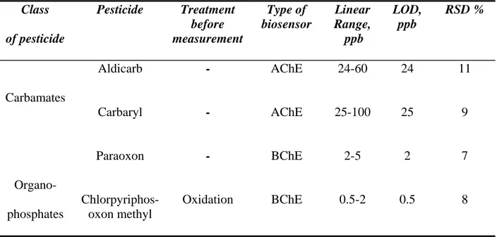
Analytical parameters such as linear range, limit of detection (LOD) and reproducibility (RDS) of the AChE and BChE biosensors were then selectively investigated. The AChE biosensor was tested with aldicarb and carbaryl due to its higher affinity toward these compounds. For the same reason, paraoxon and chlorpyrifos-methyl were tested with the BChE biosensor, which shows a higher sensitivity to these than the AChE biosensor. Table 3.3 shows the results obtained. The detection limit (LOD), defined as the concentration giving an inhibition of 20% [38], was found to be 24 and 25 ppb respectively for aldicarb and carbaryl using the AChE biosensor. With BChE biosensor, 2 and 0.5 ppb were the LOD values obtained for paraoxon and chlorpyrifos-methyl oxon, respectively. Both reproducibility and linear range (corresponding to concentrations giving 20-60% of inhibition [38]) (see table 3.3) were satisfactory with both biosensors.
Class of pesticide Pesticide Treatment before measurement Type of biosensor Linear Range, ppb LOD, ppb RSD % Aldicarb - AChE 24-60 24 11 Carbamates Carbaryl - AChE 25-100 25 9 Paraoxon - BChE 2-5 2 7 Organo-phosphates Chlorpyriphos-oxon methyl Oxidation BChE 0.5-2 0.5 8
Table 3.3 Analytical parameters for AChE and BChE biosensors relative to different pesticides.
ATChCl (1 mM) and BTChCl (5 mM) were used a substrates for AChE and BChE. Applied potential: +200 mV vs Ag/AgCl, phosphate buffer 0.05 M+ KCl 0.1 M, pH 7.4. Incubation time= 30 min.
The need for adopting a medium exchange method in the protocol for pesticide measurement has been demonstrated by testing the sensors with various interfering species such as heavy metals and fluoride (well known reversible cholinesterase inhibitors) [40,41] as well as detergents [42]. The test was performed, as reported in the experimental section, by measuring the residual enzymatic activity in the same buffer solution (with no washing step after the incubation time) and also by using the medium exchange method.
In the case of heavy metals no interference effect was observed with both the procedures adopted.
On the other hand, when SDS (200 ppb, the limit value for waste water) [43] was present in the buffer during the incubation time, measurement of the residual enzymatic activity in the same
solution (with no washing step) resulted in an inhibition of 88%. On the contrary, when the medium exchange method was adopted, and a washing step was carried out between the first incubation step and the measurement of residual enzymatic activity, no enzyme decay was observed. In our opinion, this demonstrated the need for adopting the medium exchange method, especially when handling waste water samples which are usually highly contaminated with detergents. Moreover, this procedure would avoid the presence in the measuring buffer of any potential electrochemically interfering species which could affect thiocholine determination. With this procedure, the enzyme acts as a high affinity capture agent for the pesticide, and, because of the irreversibility of the inhibition, the successive enzymatic reaction can be carried out in a fresh buffer solution, thereby circumventing the effect of reversible inhibitors present in real samples.
3.3.4 Waste and river water measurements
BChE and AChE biosensors were then tested using both waste water and river water samples from Tiber River, obtained from ACEA laboratory (municipal water company). Despite the fact that samples were taken from input tube of the softener, no inhibition was observed for either AChE or BChE biosensors. Finally, the samples were spiked with various pesticides. 48 ppb of aldicarb were spiked in waste water and river samples. Table 3.4 shows the results obtained for analysis of these spiked samples. A recovery of 79% and 125% was observed respectively for river samples and waste water thus demonstrating low matrix effect on the biosensor signal. In the case of BChE biosensor the river sample was spiked with chlorpyrifos-oxon methyl at a concentration of 1.5 ppb. Also in this case a good recovery was observed (i.e. 113%).
Type of biosensor
Source of water Pesticides Expected,
ppb Obtained, ppb Recovery, % AChE Tiber filtered Tiber Entrance water-softener filtered Entrance water-softener Aldicarb 48 48 48 48 40 38 51 60 83 79 106 125 BChE Tiber filtered Tiber Chlorpyrifos-methyl oxo 1.5 1.5 1.6 1.7 107 113
Table 3.4 Recovery studies of spiked real water samples. ATChCl (1 mM) and BTChCl (5 mM)
were used a substrates for AChE and BChE. Applied potential: +200 mV vs Ag/AgCl, phosphate buffer 0.05 M + KCl 0.1 M, pH 7.4. Incubation time = 30 min. All the values are mean of triplicate measurements.
3.4 Conclusions
The present work involved first a comparative study between four different types of modified SPEs having potential for thiocholine measurement and which are thus candidates to be used for construction of ChE-based biosensors for pesticide detection. The electrodes showed high sensitivity and low detection limit, however major drawbacks in the case of ferricyanide mediator and gold film electrode were observed due to a low stability and long preparation time. Co-phthalocyanine and Prussian Blue modified screen printed electrodes demonstrated the best results in this direction. An easiness of preparation together with a high sensitivity were observed for both sensors. Even if in the case of co-phthalocyanine electrodes a lower detection limit was observed, The Prussian Blue modified SPEs was ultimately selected because of it superior high stability. PB-modified SPEs were then used as substrate for the successive immobilisation of
two different ChE enzymes. AChE based biosensors have shown a higher sensitivity towards aldicarb and carbaryl tested as standard carbamic pesticides, while BChE demonstrated a higher affinity towards paraoxon and chlorpyrifos-oxon methyl. If confirmed with a wider number of compounds, this observation could help the researchers involved in this field to select the best ChE enzyme to be applied for certain applications. The two biosensors were then tested with different pesticides considering their different affinity. Real samples were also used in order to evaluate the matrix effect and applicability of the sensors to analytical problems giving encouraging results. Further studies are in progress to deeper investigate the different behaviour of the AChE and BChE towards the pesticide tested.
5. References
[1] FAO, Agriculture towards 2010, in: C 93/94 Document of 27th Session of the FAO Conference,
Rome, 1993.
[2] Aspelin L. Pesticides Industry Sales and Usage, 1992 and 1993 Market Estimates, US Environmental Protection Agency, Washington 1994.
[3] U.S. FDA. Pesticide Monitoring Databases; http://www.cfsan.fda.gov/~lrd/pestadd.html [4] Jury A.W., Winer A.M., Spencer W.F., Focht D.D. Environ.Contam. Toxicol., 99 (1987) 119. [5] Rotenberg M., Shefi M., Dany S., Dore I., Tirosh M., Almog S. Clin. Chim. Acta, 43 (1995) 11. [6] Bolognesi C., Morasso G. Trends Food Sci.Technol., 11 (2000) 182.
[7] American Public Health Association (Ed.), Standard methods for examination of water and wastewater 20th ed.; Washington,1998; pp 6/85-6/90.
[8] Liska I., Slobodnik J. J. Cromatogr. A, 733 (1996) 235. [9] Lacorte S., Barcelo D. Anal. Chem., 68 (1996) 2464.
[10] EPA Method 8141 A, 2000. US Environmental Protection Agency [11] Solè S., Merkoci A., Alegret S. Crit. Rev. Anal. Chem., 33 (2003) 9. [12] Hart A.L., Collier W.A., Janssen D. Biosens. Bioelectron., 12 (1997) 645. [11] Preininger C., Wolfbeis O.S. Biosens. Bioelectron., 11 (1996) 981.
[13] Bernabei M., Chiavarini S., Cremisini C., Palleschi G. Biosens. Bioelectron., 8 (1993) 265. [14] Trojanovic M., Hitchman M.L. Trends Anal. Chem.,15 (1996) 38.
[15] Gogol E.V., Evtyugin G.A., Suprun E.V., Budnikov G.K., Vinter V.G. J. Anal. Chem., 56 (2001) 963.
[17] Ivanov A.N., Evtugyn G.A., Gyurcsanyi R.E., Toth K., Budnikov H.C. Anal. Chim. Acta, 404 (2000) 55.
[18] Imato T., Ishibashi N. Biosens. Bioelectron., 10 (1995) 435.
[19] Suwansa-ard S., Kanatharana P., Asawatreratanakul P., Limsakul C., Wongkittisuksaa B., Thavarungkul D.P. Biosens. Bioelectron., 21 (2005) 445.
[20] Mascini M., Moscone D. Anal. Chim. Acta, 179 (1986) 439.
[21] Palleschi G., Bernabei M., Cremisini C. Sens. Actuators, 7 (1992) 513. [22] Collier W.A., Clear M., Hart A.L. Biosens. Bioelectron., 17 (2002) 815.
[23] Suprun E., Evtugun G., Budnikov H., Ricci F., Moscone D., Palleschi G. Anal. Bioanal. Chem., 383 (2005) 597.
[24] Trojanowicz M. Electroanalysis, 14 (2002) 131.
[25] Moore R.R., Banks C.E., Compton R.G. Analyst, 129 (2004) 755. [26] Vanderberg P.J., Johnson D.C. Anal. Chem., 65 (1993) 2713. [27] Hart J.P., Harley I.C. Analyst, 119 (1994) 259.
[28] Hart A.L., Collier W.A., Janssen D. Biosens. Bioelectron., 12 (2002) 645.
[29] Neufeld T., Eshkenazi I., Cohen E., Risphon J. Biosens. Bioelectron., 15 (2000) 323.
[30] Ricci F., Arduini F., Amine A., Moscone D., Palleschi G. J. Electroanal. Chem., 563 (2004) 229. [31] Ricci F., Arduini F., Tuta C.S., Sozzo U., Moscone D., Amine A., Palleschi G. Anal. Chim. Acta, 558 (2006) 164
[32] Ivanov A., Evtugyn G., Budnikov H., Ricci F., Moscone D., Palleschi G. Anal. Bional. Chem., 377 (2003) 624.
[33] Ricci F., Amine A., Palleschi G., Moscone D. Biosens. Bioelectron., 18 (2003) 165. [34] Lee H., Kim Y.A., Cho Y.A., Lee Y.T. Chemosphere, 46 (2002) 571.
[35] Sussman J.L., Harel M., Frowlow F., Oefner C., Goldman A., Toker L., Silman I. Science, 253 (1991) 872.
[36] Amitai G., Moorad D., Adani R., Doctor B.P. Biochemical Pharmacology, 56 (1998) 293. [37] Mohammadi H., Amine A., Cosnier S., Mousty C. Anal. Chim. Acta, 543 (2005) 143. [38] Amine A., Mohammadi H., Bourais I., Palleschi G. Biosens. Bioelectron., 21 (2006) 1405. [39] Ellman G.L. Arch. Biochem. Biophys., 82 (1959) 7l0.
[40] Danzer T., Schwedt G. Anal. Chim. Acta, 318 (1996) 275.
[41] Evtugyn G.A., Ivanov A.N., Gogol E.V., Marty J.L., Budnikov H.C. Anal. Chim. Acta, 385 (1999) 13.
[42] Jaganathan L., Boopathy R. Bioorg. Chem., 28 (2000) 242.