1
UNIVERSITY OF ROME
“TOR VERGATA”
FACULTY OF MEDICINEPHD PROGRAM IN SCIENCES AND BIOTECHNOLOGIES OF REPRODUCTION AND DEVELOPMENT
XXI CYCLE
Effects of estrogens and endocrine disrupters on mouse
embryonal germ cells and somatic gonadal cells
GINA LA SALA
Mentor: Prof. MASSIMO DE FELICI Coordinator: Prof. RAFFAELE GEREMIA
2
A Francesco per la sua immensa pazienza. Grazie
3
Desidero ringraziare il Prof. Massimo De Felici per avermi guidato in tutti questi anni che ho trascorso nel suo laboratorio, per i suoi consigli e soprattutto per avermi insegnato il lavoro del ricercatore.
Desidero ringraziare con grande affetto le persone che con me hanno diviso le gioie ed i dolori della ricerca: Luisa Campagnolo, Donatella Farini e Francesca Klinger (alias le tre ricerchine) per aver sempre dato una risposta alle mie domande “scientifiche” e per avermi dato la risposta più importante nelle situazioni difficili, la loro sensibilità.
Desidero ringraziare le compagne di viaggio: Marianna Tedesco, Lucia Scaldaferri, Elena Ievoli e Ilana Moscatelli con cui ho vissuto dei sereni momenti e le persone che sono passate per il nostro laboratorio alle quali mi lega un ricordo d’affetto, in particolare Enrico Pierantozzi.
Desidero ringraziare inoltre la Prof.ssa Susanna Dolci ed il Prof. Claudio Sette senza il cui aiuto parte dei risultati di questo lavoro non sarebbero esistiti.
Ringrazio infine il Prof. Siracusa, la Prof.ssa Camaioni e la Prof.ssa Salustri per la loro sincera disponibilità.
4
INDEX
Preface ... Pag. 6
Aims ... 6
Introduction ... 8
1. The development of the gonads in the mouse ... 8
1.1 Origin of Primordial Germ Cells and their Migration to the Gonadal Ridge ... 10
1.2 Testis Differentiation ... 13
2. Estrogens and Estrogen Receptors ... 15
2.1 ER and ERβ ... 16
2.2 Estrogen related receptors ... 18
2.3 GPR30 ... 18
2.4 Membrane estrogen receptors ... 19
3. Mechanisms of estrogens action ... 21
3.1 Nuclear genomic action ... 20
3.2 Extranuclear/Nongenomic Action of Estrogens ... 22
4. Estrogen and development of mouse gonads ... 23
4.1 Estrogen Receptor KO mice ... 25
4.2 Estrogen receptor expression and role during development of mouse testes ... 29
5. Endocrine Disrupters ... 30
Summary of results and overall conclusions ... 36
References ... 39
Publications and additional results ... 49 Paper I Rapid estrogen signalling in mouse primordial germ cells (in preparation)
Paper II Pro-apoptotic effects of lindane on mouse primordial germ cells (accepted for
publication on Toxicological Sciences)
Paper III Estrogenic assay on putative Leydig cells from mouse embryonic testes (in
5
Additional Results Effect of 17-ß-estradiol and lindane on the expression of cell cycle
genes of somatic cells of foetal testis
Additional Paper Genistein is an Efficient Estrogen in the Whole-Body throughout Mouse
6
Preface
The work presented in this thesis focuses on the effects of estrogens and endocrine disrupters termed xenoestrogens on mouse embryonal germ cells and somatic gonadal cells. The thesis is structured as follows: first, a general introduction to the thesis work; then, present the results obtained in form of one accepted paper for publication, two papers in preparation and one paper published in collaboration with the 3rd Laboratory Biotechnology Civic Hospital of Brescia; finally additional results to the thesis work.
Aims
In the recent years many progresses have been made in order to establish possible causes of anomalies of the reproductive function termed TDS (Testicular Dysgenesis Syndrome) including testicular cancer, cryptorchidism, hypospadias and sterility. Several studies indicate that these anomalies are associated to an increase in the environment of compounds acting like natural hormones named endocrine disrupters (EDs) and in particular to a class of ED that mimic estrogen estrogen action termed xenoestrogens. It has been postulated that the origin of these disorders occurs in fetal life during the delicate process of the development of reproductive system. For these reason, it is important to know how the estrogens and xenoestrogens act during the fetal development and to know the mechanism by which these compounds exert their effects.
The aim of paper I was to study the expression of estrogen receptors (ERs) in the embryonic precursors of the adult gametes termed PGCs and to analyze the existence in such cells of intracellular molecular pathways modulable by estrogens. The expression of estrogen receptors or (ER , ER ) in female and male germ cells has been reported in several species including humans and in some cases, it has been also demonstrated that the ablation of these receptors or in the absence of stimulation by the natural ligand E2 in germ cells (ERKO and ArKO mice) alter some process of their development. The molecular genomic and non genomic bases of such effects remain, however, often to be clarified and the relevance of a direct estrogen effects on germ cells is controversial. It is undoubted that the expression of ERs
7
renders germ cells a potential direct target of ED. It is mostly important to study the expression and functions of ERs in the embryonic precursors of the adult gametes termed primordial germ cells (PGCs) and to analyze the existence in such cells of molecular pathways modulated by estrogens, since PGCs are not only responsible for establishing the gamete populations but are also the tool for genome transmission to the next generation.
The aim of paper II was to investigate the effects of a prototype of xenoestrogens, the pesticide lindane, on primordial germ cells development in the mouse embryo of E12.5 dpc. Numerousreports have shown that lindane adversely affects reproductivefunction in animals, for example, in adult male rats, chronic exposure to lindane markedly reduces serum testosterone levels, epididymal sperm counts, and sperm motility, and long-lasting effects of lindane on mouse spermatogenesis following in utero exposure have been reported. In adult female mice, rabbits and rats, lindanereduces serum estrogen and progesterone levels, whereas in pregnant mice and minks, it decreases whelping rate and litter size (Beard et al., 1985; Chadwick et al., 1998; Sircar et al., 1990; Srivastava et al., 1993; Sircar et al., 1989). In an in vitro model, the pesticide abolishes oocyte directed follicle organizing activity (Li et al., 1997). Exposure to lindane during the first four days of pregnancy completelyprevents implantation in mice, but normal pregnancy resultswhen estrogen and progesterone were coadministered with the compound (Scascitelli et al., 2003). As far as we know, no works have had addressed the effect of lindane on early gametogenesis.
The aim of paper III was to verify the presence of functional ERα in embryonic testicular cells using an ERE-and AP1-Luc assay and to evaluate estrogenic activity of putative EDs on mammalian embryonic testis. We and others have reported that mouse embryonic testes from 12.5 dpc onward contain a subpopulation of somatic cells expressing estrogen receptor α (ERα), identifiable mostly as Leydig cells (Greco et al., 1992; Nielsen et al., 2000; Moe-Beherens et al., 2003). This observation marks testes as a possible target for estrogens and estrogenic compounds from early stages of embryo development. Evidence of functional ERα in embryonic testicular cells, however, was lacking and no simple assay exists to evidence estrogenic activity of compounds on such cells.
8
Introduction
1. The development of gonads in the mouse
In mammals the development of the gonads can be divided into two phases. The initial
phase is characterized by the emergence of the so-called indifferent, bipotential gonad, or
genital ridge, which is identical in males and females. The cell lines that comprise it are bipotential, being able to adopt either the male or female fate. The second phase is the development of a testis or an ovary, which is triggered solely by the expression and proper function of the testis-determining gene Sry. The indifferent gonads arise as paired structures within the intermediate mesoderm, which lies on either side of the embryo filling much of the coelomic cavity between the limb buds during the first half of development. Within this region, three segments comprising the urogenital ridge are distinguished from anterior to posterior: 1) the pronephros, which includes the adrenal primordium near its caudal end; 2) the mesonephros, the central region from which the gonad arises; and 3) the metanephros, the most posterior region from which the kidney forms.
Figure 1. Structure of the urogenital system. Schematic of the mouse urogenital system at 10.5 dpc. Epithelial structures are shown in red, mesenchymal structures are shown in blue, and the striped region denotes the genital ridge. (WD) Wolffian duct (MT) mesonephric tubules; (MD) Mullerian duct; (UB) ureteric bud; (CE) coelomic epithelia. Image from Swain A. and Lovell-Badge R., Genes Dev. 1999.
9
The gonads emerge on the ventromedial surface of the mesonephros at 10.5 days post coitum (dpc). Cells that delaminate from the coelomic epithelium seem to provide one source of cells for the growing genital ridges, while recruitment of underlying cells from the mesonephros to the epithelial population also augments the cell population in the gonadal primordium in males. From the earliest stages of gonad development, the mesonephric tubules can be seen to form continuous bridges to the epithelial cells of the gonad in male and female genital ridges (Karl and Capel, 1995).
In the mouse the gonad initially develops in a non-sex-specific manner, being morphologically identical in XX and XY embryos up until ~12.0 dpc (Fig.2). However, at ~10.5–11 dpc, Sry begins to be expressed in the male genital ridge and acts to initiate testis development. In the mouse, Sry is expressed in the genital ridge as a wave from anterior to posterior that lasts about a day and a half so that each cell sees it for a few hours only (Hacker et al., 1995).
The action of SRY is therefore thought to trigger differentiation of the Sertoli cell lineage in the testis. Once SRY triggers Sertoli cells they in turn direct the differentiation of the rest of the cell types in the testis. Therefore the decision of sex determination is essentially one of cell fate: SRY triggers Sertoli cell fate in a cell that would otherwise become a follicle cell.
The early mammalian gonad is an undifferentiated primordium composed of bipotential precursor cells that can follow one of two possible fates. Precursors for supporting cells (so named for their role in sustaining and nourishing germ cells in both sexes) and steroid-secreting cells are believed to be present in the early gonad (Merchant-Larios et al., 1993). The supporting cell lineage will give rise to Sertoli cells in the testis and ovary-specific follicle (granulosa) cells in the ovary. These cells surround the germ cells and provide an appropriate growth environment. The steroidogenic cell lineage produces the sexual hormones that will contribute to the development of the secondary sexual characteristics of the embryo. In the male these are the Leydig cells and in the female, the theca cells. The connective cell lineage will contribute to the formation of the organ as a whole. Early testis development is characterized by the formation of testicular cords that contain Sertoli and germ cells, with the Leydig cells excluded to the interstitium. The
10
connective cell lineage is a major contributor to cord formation as the peritubular myoid cells surround the Sertoli cells and together they lay down basal lamina. The testis is also characterized by rapid and prominent vascularization. Organization of the ovary takes places later than that of the testis and is less structured, with the connective tissue lineage giving rise to stromal cells and with no myoid cell equivalent.
Figure 2. Morphological changes in the gonad during differentiation. Shown are the molecular and structural changes that occur during differentiation of the mouse gonad along the male (top) and the female (bottom) pathway. The stages of embryo development that are depicted are indicated at the bottom. The genital ridge is shown as a striped structure; the mesonephros is shown in blue. Before 10.5 dpc the genital ridge of the embryo is identical between males and females. After the action of SRY, molecular differences in the gonad can be observed, as depicted at 11.5 dpc. Morphological differences between the testis and the ovary can be observed at 12.5 dpc. The structure of testicular cords, which is a consequence of migration of mesonephric cells, is shown as part of the male pathway. The different cell types of the testis and ovary are indicated, as are the genes expressed therein. Myoid cells, indicated in blue, are thought to derive from the mesonephric contribution. The different fate of germ cells between the testis and the ovary is shown. Image from Swain A. and Lovell-Badge R., Genes Dev. 1999.
1.1 Origin of Primordial Germ Cells and Their Migration to the Gonadal Ridge
The primordial germ cells (PGCs) do not arise within the genital ridge or the mesonephros but migrate from an entirely separate source. In several mammalian species, due to their characteristic property of positive staining with alkaline phosphatase, from early studies it is possible to trace their formation to the base of the allantois at the posterior end of
11
the primitive streak. In the mouse results of cell labeling experiments with fluorescent dyes suggest that a population of 45 cells is allocated to the germ line at 7 dpc in the mouse (Lawson and Hage, 1994). At 6–6.5 dpc, the precursors of the PGCs can be found in the epiblast close to the extraembryonic ectoderm, but are evidently not yet restricted to a germ cell fate because they can also form extraembryonic mesoderm. When PGCs are first seen in the mouse at 7 dpc, they are in the region of the forming hindgut. As development proceeds, the hindgut invaginates and the germ cells are swept into the embryo. By 9.5 dpc, PGCs begin to leave the hindgut and pass into the forming urogenital ridges, which are in close proximity at this time. As development proceeds, the hindgut descends into the coelomic cavity and PGCs arriving later must migrate through the dorsal mesentery before entering the developing gonads (Fig. 3).
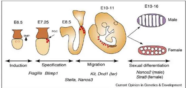
Figure 3. Schematic representation of germ cell development in the mouse embryo. The stages of mouse germ cell development and major genes involved in each process are indicated. Primordial germ cells (PGCs) are induced in the epiblast by BMP signaling at E5.5–E6.5. PGCs are specified through the function of Blimp1 by E7.25. PGCs are protected from apoptotic signals during their migration via Kit-mediated signaling, and through the functions of Nanos3 and Dead end1. Once PGCs reach and enter the genital ridge at E11.5, they differentiate according to the somatic sex of the embryo. Nanos2 promotes the male germ cell fate by suppressing the female fate. The locations of the PGCs are indicated by red circles. Image from Saga Y.,
12
Survival of the PGCs during migration is dependent on an interaction between the tyrosine kinase receptor c-KIT, which is present on the surface of PGCs, and its ligand, stem cell factor (SCF) also called Kit ligand (Kl), which is produced by the surrounding tissues (reviewed in Bendel-Stenzel et al., 1998). During migration the PGCs also undergo several rounds of cell division to achieve a population of 3,000 cells by 11.5 dpc, when almost all the PGCs have arrived at their destination. Once inside the gonadal ridge, the germ cells lose their motility and begin to aggregate with one another. They continue to proliferate within the indifferent gonad and maintain their bipotentiality until 13 dpc, where upon germ cells within the male gonad become enclosed within the forming testis cords and enter mitotic arrest as T1 prospermatogonia. In the female, proliferation continues for a short while longer before the germ cells enter meiosis at 13.5 dpc. PGCs thus have the potential to develop either as meiotic oocytes, progressing through the first meiotic prophase and arresting in diplotene just after birth, or as prospermatogonia, mitotically arrested in G1/G0 until a few days after birth, when they resume proliferation (Hilscher et al., 1974; MacLaren, 1995). This developmental switch, which has occurred by 13.5 dpc, is dependent on the sex of the somatic cells in the gonad, rather than the chromosomal sex of the PGCs: XY PGCs can develop as oocytes in female embryos, and XX PGCs can develop as prospermatogonia in male embryos (Palmer and Bourgoyne, 1991).
13
1.2 Testis Differentiation
Testis differentiation is induced by the expression of Sry in a subset of somatic cells that are induced to differentiate into Sertoli cells. Sertoli cells are believed to act as the organizing center of the male gonad and orchestrate the differentiation of all other cell types (Fig 4).
Sertoli cells
Sertoli cells are somatic cells that associate with germ cells and nurture their development into sperm. They are the first cell type known to differentiate within the gonad from bipotential precursors of the supporting cell lineage and are therefore the first indicator that the gonad has passed from the indifferent stage into testis development. Once Sertoli cell fate is triggered by SRY, genes involved in Sertoli cell function become activated. One of these genes is Amh. Its product, AMH induces the regression of the Mullerian ducts, which in the female give rise to the oviducts and uterus. Sertoli cells polarize, aggregate around germ cells and reorganize the gonad into two compartments: the tubular testis cods composed of Sertoli and germ cells, and the interstitial space between the cords. Peritubular myoid cells surround Sertoli cells and cooperate to deposit the basal lamina at the periphery of tubule structure. A central role of Sertoli cells is to sustain germ cells during development and later during spermatogenesis. They do so by forming close cell–cell contacts and providing factors involved in growth and differentiation. A candidate factor involved in cell– cell interactions between Sertoli and germ cells is Desert hedgehog (Dhh), a member of the hedgehog family of molecules that signal at close range.
Peritubular myoid cells
One of the three cell types that migrate from the mesonephros into the male gonad is the peritubular myoid (PM) cell. These cells form a single layer of flattened cells surrounding the Sertoli cells, circumscribing the testis cords. They are thought to have two main functions: 1) to contribute structurally to the formation of the testis cords in conjunction with Sertoli cells and 2) to promote the movement of mature sperm through the seminiferous tubules of the adult testis for export to the seminal vesicles, a function
14
mediated by their smooth musclelike character. PM cells express smooth muscle actin ( -Sma) and desmin. PM cells represent the only cell type in the testis so far for which no counterpart can be identified in the ovary. This might be due to their origin from immigrating cells from the mesonephros, which only occurs in an XY gonad after the expression of Sry (Capel et al., 1999; Martineau J et al., 1997).
Leydig cells
Within the second compartment of the testis, the interstitium, steroidogenic Leydig cells differentiate. These cells secrete a hormone that plays a role in establishing and maintaining the secondary male sex characteristics. Leydig cells often lie in clusters close to blood vessels, in line with their steroidogenic role. In mammals there are two types of Leydig cells. The fetal Leydig cells originate, at least in part, in the mesonephros, and are responsible for the production of androgen for the fetal masculinization; these cells probably degenerate postnatally. The adult Leydig cells, which differentiate after birth, appear to be unrelated to their fetal counterparts. Studies indicated that they arise from undifferentiated precursor cells that are part of the mesenchymal cells of the interstitium (Hardy, 1993). The origin and roles of Leydig cells are discussed comprehensively in Chase 1982.
Vascular and other interstitial cells
Although Leydig cells are often considered the main component of the testicular interstitium, probably because of their essential and obvious male-specific endocrine roles, several other interstitial cell types can be found. These include endothelial cells, fibroblasts, and blood-derived cells such as macrophages, lymphocytes, plasma cells, monocytes, and mast cells. Endothelial cells, alongside PM and Leydig cells, represent a third cell type that migrates into the testis from the mesonephros (Martineau et al., 1997). They form the male-specific vasculature with the prominent coelomic vessel on the surface of the gonad and side branches in between the testis cords.
15
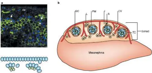
Figure 4. Compartmentalization of the testis. a At the earliest stages of testis organogenesis (11.75–12.0
days post coitum(dpc), Sertoli cells (stained with SF1 antibody; blue) polarize and begin to aggregate around clusters of primordial germ cells (stained with PECAM antibody; asterisk) to initiate development of testis cords. ce, coelomic epithelium. b Between 11.5–12.5 dpc, the cells of the testis are organized into two functional compartments: testis cords (TC) and the interstitial space (I) outside the cords. Within testis cords, Sertoli cells (S; blue) surround germ cells (GC; green). A basal lamina is deposited between Sertoli cells and peritubular myoid cells (PM). The interstitial compartment contains Leydig cells (L; yellow) and the coelomic vessel (CV; red), with branches that extend between cords. Image from Brennan J. and Capel B., Nature Reviews 2004.
2. Estrogens and Estrogen Receptors
Estrogens (U.S., otherwise oestrogens) are a group of steroid compounds, named for
their importance in the estrous cycle, and functioning as the primary female sex hormone. The three major naturally occurring estrogens in women are estradiol, estriol, and estrone. Estradiol, like other steroids, is derived from cholesterol. After side chain cleavage and utilizing the delta-5 pathway or the delta-4 pathway androstenedione is the key intermediary. A fraction of the androstenedione is converted to testosterone, which in turn undergoes conversion to estradiol by an enzyme called aromatase. Estrogen is produced primarily by developing follicles in the ovaries, the corpus luteum, and the placenta. Follicle-stimulating hormone (FSH) and luteinizing hormone (LH) stimulate the production of estrogen in the ovaries. Some estrogens are also produced in smaller amounts by other tissues such as the
16
liver, adrenal glands, and the breasts, fat cells are active to convert precursors to estradiol, and will continue to do so even after menopause. They are usually present at significantly higher levels in women of reproductive age. They promote the development of female secondary sex characteristics, such as breasts, and are also involved in the thickening of the endometrium and other aspects of regulating the menstrual cycle. Also men produce estrogens that are present in adult where they regulate certain functions of the reproductive system important to the maturation of sperm (O‟Donnell et al., 2001).
Estradiol enters cells freely but the pleiotropic effects of estrogens are transduced trough multiple ER receptor subtypes as ER /β or GPR30 (G-protein coupled receptors-30) that have multiple subcellular locations (Filardo and Thomas, 2005; Watson and Gametchu, 2003). These receptors inhabit nuclei and cytoplasm, plasma membranes or perimembrane spaces (Clarke et al., 2000), endoplasmic reticulum (Revankar et al., 2005) and mitochondria; they also sometimes change locations or arrangements within their locations, depending upon liganding or other circumstances (Song et al. 2002).
2.1 ER and ERβ
The two mammalian ERs exhibit modular structures characteristic of the nuclear receptor superfamily. The two receptor are not isoforms of each other, but rather different proteins encoded by separate genes located on different chromosomes (10 and 12 respectively in mouse, 1and 6 in rat, and 6 and 14 in humans). Hormone activated estrogen receptors form dimers, and since the two forms could be are coexpressed in many cell types, the receptors may form ER ( ) or ERβ (ββ) homodimers or ERαβ (αβ) heterodimers. The ER proteins are each composed of six functional domains labeled A–F, a signature characteristic of the entire superfamily (Fig. 5).
17
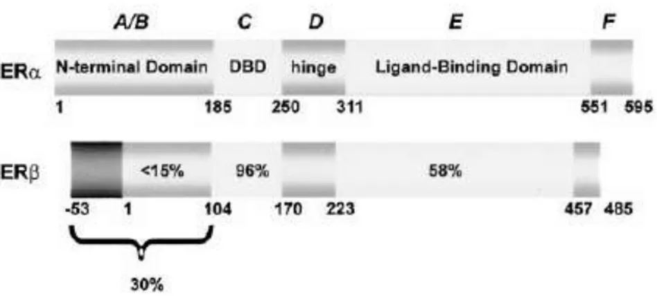
Figure 5. Comparison of the structure of ER and ERβ proteins. The functional domains A–F and the percentage homology of ERβ compared with ER are shown. Indicated are the N-terminal domain (A/B domain), the DNA binding domain (DBD) (C domain), the hinge region (D domain), the ligand-binding domain (E domain), and the C-terminal region (F domain). The two potential start sites on ERβ are designated 253 and 11.Image from O‟Donnell et al., Endocrine Reviews 2001.
The N‟-terminal A/B domain is the least conserved among all members and demonstrates only 17% homology between the two ERs. It contains the activation function 1 (AF1) region, which is one of two regions critical for the transactivation function of the members of the receptor family. By contrast, the C domain is the most highly conserved region, being the DNA binding domain that contains the zinc-finger motifs. The E domain, or ligand-binding domain, is modestly conserved throughout the superfamily and confers ligand specificity on the members. Conservation of amino acid sequence in this region is 60% between the ER and ERβ; however, each binds estradiol with about equal affinity, although the relative binding of other ligands differs substantially between them (Kuiper et al., 1997; Kuiper et al.1998). The E domain also contains the major dimerization surface of the receptors, and the second transactivation function, activation function 2 (AF-2), is also located in this region of the C‟-terminus. Transcription of the mouse ER gene in vivo leads predominantly to a single transcript of approximately 6.3 kb, encoding a protein of 599 amino acids. Initial studies of the rodent ERβ transcript indicated it was substantially shorter than the ER , namely 485 amino acids (Kuiper et al., 1996). This is largely due to a significantly shorter N‟-terminal region . Due to alternative RNA splicing, several ER isoforms are known to
18
exist. At least three ER and five ERβ isoforms have been identified. The ERβ isoforms receptor subtypes can only transactivate transcription when a heterodimer with the functional ERß1 receptor of 59 kDa is formed. The ERß3 receptor was detected at high levels in the testis. The two other ER isoforms are 36 and 46kDa (Nilsson et al., 2001). Only in fish, but not in humans, an ERγ receptor has been described (Hawkins et al., 2000). There is considerable tissue specificity in the expression of ER and ERβ. Thus, ER is the dominant species expressed in uterus, liver, adipose, skeletal muscle, pituitary, and hypothalamus, whereas ERβ is the major form in ovary and prostate, as well as other regions of the brain including the limbic system, cerebellum, and cerebral cortex (Couse et al., 1997).
2.2 Estrogen related receptors
At the superfamily of the nuclear receptors belong putative receptor molecules for which no known ligands are identified and these are classified as orphan nuclear receptors. Estrogen-related receptors belong to a subfamily of such orphan nuclear receptors, and comprise of three members, ERR- , -β, and -γ (Giguère et al., 1988 and Giguère, 2002). These ERR proteins are, as their names indicate, closely related to the ERs in their structures and bind to the estrogen response elements, but they are not activated by estrogen (Giguère, 1988). Three ERR proteins display a high degree of structural similarities within binding domains for their ligand and DNA, suggesting that they would bind to similar ligands and targets.
2.3 GPR30
GPR30 is an orphan member of the G-protein-coupled receptor superfamily, that has been reported to trigger rapid signaling by estrogen. GPR30 is localized mainly into endoplasmic reticulum and binds E2 with nanomolar affinity (Filardo et al., 2000; Revankar et al., 2005). The biological relationship between GPR30 and conventional ERs is currently unknown and further studies are required to determine the relative contribution of either or
19
both of these pathways to estrogen signaling. Many studies have indicated that this receptor through a cross-talk between ER activate the rapid Egfr/Erk/Fos pathway that in turn stimulate mouse spermatogonial cells (GC-1) or ovarian cancer cells to proliferate (Sirianni et al., 2008; Albanito et al., 2007). It is possible however that the rapid E2 signaling is mediated by a complex network of proteins that consists of conventional steroid receptors and other steroid binding proteins such as GPR30 (Cheskis, 2004).
2.4 Membrane estrogen receptors
Classical steroid receptors, localized in the cytosol and/or nucleus, traditionally mediate their primary effects at the genomic level. In recent years, a large number of reports have described membrane-associated estrogen receptors, either similar to or distinct from the classical nuclear estrogen receptors (Acconcia et al., 2004; Razandi et al., 2003). These receptors have been postulated to mediate aspects of cellular estrogen function, including traditional genomic (transcriptional) signaling as well as novel non-genomic (rapid) signaling.
Although the majority of ER is localized in the nucleus, there is evidence that a small fraction of the receptor is localized at or near the cell membrane in either the presence or absence of E2. While the precise mechanism(s) are currently unknown, it has been proposed that ER translocation to cell membrane is mediated by its interactions with membrane proteins. Candidate interacting proteins include caveolin-1/-2 and the 110-kDA caveolin-binding protein–striatin. Caveolae are specialized regions of the plasma membrane that assemble and organize signaling protein complexes (Shaul, 1998). Striatin is a calmodulin-binding member of the WD-repeat family of proteins. It has been reported to anchor ER to the cell membrane, and to serve as a scaffold for the formation of an ER – Gai complex, which is critical for E2 activation of eNOS (Lu et al., 2004). It has been also proposed that ER can be targeted to the cell membrane by the adaptor protein Shc (Pelicci et al., 1996). The SH2 domain of Shc can directly interact with the N-terminal part of ER (Song et al., 2002). Recently another membrane adaptor protein the p130Cas
(Crk-20
associated substrate) has also been reported to interact with ER -cSrc complex in T47D breast cancer cells and to potentiate the E2 activation of Src (Cabodi et al., 2004). Finally, palmitoyl-acyl-transferase (PAT)-dependent S-palmitoylation of ERα was recently reported to promote Er association with the plasma membrane and interaction with caveolin-1 (Acconcia et al., 2004, 2005). Furthermore, cystine 447-mutated ER did not stimulate activation of MAP and PI3 kinases (Acconcia et al., 2005). A terminally truncated 46 kDa variant of ER has been found to be preferentially palmitoylated and enriched in plasma membrane of several cell types (endothelial, osteoblasts, and MCF-7 cells) (Denger et al., 2001).
In conclusion, several membrane proteins have been identified that interact with classical receptors and influence their non-genomic action. However, the precise role of these proteins in receptor regulation of the cell signaling remains to be further investigated. It is possible that the composition of ER complexes at the plasma membrane is cell context dependent, which may potentially explain the cell type selectivity of non-genomic action.
21
3. Mechanisms of estrogens action
3.1 Nuclear genomic action
ERE-dependent transcriptional regulation
The classical mechanism of steroid hormone action involves nuclear interactions of intracellular ERs receptors, which are either cytoplasmic or nuclear. Binding of hormone to ER releases the receptor from an inhibitory complex with HSPs (Heat Shock Proteins) and triggers conformational changes that allow ER to bind the responsive elements in the target gene promoters (Fig.6). Subsequently, the receptor-ligand complex binds to the a 15-bp palindromic ERE (Estrogen Response Element) located in the target gene promoters, and stimulates gene transcription. The ERE is a 13 base pair inverted repeat sequence (GGTCAnnnTGACC), and the ER binds as a dimer, with one ER molecule contacting each 5 base pair inverted repeat (Klinge et al., 2001). Maximum transcriptional activity requires the concerted actions of the ligand-independent AF1 domain and the ligand-dependent AF2 domain.
The transcriptional activity is also affected by a number of regulatory cofactors including chromatin-remodeling complexes, coactivators, and corepressors. Coactivators generally do not bind to the DNA but are recruited to the target gene promoters through protein-protein interactions with the ER. Examples of ER coactivators include, members of the p160/SRC (Steroid Receptor Coactivator) family: SRC1/NCoA1 (Nuclear Receptor Coactivator-1); NCoA2; NCoA3/AIB1/TRAM1/RAC3; the cointegrators: CBP (CREB-Binding Protein) and p300; the family of CITED (CBP/P300-Interacting Transactivator,With Glu/Asp-Rich Carboxy-Terminal Domain) proteins. Corepressors like NCoR (Nuclear Receptor Co-Repressor) and MTA1 (Metastasis Associated-1) protein have been implicated in the transcriptional silencing. In addition, a few bifunctional coregulators such as PELP1 (Proline Glutamic Acid-Rich Nuclear Protein) also exist that can act both as coactivators and corepressors of ER. It is the relative balance of receptors, coactivator and
22
corepressor proteins, which is a critical determinant of the ability of this classical pathway to initiate responses. Since the relative concentrations of these molecules is cell specific, sex steroid hormones can have vastly different functions in different tissues of the same organism
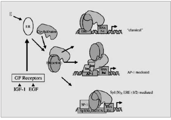
Figure 6. Mechanism of ER mediated transcription ER-mediated transcription is initiated following E2 binding or ligand-indipendent activation resulting from growth factor receptor pathway signalling and cross-talk with ER. Once activated, ER dimers recruit coactivators and can mediated transcription of genes via direct binding to EREs in target genes (classical mechanism). Alternatively, ER can recruit coactivators to an AP-1 complex (AP-1 mediated mechanism). Finally, ER can interact with promoters containing Sp1 binding sequences and ERE-half sites (Sp1(N)x ERE(1/2)mediated). Image from Hewitt and Korach. Reviews in Endocrine and Metabolic Disorders 2002.
23 ERE-indipendent Genomic action of ER
In recent years, mechanisms of gene regulation by ERs that deviate from this „„classical model‟‟ have also been described. These include gene regulation by ERs that does not involve direct receptor binding to DNA, but rather via ERs participation in the formation of the pre-initiation complex via protein–protein interactions, such as the AP1 complex (Webb et al., 1999), as in the case of the collagenase and IGF-I genes. First, binding of Jun and Fos to the AP-1 site is needed for ER action, and ER appears to increase the intrinsic transcriptional activity of Jun:Fos when bound to the site (Fig.6). Estrogen regulation is also seen in genes such a c-FOS and TGFa which lack a full ERE sequence. This regulation is mediated by an interaction between ERα and SP1 proteins, which bind ERE-half sites and GC rich sequence, respectively, in the regulatory regions of these genes. ERα interacts with Sp1 protein to transactivate genes through binding Sp1(N)xERE or Sp1(N)xERE half-site (1/2) motifs where both ERs and Sp1 bind DNA elements (Safe S. 2001).
3.2 Extranuclear/Nongenomic Action of Estrogens
In addition to transcriptional regulation, which occurs on a time scale of hours, estrogen also mediates cellular effects with response times from seconds to minutes. These rapid non-genomic estrogen signaling events include the generation of the second messengers Ca2+, cAMP, and NO, as well as activation of receptor tyrosine kinases, such as EGFR and IGF-1R, and protein/lipid kinases (e.g. PI 3-kinase, Akt, MAPK family members, Src family members, and PKA/PKC). In many reports, the estrogen-responsive receptor is proposed to be ER itself (either α or β), or a modified form of the protein (Acconcia et al., 2004) (Fig.7). Complexes between the classical ERs and G proteins (Navarro et al., 2003) as well as with PI3 kinase (Simoncini et al., 2003) have been described. Recently, ER associations with plasma membrane Gi proteins have been reported to mediate NO production and cAMP inhibition (Navarro et al., 2003). The nature of the upstream receptor targets remains to be better established.
24
Multiple lines of evidence suggest that activation of the tyrosine kinase cSrc represents one of the initial steps in ER -mediated cell signaling in MCF7 cells (Migliaccio et al., 2002). cSrc can be initially activated either by dephosphorylation of the C-terminal inhibitory phosphotyrosine site (Tyr 529) or in oncogenic variants by loss of the C-terminal tail), or by binding of high affinity ligands to the SH2 or SH3 domains. These domains are modular polypeptide units that mediate protein–protein interactions and are found together on many proteins, suggesting that their activities can be coordinated and that they can cooperate in Src regulation (Cohen et al., 1995). After this initial activation, Src autophosphorylation loop leads to phosphorylation in Tyr 418 and stimulate Src activity (Superti-Furga et al., 1995).
Figure 7. Signaling by estrogens mediated by a complex interface of direct control of gene expression (genomic action) and by regulation of cell signalling/phosphorylation (non genomic action).
25
Recently, an adaptor protein Modulator of Nongenomic Action of Estrogen Receptor (MNAR) has been identified that is required for E2 induced ER activation of cSrc and downstream MAP kinase pathway (Wong et al., 2002). It has been recently demonstrated that in MCF7 cells treated with E2, endogenous MNAR, ER and cSrc also interacted with p85, the regulatory subunit of the PI3 kinase. Further, ER -MNAR activation of cSrc, led to MNAR phosphorylation on Tyr 920 which was required for its interaction with SH2 domain of p85 and activation of the PI3K/Akt pathway. Existing data indicate that MNAR is a scaffold, which is promoting receptor binding to Src and stabilizing ER -Src complex.
Crosstalk between growth factors and ERs takes place in both nuclear and cytoplasmic compartments. Both, EGF and IGF can also activate ER transcriptional activity in presence of E2. E2 may rapidly activate the two main signaling cascades coupled to the IGF-I and the EGF receptors: the PI3K/Akt and the Src/MAPK signaling pathways. Initially an indirect mechanism of ER activation of epidermal growth factor receptor (EGFR) has been proposed to explain these effects. According to this hypothesis, ER bound to caveolin-1 in the cell membrane could interact with a Gprotein- coupled receptor, which in response to estrogen- or tamoxifen-binding may directly or indirectly interact with and activate specific G proteins. The subsequent activation of cSrc leads to activation of matrix metallo-proteinases, which in turn cleave heparin-binding epidermal growth factor (EGF) from the membrane. This form of EGF then binds to surface EGFR in an autocrine or paracrine manner to activate the receptor and its downstream kinases including ERK 1/2 MAPK and Akt (Levin, 2002; Levin 2003). Recent evidence however indicates that ER and Src may play a direct role in EGF activation. Direct interaction has also been documented for ER and IGF receptor both in vitro and in vivo (Mendez et al., 2003). The interaction was coincident with the increase in tyrosine phosphorylation of IGF-I receptor, suggesting a possible causal relationship (Mendez et al., 2003).
One of the best-characterized extranuclear actions of estrogens is the rapid activation of the Ras/Raf/MAPK pathway. In neuronal cells, E2 rapidly triggers Erk 1/2 activation, leading to cFos gene expression. E2 activated growth of human colon carcinoma-derived
26
Caco-2 cell was found to be mediated through rapid and reversible stimulation of the cSrc and cYes, and subsequent activation of ERK1 and ERK2 kinases. E2-mediated stimulation of Ras/Raf/ERK pathway promotes MCF7 cell proliferation (Migliaccio et al., 1996). The MAPK pathway is involved in the control of many fundamental cellular functions that include cell proliferation, survival, differentiation, apoptosis, motility, and metabolism. Another well-characterized and biologically important action of E2 is the acute effect on blood vessels to stimulate vasodilatation and protect against vascular injury. This action is mediated by a subpopulation of ER in plasma membrane of endothelial cells through the activation of eNOS and the stimulation of NO production via the PI3 kinase/Akt signaling pathway. One of the important down stream targets of PI3K is the threonine-serine kinase Akt/ protein kinase B. Activation of PI3K/Akt by E2 has also been shown to be important in breast cancer cells in mediating E2- stimulation of cell cycle progression (Castoria et al., 2001) and inhibition of apoptosis. Many cell-signaling pathways converge upon and regulate the phosphorylation status and hence activity of multiple transcription factors, which affects gene expression. Several examples of this mode of regulation have been reported, including ER -dependent E2 regulation of the c-fos gene mediated by Src/MAP and Src/PI3 kinase pathways converging on Elk-1 and SRF, respectively; E2 regulation of cyclin D1 mediated by PI3K/Akt pathway and E2 regulation of the Egr-1 gene mediated by MAP kinase activation of SRF.
4. Estrogens and development of mouse gonads
4.1 Estrogen Receptor KO mice
The advent of gene deletion techniques has allowed the generation of mice lacking ER (Couse and Korach, 1999), ER (Krege et al., 1998) or both ER and ER (Couse and Korach, 1999), as well as mice that lack the ability to synthesize estrogens due to deletion of the aromatase gene (Fisher et al., 1998). Characterization of the phenotypes exhibited by these models exert that both sexes of the ERKO mice are infertile, whereas only the
27
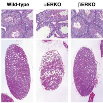
ERKO female has shown impaired fertility. In the male ERKO mice, infertility is due to deficits at several points in the reproductive process, including severe reduction in sperm numbers and lack of sperm function, as well as abnormal sexual behavior. The seminiferous tubules of the ERKO testes show progressive dilation that is accompanied by degeneration of the seminiferous epithelium (Fig. 8). In contrast, the testes of the ERKO mice appear normal (Fig. 8), and produce sufficient and functional sperm to allow fertility, resulting in production of offspring in mice examined to date. Therefore, ER appears to be more critical than ER in mediation of the estrogen actions necessary for maintenance of healthy testicular structures and the somatic cell function required for successful sperm maturation. Double mutant for both receptors and β ( βERKO) are infertile and show phenotype and alterations of reproductive tract functions comparable to ERKO (Couse et al., 1999).
Figure 8. Pathology of adult ERKO and ERKO testes. The wild-type and ERKO are indistinguishable, while the ERKO testis shows degeneration of the testicula structures. Image from Hewitt S.C. et al., Breast
28
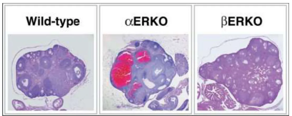
Normally, the female rodent reproductive tract grows and matures in response to cycling ovarian hormones, including estradiol. The ovarian phenotypes are a major component of the infertility in the ERKO mice and the subfertility in the ERKO mice. The ERKO female does not ovulate, while the βERKO female is subfertile with reduced litter numbers and smaller litter sizes compared with wild-type littermates. Interestingly, although both ER and ER are detected in the ovary, their localization differs with ER in the granulosa cells and ER in the theca and interstitial cells of the ovary. The hallmark phenotype of the ERKO female is the enlarged hemorrhagic cystic ovary (Fig. 9), although the prepubertal ERKO ovary looks similar to its wild-type littermate. This phenotype begins to develop progressively as the animal matures and is apparently due to a lack of estradiol feedback inhibition in the pituitary, which results in chronically elevated LH and subsequent hyperstimulation of the ovary.
.
Figure 9. Ovarian pathology of the ERKO mice. Histological analysis of the wildtype ovary shows normal follicular development and indications of ovulation. The ERKO ovary shows large cystic structures and arrested follicle development with no indication of ovulation, while the βERKO ovary shows development of follicles is occurring but with little indication of successful ovulation. Image from Hewitt S.C. et al., Breast
Cancer Res 2000.
This indicates that ER is responsible for mediating the LH feedback inhibition in the hypothalamic–pituitary axis. The constant LH stimulation in the ERKO mice results in an abnormal endocrine environment in the ERKO female, with elevated estradiol and
29
testosterone, and chronic preovulatory basal progesterone levels. The ERKO ovaries produce normal serum levels of estradiol and testosterone, and the circulating serum gonadotropin levels are also normal. However, the ERKO ovaries function suboptimally, as illustrated by the appearance of numerous unruptured follicles following superovulation. Attempts to superovulate the ERKO female results in some ovulation, but the number of oocytes released is reduced compared with wild-type females. A role for ER in ovulation is thus indicated, but the mechanism is still being defined. Female βERKO are infertile, the ovaries exhibit follicle transdifferentiation to structures resembling seminiferous tubules of the testis with postnatal sex reversal (Couse et al., 1999).
4.2 Estrogen receptor expression and role during development of mouse testes
ER and ERß are present in the testis very earlyin development and their distribution in various types of testicularcell has been extensively studied in mammals. In the mouse, immunohistochemical data have shown that ER protein is present in the undifferentiated gonad as early as 10.5 dpc (Greco et al. 1992)and is localized mainly in the fetal Leydig cells until birth in rodents(reviewed in O‟Donnell et al. 2001). Only one study has shown some staining in the seminiferous cords (Greco et al. 1992).ERß mRNA is detected in the testis as early as 14dpc in the mouse and is present primarilyin the gonocytes, but also in the Sertoli and Leydig cells,as early as 16 dpc in the rat, while in the rat ERßprotein is present at 16 dpc in the three main types of testicular cells and it is found exclusively in the gonocytes in the mouse (Saunders et al. 1998). Immunohistochemicalanalysis has shown that in humans, ERα is not presentin the testis but ERß is expressed in germ cells,Sertoli cells, and Leydig cells (Saunders et al. 2001). There are few papers on mouse that describe the role of estrogen during the fetal life. Geraldine Delbès provided the first demonstration, utilizing the mice inactivated for ERα and ERβ (Delbès et al. 2004, Delbès et al., 2005), that endogenous estrogensphysiologically regulate testicular development in a negativemanner during fetal and neonatal life by controlling the two main functions of the testis, gametogenesis and steroidogenesis. In fact, the inactivation of the ERß gene induced a 50%
30
increase in the number of gonocytes observed 2 and 6 days afterbirth due to an increase in the proliferation and adecrease in the apoptosis of these cells, with no change inSertoli cell or Leydig cell number. The inactivation of the ER caused fetal Leydig cellhypertrophy and induced higher levels of StAR, P450scc, and P450 c17 mRNA in such cells. These data clearly show that endogenous estrogens inhibit some important process of testicular development and function duringfetal and neonatal life. In summary ERß is involved in the control of germ cell proliferation/apoptosis, consistent with its location within the seminiferouscords, whereas ER is mainly present in the fetal Leydig cells and regulates steroidogenesis.
5 Endocrine Disrupters
During the last decades, epidemiological studies in many countries have shown trends of increased incidences in disorders of the male reproductive system which include testicular cancer, cryptorchidism, hypospadias/intersex, and subfertility (Toppari et al., 1996). These four reproductive disorders have been suggested to be symptoms of one underlying entity, the testicular dysgenesis syndrome (TDS) (Skakkebaek et al., 2001) (Fig. 10). It is currently thought that TDS is caused by changes occurring during the fetal period, because the origins of the four disorders can be traced during the fetal development. For example, it is know that cryptorchidism results from the abnormalities in the production or activity of Insl3 or the androgens regulating the transbdominal descendent of the testes (Kubota et al., 2002). Hypospadias results from a defect in androgens production or action during fetal development. In effect, Hypospadia is caused by a defect on production of testosterone and how consequence of its action. The etiology of the testis cancer remain unclear, but there is considerable evidence to suggest that it originates, at least some type of germ cell tumors such as teratoma, early in development (Skakkebaek et al., 2001) when gonocytes would normally have differentiated into spermatogonia.
The increased incidence of TDS cannot explained by genetic factors alone, and instead environmental and life-factors have been suggested as causes of TDS, like exposure
31
to environmental chemicals that possess endocrine-disrupting activity (Toppari et al., 1996; Skakkeaek et al., 1998).
Figure 10. Schematic illustration of the pathogenesis and aetiology of the testicular dysgenesis syndrome (TDS). Image from Skakkebaek et al, Human reproduction 2001
The Endocrine disrupters are exogenous substances that act like hormones in the endocrine system and disrupt the physiologic function of endogenous hormones. Endocrine disrupting compounds encompass a variety of chemical classes, including hormones, plant constituents (phytoestrogens), pesticides, compounds used in the plastics industry, in consumer products, and other industrial by-products and pollutants (Table 1). EDs that mimic the action of natural estrogens are termed xenoestrogens. The most common mode of exposure to EDs is through dietary sources. Human ingestion of phytoestrogens, for example, is quite significant, especially in soy supplemented diets. The most important of these compounds in terms of human consumption are the isoflavones (genistein and
32
daidzein), found mainly in soy products, but also present in fruits and nuts. Phytoestrogens such as these are increasingly marketed as over-the-counter, natural products, for use as an alternative to hormone replacement therapy in post-menopausal women. Zearalenone (6-[10-hydroxy-6-oxo-trans-1-undecenyl]-Bresorcyclic acid lactone) is a mycoestrogen biosynthesized by the fungi Fusarium graminearum, Fusarium culmorum, Fusarium equiseti, and Fusarium crookwellense (Bennett and Klich, 2003). These fungi are commonly found in cereal crops and it has also been patented as an oral contraceptive (Bennett and Klich, 2003).
Bisphenol-A (BPA) is an industrial monomer used in production of polycarbonates and
epoxy resins. There is considerable potential for human exposure to this compound because traces of it are known to leach from the lining of food cans, plastic ware, and from dental sealants. BPA is less likely to bioaccumulate than some xenoestrogens because it is readily metabolized through glucuronidation followed by excretion. Nonetheless, there have been numerous in-vitro and in-vivo studies of the estrogen-like effects of BPA, which illustrate its potential for endocrine disruption, and adverse effects on development. Phthalates called “plasticizers,” are a group of industrial chemicals used to make plastics like polyvinyl chloride (PVC) more flexible. The most widely-used phthalates is the di-2-ethyl hexyl phthalate (DEHP). Recently it has been demonstrated that phthalate, when administered orally, are rapidly hydrolyzed in the gut and other tissues to produce the corresponding monoesters mono-(2-ethylhexyl) phthalate (MEHP), one of the metabolites of DEHP, showed the most potent testicular toxicity. Some of the more prevalent synthetic estrogens in the environment include DDT metabolites and polychlorinated biphenyls (PCBs) or organohalogenated pesticide like the γ-isomer of hexachlorocyclohexane lindane. Lindane ( -HCC) has been largely used as an insecticide and disinfectant in agriculture and entered also in the composition of some lotions, creams and shampoos used against parasites (lice and scabies). These organochlorines, for their physical and chemical characteristics, are known to accumulate and persist in biological matrices, human blood, adipose tissues and milk (termed bioaccumulation).
In table 2 and 3 are reported some of the in vitro and in vivo effects exerted by estrogens and estrogenic compound in rodents fetal testis .
33
Table.1. Chemicals with endocrine disrupting activity. Compounds used in our studies
34
TABLE.2 In vitro effects of estrogenic compounds on fetal rat testis or testicular cells.
Protocol Age Compound Effects References
Organ culture 13.5 dpc 14.5 dpc 20.5 dpc E2 DES E2 DES ↓ number of gonocytes ↓ number of sertoli ↓ number of leydig ↓ testosterone secretion No modification of number of gonocytes Lassurguere et al.,2003 Delbès et al., 2006
Leydig cell culture 16.5 dpc 20.5 dpc
E2 DES
↓ testosterone secretion Delbès et al., 2006
Purified gonocyte culture
3 dpp
E2 ↑ gonocyte proliferation Li et al., 1997
Leydig cell culture Lindane ↓ testosterone secretion ↓ StAR
Ronco et al., 2001 Walsh et al., 2000 Myoid cell culture Lindane Membrane depolarization Silvestroni et al.,
1999 Sertoli cell culture Phthalate ↑ activity of
mitochondrial enzyme Heindel et al., 1992 Organ culture Rat Testis 13 dpc 18 dpc
35
TABLE.3 Effects of in vivo treatment with estrogenic compounds during fetal on testicular
development in rodents. Protocol Compound Treatment
age
Observation age
Effects References
Gavage (rat) Ethinyl estradiol
11-17 dpc 18 dpc Ootestis, cryptorchidism ↑ number gonocyte ↓ number of Sertoli Leydig cell hyperplasia
Yasuda et al., 1985 Subcutaneou s injection (mice) ZEA DES 9-10 dpc 12-18 Change in gonocyte differentiation
Leydig cell hyperplasia
Perez-Martin et al., 1996
Gavage (rat) Lindane DEA
9-16 dpc 60 dpp ↓ number spermatids Traina et al., 2003
Gavage (rat) DES Bpa Genistein 14-21 dpc 0-3 dpp ↑ hsp90 levels in gonocytes ↑ PDGFR Tuillier et al., 2003 Wang et al., 2004 Subcutaneou s injection (rat) DES 11 and 15 dpc 17 ↓ SF-1 mRNA testis ↓ SF-1 protein sertoli Saunders et al., 1997
36
Summary of results and overall discussion
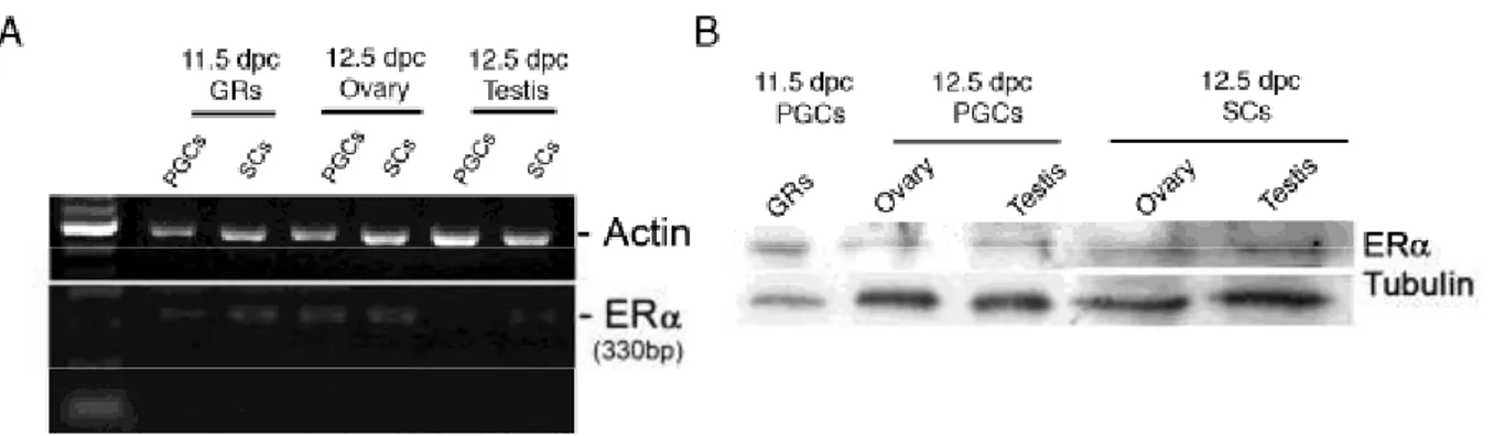
Some of the most relevant results reparted in the present thesis are that in the mouse embryonic gonads besides gonadal somatic cells, primordial germ cells (PGCs) the precursors of adult gametes, express estrogen receptor (ER) and that 17-β-estradiol (E2) via such receptor is able to modulate molecular signalling known to be crucial for their development. Specifically, we demonstrated that PGCs from 11.5-12.5 days post coitum (dpc) mouse embryos express ERα transcripts and protein and that at concentrations of 1-10 nM E2 stimulates rapid (within 20 min) about 3-fold AKT (Ser473) and 2-fold ERK1/2 (Thr202/Tyr204) and SRC (Tyr418) phosphorylation. In addition, the E2 stimulatory effects were associated with increased phosphorylation of the tyrosine kinase KIT (Tyr568/570). While the ER antagonist ICI 182780 was able to abolish all these E2 effects, AKT phosphorylation was inhibited by the PI3K inhibitor LY 294002 and the SRC family inhibitor (PP2). This latter beside SRC phosphorylation was also able to abolish the increased phosphorylation of KIT and ERKs caused by E2. Taken together these results suggest that E2 may modulate via ERα non genomic signalling/phosphorylation cascade in mouse PGCs. This was also supported by the finding that PGCs express MNAR (Modulator of Non genomic Action of estrogen Receptor), a scaffold protein that regulate ER activation in other cell types in which non genomic estrogen action has been demonstrated. Finally, we found that culturing of PGCs in the presence of 10 nM E2 resulted in significant ERα-dependent increase of their number. The results presented in this thesis, provides evidence for novel direct non genomic actions of estrogens on PGCs and suggest that these cells can represent putative target for estrogens and estrogenic compounds during early stages of embryo development in mammals.
In order to study the effect of a prototype of xenoestrogens the pesticide lindane (γ-HCH), an ED, on PGC development in the mouse embryo, we exposed by gavage pregnant mice to 15-30 mg/Kg/bw lindane during the period of PGC migration and gonad colonization (from 8.5 to 11.5 days post coitum, dpc). This treatment resulted in a significant reduction of the number of germ cells within 12.5 dpc testis and ovaries (a maximum of about 25% and 40%, respectively). Similarly, lindane caused a dose-dependent decrease of the PGC number in an in
37
vitro culture model. Further experiments showed that in such model, lindane induced features of apoptotic cell death in PGCs such as increase in caspase-3 activity, PARP cleavage and TUNEL positivity. A marked increase of the number of PGCs positive for TUNEL staining was also observed in 12.5 dpc gonads of embryos from pregnant mice subjected one day before to acute lindane treatment (60 mg/Kg/bw). Finally, we show that a brief incubation of isolated PGCs with 10-5 M lindane resulted in a marked decrease of the basal and KL-induced phosphorylation level of the AKT kinase, known to be crucial for PGC survival. Taken together these results demonstrated that embryo exposure to lindane during early stages of gametogenesis can severely impair the number of germ cells in the foetal gonads; the compound appears to affect PGC survival through a direct pro-apoptotic action likely resulting from its adverse effect on AKT activity in such cells.
The lack of a functional assay able to detect the estogenic activity of estrogens and EDs on somatic cells of the embryonal testis know to express ER , prompted us to devise a protocol for the expansion of the testis somatic cells expressing estrogen receptor α (ERα) from 12.5 dpc embryos and for their transfection with a plasmid that contains the classical estrogen responsive element (ERE) or the alternative estrogen AP-1 responsive element upstream of the luciferase reporter gene (ERE-Luc and AP1-Luc). StAR immunopositivity of the most part of the ERα+ cells grown in culture, allowed their identification as putative Leydig cells. Using the luciferase assay, we evaluated the estrogen activity of 17-ß estradiol (E2), the natural ligand of ERα, on such cells. For comparison, the same assay was carried out on a MCF-7 human breast cancer cell line expressing ERα. The results showed that 24 hr incubation in the presence of E2 resulted in a dose-dependent increase of ERE-Luc activity. At 10-8 M E2 concentration ERE-Luc activity increased from 1.7 to 3-fold in the putative Leydig cells and 2.3 to 5.7-fold in MCF-7 cells. These effects were abolished when 10-5 M ICI 182.80, an inhibitor of E2 binding, was present in the assay. AP-1-Luc activity was less sensitive to E2 stimulation in both cell types (10-8 M E2: putative Leydig cells= 1.2 to 2.7-fold, MCF-7= 3-fold) and the effect of E2 was not abolished by ICI 182.80. Eventually to validate the assay with a xenoestrogen compound, we stimulated the transfected putative Leydig cells and MCF-7 cells with lindane ( -HCH). Taken together the reported results represent the first evidence of a functional ERα pathway in putative Leydig cells
38
from early stage of testis development and describe an in vitro assay that can be used to evaluate estrogenic activity of compounds on mammalian embryonic testis.
In conclusion the present data report evidence for the existence of functional estrogen-dependent pathways in embryonic mouse gonads in particular in testis, both germ and somatic cells. The findings that E2 is able to activate via ER multiple intracellular signalling in PGCs and that the xenoestrogens, lindane negatively interfere with one of these pathways, namely AKT activation are specifically relevant to support the notion of the TDS origin during early stages of testis development. While data are accumulating showing direct effect of estrogens and EDs on gene expression and specific functions of somatic cells of the embryonic testes, in particular Leydig cells, such results on germ cells are lacking and further studies are needed to investigate the effects of these compounds on embryonic germ cell function including epigenetic regulation.
39
References
Acconcia F, Ascenzi P, Bocedi A, Spisni E, Tomasi V, Trentalance A, Visca P, Marino M. 2005 Palmitoylation-dependent estrogen receptor alpha membrane
localization: regulation by 17beta-estradiol. Mol Biol Cell. Jan;16(1):231-7. Epub 2004 Oct 20.
Acconcia F, Ascenzi P, Fabozzi G, Visca P, Marino M. 2004 S-palmitoylation
modulates human estrogen receptor-alpha functions. Biochem Biophys Res Commun. Apr 9;316(3):878-83
Albanito L, Madeo A, Lappano R, Vivacqua A, Rago V, Carpino A, Oprea TI, Prossnitz ER, Musti AM, Andò S, Maggiolini M. 2007 G protein-coupled
receptor 30 (GPR30) mediates gene expression changes and growth response to 17beta-estradiol and selective GPR30 ligand G-1 in ovarian cancer cells. Cancer Res. Feb 15;67(4):1859-66
Albro P.W. 1987 The biochemical toxicology of di-(2-ethylhexyl) phthalate and related
phthalates: testicular atrophy and hepatocarcinogenesis, Rev. Biochem. Toxicol. 8 (1987), pp. 73–119
Bendel-Stenzel M, Anderson R, Heasman J, Wylie C. 1998 The origin and migration of
primordial germ cells in the mouse. Semin Cell Dev Biol. Aug;9(4):393-400. Review
Beard, A.P., and Rawlings, N.C. 1998 Reproductive effects in mink (Mustela vison)
exposed to the pesticides Lindane, Carbofuran and Pentachlorophenol in a multigeneration study. J Reprod Fertil. 113, 95–104
Cabodi S, Moro L, Baj G, Smeriglio M, Di Stefano P, Gippone S, Surico N, Silengo L, Turco E, Tarone G, Defilippi P. 2004 p130Cas interacts with estrogen receptor
alpha and modulates non-genomic estrogen signaling in breast cancer cells. J Cell Sci. Mar 15;117(Pt 8):1603-11
40
Capel B, Albrecht KH, Washburn LL, Eicher EM. 1999 Migration of mesonephric
cells into the mammalian gonad depends on Sry. Mech Dev. Jun;84(1-2):127-31.
Castoria G, Migliaccio A, Bilancio A, Di Domenico M, de Falco A, Lombardi M, Fiorentino R, Varricchio L, Barone MV, Auricchio F. 2001 PI3-kinase in concert
with Src promotes the S-phase entry of oestradiol-stimulated MCF-7 cells. EMBO J. Nov 1;20(21):6050-9 Cell. Jan 27;80(2):237-48. Review.
Chadwick, R.W., Cooper, R.L., Chang, J., Rehnberg, G.L., and McElroy, W.K. 1988
Possible antiestrogenic activity of lindane in female rats. J Biochem Toxicol. 3, 147-58.
Chase DJ, Dixon GE, Payne AH. 1982 Development of Leydig cell function Prog Clin
Biol Res.;112:209-19. Review.
Cheskis BJ. 2004 Regulation of cell signalling cascades by steroid hormones. J Cell
Biochem. Sep 1;93(1):20-7. Review.
Chowdhury, A.R., Venkatakrishna-Bhatt, H., and Gautam, A.K. 1987 Testicular
changes of rats under lindane treatment. Bull Environ Contam Toxicol. 38, 154–156.
Chowdhury, A.R., Gautam, A.K., and Bhatnagar, V.K. 1990 Lindane induced changes
in morphology and lipids profile of testes in rats. Biomed Biochim Acta. 49, 1059– 1065.
Chowdhury, A.R., and Gautam, A.K. 1994 Steroidogenic impairment after lindane
treatment in male rats. J UOEH.16, 145–152
Clarke CH, Norfleet AM, Clarke MS, Watson CS, Cunningham KA, Thomas ML.
2000 Perimembrane localization of the estrogen receptor alpha protein in neuronal processes of cultured hippocampal neurons. Neuroendocrinology. Jan;71(1):34-42
Cohen GB, Ren R, Baltimore D. 1995 Modular binding domains in signal transduction
proteins. Cell. 1995 Jan 27;80(2):237-48. Review
Couse JF, Lindzey J, Grandien K, Gustafsson JA, Korach KS. 1997 Tissue


![(1)Bibliography [1] Schr¨oder, D](data:image/gif;base64,R0lGODlhAQABAIAAAP///wAAACH5BAEAAAAALAAAAAABAAEAAAICRAEAOw==)