Unacylated Ghrelin Enhances Satellite Cell
Function and Relieves the Dystrophic Phenotype in
Duchenne Muscular Dystrophy mdx Model
S
IMONER
EANO,
a* E
LIAA
NGELINO,
a* M
ICHELEF
ERRARA,
aV
ALERIAM
ALACARNE,
aH
ANAS
USTOVA,
aO
MARS
ABRY,
aE
MANUELAA
GOSTI,
aS
ARAC
LERICI,
aG
IULIAR
UOZI,
bL
ORENAZ
ENTILIN,
bF
LAVIAP
RODAM,
cS
TEFANOG
EUNA,
dM
AUROG
IACCA,
bA
NDREAG
RAZIANI,
a*
N
ICOLETTAF
ILIGHEDDU a*
Key Words. Satellite cell self-renewal•Skeletal muscle regeneration•Ghrelin• Duchenne muscular dystrophy•mdx dystrophic mice
A
BSTRACTMuscle regeneration depends on satellite cells (SCs), quiescent precursors that, in consequence of injury or in pathological states such as muscular dystrophies, activate, proliferate, and differ-entiate to repair the damaged tissue. A subset of SCs undergoes self-renewal, thus preserving the SC pool and its regenerative potential. Unacylated ghrelin (UnAG) is a circulating hormone that protects muscle from atrophy, promotes myoblast differentiation, and enhances ischemia-induced muscle regeneration. Here we show that UnAG increases SC activity and stimulates Par polarity complex/p38-mediated asymmetric division, fostering both SC self-renewal and myo-blast differentiation. Because of those activities on different steps of muscle regeneration, we hypothesized a beneficial effect of UnAG in mdx dystrophic mice, in which the absence of dys-trophin leads to chronic muscle degeneration, defective muscle regeneration, fibrosis, and, at later stages of the pathology, SC pool exhaustion. Upregulation of UnAG levels in mdx mice reduces muscle degeneration, improves muscle function, and increases dystrophin-null SC self-renewal, maintaining the SC pool. Our results suggest that UnAG has significant therapeutic potential for preserving the muscles in dystrophies.STEMCELLS2017;35:1733–1746
S
IGNIFICANCES
TATEMENTMuscle dystrophies (MDs) are pathologies characterized by chronic degeneration of muscles. This induces a sustained and continuous regeneration that depends on satellite cells (SCs). Defective regeneration and SC pool exhaustion ultimately lead to the replacement of muscles with scar tissue and loss of functionality. Unacylated ghrelin (UnAG) is a circulating hormone with protective activities on skeletal muscles. Here we demonstrated that UnAG helps to main-tain the pool of SCs and that it has a beneficial effect on muscles of dystrophic mice. UnAG could represent a novel treatment for MDs, either alone or as an adjuvant therapy for one of the promising stem cell-based therapies.
I
NTRODUCTIONInjuries or pathological states such as muscular dystrophies trigger regeneration in the adult skeletal muscle. Muscle regeneration is mainly sustained by a heterogeneous population of quiescent resident precursors, called satellite cells (SCs), characterized by the expression of the transcriptional factor Pax7 [1]. In conse-quence of injury, SCs activate, proliferate, and eventually differentiate to repair the damaged tissue and restore muscle function. A portion of SCs undergoes self-renewal through asym-metric division, thus maintaining the quiescent SC pool and allowing the muscle to retain its
regenerative potential [2, 3]. The asymmetric division generates two daughter cells with diver-gent fates: one proliferating myoblast expressing the marker of myogenic commitment MyoD (MyoD1) and one MyoD– quiescent SC preserv-ing stem features. The differential expression of MyoD depends on the asymmetric segregation of the Par polarity complex during SC activation that leads to a polarized activation of p38 MAPK pathway, triggering MyoD expression in only one daughter cell [4, 5].
Ghrelin and unacylated ghrelin (UnAG) are circulating peptide hormones mainly produced by the stomach. Ghrelin derives from the
aDepartment of Translational
Medicine, University of Piemonte Orientale, Novara, Italy and Istituto
Interuniversitario di Miologia (IIM);dDepartment of Clinical
and Biological Sciences, University of Torino and Neuroscience Institute Cavalieri Ottolenghi (NICO), Orbassano (TO), Italy;
bInternational Centre for
Genetic Engineering and Biotechnology (ICGEB), Trieste, Italy;cDepartment of
Health Sciences, University of Piemonte Orientale, Novara, Italy *Contributed equally. Correspondence: Nicoletta Filigheddu, Ph.D., Department of Translational Medicine, University of Piemonte Orientale, Via Solaroli 17, 28100 Novara, Italy. Telephone: 1 39-0321-660-529; e-mail: nicoletta. [email protected]; or Andrea Graziani, Ph.D., Universit!a Vita-Salute San Raf-faele, via Olgettina 58, 20132, Milano, Italy. Telephone: 139-02-2643-3823; e-mail: graziani. [email protected]
Received August 10, 2016; accepted for publication April 6, 2017; first published online in STEMCELLSEXPRESSApril 24, 2017.
VC AlphaMed Press
1066-5099/2017/$30.00/0 http://dx.doi.org/ 10.1002/stem.2632
S
TEMC
ELLS2017;35:1733–1746 www.StemCells.com
octanoylation of the preprohormone by the ghrelin-O-acyltransferase (GOAT) enzyme [6, 7]. Acylation is required for the binding to the growth hormone secretagogue receptor-1a (GHSR-1a) to induce growth hormone release and to perform multiple endocrine functions [8, 9]. UnAG, the main circulat-ing form of the peptide, does not bind to GHSR-1a but fea-tures several biological activities, including the enhancement of skeletal muscle regeneration induced by hindlimb ischemia [10, 11] and improvement of insulin sensitivity in skeletal muscle [12, 13]. Besides, UnAG shares with ghrelin numerous biological effects, among which the protection of skeletal muscle from atrophy [14, 15] and the promotion of myoblast differentiation [16].
While the mechanisms through which UnAG protects skel-etal muscle from atrophy and insulin resistance have been described [12–14], the cellular and molecular mechanisms mediating UnAG ability to enhance muscle regeneration remain to be elucidated. Here we show that UnAG affects multiple stages of muscle regeneration, including SC activa-tion, proliferaactiva-tion, and self-renewal, the latter through induc-tion of SC asymmetric division mediated by PKCk/i-Par6 complex formation and asymmetric activation of p38. More-over, UnAG induces the differentiation of committed myo-blasts, thus promoting the regeneration of injured muscles. Based on UnAG ability to enhance skeletal muscle regenera-tion, we hypothesized that UnAG could have a therapeutic importance for muscle dystrophies. Duchenne muscular dystro-phy (DMD) is characterized by the absence of the dystrophin protein, whose main function is to connect the myofiber cyto-skeleton to the extracellular matrix through the dystrophin-associated glycoprotein complex. In the absence of dystrophin, myofibers are extremely susceptible to contraction-induced damage, with the consequent chronic degeneration [17]. More-over, dystrophin-null SCs display an impairment of self-renewal and asymmetric division that results in a faulty myogenic pro-gression and, thus, in a defective regenerative process [18, 19]. We show that upregulation of circulating or local UnAG levels in mdx mice improves the dystrophic phenotype, including muscle architecture and functionality. Moreover, UnAG blunts the self-renewal defect of dystrophin-null SCs, thus preserving the SC pool at later stages of the pathology.
M
ATERIALS ANDM
ETHODSAnimals
Animal experiments were performed according to procedures approved by the Institutional Animal Care and Use Committee at the University of Piemonte Orientale. Male mice, matched for age and weight, were used for all experiments. Dystrophin-deficient mdx mice (C57BL/10ScSn-Dmdmdx/J) and C57BL/6-Tg(CAG-EGFP)131Osb/LeySopJ mice with ubiquitous GFP expres-sion were from The Jackson Laboratory (Bar Harbor, ME, https://www.jax.org/); FVB1-Myh6/Ghrl and C57BL/6-Myh6/ Ghrl transgenic mice were generated as previously described [14]. Animals were fed ad libitum and had unrestricted access to drinking water. The light/dark cycle in the room consisted of 12/12 hours with artificial light. To generate dystrophic mice overexpressing the ghrelin gene, C57BL/6J hemizygous Myh6/ Ghrl male mice were bred to homozygous Dmdmdx/mdx female
mice to yield an equal proportion of male mdxTg1and mdxTg–
littermate controls. mdx mice bearing Myh6/Ghrl transgene were identified by PCR genotyping. High levels of plasmatic UnAG in mdxTg1 were confirmed by EIA kit (SPIbio Bertin
Pharma, Montigny le Bretonneux, France, http://www.bertin-pharma.com/) according to the manufacturer’s instructions. The numbers of mice estimated sufficient to detect a difference between two means as large as 1 SD unit with 80% power and a significance level of 95% at Student’s t test were calculated with the program by R.V. Lenth (www.stat.uiowa.edu/~rlenth/ Power/index.html). The investigators conducting the experi-ments were blind to the experimental group assessed. The investigators quantifying the experimental outcomes were maintained blinded to the animal group or intervention. Finally, the statistic evaluation of the experimental data was performed by another investigator not directly involved in data collection and parameter measurement.
Reagents
Rat UnAG peptide was purchased from PolyPeptide Laborato-ries (Strasbourg, France, http://www.polypeptide.com/). Media and fetal bovine serum (FBS) were from Gibco (Thermo Fisher Scientific, Ashford, UK, https://www.thermofisher.com/), horse serum (HS) from PAA (GE Healthcare, Little Chalfont, UK, http://www3.gehealthcare.com/), and media supple-ments, unless otherwise specified, were from Sigma-Aldrich (Paisley, UK, http://www.sigmaaldrich.com).
Cardiotoxin-Induced Muscle Regeneration
Experiments on muscle regeneration were conducted on adult male FVB1 and FVB1-Myh6/Ghrl mice matched for age and weight. Cardiotoxin (CTX) from Naja mossambica mossambica (Latoxan, Portes-le`s-Valence, France, http://www.latoxan.net/) was dissolved in sterile saline to a final concentration of 10 lM. Mice were anesthetized by isofluorane inhalation and hindlimbs were shaved and cleaned with alcohol. Tibialis ante-rior (TA) muscles were injected with 45 ll of CTX with a 30-gauge needle, with 15 microinjections of 3 ll CTX each in the mid-belly of the muscle to induce a homogeneous damage. The TA muscles of the contralateral hindlimbs were injected with saline. After injection, animals were kept under a warm-ing lamp until recovery.
For some experiments, immediately after CTX administration, a single intraperitoneal injection of 5-bromo-20-deoxyuridine (BrdU) (6 lg/g mouse) was given, followed by BrdU adminis-tered ad libitum in drinking water (2.5 mg/ml) for 7 days.
Histological Analysis
Muscles were trimmed of tendons and adhering nonmuscle tissue, mounted in Killik embedding medium (Bio-optica, Milan, Italy, http://www.bio-optica.it/), frozen in liquid-nitrogen-cooled isopentane, and stored at 2808C. Transverse muscle sections (7 mm) were cryosectioned from the mid-belly of each muscle. Sections were stained with hematoxylin/ eosin to reveal general muscle architecture. Images of whole muscle sections were acquired with the slide scanner Pannor-amic Midi 1.14 (3D Histech, Budapest, Hungary, http://www. 3dhistech.com/) and cross-sectional areas (CSA) of centro-nucleated fibers quantified with ImageJ software (v1.49o). Muscle collagen content was assessed with Masson trichromic staining.
To quantify muscle damage and areas of focal necrosis, 1% wt/vol Evans blue dye (EBD) was injected intraperitoneally (5 ml/g of animal weight). Muscles were collected 20 hours after EBD injection. Sections 7-lm thick were cryosectioned, and EBD uptake was detected as red epifluorescence and quantified as above.
SC Isolation and Culture
Primary myoblasts were isolated from the main hindlimb muscles (TA, gastrocnemius, quadriceps [QUAD], extensor digi-torum longus [EDL], soleus) and diaphragm. Muscles were cut with a lancet into small fragments (about 3 mm3) and further inspected to eliminate, as much as possible, any remaining connective tissue. The mass was resuspended in 3 ml of 0.1% pronase and incubated for 1 hour at 378C for digestion. The suspension was then centrifuged at 400g for 5 minutes and the pellet resuspended in DMEM 10% HS medium, passed several times through a serological pipette, filtered through 40 lm strainers, and further centrifuged at 400g for 10 minutes. SCs were separated from fibroblasts and other cells using the SC Isolation Kit (MACS Miltenyi Biotec, Milan, Italy) following the manufacturer’s instructions. After isolation, SCs were either plated on gelatin-coated dishes or immediately used in muscle transplantation experiments.
Plated cells were cultured in growth medium (GM), Dul-becco’s Modified Eagle’s Medium (DMEM) with 20% FBS, 10% HS, 1% chicken embryo extract (CEE, USBiological Life Scien-ces, Salem, MA, https://www.usbio.net/), and 10 ng/ml FGF-2 (Peprotech Inc., London, U.K., http://www.peprotech.com). When cells reached 70%–80% of confluence, GM was shifted into differentiation medium (DM), DMEM with 5% HS for 3 days. UnAG (100 nM) was added simultaneously to DM.
SC Transplantation
To facilitate cell engraftment, one day before muscle trans-plantation, CTX injection was performed in the mid-belly of TA muscles of recipient mice. SCs were isolated from C57BL/ 6-Tg(CAG-EGFP)131Osb/LeySopJ (green fluorescence protein, GFP) mice and 100,000 cells, resuspended in serum-free DMEM, were injected in the previously injured recipient muscles. Contralateral TA muscles were injected with cell-free DMEM. Muscles were harvested 30 days after injection, fixed in 4% paraformaldehyde (PFA), and analyzed.
Myofiber Isolation and Culture
EDL muscles were digested in 0.2% collagenase type-I in DMEM for 60–70 minutes at 378C. Muscles were mechanically dissociated, and single fibers liberated. After extensive wash-ing, myofibers were either immediately fixed or cultured in low proliferation medium (LPM, DMEM supplemented with 10% HS and 0.5% CEE) in suspension. UnAG was added in LPM immediately after fiber seeding. At different time points after plating (6-72-96 hours) fibers were fixed in 4% PFA for 10 minutes.
For experiments with the chemotherapic drug AraC (Cyto-sine b-D-arabinofuranoside), myofibers were cultured for 72 hours in F12 medium supplemented with 15% HS and 1 nM FGF-2 in the presence or absence of 100 nM UnAG and then incubated with or without 100 lM AraC for 48 hours and fixed (day 5).
Immunofluorescence
For Pax7 and BrdU detection, tissue sections were fixed in 4% PFA for 20 minutes, washed, permeabilized with cold methanol for 6 minutes, and then antigen-retrieved with sodium citrate (10 mM, 0.05% Tween in PBS) at 958C for 30 minutes. For blocking the unspecific binding sites, slices were incubated in 4% bovine serum albumin (BSA) for 2 hours at RT and then with M.O.M. blocking reagent (Vector Laboratories, Burlingame, CA, https://vectorlabs.com/) for 1 hour at RT. Sections were stained with an anti-Pax7 antibody (1:100; Developmental Stud-ies Hybridoma Bank, Iowa City, IA, http://dshb.biology.uiowa. edu/) and with anti-BrdU antibody (1:300; Bio-Rad, Segrate, MI, Italy, http://www.bio-rad.com/) overnight at 48C. After washing, sections were incubated with the appropriate Alexa Fluor Dyes-conjugated secondary antibody (488-anti-mouse/anti-rabbit or 568-anti-rabbit; Thermo Fisher Scientific) for 1 hour at RT. 40 ,6-diamidino-2-phenylindole (DAPI) was incubated for 5 minutes.
For immunofluorescence with anti-laminin (1:200; Dako, Agilent Technologies, Santa Clara, CA, http://www.agilent. com/), GFP (1:200; Thermo Fisher Scientific), and anti-embryonic MyHC (1:20; Developmental Studies Hybridoma Bank), after fixing, slices were permeabilized with 0.2% triton in 1% BSA for 15 minutes and blocked with 4% BSA for 30 minutes. One hour of incubation with primary antibodies was followed by 45 minutes of secondary antibody incubation at RT.
Images were acquired using the slide scanner Pannoramic Midi Scanner 1.14 (3D Histech) and quantified with Pannor-amic viewer software or ImageJ v1.49o software. For immuno-fluorescence on isolated fibers and on cultured SCs, samples were fixed in 4% PFA for 10 minutes, permeabilized with 0.5% triton for 6 minutes and blocked with 4% BSA for 30 minutes. Primary antibodies to detect Pax7, MyoD (1:500; Santa Cruz Biotechnology, Dallas, TX, https://www.scbt.com/), myogenin (1:100; Developmental Studies Hybridoma Bank), and MyHC (1:100; Developmental Studies Hybridoma Bank) were incu-bated overnight at 48C, and the secondary antibodies for 45 minutes at RT, followed by 5 minutes of DAPI. Images were acquired with a Leica CTR5500 B fluorescent microscope (Leica Biosystems, Wetzlar, Germany, http://www.leicabiosys-tems.com/) with the Leica Application SuiteX 1.5 software, and quantification was performed with ImageJ.
To evaluate the asymmetric division events of SC pairs, MyoD levels in each cell were obtained by subtracting the background from the nuclear fluorescence intensity (deter-mined by overlap with DAPI staining). Cell pairs were scored “asymmetric” when the MyoD nuclear intensity of one daugh-ter cell was ! 1% (“MyoD–“) and the other one was >1% (“MyoD1”) of the maximal intensity.
Asymmetric distribution of active p38 and PKCk/i was eval-uated by SC incubation with rabbit or mouse anti-phospho-p38T180/Y182 (1:200, Cell Signaling Technology, Beverly, MA,
https://www.cellsignal.com/), rabbit anti-p38 (1:200, Cell Signal-ing Technology), rat anti-CD34 (1:200, BD Biosciences, Cowley, UK, http://www.bdbiosciences.com/), rabbit anti-PKCk/i (1:200, Santa Cruz Biotechnology), rabbit anti-syndecan 4 (1:200, BioVision, Milpitas, CA, http://www.biovision.com/), and mouse anti-Pax7 (1:100; Developmental Studies Hybridoma Bank) fol-lowed by the appropriate Alexa Fluor Dyes-conjugated second-ary antibodies (546 or 647 anti-mouse, 488 anti-rabbit, and 546
anti-rat; Thermo Fisher Scientific). Nuclei were counterstained with TO-PRO-3 iodide (1:100 Thermo Fisher Scientific). Images were acquired with Leica confocal microscope TCS SP2 using a 63X objective, NA 5 1.32, equipped with LCS Leica confocal soft-ware. Asymmetry of phospho-p38T180/Y182 and PKCk/i was
quantified with ImageJ.
For the proximity ligation assay (PLA; Duolink from Sigma-Aldrich), myofiber-associated SCs were incubated with mouse anti-PKCk/i (1:200, BD Bioscience) and rabbit anti-PAR3 (1:200, Merck Millipore) then processed according to the manufacturer’s instructions.
Gene Expression Analysis
Total RNA from muscles was extracted by RNAzol. RNA was retro-transcribed with High-Capacity cDNA Reverse Transcription Kit (Thermo Fisher Scientific), and real-time PCR was performed with the StepOnePlus Real-Time PCR System (Thermo Fisher Sci-entific) using Mm00445450_m1 (Ghrl) and Mm00506384_m1 (Ppif) TaqMan assays.
Hanging Test
A wire-hanging test was used to assess whole-body muscle strength and endurance. The test was performed as previously described [20]. Briefly, mice were subjected to an 180 seconds-hanging test, during which “falling” and “reaching” scores were recorded. When a mouse fell or reached one of the sides of the wire, the falling score or reaching score was diminished or increased by 1, respectively. A Kaplan-Meier-like curve was created afterward. Moreover, the longest time between two falls was taken as the latency-to-fall value [21].
Statistical Analysis
All data were expressed as mean 6 SEM, absolute values, or percentages. For continuous variables, the variation between groups was compared by means of nonparametric Wilcoxon and Mann–Whitney U tests, as appropriate. When analyzing experiments acquired with different instruments, analysis of covariance (ANCOVA) was used to determine differences between groups by using the instrument as a covariate. Multi-ple logistic regression was used for trends. Statistical signifi-cance was assumed for p < .05. The statistical analysis was performed with SPSS for Windows version 17.0 (SPSS Inc; Chi-cago, IL).
R
ESULTSUnAG Upregulation in Myh6/Ghrl Transgenic Mice
Enhances Muscle Regeneration
Muscle damage induces the release, within the muscle, of several factors that activate SCs, triggering the expression of myogenic genes, such as Myf5 and MyoD [22, 23] that even-tually lead to the terminal differentiation of muscle precursors and their fusion among themselves or to the existing fibers. Muscle damage also induces, within the muscle, the expres-sion of the ghrelin gene (Supporting Information Fig. 1) and the preproghrelin protein [24], suggesting that its products— ghrelin, UnAG, and obestatin—may participate in the repair process. Accordingly, exogenously administered obestatin and UnAG enhance muscle regeneration in CTX-injured gastrocne-mii and in hindlimb ischemia, respectively [10, 11, 24].
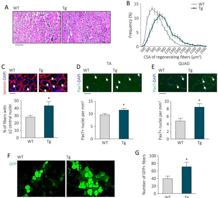
Consistently, high levels of circulating UnAG in Myh6/Ghrl transgenic mice (Tg) [14] improved muscle regeneration of TA muscle after CTX injury (Fig. 1A–1C).
Despite no differences were noticeable between WT and Tg in noninjured muscle CSA distribution (Supporting Informa-tion Fig. 2A), at day 7 post CTX injury improved muscle regen-eration in Tg mice was evidenced by a shift toward bigger areas of regenerating (i.e., centronucleated) fibers (Fig. 1B). This regeneration was accompanied by an increment in the number of regenerating fibers with "2 nuclei (Fig. 1C), sug-gesting an increase in myoblast differentiation and fusion dur-ing regeneration, in agreement with the pro-differentiative activity of UnAG in C2C12 myoblasts [16]. The shift toward bigger areas and the increased myoblast fusion did not trans-late into a hypertrophic phenotype, as at 15 days post-injury fiber distributions of WT and Tg overlapped (Supporting Infor-mation Fig. 2B). Consistently with the hypothesis that UnAG induces faster recovery, injured Tg muscles displayed more embryonal MyHC (eMyHC)-positive myofibers at day 3 post-CTX, although at day 7 no significant differences were observed in the number of eMyHC-expressing fibers (Support-ing Information Fig. 2C–2E). Also, transient collagen deposi-tion during regeneradeposi-tion tended to disappear more rapidly in Tg muscles between days 7 to 15 post-injury (Supporting Information Fig. 2F, 2G).
Though Tg mice do not overtly differ from their WT litter-mates [14], a closer examination of their not-injured muscles revealed a larger number of SCs, seen as Pax71 nuclei, in both TA and QUAD from Tg animals (Fig. 1D, 1E). However, during the phase of intense proliferation following injury, SC number in muscles from Tg and WT were comparable (Sup-porting Information Fig. 2H–2I). This finding suggests that a UnAG-rich environment may confer a regenerative advantage, at least partially by promoting post-natal SC pool formation. In addition, transplant of SCs from GFP donor mice in muscles of Tg or WT recipient mice resulted, 30 days later, in #80% increase in GFP1 fibers in Tg than in WT mice (Fig. 1F, 1G), suggesting that UnAG promotes skeletal muscle regeneration plausibly by acting on the transplanted SCs.
UnAG Promotes SC Activity and Their Asymmetric
Division
To explore in detail the effects of UnAG on SCs, we isolated single fibers from WT muscles, thus maintaining SCs in an original niche-like environment [25], and we cultured them in the presence or absence of 100 nM UnAG. When cultured, SCs undergo activation and turn on MyoD expression (Pax71/ MyoD1). After 72 hours in culture, several clusters of myo-blasts originated from a single SC are visible on myofibers. During this phase, the majority of activated SCs turns off Pax7 and commits to terminal differentiation (Pax7–/MyoD1), while a small subset undergoes self-renewal, retaining Pax7 but not MyoD expression (Pax71/MyoD–) [26, 27]. UnAG treatment within 6 hours expanded the portion of activated SCs (Fig. 2A; Supporting Information Fig. 3A), within 72 hours it increased the number of cells in each cluster (Fig. 2B; Sup-porting Information Fig. 3B), and within 96 hours raised the portion of SCs that underwent self-renewal (Fig. 2C; Support-ing Information Fig. 3C). Cotreatment with BrdU at the begin-ning of the experiment demonstrated that all cells underwent DNA replication, as the totality of Pax71 cells within the
clusters after 96 hours of UnAG treatment were also BrdU1 (Supporting Information Movies 1 and 2). Altogether, these data indicate that UnAG enhances SC activity by promoting their activation, expansion, and self-renewal.
SCs undergo self-renewal through asymmetric division that gives rise to a proliferating daughter cell and a quiescent daugh-ter cell [4]. In culture, asymmetric division generally occurs dur-ing the first cellular division. This has been demonstrated by culturing myofibers with cytosine b-D-arabinofuranoside (AraC), a chemotherapic drug that selectively kills cycling cells and spares quiescent cells. Incubation of isolated myofibers with AraC during the first three days kills all myofiber-associated SCs, while the addition of AraC to the culture medium from day 3
to day 5 allows the detection of AraC-resistant Pax71 SCs deriv-ing from cells that divided at least once (Fig. 2D; [4, 28]). A higher number of Pax71/MyoD– SCs was found in UnAG-treated myofibers after incubation with AraC from day 3 to 5 (Fig. 2E), suggesting that UnAG acts during SC first replications, likely regulating SC asymmetric division. Asymmetric division can be assessed by the quantification of myofiber-associated SC doublets, bona fide derived from a single SC after the first cell division, in which only one of the two daughter cells is MyoD1 [4]. UnAG treatment induced a sixfold increase in the percent-age of SC doublets in which only one cell is MyoD1 (Fig. 2F, 2G), indicating that UnAG actually promotes SC asymmetric divi-sion. To verify whether UnAG induces self-renewal also in vivo,
Figure 1. UnAG upregulation in Myh6/Ghrl transgenic mice enhances muscle regeneration, increases satellite cell (SC) number in non-injured muscles, and improves SC engraftment. (A): H&E representative images of WT and Myh6/Ghrl (Tg) TA muscle sections 7 days after cardiotoxin injury. Scale bar, 100 lm. (B): CSA frequency distribution of regenerating fibers in TA. Chi-square test was used to com-pare distributions. Trend p < .01. (CSA mean mm2: Tg 837.02 6 55.41; WT 559.28 6 15.81; p < .05). N " 4. (C): Representative images of laminin IF with DAPI staining of WT and Tg regenerating fibers and percentage of fibers with "2 central nuclei (arrows) over the total of regenerating fibers. Scale bar, 30 lm. Mean 6 SEM *, p < .05; n " 4. (D, E): Representative images and quantification of Pax71 SCs/ mm2(arrows) in noninjured TA (D) and QUAD (E) of WT and Tg mice. Mean 6 SEM Scale bars, 30 lm. *, p < .05; TA n 5 10; QUAD n 5 5. (F, G): Representative images (F) and quantification (G) of GFP1 myofibers in transplanted TA of WT and Tg mice. Scale bar, 200 lm. Mean 6 SEM *, p < .05; n 5 11 (WT) and 12 (Tg). Abbreviations: CSA, cross-sectional areas; DAPI, 40,6-diamidino-2-phenylindole; GFP, green fluorescent protein; QUAD, quadriceps; TA, tibialis anterior; Tg, transgenic mice; WT, wild type mice.
Figure 2. UnAG induces activation, proliferation, and self-renewal of SCs. (A): Percentage of MyoD1 SCs after 6 hours of treatment of isolated myofibers with 100 nM UnAG in low proliferation medium. "20 myofibers/treatment, "40 SCs/treatment. (B): Cells per cluster after 72 hours of treatment. "20 myofibers/treatment, "16 clusters/treatment, "2 SCs/cluster; n 5 3 independent experiments. (C): Per-centage of Pax71/MyoD– SCs after 96 hours of treatment. Mean 6 SEM *, p < .05. "25 myofibers/treatment, "30 clusters/treatment, "3 SCs/cluster; n 5 3 independent experiments. (D): Schematic of experiments with AraC to identify quiescent daughter SCs. (E): AraC-resistant Pax71 cells. Mean 6 SEM *, p < .05; "25 myofibers/treatment, total araC-AraC-resistant Pax71 SC n 5 13 (control), 43 (UnAG); n 5 3 independent experiments. (F): Representative images of SCs that underwent symmetric (control) or asymmetric (UnAG-treated) division. Scale bar, 20 lm. (G): Percentage of asymmetric division events in SC doublets. Mean 6 SEM *, p < .05; for each experiment, "22 fibers/ treatment; "22 doublets/treatment; n 5 3 independent experiments. (H): Experimental design schematic: mice were daily treated with BrdU for the first 7 days after CTX injection. Muscles were harvested 50 days after injury. (I): Representative images of tibialis anterior transverse sections, arrow: Pax71/BrdU1 nucleus. Scale bar, 40 lm. (J): Pax71/BrdU1 nuclei normalized to the contralateral SC number. Mean 6 SEM *, p < .05; n " 8. (K): SC forced exhaustion design schematic: 10 days after three injections of CTX at 5-days intervals, EDL fibers were isolated from injured hindlimbs and immediately fixed. (L): Number of Pax71/Myf5– SCs in 100 isolated fibers. Mean 6 SEM *, p < .05 versus multiple injured WT; §, < .05 versus not injured WT; n " 8. Abbreviations: AraC, cytosine b-D-arabinofuranoside; BrdU, 5-bromo-20-deoxyuridine; CTX, cardiotoxin; DAPI, 40,6-diamidino-2-phenylindole; EDL, extensor digitorum longus; SC, satellite cell; UnAG, unacylated ghrelin.
we administered BrdU to WT and Myh6/Ghrl mice during the phase of intense myoblast proliferation post-injury (Fig. 2H). Since BrdU is incorporated in every cycling cell, when muscle regeneration is fully achieved, and SC proliferation no longer occurs, any cell positive for both BrdU and Pax7 (Fig. 2I) is a SC that cycled at least once and then underwent self-renewal [29]. Fifty days post-injury the number of Pax71/BrdU1 SCs—nor-malized on SC number in the contralateral, noninjured muscle— was higher in Myh6/Ghrl than in WT muscles (Fig. 2J), demon-strating that upregulation of UnAG enhanced SC self-renewal also in vivo. SC self-renewal is of particular importance when skeletal muscle is subjected to repeated cycles of degeneration/ regeneration that could lead to the progressive depletion of the SC pool; therefore, we assessed the impact of UnAG on the compartment of Myf5– SCs, a subpopulation of SCs that under-goes depletion in an artificial model of SC pool exhaustion, obtained by multiple rounds of muscle injury (Fig. 2K, [30]). EDL fibers isolated from injured hindlimbs of WT mice displayed a 50% loss of Myf5– SCs, while fibers from Tg mice maintained the number of Myf5– SCs (Fig. 2L), demonstrating that UnAG helps to maintain the SC pool also upon repeated injuries.
UnAG Induces Satellite Cell Self-Renewal Through Par
Complex Assembly and Activation of p38
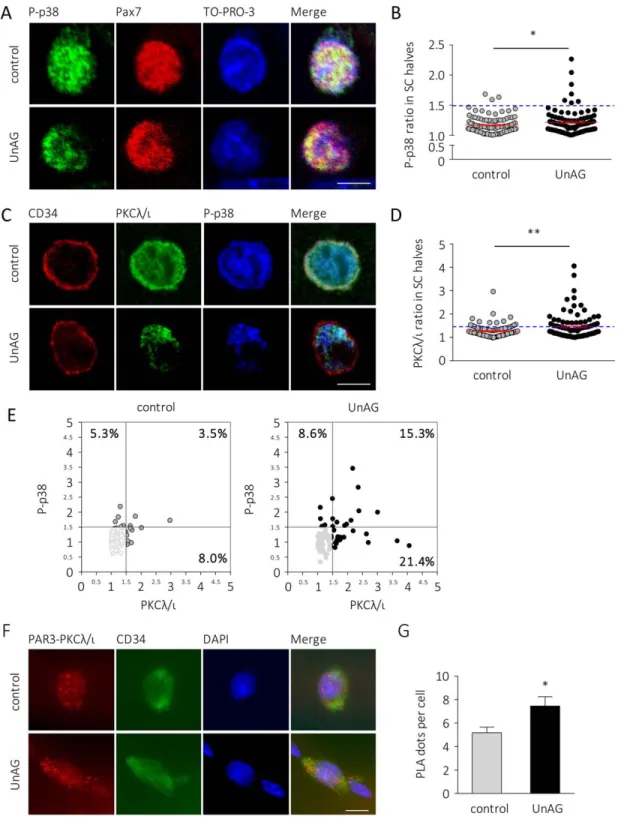
UnAG exerts its anti-atrophic and pro-differentiative effects on skeletal muscle through activation of p38 [14, 16], a mas-ter regulator of SC activities, as its activation mediates either SC proliferation or differentiation [5, 31]. The role of p38 is particularly relevant in SC self-renewal, since the asymmetric segregation of phosphorylated p38 in dividing SCs regulates their asymmetric division, triggering MyoD protein expression in only one daughter cell [4, 5]. Treatment of myofibers with UnAG for 36 hours increased the asymmetric distribution of phosphorylated p38 in SCs without affecting total p38 uniform distribution (Fig. 3A, 3B; Supporting Information Fig. 4).
Since in the dividing SC phospho-p38 colocalizes with the atypical PKCk/i [4], we assessed if UnAG could also enhance the asymmetric distribution of PKCk/i. Indeed, UnAG treat-ment increased PKCk/i localization in one of the two SC halves (Fig. 3C, 3D). Moreover, UnAG enhanced the asymmet-ric cosegregation of PKCk/i and phospho-p38, seen as the increased percentage of SCs with asymmetric distribution of the two proteins in the same side of the cell (Fig. 3C, 3E).
Asymmetric localization of atypical PKCk/i and phospho-p38 during SC division is closely related to the Par3-Par6-PKCk/ i complex (“Par complex”) formation during SC asymmetric division [4]. The increased localization of both PKCk/i and phospho-p38 observed in UnAG-treated SCs could depend on an enhanced Par complex formation. Therefore, we assessed if UnAG enhanced PKCk/i and PAR3 complex formation within SCs using a PLA. We observed that indeed UnAG-treated SCs displayed an increased number of PLA dots compared to con-trol (Fig. 3F, 3G), indicating that UnAG enhances Par complex assembly.
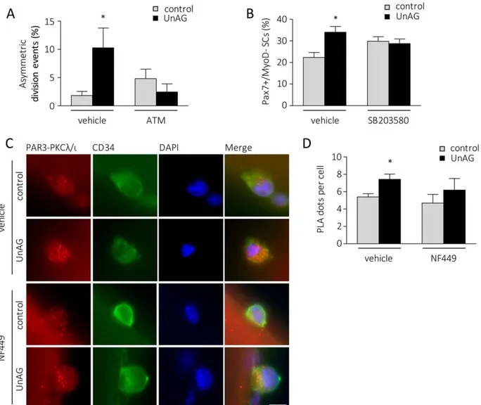
Asymmetric division depends on Par complex formation and its asymmetric localization [4] and, accordingly, incubation with 10 mM aurothiomalate, an inhibitor of Par complex assembly [32], prevented the effect of UnAG on SC asymmet-ric division (Fig. 4A).
The activation of p38 in dividing SCs affects their ability to undergo self-renewal and, indeed, incubation of
UnAG-treated myofibers with 5 mM of p38 inhibitor SB203580, completely abrogated UnAG effect on self-renewal (Fig. 4B). Altogether these data suggest that UnAG promotes SC asym-metric division and self-renewal through asymasym-metric phos-phorylation of p38.
Incubation of UnAG-treated myofibers with 10 mM NF449, a compound able to abolish the anti-atrophic activity of UnAG in C2C12 myotubes by uncoupling Gas from G
protein-cou-pled receptors (GPCRs) [14], abrogated the increment of atyp-ical PKC/PAR3 complex induced by UnAG (Fig. 4C, 4D), supporting the hypothesis that UnAG acts through a Gas
–cou-pled GPCR, as previously suggested [14].
Upregulation of Circulating UnAG Protects Dystrophic
Muscles Architecture and Functionality
The improved muscle regeneration in Myh6/Ghrl mice and enhanced SC activity within the muscle of Myh6/Ghrl mice and in response to UnAG treatment suggest that increase in UnAG may be beneficial for muscle diseases such as dystro-phies, in which the lack of dystrophin impacts both on muscle fragility and on SC function, leading to chronic degeneration and impaired regeneration [18, 33]. To test the hypothesis that upregulation of UnAG circulating levels protects dystro-phic muscles from deterioration, we crossed dystrophin-null mdx mice with hemizygous Myh6/Ghrl mice, producing mdxTg1mice and mdxTg–littermate controls. Histological
anal-ysis revealed that muscles of both animal groups were under-going regeneration, seen as high density of Pax71 SC and the presence of eMyHC1 fibers in diaphragms (Supporting Infor-mation Fig. 5A–5D). However, in mdxTg1 mice compared to
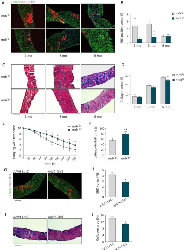
the mdxTg– littermates, displayed lower fiber damage in
dia-phragms of 1- and 3-month-old mice, while no differences were evident at 6 months of age (Fig. 5A, 5B). During the progression of the pathology, the gradual replacement of functional muscle with collagen in diaphragm was delayed in mdxTg1 mice compared to the mdxTg– littermates, becoming
significantly lower at 6 months of age (Fig. 5C, 5D), indicating that upregulation of UnAG ameliorates the dystrophic pheno-type. Consistently, hanging-wire-test scores and latency-to-fall time, assessments of muscular functionality and endurance, were improved in mdxTg1mice starting from 4 months of age
(Fig. 5E, 5F), showing that the differences highlighted by his-tological analysis translated into enhanced functional perfor-mance. Altogether, these data show that upregulation of circulating UnAG partially relieves the pathological condition of mdx dystrophic mice.
To assess if these UnAG activities could have clinical rele-vance, as a proof of concept, we exogenously administered UnAG to dystrophic mice using adeno-associated virus (AAV)-mediated delivery. We used the AAV9-Ghrl vector that has been demonstrated to protect the muscle from ischemic injury as the UnAG peptide does [11]. We injected 3.5 3 1011
vg of either AAV9-Ghrl or AAV9-LacZ in the tail vein of 3-week-old mdx mice, when the first round of muscle degenera-tion occurs [34], and analyzed the effect on muscles at 3 months of age. Ghrelin upregulation in muscles was con-firmed by real-time RT-PCR (Supporting Information Fig. 6A, 6B). Analysis of diaphragms revealed less muscle damage (Fig. 5G, 5H) and collagen deposition (Fig. 5I, 5J) in AAV9-Ghrl-injected mice compared to the AAV9-LacZ-AAV9-Ghrl-injected controls. Altogether, these data indicate that UnAG treatment is
Figure 3. UnAG induces asymmetric localization of phospho-p38 and PKCk/i within the satellite cell and increases PAR3-PKCk/i complex for-mation. (A): Representative images and (B) quantification of asymmetric distribution of phospho-p38T180/Y182 in UnAG-treated versus untreated (control) SCs. Cells over the dashed line display at least 50% more phospho-p38T180/Y182in one half of the nucleus compared to the other half. Mean 6 SEM *, p < .05; for each experiment "30 fibers/treatment, "34 SCs/treatment; n 5 3 independent experiments. Scale bar, 5 lm. (C): Representative images of PKCk/i and phospho-p38T180/Y182distribution in control (top) and UnAG-treated (bottom) SCs (CD341). Scale bar, 5 lm. (D): Quantification of asymmetric distribution of PKCk/i in UnAG-treated versus untreated (control) SCs. Cells over the dashed line display at least 50% more PKC in one half of the nucleus compared to the other half. **, p < .01; for each experiment "30 fibers/treat-ment, "28 SCs/treatment; n 5 3 independent experiments (E): Correlation between the asymmetric distribution of PKCk/i (x-axis) and phos-pho-p38T180/Y182(y-axis) in control (left) versus UnAG-treated (right) SCs. Each dot denotes a single satellite cell. The bottom-left shadowed quadrants contain the SCs with both phospho-p38T180/Y182and PKCk/i uniformly distributed. Total SCs counted n 5 202 (control), 188 (UnAG-treated). (F): Representative images of the PLA-detected complexes of PAR3 and PKCk/i (red) in control (top) versus UnAG-treated (bottom) SCs (CD341, green). Nuclei stained with DAPI. Scale bar, 5 lm. (G): Quantification of the PLA dots per single satellite cell in control versus UnAG-treated SCs. Mean 6 SEM *, p < .05; for each experiment, "30 fibers, nuclei " 69 (control), "70 (UnAG); n 5 3 independent experi-ments. Abbreviations: DAPI, 40,6-diamidino-2-phenylindole; PLA, proximity ligation assay; SC, satellite cells; UnAG, unacylated ghrelin.
effective even after the onset of muscle degeneration, sup-porting the idea that UnAG administration could represent a potential treatment for muscular dystrophies.
UnAG Enhances Dystrophin-Null Satellite Cell
Self-Renewal and Myogenic Commitment
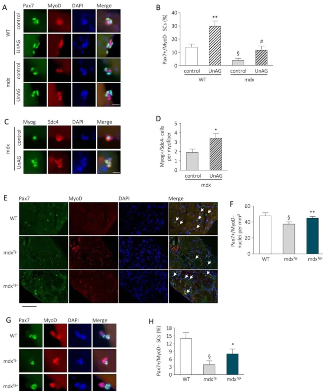
To assess the direct effect of UnAG on dystrophic SCs, we iso-lated EDL fibers from dystrophin-null mdx and WT mice, and we cultured them in the presence or absence of 100 nM UnAG for 96 hours. The number of Pax71/MyoD– SCs was 60% lower in mdx fibers compared to the WT ones, indicating an intrinsic self-renewal defect of dystrophin-deficient SCs, in agreement with previously reported data [19]; however, UnAG significantly raised the Pax71/MyoD– SC portion in both WT and mdx fibers (Fig. 6A, 6B). The relative increase of quiescent SCs, of about 50%, was the same in both WT and mdx fibers,
indicating that UnAG promotes SC self-renewal independently from the presence of dystrophin. Also, UnAG enhanced the number of myogenin-expressing SCs in mdx myofibers cul-tured for 72 hours (Fig. 6C, 6D), indicating that the increase of quiescent SCs does not imply an unbalance between self-renewal and myogenic commitment.
The sustained muscle regeneration in mdx mice is empha-sized by the huge expansion of SCs that, however, in the long run, leads to the depletion of the SC pool. Although no signif-icant differences in the number of SCs are evident in mdxTg1
mice compared to mdxTg–muscles at 1- to 6-months-old
ani-mals (Supporting Information Fig. 5A, 5B), in diaphragms of 12-month-old mice we observed that the exhaustion of SC pool, characteristic of the advanced pathology [19], was less pronounced in mdxTg1 mice compared to mdxTg– mice (Fig.
6E, 6F), coherently with the concept that UnAG promotes SC
Figure 4. UnAG-induced satellite cell asymmetric division and self-renewal is mediated by Par complex assembly-p38 activation and occurs through a Gas-coupled GPCR. (A): Percentage of asymmetric division events in SC doublets after 48 hours of UnAG treatment in the presence/absence of 10 lM ATM, an inhibitor of atypical PKCk/i-Par6 complex formation. Mean 6 SEM *, p < .05; "20 fibers/treatment, "22 doublets/treatment; n 5 4 independent experiments. (B): Percentage of Pax71/MyoD–SCs after 96 hours of UnAG treatment in the presence/absence of 5 lM p38 inhibitor SB203580. Mean 6 SEM *, p < .05 versus DMSO-treated control; "25 myofibers/treatment, "50 clusters/treatment, "3 SCs/cluster; n 5 2 independent experiments. (C): Representative images of PLA-detected complexes of PAR3 and PKCk/i (red) in control versus UnAG-treated SCs (CD341, green) in the presence or absence of 10 lM NF499. Nuclei stained with DAPI. Scale bar, 5 lm. (D): Quantification of the PLA dots per satellite cell. Mean 6 SEM *, p < .05; "30 fibers; n nuclei 5 72 (control), 70 (UnAG), 81 (NF499), 45 (NF499 1 UnAG); n 5 2 independent experiments. Abbreviations: ATM, aurothiomalate; DAPI, 40,6-diamidino-2-phenylindole; PLA, proximity ligation assay; SC, satellite cells; UnAG, unacylated ghrelin.
Figure 5. Unacylated ghrelin upregulation in mdx mice attenuates the dystrophic phenotype. (A): Representative images and (B) quan-tification of EBD uptake in diaphragms of 1- to 6-month-old mdxTg–and mdxTg1mice. Scale bar, 200 lm. Mean 6 SEM **, p < .01 ver-sus mdxTg–; n 5 7 (mdxTg–) and 6 (mdxTg1). (C): Representative images of Masson trichrome staining and (D) quantification of collagen deposition in the diaphragm of 1- to 6-month-old mdxTg1and mdxTg–mice. Scale bar, 200 lm. Mean 6 SEM *, p < .05; n 5 7. (E, F): Muscular functionality measured by hanging wire test scores (E) and average latency-to-fall time (F) of 4-month-old mdxTg–and mdxTg1 mice. Mean 6 SEM; *, p < .05 and **, p < .01 versus mdxTg–; n 5 20. (G): Representative images and (H) quantification of Evan Blue Dye uptake in AAV9-LacZ- or AAV9-Ghrl-transduced diaphragms of 3-month-old mdx mice. Scale bar, 200 lm. Mean 6 SEM *, p < .05 versus LacZ-transduced muscles; n 5 5. (I): Representative images and (J) quantification of collagen deposition in LacZ- and AAV9-Ghrl-transduced diaphragms of 3-month-old mdx mice. Scale bar, 200 lm. Mean 6 SEM *, p < .05 versus AAV9-LacZ-transduced muscles; n 5 5. Abbreviations: DAPI, 40,6-diamidino-2-phenylindole; EBD, Evans blue dye.
Figure 6. UnAG improves dystrophin-null SCs activity. (A): Representative images of Pax7 and MyoD IF and DAPI counterstaining of SCs on myofibers isolated from WT or mdx extensor digitorum longus (EDL) muscles and treated with 100 nM UnAG for 96 hours. Scale bars, 20 lm. (B): Percentage of Pax71/MyoD– SCs after 96 hours of treatment of WT or mdx myofibers with 100 nM UnAG. Mean 6 SEM **, p < .01 versus. WT control, §, p < .01 versus WT control, and #, p < .01 versus mdx control. "25 myofibers/treatment, "14 cluster/treatment, "3 SCs/cluster; single experiment. (C): Representative images of Myog and Sdc4 IF and DAPI counterstaining of SCs on myofibers isolated from mdx EDL muscles and treated with 100 nM UnAG for 72 hours. Scale bar, 20 lm. (D): Number of Myog1/ Sdc4- SCs per fibers after 72 hours of treatment of mdx myofibers with 100 nM UnAG. *, p < .01 versus mdx, "45 myofibers/treatment, total Myog1/Sdc4- SCs 5 95 (control), 140 (UnAG); n 5 3 independent experiments. (E): Representative images (scale bar, 200 lm) and (F) quantification of Pax71/MyoD– (arrows) cells/mm2in diaphragm sections of 12-month-old mdxTg1and mdxTg–
mice. Mean 6 SEM §, p < .05 versus WT; *, p < .05 versus mdxTg–; n 5 7. (G): Representative images (scale bar, 20 lm) and (H) percentage of Pax71/MyoD– SCs on myofibers isolated from aged (12 months) WT, mdxTg–, and mdxTg1cultured for 96 hours in low proliferation medium. §, p < .01 versus WT control and *, p < .05 versus mdxTg–
, "23 myofibers/group, "23 cluster/group, "3 SCs/cluster; single experiment. Abbreviations: DAPI, 40,6-diamidino-2-phenylindole; Myog, myogenin; SCs, satellite cells; Sdc4, syndecan 4; UnAG, unacylated ghrelin; WT, wild type mice.
self-renewal. Consistently, after 96 hours in culture, myofibers from mdxTg1 mice displayed twice as much Pax71/MyoD–
SCs, likely reflecting an initial higher content of functional SCs (i.e., able to undergo self-renewal) compared to mdxTg–mice
(Fig. 6G, 6H).
D
ISCUSSIONThe data herein presented demonstrate that UnAG acts on SCs enhancing their activation, differentiation, and self-renewal. SC self-renewal depends on either symmetric or asymmetric division of a subpopulation of noncommitted pro-genitors. For instance, Wnt7a promotes SC self-renewal through induction of their symmetric division via a noncanoni-cal, planar-cell-polarity pathway [35] and without affecting SC differentiation. On the contrary, UnAG promotes SC self-renewal enhancing at the same time their terminal differenti-ation, as shown by the increase of fusion index on cultured SCs (Supporting Information Fig. 7). This effect is likely a con-sequence of UnAG-induced increase on SC asymmetric divi-sion that simultaneously maintains the stem compartment of MyoD– SCs and expands the number of the committed MyoD1 myoblasts able to respond to the pro-differentiative activity of UnAG, in agreement with the effect observed on C2C12 [16].
SC asymmetric division is sustained by the Par polarity complex that includes the atypical PKCk/i, which controls the asymmetric activation of p38 that, in turn, triggers MyoD expression and myogenic commitment in only one daughter cell [4, 5]. Phosphorylated p38 plays a key role in asymmetric division [4]; indeed, loss or reduction of asymmetric segrega-tion of phosphorylated p38 and its diffuse activasegrega-tion within the SC from aged mice determines a strong decline of asym-metric division events and the consequent impairment of SC self-renewal ability [28].
UnAG promotes the asymmetric cosegregation of PKCk/i and phospho-p38 and the Par complex assembly in myofiber-associated SCs. In addition, uncoupling of PKCk/i from the Par complex and inhibition of p38 activity impair UnAG-induced asymmetric division and self-renewal, indicating that UnAG trig-gers signaling pathways contributing to the formation of the polarity complex. The finding that uncoupling of Gas from
GPCRs impairs the UnAG-stimulated assembly of the polarity complex suggests that UnAG acts in SCs through a similar or the same receptor mediating its anti-atrophic activity in C2C12 myotubes [14]. The lack of knowledge on the identity of UnAG receptor hinders deeper investigations on the signaling mecha-nisms mediating its activity in SCs. Nevertheless, UnAG stimu-lates cAMP and PKA [36], which are linked to the activation of the polarity kinase LKB1 and to the formation of the polarity complex [37–39], thus leading to the speculation that cAMP/ PKA pathway may contribute to UnAG polarizing signal in SCs.
The expansion of SCs through asymmetric division and the enhancement of their differentiation elicited by UnAG underline its ability to enhance skeletal muscle regeneration (Fig. 1A–1C, [10, 11]), consistently with the hypothesis that ghrelin induction in the injured muscle contributes to the repair process. Indeed, ghrelin is rapidly and transiently induced upon muscle damage [24]. However, the identification of the cells responsible for ghrelin expression is hampered by the poor specificity of the
available anti-preproghrelin antibodies, which recognize positive signals in injured skeletal muscle of ghrelin KO mice (data not shown). As ghrelin is highly expressed in neutrophils, which are recruited to the site of damage with similar kinetics as ghrelin induction [40, 41], we can speculate that neutrophils may be responsible for ghrelin upregulation observed in muscles after injury. Altogether these findings suggest that UnAG is part of the damage-induced tissue repair process.
The more efficient engraftment of donor SCs in Tg mice (Fig. 1F, 1G) is consistent with a direct effect on SC functional-ity as well. However, the better engraftment may also depend on UnAG anti-inflammatory activity. Indeed, in skeletal muscle, UnAG inhibits TNF-a expression following either burn injury or high-fat diet [12, 15]. Furthermore, we cannot rule out that the increased engraftment of SCs in Tg muscle may partially be due to an anti-apoptotic effect of UnAG on transplanted SCs since UnAG inhibits apoptosis in both cardiomyocytes and myo-blasts through activation of autophagy [11, 12].
Altogether, these findings indicate that UnAG regulates multiple steps of muscle regeneration by stimulating asym-metric division-mediated SC self-renewal and by promoting terminal differentiation and fusion of proliferating myoblasts. The capacity of UnAG to induce SC self-renewal also in vivo translates in the ability to preserve the quiescent SC pool upon repeated cycles of injury/regeneration.
UnAG pro-regenerative effect on skeletal muscle and its activity on SCs may account for the less severe phenotype observed in dystrophic mice with high levels of circulating or local UnAG in mdxTg1 or AAV-Ghrl-treated mdx mice,
respec-tively. However, the anti-inflammatory, and, consequently, the anti-fibrotic activities of UnAG [42, 43] could be likewise rele-vant to explain the protection of tissue architecture and the amelioration of muscle performance. In addition, as defective basal autophagy contributes to the dystrophic phenotype [44, 45], UnAG-enhanced autophagy may likewise contribute to its protective activity in mdx mice.
In dystrophy, the exhaustion of the SC pool has been assumed to cause the failure of regeneration to keep up with muscle damage. However, in both human and mice SC pool exhaustion likely sets in only at late stages of the pathology as a consequence of defective SC self-renewal [19]. Furthermore, dys-trophic muscles show an increased overall number of SCs, although, within this figure, the portion of quiescent SCs is reduced, reflecting an ongoing regeneration [18, 19, 46]. It is plausible that the defect in muscle regeneration of mdx mice resides at least in part in the defective asymmetric division of dystrophin-null SC that translates in an imbalance between SCs and committed myoblasts able to terminally differentiate and repair the damaged muscle [18, 33]. The finding that UnAG, in dystrophin-null SCs, enhances their self-renewal and increases the number of committed myoblasts suggests that UnAG promotes SC asymmetric division by activating pathways that are indepen-dent of dystrophin expression. Thus, we can speculate that the increase in the absolute number of functional SCs triggered by UnAG in mdx mice may increase the number of committed pro-genitors, thus sustaining the better muscle regeneration and improved dystrophic phenotype observed in mdxTg1mice.
Altogether, these data suggest that increase in either circu-lating or local UnAG levels could delay the progression of the disease. A therapeutic approach would presumably involve the chronic administration of UnAG to dystrophic patients.
Although the receptor through which UnAG exerts its biological activities remains elusive, UnAG has been recently used in clini-cal trials to assess its metabolic effects, and it was reported that the peptide was well tolerated, and no serious adverse events occurred during the studies [47–51]; therefore, UnAG could be a realistic adjuvant treatment in the near future to help to preserve muscles of dystrophic patients.
S
UMMARYUnAG affects in multiple ways SC physiological behavior and enhances skeletal muscle regeneration. These activities result in an overall beneficial effect in dystrophic mdx mice, seen as an improved condition of muscles and a better physical per-formance, and suggest that UnAG could be regarded as a real-istic therapeutic strategy for muscular dystrophies, either as a self-sufficient treatment or as an adjuvant with other therapies.
A
CKNOWLEDGMENTSThis study was supported by research grants from the Muscular Dystrophy Association (Grant MDA294617 to N.F. and A.G.),
AFM-T"el"ethon (Grant 16437 to A.G.) and Compagnia di San Paolo (to A.G. and N.F.). E.A., M.F., V.M., S.C., and A.G. are cur-rently affiliated with the Universit!a Vita-Salute San Raffaele, Milano, Italy.
A
UTHORC
ONTRIBUTIONSS.R., E.A., and M.F.: conception and design; collection and/or assembly of data; data analysis and interpretation; manuscript writing; V.M. and H.S.: conception and design; collection and/ or assembly of data; data analysis and interpretation; final approval of manuscript; O.S., E.A., S.C., and G.R.: collection and/or assembly of data; final approval of manuscript; L.Z. and S.G.: provision of study material; final approval of manu-script; F.P.: data analysis and interpretation; final approval of manuscript; M.G.: conception and design; final approval of manuscript; A.G. and N.F.: conception and design; data analy-sis and interpretation; manuscript writing.
D
ISCLOSURE OFP
OTENTIALC
ONFLICTS OFI
NTERESTThe authors indicated no potential conflicts of interest.
R
EFERENCES1 Seale P, Sabourin LA, Girgis-Gabardo A et al. Pax7 is required for the specification of myogenic satellite cells. Cell 2000;102:777– 786.
2 Collins CA, Olsen I, Zammit PS et al. Stem cell function, self-renewal, and behav-ioral heterogeneity of cells from the adult muscle satellite cell niche. Cell 2005;122: 289–301.
3 Kuang S, Kuroda K, Le Grand F et al. Asymmetric self-renewal and commitment of satellite stem cells in muscle. Cell 2007;129: 999–1010.
4 Troy A, Cadwallader AB, Fedorov Y et al. Coordination of satellite cell activation and self-renewal by Par-complex-dependent asym-metric activation of p38a/b MAPK. Cell Stem Cell 2012;11:541–553.
5 Jones NC, Tyner KJ, Nibarger L et al. The p38alpha/beta MAPK functions as a molecu-lar switch to activate the quiescent satellite cell. J Cell Biol 2005;169:105–116.
6 Gutierrez JA, Solenberg PJ, Perkins DR et al. Ghrelin octanoylation mediated by an orphan lipid transferase. Proc Natl Acad Sci USA 2008;105:6320–6325.
7 Yang J, Brown MS, Liang G et al. Identifi-cation of the acyltransferase that octanoy-lates ghrelin, an appetite-stimulating peptide hormone. Cell 2008;132:387–396.
8 Kojima M, Hosoda H, Date Y et al. Ghrelin is a growth-hormone-releasing acylated pep-tide from stomach. Nature 1999;402:656–660.
9 M€uller TD, Nogueiras R, Andermann ML et al. Ghrelin. Mol Metab 2015;4:437–460. 10 Togliatto G, Trombetta A, Dentelli P et al. Unacylated ghrelin promotes skeletal muscle regeneration following hindlimb ischemia via SOD-2-mediated miR-221/222 expression. J Am Heart Assoc 2013;2:e000376.
11 Ruozi G, Bortolotti F, Falcione A et al. AAV-mediated in vivo functional selection of
tissue-protective factors against ischaemia. Nat Commun 2015;6:7388.
12 Gortan Cappellari G, Zanetti M, Semolic A et al. Unacylated ghrelin reduces skeletal muscle reactive oxygen species generation and inflammation and prevents high-fat diet induced hyperglycemia and whole-body insu-lin resistance in rodents. Diabetes 2016;65: 874–886.
13 Tam BT, Pei XM, Yung BY et al. Unacy-lated ghrelin restores insulin and autophagic signaling in skeletal muscle of diabetic mice. Pfl€ugers Arch 2015;467:2555–2569.
14 Porporato PE, Filigheddu N, Reano S et al. Acylated and unacylated ghrelin impair skeletal muscle atrophy in mice. J Clin Invest 2013;123:611–622.
15 Sheriff S, Kadeer N, Joshi R et al. Des-acyl ghrelin exhibits pro-anabolic and anti-catabolic effects on C2C12 myotubes exposed to cytokines and reduces burn-induced mus-cle proteolysis in rats. Mol Cell Endocrinol 2012;351:286–295.
16 Filigheddu N, Gnocchi VF, Coscia M et al. Ghrelin and des-acyl ghrelin promote differentiation and fusion of C2C12 skeletal muscle cells. Mol Biol 2007;18:986–994. 17 Wallace GQ, McNally EM. Mechanisms of muscle degeneration, regeneration, and repair in the muscular dystrophies. Annu Rev Physiol 2009;71:37–57.
18 Dumont NA, Wang YX, von Maltzahn J et al. Dystrophin expression in muscle stem cells regulates their polarity and asymmetric division. Nat Med 2015;21:1455–1463. 19 Jiang C, Wen Y, Kuroda K et al. Notch signaling deficiency underlies age-dependent depletion of satellite cells in muscular dystro-phy. Dis Model Mech 2014;7:997–1004. 20 Raymackers JM, Debaix H, Colson-Van Schoor M et al. Consequence of parvalbumin deficiency in the mdx mouse: Histological, biochemical and mechanical phenotype of a
new double mutant. Neuromuscul Disord 2003;13:376–387.
21 van Putten M, de Winter C, van Roon-Mom W et al. A 3 months mild functional test regime does not affect disease parame-ters in young mdx mice. Neuromuscul Disord 2010;20:273–280.
22 Yablonka-Reuveni Z, Rivera AJ. Temporal expression of regulatory and structural mus-cle proteins during myogenesis of satellite cells on isolated adult rat fibers. Dev Biol 1994;164:588–603.
23 Crist CG, Montarras D, Buckingham M. Muscle satellite cells are primed for myogenesis but maintain quiescence with sequestration of Myf5 mRNA targeted by microRNA-31 in mRNP granules. Cell Stem Cell 2012;11:118–126. 24 Gurriar"an-Rodr"ıguez U, Santos-Zas I, Al-Massadi O et al. The obestatin/GPR39 system is up-regulated by muscle injury and func-tions as an autocrine regenerative system. J Biol Chem 2012;287:38379–38389. 25 Bischoff R. Regeneration of single skele-tal muscle fibers in vitro. Anat Rec 1975;182: 215–235.
26 Olguin HC, Olwin BB. Pax-7 up-regulation inhibits myogenesis and cell cycle progression in satellite cells: A potential mechanism for self-renewal. Dev Biol 2004; 275:375–388.
27 Zammit PS, Golding JP, Nagata Y et al. Muscle satellite cells adopt divergent fates: A mechanism for self-renewal?. J Cell Biol 2004;166:347–357.
28 Bernet JD, Doles JD, Hall JK et al. p38 MAPK signaling underlies a cell-autonomous loss of stem cell self-renewal in skeletal mus-cle of aged mice. Nat Med 2014;20:265–271. 29 Shea KL, Xiang W, LaPorta VS et al. Sprouty1 regulates reversible quiescence of a self-renewing adult muscle stem cell pool during regeneration. Cell Stem Cell 2010;6: 117–129.
30 Buono R, Vantaggiato C, Pisa V et al. Nitric oxide sustains long-term skeletal mus-cle regeneration by regulating fate of satellite cells via signaling pathways requiring Vangl2 and cyclic GMP. STEMCELLS2012;30:197–209. 31 Palacios D, Puri PL. The epigenetic net-work regulating muscle development and regeneration. J Cell Physiol 2006;207:1–11. 32 Stallings-Mann M, Jamieson L, Regala RP et al. A novel small-molecule inhibitor of pro-tein kinase Ci blocks transformed growth of non–small-cell lung cancer cells. cancer. Res 2006;66:1767–1774.
33 Chang NC, Chevalier FP, Rudnicki MA. Satellite cells in muscular dystrophy – lost in polarity. Trends Mol Med 2016;22:479–496. 34 Grounds MD, Radley HG, Lynch GS et al. Towards developing standard operating pro-cedures for pre-clinical testing in the mdx mouse model of Duchenne muscular dystro-phy. Neurobiol Dis 2008;31:1–19.
35 Le Grand F, Jones AE, Seale V et al. Wnt7a activates the planar cell polarity path-way to drive the symmetric expansion of satel-lite stem cells. Cell Stem Cell 2009;4:535–547. 36 Granata R, Settanni F, Biancone L et al. Acylated and unacylated ghrelin promote proliferation and inhibit apoptosis of pancre-atic beta-cells and human islets: Involvement of 3’,5’-cyclic adenosine monophosphate/pro-tein kinase A, extracellular signal-regulated kinase 1/2, and phosphatidyl inosit. Endocri-nology 2007;148:512–529.
37 Collins SP, Reoma JL, Gamm DM et al. LKB1, a novel serine/threonine protein kinase and potential tumour suppressor, is phos-phorylated by cAMP-dependent protein kinase (PKA) and prenylated in vivo. Biochem J 2000;345:673–680.
38 Bernard LP, Zhang H. MARK/Par1 kinase is activated downstream of NMDA receptors through a PKA-dependent mechanism. PLoS One 2015;10:1–11.
39 Shen YA, Chen Y, Dao DQ et al. Phosphor-ylation of LKB1/Par-4 establishes Schwann cell polarity to initiate and control myelin extent. Nat Commun 2014;5:4991.
40 Hattori N, Saito T, Yagyu T et al. GH, GH receptor, GH secretagogue receptor, and Ghrelin expression in human T cells, B cells, and neutrophils. J Clin Endocrinol Metab 2001;86:4284–4291.
41 Tidball JG, Villalta SA. Regulatory inter-actions between muscle and the immune system during muscle regeneration. Am J Physiol Regul Integr Comp Physiol 2010;298: R1173–R1187.
42 Prodam F, Filigheddu N. Ghrelin gene products in acute and chronic inflammation. Arch Immunol Ther Exp (Warsz) 2014;62: 369–384.
43 Angelino E, Reano S, Ferrara M et al. Anti-fibrotic activity of acylated and unacylated ghrelin. Int J Endocrinol 2015;2015:38568. 44 De Palma C, Morisi F, Cheli S et al. Autophagy as a new therapeutic target in
Duchenne muscular dystrophy. Cell Death Dis 2012;3:e418.
45 Pal R, Palmieri M, Loehr JA et al. Src-dependent impairment of autophagy by oxida-tive stress in a mouse model of Duchenne muscular dystrophy. Nat Commun 2014;5:4425. 46 Kottlors M, Kirschner J. Elevated satellite cell number in Duchenne muscular dystro-phy. Cell Tissue Res 2010;340:541–548. 47 Broglio F, Gottero C, Prodam F et al. Non-acylated ghrelin counteracts the metabolic but not the neuroendocrine response to acylated ghrelin in humans. J Clin Endocrinol Metab 2004;89:3062–3065.
48 Kiewiet RM, Van Aken MO, Van Der Weerd K et al. Effects of acute administration of acylated and unacylated ghrelin on glu-cose and insulin concentrations in morbidly obese subjects without overt diabetes. Eur J Endocrinol 2009;161:567–573.
49 Benso A, St-Pierre DH, Prodam F et al. Metabolic effects of overnight continuous infusion of unacylated ghrelin in humans. Eur J Endocrinol 2012;166:911–916.
50 €Ozcan B, Neggers SJCMM, Miller AR et al. Does des-acyl ghrelin improve glycemic control in obese diabetic subjects by decreas-ing acylated ghrelin levels? Eur J Endocrinol 2014;170:799–807.
51 Tong J, Davis HW, Summer S et al. Acute administration of unacylated ghrelin has no effect on basal or stimulated insulin secretion in healthy humans. Diabetes 2014;63:2309–2319.