KU0063794, a Dual mTORC1 and mTORC2 Inhibitor, Reduces
Neural Tissue Damage and Locomotor Impairment After Spinal
Cord Injury in Mice
Marika Cordaro1&Irene Paterniti1&Rosalba Siracusa1&Daniela Impellizzeri1& Emanuela Esposito1&Salvatore Cuzzocrea1,2
Received: 18 January 2016 / Accepted: 3 March 2016 / Published online: 10 March 2016 # Springer Science+Business Media New York 2016
Abstract Autophagy is an intracellular catabolic mechanism for the degradation of cytoplasmic constituents in the autophagosomal–lysosomal pathway. This mechanism plays an important role in homeostasis and it is defective in certain diseases. Preceding studies have revealed that autophagy is developing as an important moderator of pathological re-sponses associated to spinal cord injury (SCI) and plays a crucial role in secondary injury initiating a progressive degen-eration of the spinal cord. Thus, based on this evidence in this study, we used two different selective inhibitors of mTOR activity to explore the functional role of autophagy in an in vivo model of SCI as well as to determine whether the autophagic process is involved in spinal cord tissue damage. We treated animals with a novel synthetic inhibitor temsirolimus and with a dual mTORC1 and mTORC2 inhib-itor KU0063794 matched all with the well-known inhibinhib-itor of mTOR the rapamycin. Our results demonstrated that mTOR inhibitors could regulate the neuroinflammation associated to SCI and the results that we obtained evidently demonstrated that rapamycin and temsirolimus significantly diminished the expression of iNOS, COX2, GFAP, and re-established nNOS levels, but the administration of KU0063794 is able to blunt the neuroinflammation better than rapamycin and temsirolimus. In addition, neuronal loss and cell mortality in
the spinal cord after injury were considerably reduced in the KU0063794-treated mice. Accordingly, taken together our re-sults denote that the administration of KU0063794 produced a neuroprotective function at the lesion site following SCI, representing a novel therapeutic approach after SCI.
Keywords KU0063794 . Temsirolimus . SCI . Autophagy . Inflammation
Introduction
Spinal cord injury (SCI) is one of the most primary reasons of long-term disability among young adults worldwide that consist of primary and secondary mechanisms that may explain the dif-ficulty in finding a suitable therapy [1] for the primary injury while the secondary injury is responsible for the extension of injury and produces further tissue loss and dysfunction, including a cascade of stress response. Plus the stress response, autophagy is a highly essential cellular response to damage and influences the improvement and progression of post-traumatic disease [2].
The term autophagy, from GreekBself-eating^ refers to a range of processes, including chaperone-mediated autophagy, microautophagy, and macroautophagy, that regulated process of degradation and recycling of cellular constituents, partici-pated in organelle turnover and the bioenergetic management of starvation of spinal cord injury.
The mammalian target of rapamycin (mTOR), a conserved serine/threonine kinase, is the catalytic subunit of two funda-mentally distinct complexes: complexes-mTOR complex 1 (mTORC1) and complexes-mTOR complex 2 (mTORC2) that individually plays an essential role in the control of cell proliferation. Both complexes localized to distinctive subcel-lular sections, thus affecting their initiation and role [3,4]. The mTORC1 stimulate protein synthesis by mRNA translation
Marika Cordaro and Irene Paterniti contributed equally to this work. * Salvatore Cuzzocrea
1
Department of Chemical, Biological, Pharmaceutical and Environmental Sciences, University of Messina, Viale Ferdinando Stagno D’Alcontres, 31, 98166 Messina, Italy
2 Department of Pharmacological and Physiological Science, Saint
and cell development by entering the G1 phase of the cell cycle; however, mTORC2, firstly identified as a regulator of the actin cytoskeleton, has been indicated to phosphorylate members of the AGC kinase family, including Akt, which is linked to several pathological conditions [5–7]. They have distinctive downstream targets, different biological functions and importantly, different sensitivity to the drug rapamycin. mTORC1 is pharmacologically inhibited by short-term rapamycin management, whereas mTORC2 is resistant to short-term rapamycin treatment, although long-term treatment can prevent mTORC2 complex assembly [8,9].
In these years, a number of inhibitors of the PI3K/AKT/ mTOR pathway has been identified such as temsirolimus and KU0063794.
Temsirolimus was the first mTORC1 inhibitor investigated in clinical trials in the late 1990s in patients with cancer. It is an ester derivative of rapamycin and a specific inhibitor of mTORC1 that interferes with the synthesis of proteins that regulate prolifera-tion, growth, and survival of tumor cells. Treatment with temsirolimus leads to cell cycle arrest in the G1 phase and also stops tumor angiogenesis by reducing synthesis of VEGF [10].
KU0063794 is a second generation mTOR inhibitor targeting mTORC1 and mTORC2, including p70S6K, 4E-BP1, and Akt. Specifically, inhibits the phosphorylation of S6K1 and 4E-BP1, which are downstream substrates of mTORC1, and it inhibits Akt phosphorylation on Ser473, which is the target of mTORC2 [11,12]. In a recent study, it has been demonstrated that KU0063794 decreasing the via-bility and growth of renal cell carcinoma cell lines, Caki-1 and 786-O, and showed anti-fibrotic activity in keloid disease [12, 13]. Previously, it has been showed that mTOR plays a key role in modulation of macrophage/microglia activation, reduc-tion of IL-1β and TNFα producreduc-tion, expression of nitric oxide synthase, prevention of apoptosis neuronal loss, and demye-lination both in the first and second phases of the damage after injury [14]. However, the mechanism of autophagy-related inflammation after SCI is still unclear.
Moreover, it has been demonstrated that rapamycin treat-ment significantly improved the neurological recovery from SCI and increased the number of surviving neurons at the lesion epicenter [15]. So, in this regard, we evaluated Ku0063794, in parallel with temsirolimus, as potential treat-ments for inflammation in SCI in ex vivo and in vivo models.
Materials and Method
In Vivo Procedures Animals
Male adult CD1 mice (male 25–30 g; Harlan Nossan, Milan, Italy) were accommodated in a controlled environment and
provided with standard rodent food and water. Mice were housed in stainless steel cages in a room kept at 22 ± 1 °C with a 12-h light, 12-h dark cycle. The animals were acclimatized to their location for 1 week and had ad libitum access to water and rodent standard diet. The study was permitted by the University of Messina Review Board for the care of animals. All animal experiments complied with regulations in Italy (D.M. 116192) as well as the EU regulations (O.J. of E.C. L 358/1 12/18/1986).
Spinal Cord Injury
Surgical anesthesia was induced by ketamine and xylazine (2.6 and 0.16 mg/kg body weight, respectively) administered intraperitoneally (i.p.). A longitudinal cut was made on the midline of the back and the paravertebral muscles were un-covered. The spinal cord was uncovered via a four-level T6– T7 laminectomy; injury was produced by extradural compres-sion of the spinal cord by an aneurysm clip with a closing force of 24 g. In all damaged groups, the spinal cord was compressed for 1 min. Sham animals were just subjected to laminectomy. After surgery, 1 ml of saline was administered subcutaneously to replace the blood volume lost during the operation. Throughout recuperation from anesthesia, mice were sited on a warm heating pad and covered with a warm towel. Food and water were offered to the mice ad libitum. During this period, the animal’s bladders were physically voided twice a day until the mice were able to regain normal bladder function.
Experimental Groups
The mice were randomly allocated into the following groups: i. Sham + vehicle group: mice were subjected to the surgical procedures as the above groups except that the aneurysm clip was not applied.
ii. Sham + rapamicyn (1 mg/kg): same as the sham plus intraperitoneal administration of rapamicyn (1 mg/kg) 1 and 6 h after injury.
iii. Sham + temsirolimus (0.6 mg/kg): same as the sham plus intraperitoneal administration of temsirolimus (0.6 mg/kg) 1 and 6 h after injury.
iv. Sham + KU0063794 (8 mg/kg): same as the sham plus intraperitoneal administration of KU0063794 (8 mg/kg) 1 and 6 h after injury.
v. SCI + vehicle group: mice were subjected to SCI plus intraperitoneal administration of vehicle (saline). vi. SCI + rapamicyn (1 mg/kg): same as the SCI plus
intra-peritoneal administration of rapamicyn (1 mg/kg) 1 and 6 h after injury.
vii. SCI + temsirolimus (0.6 mg/kg): same as the SCI plus intraperitoneal administration of temsirolimus (0.6 mg/kg) 1 and 6 h after injury.
viii. SCI + KU0063794 (8 mg/kg): same as the SCI plus intraperitoneal administration of KU0063794 (8 mg/kg) 1 and 6 h after injury.
Ten mice from each group for each parameter were killed at 24 h after SCI in order to evaluate the various considerations. Tissue Processing and Histology
Briefly, paraffin tissue sections (thickness, 7 μm) were deparaffinized with xylene, stained with hematoxylin and eo-sin, and studied using light microscopy (AxioVision, Zeiss, Milan, Italy) by an experienced histopathologist. Damaged neurons were counted and the histopathologic alterations of the gray matter were scored on a six-point scale [16]: 0, no lesion detected; 1, gray matter contained one to five eosino-philic neurons; 2, gray matter contained five to ten ic neurons; 3, gray matter contained more than ten eosinophil-ic neurons; 4, small infarction (less than one third of the gray matter area); 5, moderate infarction (one third to one half of the gray matter area); 6, large infarction (more than half of the gray matter area). The scores from all the sections of every spinal cord were averaged to give a final score for distinct mice. All the histological analyses were completed in a blinded fashion.
Grading of Motor Disturbance
Recovery from motor impairment was classified using the Basso mouse scale (BMS) open-field score [17]. After injury, the motor function of mice exposed to compression trauma was evaluated once a day for 10 days. The evaluations were completed by two blind observers for totally analyzed groups. The BMS scale ranges from 0 (indicating complete paralysis) to 9 (indicating normal hind limb function), and rates locomo-tion on such aspects of hind limb funclocomo-tion as weight support, stepping ability, coordination, and toe clearance. The BMS score was determined for ten mice in each group.
Immunohistochemical Localization of COX2, iNOS, Bax, Bcl-2, and GFAP
At the end of the experiment, 24 h following trauma, spinal cord tissues were taken and fixed for 24 h in paraformalde-hyde mix (4 % in PBS 0.1 M) at room temperature, dehydrated by graded ethanol and xylene and embedded in Paraplast (Sherwood Medical, Mahwah, NJ). Thereafter, 7 μm sections thick were cut from the paraffin-embedded tissue. Following deparaffinization with xylene and graded ethanol, endogenous peroxidase was quenched with 0.3 %
(v/v) hydrogen peroxide in 60 % (v/v) methanol for 30 min. Slices were permeablized with 0.1 % (w/v) Triton X-100 in PBS for 20 min. Non-specific adsorption was minimized by incubating the section in 2 % (v/v) normal goat serum in PBS for 20 min. Endogenous biotin and avidin binding sites were blocked by progressive incubation for 15 min with biotin and avidin (Vector Laboratories, Burlingame, CA, USA), respec-tively. Subsequently, slices were incubated overnight with anti-COX2 mouse polyclonal antibody (Cayman 1:500 in PBS, v/v), or anti-iNOS mouse polyclonal antibody (BD Trasduction 1:500 in PBS, v/v), anti-Bax rabbit polyclonal antibody (Santa Cruz Biotechnology 1:500 in PBS, v/v), anti-Bcl-2 rabbit polyclonal antibody (Santa Cruz Biotechnology 1:500 in PBS, v/v), and with anti-glial fibrillary acidic protein (anti-GFAP) mouse monoclonal antibody (1:500; Cell Signaling Technology). Sections were cleaned with PBS and incubated with peroxidase-conjugated bovine anti-mouse immunoglobulin G (IgG) secondary antibody or peroxidase-conjugated goat anti-rabbit IgG (1:2000 Jackson Immuno Research, West Grove, PA, USA). Specific labeling was detected with a biotin-conjugated goat anti-rabbit IgG or biotin-conjugated goat anti-mouse IgG and avidin-biotin per-oxidase complex (Vector Laboratories, Burlingame, CA, USA). To verify the binding specificity for COX2, iNOS, Bax, Bcl-2, and GFAP, control slices were also incubated with the single primary antibody (no secondary) or with just the secondary antibody (no primary). In these controls, no posi-tive staining was detected, indicating that the immunoreaction was positive in all the experiments. The immunohistochemi-cal pictures were collected by Zeiss microscope using Axio Vision software. For graphic representation of densitometric analysis, we measured the intensity of positive staining (brown staining) by computer-assisted color image analysis (Leica QWin V3, UK). The percentage area of immunoreac-tivity (determined by the number of positive pixels) was expressed as percent of total tissue area (red staining). Replicates for each experimental condition and histochemical staining were obtained from each mouse in each experimental group. In sham-operated mice, the central areas of equivalent tissue sections were taken as reference points, and a compa-rable number of optical fields were counted [18]. All the his-tological examinations were carried out without knowledge of the treatments.
Western Blot Analysis for nNOS, Fas-Ligand, IL-1β, TNF-α, andβ-actin
Cytosolic and nuclear extracts were made as previously de-scribed with minor modifications [19]. Spinal cord tissue from each mouse were suspended in extraction Buffer A containing 0.2 mM PMSF, 0.15 mM pepstatin A, 20 mM leupeptin, 1 mM sodium orthovanadate, homogenized at the maximum setting for 2 min, and centrifuged at 12,000 rpm for 4 min at
4 °C. Supernatants represented the cytosolic fraction. The pel-lets, containing enriched nuclei, were resuspended in buffer B containing 1 % Triton X-100, 150 mM NaCl, 10 mM Tris– HCl pH 7.4, 1 mM EGTA, 1 mM EDTA, 0.2 mM PMSF, 20 mM leupeptin, and 0.2 mM sodium orthovanadate. After centrifugation 10 min at 12,000 rpm at 4 °C, the supernatants containing the nuclear protein were stored at−80 °C for fur-ther analysis. The levels of nNOS, Fas-Ligand, IL-1β, TNF-α, and β-actin were calculated in cytosolic fraction from spinal cord tissue collected after 24 h following SCI. The filters were blocked with 1× PBS, 5 % (w/v) non-fat dried milk (PM) for 40 min at room temperature and successively probed with specific anti-nNOS (1:1000; BD transduction), anti-Fas-Ligand (1:500; Santa Cruz Biotechnology), anti-IL-1β (1:500; Santa Cruz Biotechnology), anti-TNF-α (1:1000; AbCam), in 1× PBS, 5 % (w/v) non-fat dried milk, 0.1 % Tween-20 (PMT) at 4 °C overnight. Membranes were incu-bated with peroxidase-conjugated bovine anti-mouse IgG sec-ondary antibody or peroxidase-conjugated goat anti-rabbit IgG (1:2000, Jackson Immuno Research, West Grove, PA) for 1 h at room temperature. To establish that blots were load-ed with equivalent amounts of proteic lysates, they were sim-ilarly incubated in the presence of the antibody againstβ-actin (1:5000; Santa Cruz Biotechnology). The relative expression of the protein bands of for nNOS (155 kDa), Fas-Ligand (31 kDa), IL-1β (17 kDa), TNF-α (17 kDa) and β-actin (42 kDa) were detected with enhanced chemiluminescence detection system reagent according to manufacturer’s instruc-tions (SuperSignal West Pico Chemiluminescent Substrate, Pierce). The relative expression of the protein bands was cal-culated by densitometry with Bio-Rad ChemiDoc™ XRS + software and standardized toβ-actin levels. Images of blot signals (8-bit/600-dpi resolution) were imported to analysis software (Image Quant TL, v2003). A preparation of commer-cially available molecular weight markers made of proteins of molecular weight 10 to 250 kDa was used to define molecular weight positions and as indication of concentrations for each molecular weight.
Ex Vivo Procedures
Preparation of Spinal Cord Organotypic Slice Cultures Spinal cord slice cultures were taken from mouse spinal cord at postnatal day 6 as previously described by Esposito et al. [20]. Briefly, the dorsal skin as well as the musculature of the trunk were disconnected nearby the midline, after decapitation with large bandage scissors. Successively, the dura mater was incised after a total longitudinal laminectomy and the spinal cord was separated from the denticulate ligaments and imme-diately sited in ice-cold dissecting media (pH 7.15). Residues of the adjacent dura mater were removed under microscopic control. Next, the spinal cord was cut into transverse slices of
400 μm thickness by a tissue chopper (McIlwain, ON, Canada) to make the spinal cord organotypic slice cultures and positioned into a sterilized petri dish with Earle’s balanced salt solution. To acquire reliable data with analysis of cell death, we cultured just thoracic slices that were very consistent in cross-sectional dimension and in this way each animal gen-erated up to eight usable slices. Ultimately, spinal slices were transferred into Millicell-CM cultured plate inserts (Millipore, Billerica, MA, USA). The inserts were located into wells of a 6-well plate containing 1.5 ml of antibiotic-free medium, con-taining: 50 % MEM with Earle’s balanced salt solution and glutamine; 25 % Hank’s balanced salt solution; and 25 % horse serum supplemented with 20 mM 4-(2-hydroxyethyl)-1-piperazineethanesulfonic acid (HEPES) sodium salt and 6 mg/mlD-glucose (Gibco, Carlsbad, CA, USA). Slices were incubated at 37 °C for 7 days and the medium was changed every 2 days. Organotypic cultures were examined day-to-day to observe general structural integrity (white and gray matter) and neurite outgrowth.
KU0063794 Treatments
Seven days after stabilization of development, cultures were distributed into the following groups:
1. Control (CTR): spinal cord slices were cultured with stan-dard culture medium and treated only with vehicle. 2. Damage: spinal cord slices were sagittally cut with a blade
under microscopic control [20].
3. Damage + KU0063794 (KU0063794): spinal cord slices were sagittally cut, as previously described, and KU0063794 0.5μM, was placed in culture medium 1 h before injury.
KU0063794 was left in a culture medium for 24 h after injury. Spinal cord slices were then used for 3-(4,5-dimethyl-thiazol-2-yl)-2,5-diphenyltetrazolium bromide (MTT) assay and nitrite production.
Fig. 1 The severity of tissue damage following SCI is decreased in KU0063794 and temsirolimus treatment mice. As shown in panel 1, an extensive damage to the spinal cord was observed in SCI mice group (b) compared to sham-operated mice (a). Figure d and relative quantification in figure f showed a decrease in the severity of trauma in mice treated with temsirolimus. Indeed, treatment with the KU0063794 (e) reduced histological alterations more effectively than the treatment with temsirolimus or rapamicyn (c). Also, mice subjected to SCI showed significant deficits in hind limb movement (g). In the KU0063794-treated mice group, the neurological score better improved than temsirolimus-treated or Rapamicyn-treated mice group, and persisted up to 10 days after SCI (g). The figures are representative of at least three experiments performed on different experimental days. Figures are representative of all the animals in each group. Values are given as mean ± SEM of 20 animals for each group. #P < 0.05 vs SCI group; ***P < 0.001 vs sham group; ###P < 0.001 vs SCI group
Viability of Organotypic Cultures by Tetrazolium Dye The MTT colorimetric assay was used to measure cell growth and viability, as previously described by Abe and Matsuki [21]. Twenty-four hours after mechanical damage, viability
of organotypic cultures was calculated by using a mitochondria-dependent dye for live cells (tetrazolium dye; MTT) to formazan. Cultures were incubated at 37 °C with MTT (0.2 mg/ml) for 1 hour. Culture medium was aspired by aspiration and the cells were lysed with dimethyl sulfoxide
(DMSO; 100μl). The extent of reduction of MTT to formazan within cells was quantified by the measurement of optical density at 550 nm (OD550) with the microplate reader. Measurement of Nitrite Levels
Total nitrite levels, as an indicator of nitric oxide (NO) syn-thesis, were measured in the supernatant. The nitrate in the medium was reduced to nitrite by incubation with nitrate re-ductase (670 mU/ml) andβ-nicotinamide adenine dinucleo-tide 3-phosphate (160 mM) at room temperature for 3 hours. The entire nitrite concentration was then measured using the Griess reaction by addition 100μl Griess reagent (0.1 % (w/v) N-(1-Naphthyl) ethylenediamine dihydrochloride in H2O and 1 % (w/v) sulfanilamide in 5 % (v/v) concentrated H3PO4; volume 1:1) to the 100μl sample. OD550was measured using an enzyme-linked immunosorbent assay (ELISA) microplate reader (Tecan, Männedorf, Switzerland). Nitrite concentra-tions were analyzed by comparison with OD550of standard solutions of sodium nitrite prepared in H2O.
Materials
Except otherwise stated, all compounds were obtained from Sigma-Aldrich Company Ltd (Milan, Italy). All stock solu-tions were prepared in nonpyrogenic saline (0.9 % NaCl; Baxter, Italy) or 10 % dimethyl sulfoxide.
Statistical Evaluation
All values in the figures and text are expressed as mean ± stan-dard error of the mean (SEM) of N observations. For the ex vivo studies N represents the number of animals studied. In the experiments involving histology and immunohisto-chemistry, the figures shown are representative of at least three experiments performed on different experimental days. A p value of less than 0.05 was considered significant. The results were analyzed by one-way ANOVA followed by a Bonferroni post hoc test for multiple comparisons.
Results
In Vivo Study
The Severity of Tissue Damage Following SCI Is Decreased in KU0063794 and Temsirolimus Treatment Mice
Twenty-four hours after SCI, the sections obtained from each group were stained with hematoxylin and eosin (H&E) for histologic assessment of contusion areas. A substantial dam-age to the spinal cord, at the level of the perilesional area, considered by the presence of edema as well as alteration of
the white matter, was observed in SCI mice group (Fig.1b, see histological score F) compared to sham-operated mice (A). As shown in Fig.1dand relative quantification in Fig.1f, a de-crease in the severity of trauma was observed in mice treated with temsirolimus. Indeed, treatment with the KU0063794 (Fig.1e, see histological score F) reduced histological alter-ations more effectively than the treatment with temsirolimus or rapamicyn (Fig.1c, dsee histological score F). To evaluate whether histological damage to the spinal cord was associated with a loss of motor function, the BMS open-field score was used. Motor function was not impaired in sham mice. Mice subjected to SCI showed significant deficits in the hind limb movement (Fig.1g). The treatment of SCI-operated mice with KU0063794 leads to a significant improvement of the neuro-logical score in comparison with the treatment with temsirolimus or rapamicyn. Moreover, the motor function im-provement observed after the treatment with KU0063794 sig-nificantly persisted up to 10 days after SCI.
KU0063794 and Temsirolimus Modulates COX2 and iNOS Expression and nNOS Formation After SCI
Two important landmarks of secondary tissue loss following SCI injury are the expression of inducible nitric oxide (iNOS) and cyclooxygenase-2 (COX-2). For this reason iNOS and COX-2 expression were evaluated by immunohistochemical analysis in the spinal cord sections 24 h after SCI. Spinal cord sections obtained from sham-operated mice did no stain either for COX2 and iNOS (Fig.2a, b, respectively, see densitome-try analysis K). A substantial increase in COX2 and iNOS expression was observed in spinal cord section obtained at 24 h after SCI (Fig.2c, d, respectively, see densitometry anal-ysis K), while temsirolimus treatment significantly reduced the degree of positive staining (Fig. 2g, h, respectively, see densitometry analysis K). However, in KU0063794-treated mice group the decrease of positive staining is more effective-ly than the treatment with temsirolimus or rapamicyn (Fig.2i,
Fig. 2 KU0063794 and temsirolimus modulates COX2 and iNOS expression and nNOS formation after SCI. Spinal cord sections obtained from sham-operated mice did no stain either for COX2 and iNOS (a, b). A substantial increase in COX2 and iNOS expression was observed in spinal cord section obtained at 24 h after SCI, while temsirolimus treatment significantly reduced the degree of positive staining (g, h). However, in KU0063794-treated mice group the decrease of positive staining is more effectively than the treatment with temsirolimus or rapamicyn (i, j). Additionally, as shown in Fig.2l, we observed basal levels of nNOS in the spinal cord section obtained from sham-operated animals. On the contrary, at 24 h after SCI a loss of nNOS was detected. The treatment with KU0063794 significantly restored the levels of nNOS up to that of uninjured mice. The figures are representative of at least three experiments performed on different experimental days. Figures are representative of all the animals in each group. Values are given as mean ± SEM of 20 animals for each group. ##P < 0.01 vs SCI group; **P < 0.01 vs sham group; ***P < 0.001 vs sham group; ###P < 0.001 vs SCI group
j, respectively, see densitometry analysis K). Besides, to in-vestigate whether the KU0063794 had also effect on neuronal nitric oxide synthase (nNOS) we assessed by Western blot the nNOS expression. As show in Fig.2l, we observed basal levels of nNOS in the spinal cord section obtained from sham-operated animals (see densitometry analysis L’). On the contrary, at 24 h after SCI a loss of nNOS was detected. The treatment with KU0063794 significantly restored the levels of nNOS up to that of uninjured mice.
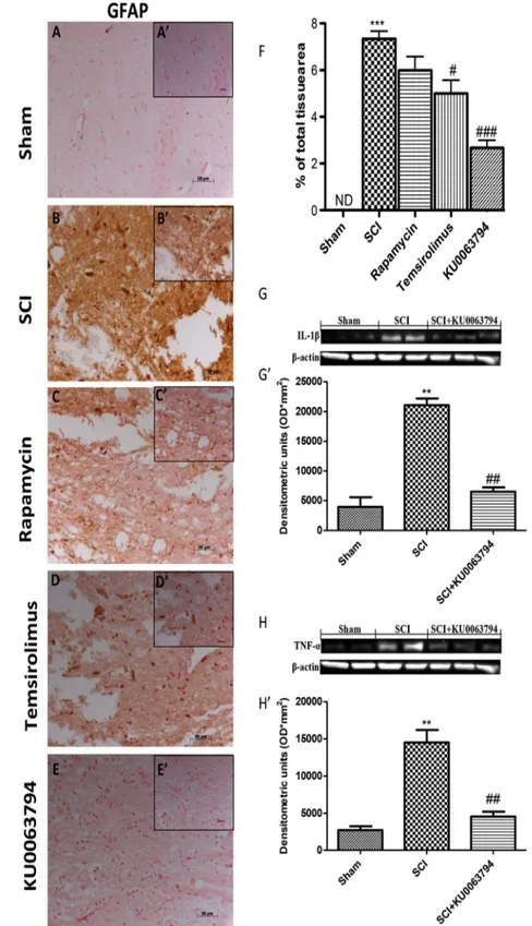
Effect of KU0063794 and Temsirolimus on Astrocyte Activation and Cytokines Production
The primary mechanical damage to the spinal cord initiates a secondary damage that includes microglia and astrocytes ac-tivation that release a large number of proinflammatory cyto-kines. To evaluate if in astrocytes, KU0063794, may induce indirectly, by activating mTORC2, the activation of p-Akt and cell survival we investigated GFAP, IL-1β, and TNF-α pro-duction, both inflammation sign. A substantial increase in GFAP expression was found in spinal cord tissues collected at 24 h after SCI (Fig.3bsee relative densitometric analysis shown in Fig.3f). Spinal activation of astrocytes was signifi-cantly attenuated in KU0063794-treated mice (Fig.3e see relative densitometric analysis shown in Fig.3f) compared to temsirolimus-treated or rapamicyn-treated mice group (Fig.3c, d see relative densitometric analysis shown in Fig.3f). Furthermore, Western blot analysis revealed a mark-edly increase of expression of IL-1β and TNF-α production in the spinal tissues collected at 24 h after SCI compared to sham group. KU0063794 treatment significantly diminished the post-SCI expression of IL-1β and TNF-α (Fig.3g, h and relative densitometric analysis shown in Fig.3g’ and 3h’). Effect of KU0063794 and Temsirolimus on Apoptosis Pathway
The connection between autophagy and apoptosis is an in-creasing area of research. The molecular relationship between autophagy and cell death are intricate, complex, and still poor-ly understood. To test whether spinal cord damage was asso-ciated with autophagy and apoptosis, we determined Bax and Bcl-2 expression by immunohistochemical staining. Immunohistochemistry for Bax and Bcl-2 showed that spinal cord sections from sham-operated mice did not stain for Bax (Fig.4a), whereas SCI-operated mice exhibited positive stain-ing for Bax (Fig.4c). Temsirolimus treatment significantly reduced the degree of positive staining (Fig.4g). However, in KU0063794-treated mice group, the decrease of positive staining is more effectively than the treatment with temsirolimus or rapamicyn (Fig.4g, i see densitometry anal-ysis K). Vice versa, spinal cord sections from sham-operated mice demonstrated Bcl-2 positive staining (Fig.4b), whereas
in SCI mice, the staining was significantly reduced (Fig.4d). Treatment with temsirolimus significantly increased the de-gree of positive staining (Fig.4d). However, in KU0063794-treated mice group the increase of positive staining for Bcl-2 is more effectively than the treatment with temsirolimus or rapamicyn (Fig.4f, hsee densitometry analysis K). In addi-tion, we investigated the expression of Fas-Ligand a mediator of apoptosis. A low basal expression of Fas-Ligand was de-tected in spinal cord samples from sham-operated mice, whereas Fas-Ligand levels were substantially increased in SCI mice (Fig.4l, see densitometry analysis 4 L’). As showed by densitometric analysis KU0063794 drastically decreased Fas-Ligand expression (Fig.4).
Ex Vivo Study
Effect of KU0063794 on Cell Viability and Nitrite (NO2−) Concentration in Spinal Cord Slices
Slices were effectively cultured for up to 7 days. Viable cells within the slices, recognized using MTT tetrazolium dye, were visualized under light microscopy. The level of cell death was evaluated in each slice at 24 h after damage. Slices subject to mechanical damage showed a significantly reduced viability compared to the uninjured group (Fig.5a). Pre-treatment with KU0063794 0.5μM, 1 h before injury, considerably reduced cell death compared to the injured group. Also, we investigat-ed the levels of nitrite liberatinvestigat-ed into the culture minvestigat-edium by Griess reagent. The untreated control group released very low levels of NO2−; instead, damage significantly enhanced the levels of NO2− production (Fig.5b). KU0063794 treatment decreased the injury-induced NO2− production.
Discussion
SCI is the consequence of an initial physical damage followed by a secondary degenerative process that leads to the obliter-ation of ascending and descending axonal tracts that control motor, sensory, and autonomic functions. SCI can be divided into a primary damage, characterized by time of impact, and secondary damage that occurs due to pathophysiologic pro-cesses that follow. In particular, among all the secondary in-jury mechanisms, the inflammatory response is the key con-tributor and results in expansion of the lesion and further loss of neurologic function [22].
Autophagy, a highly complex process that cells induced in response to a wide range of stressful conditions in order to maintain cellular homeostasis was also been connected in var-ious neuronal damage models. However, the role of autopha-gy in SCI is still controversial and its interrelationship with inflammation and apoptosis remains unclear [23].
The connection between autophagy and inflammation are complex. Each regulates the other by different mechanisms such as toll-like receptors (TLRs) and NOD-like receptor (NLRs) can elicit autophagy for pathogen clearance [24]. Moreover, recent studies have
hypotized that autophagy acts by at least two means to protect cells from excessive long lasting inflammation: indirectly by allowing efficient clearance of damaged organelles and directly by suppressing proinflammatory complexes [25].
Fig. 3 Effect of KU0063794 and temsirolimus on astrocyte activation and cytokines production. As shown in panel 3, a substantial increase in GFAP expression was found in spinal cord tissues collected at 24 h after SCI (b). Spinal activation of astrocytes was significantly attenuated in KU0063794-treated mice (e) compared to
temsirolimus-treated or rapamicyn-treated mice group (c, d). Furthermore, Western blot analysis revealed a markedly increase of expression of IL-1β and TNF-α production in spinal tissues collected at 24 h after SCI compared to sham group. KU0063794 treatment significantly diminished the post-SCI expression of IL-1β and TNF-α (g, h). The figures are representative of at least three experiments performed on different experimental days. Figures are representative of all the animals in each group. Values are given as mean ± SEM of 20 animals for each group. #P < 0.05 vs SCI group; ##P < 0.01 vs SCI group; **P < 0.01 vs sham group; ***P < 0.001 vs sham group; ###P < 0.001 vs SCI group
Previous study conducted by Chen et al. have demonstrated that treatment with rapamycin enhanced autophagy has anti-inflammatory and neuroprotective effect and improved motor function suggesting that it can be applied during the acute phase after SCI [15]. Based on these observations, we per-formed studies in the attempt to determine if the new second generation mTOR inhibitor targeting mTORC1 and mTORC2, KU0063794, could be more efficient as a novel anti-inflammatory treatment with respect to the first genera-tion mTOR inhibitor targeting only mTORC1, rapamicyn, and temsirolimus. For that purpose, we used an experimental mod-el of SCI induced by extradural compression of the spinal cord (T6-T7) using an aneurysm clip with a closing force of 24 g via a four-level T5-T8 laminectomy and using spinal cord organotypic slice culture.
In the first step, using H&E staining, we analyzed the se-verity of the trauma at the level of the perilesional area. Our results clearly established important damage in the spinal cord tissue collected from SCI animals compared with sham-operated mice. Protection against tissue damage and edema formation was observed in the group of mice treated with temsirolimus. Indeed, treatment with KU0063794 reduced histological alterations more effectively than the treatment with temsirolimus or rapamicyn.
Moreover, motor disturbance was measured every day until 10 days after SCI by the BMS score. In mice subjected to SCI and than treated with KU0063794, we detected an increased neurological recovery than temsirolimus-treated or rapamicyn-treated mice group.
Previous studies indicated an improved iNOS and peroxynitrite in the injured spinal cord. In fact, NO pro-duction can contribute to cell death, tissue damage, and degeneration observed in SCI. Moreover, high PGE2
concentrations reflecting an increased activity of cycloox-ygenase (COX)-2 in the damaged spinal cord [26, 27]. Thus, in this study, we analyzed the expression of iNOS and COX-2 by immunohistochemical staining and nNOS by Western blot analysis. Our results demonstrated that temsirolimus treatment significantly reduced the degree of positive staining, but in KU0063794-treated mice group the decrease of positive staining was more effective. Also, we assessed that the expression of nNOS was significantly restored after treatment with KU0063794.
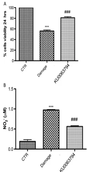
Fig. 5 Effect of KU0063794 on cell viability and nitrite (NO2−)
concentration in spinal cord slices. The level of cell death assessed in each slice at 24 h after damage showed that slices subject to mechanical damage showed a significantly reduced viability compared to the uninjured group (a). Pre-treatment with and KU0063794 1 h before injury significantly reduced cell death compared to the injured group. Moreover, the untreated control group released very low levels of NO2−; instead, damage significantly enhanced the levels of NO2−
production (b). KU0063794 treatment decreased the injury-induced NO2− production. The figures are representative of at least three
experiments performed on different experimental days. Figures are representative of all the animals in each group. Values are given as mean ± SEM of 20 animals for each group. ***P < 0.001 vs sham group; ###P < 0.001 vs SCI group
Fig. 4 Effect of KU0063794 and temsirolimus on apoptosis pathway. Immunohistochemistry for Bax and Bcl-2 showed that spinal cord sections from sham-operated mice did not stain for Bax (a), whereas SCI-operated mice exhibited positive staining for Bax (c). Temsirolimus treatment significantly reduced the degree of positive staining (g). However, in KU0063794-treated mice group, the decrease of positive staining is more effectively than the treatment with temsirolimus or rapamicyn (g, i). Vice versa, spinal cord sections from sham-operated mice demonstrated Bcl-2 positive staining (b), whereas in SCI mice, the staining was significantly reduced (d). Treatment with temsirolimus significantly reduced the degree of positive staining (d). However, in KU0063794-treated mice group the increase of positive staining for Bcl-2 is more effectively than the treatment with temsirolimus or rapamicyn (f, h). Moreover, Western blot analysis showed a low basal expression of Fas-Ligand in spinal cord samples from sham-operated mice, whereas Fas-Ligand levels was substantially increased in SCI mice (l). As shown by densitometric analysis, KU0063794 drastically decreased Fas-Ligand expression. The figures are representative of at least three experiments performed on different experimental days. Figures are representative of all the animals in each group. Values are given as mean ± SEM of 20 animals for each group. #P < 0.05 vs SCI group; ##P < 0.01 vs SCI group; **P < 0.01 vs sham group; ***P < 0.001 vs sham group; ###P < 0.001 vs SCI groupAfter SCI, reactive astrocytes contributes to the inhibitory environment within the injured spinal cord, denoted by in-creased immunoreactivity of glial fibrillary acid protein, GFAP as well as gene expression of proinflammatory cytokines and their receptors, including IL-1β, TNF-α is significantly increased [28,29]. Our results demonstrate that spinal activa-tion of astrocytes was significantly attenuated in KU0063794-treated mice compared to temsirolimus-KU0063794-treated or rapamicyn-treated mice group as well as KU0063794 treatment significant-ly diminished the post-SCI expression of IL-1β and TNF-α.
Proinflammatory cytokines stimulate other biochemical signals leading to the degeneration of myelin and apoptosis of neurons that cause the neurological deficit [30,31]. We report in this study that in KU0063794-treated mice group the decrease of positive staining for Bax was more effectively than the treatment with temsirolimus or rapamicyn, converse-ly, Bcl-2 is expressed much more in mice treated with KU0063794. Additionally, FasL signaling plays a central role in SCI [32]. We found that KU0063794 treatment lead to a substantial reduction of FasL activation.
To better understand the role of KU0063794, we used an ex vivo model of organotypic spinal cord slices cultures. This model offers the advantage of improved knowledge about the mechanism of action of a dual mTORC1 and mTORC2 inhibitor.
To validate our data in this study, using spinal cord organotypic slice cultures, we showed a significant decrease in cell death and in the injury-induced NO2−production fol-lowing KU0063794 pre-treatment.
The necessity for emerging new therapeutics for SCI treat-ment and the current scarcity of specific therapy for this indi-cation underscore the importance of connection and charac-terization of novel neuroprotective compounds. Considering that the capacity of autophagy to contain detrimental side ef-fects of inflammation might contribute to its positive role on health and proposed the possibility that targeting the autoph-agy pathway might offer a significant approach to SCI treat-ment. However, considering the multiple roles of autophagy, further investigations are needed to examine in which situa-tions stimulated autophagy is beneficial and does not generate detrimental side effects.
Acknowledgments The authors would like to thank Maria Antonietta Medici for her excellent technical assistance during this study and Mr Francesco Soraci for his secretarial and administrative assistance and Miss Valentina Malvagni for her editorial assistance with the manuscript.
This work was supported by L’Oréal award.
Compliance with Ethical Standards The study was permitted by the University of Messina Review Board for the care of animals. All animal experiments complied with regulations in Italy (D.M. 116192) as well as the EU regulations (O.J. of E.C. L 358/1 12/18/1986).
Competing Interests The authors declare that they have no competing interests.
References
1. Blesch A, Tuszynski MH (2009) Spinal cord injury: plasticity, re-generation and the challenge of translational drug development. Trends Neurosci 32(1):41–47. doi:10.1016/j.tins.2008.09.008
2. Wang W, Guo Z, Xu Z, Meng Q, Chen C, Zhang Y, Cao X (2015) Effect of pollen typhae on inhibiting autophagy in spinal cord injury of rats and its mechanisms. Int J Clin Exp Pathol 8(3):2375–2383 3. Wullschleger S, Loewith R, Hall MN (2006) TOR signaling in
growth and metabolism. Cell 124(3):471–484. doi:10.1016/j.cell. 2006.01.016
4. Betz C, Hall MN (2013) Where is mTOR and what is it doing there? J Cell Biol 203(4):563–574. doi:10.1083/jcb.201306041
5. Menon S, Manning BD (2008) Common corruption of the mTOR signaling network in human tumors. Oncogene 27(Suppl 2):S43– 51. doi:10.1038/onc.2009.352
6. Foster KG, Fingar DC (2010) Mammalian target of rapamycin (mTOR): conducting the cellular signaling symphony. J Biol Chem 285(19):14071–14077. doi:10.1074/jbc.R109.094003
7. Sparks CA, Guertin DA (2010) Targeting mTOR: prospects for mTOR complex 2 inhibitors in cancer therapy. Oncogene 29(26): 3733–3744. doi:10.1038/onc.2010.139
8. Phung TL, Ziv K, Dabydeen D, Eyiah-Mensah G, Riveros M, Pe rruzzi C, Sun J, M onahan-Earley RA et a l (2006) Pathological angiogenesis is induced by sustained Akt signal-ing and inhibited by rapamycin. Cancer Cell 10(2):159–170. doi:10.1016/j.ccr.2006.07.003
9. Sarbassov DD, Ali SM, Sengupta S, Sheen JH, Hsu PP, Bagley AF, Markhard AL, Sabatini DM (2006) Prolonged rapamycin treatment inhibits mTORC2 assembly and Akt/PKB. Mol Cell 22(2):159– 168. doi:10.1016/j.molcel.2006.03.029
10. Duran I, Siu LL, Oza AM, Chung TB, Sturgeon J, Townsley CA, Pond GR, Seymour L et al (2006) Characterisation of the lung toxicity of the cell cycle inhibitor temsirolimus. Eur J Cancer 42(12):1875–1880. doi:10.1016/j.ejca.2006.03.015
11. Garcia-Martinez JM, Moran J, Clarke RG, Gray A, Cosulich SC, Chresta CM, Alessi DR (2009) Ku-0063794 is a specific inhibitor of the mammalian target of rapamycin (mTOR). Biochem J 421(1): 29–42. doi:10.1042/BJ20090489
12. Zhang H, Berel D, Wang Y, Li P, Bhowmick NA, Figlin RA, Kim HL (2013) A comparison of Ku0063794, a dual mTORC1 and mTORC2 inhibitor, and temsirolimus in preclinical renal cell carci-noma models. PLoS One 8(1):e54918. doi:10.1371/journal.pone. 0054918
13. Syed F, Sanganee HJ, Singh S, Bahl A, Bayat A (2013) Potent dual inhibitors of TORC1 and TORC2 complexes (KU-0063794 and KU-0068650) demonstrate in vitro and ex vivo anti-keloid scar activity. J Invest Dermatol 133(5):1340–1350. doi:10.1038/jid. 2012.483
14. Kanno H, Ozawa H, Sekiguchi A, Yamaya S, Tateda S, Yahata K, Itoi E (2012) The role of mTOR signaling pathway in spinal cord injury. Cell Cycle 11(17):3175–3179. doi:10.4161/cc.21262
15. Chen HC, Fong TH, Hsu PW, Chiu WT (2013) Multifaceted effects of rapamycin on functional recovery after spinal cord injury in rats through autophagy promotion, anti-inflammation, and neuroprotec-tion. J Surg Res 179(1):e203–210. doi:10.1016/j.jss.2012.02.023
16. Lang-Lazdunski L, Blondeau N, Jarretou G, Lazdunski M, Heurteaux C (2003) Linolenic acid prevents neuronal cell death and paraplegia after transient spinal cord ischemia in rats. J Vasc Surg 38(3):564–575
17. Basso DM, Fisher LC, Anderson AJ, Jakeman LB, McTigue DM, Popovich PG (2006) Basso Mouse Scale for locomotion detects differences in recovery after spinal cord injury in five common
mouse strains. J Neurotrauma 23(5):635–659. doi:10.1089/neu. 2006.23.635
18. Shea TB (1994) Technical report. An inexpensive densitometric analysis system using a Macintosh computer and a desktop scanner. Biotechniques 16(6):1126–1128
19. Bethea JR, Castro M, Keane RW, Lee TT, Dietrich WD, Yezierski RP (1998) Traumatic spinal cord injury induces nuclear factor-kappaB activation. J Neurosci 18(9):3251– 3260
20. Esposito E, Paterniti I, Meli R, Bramanti P, Cuzzocrea S (2012) GW0742, a high-affinity PPAR-delta agonist, mediates protec-tion in an organotypic model of spinal cord damage. Spine ( P h i l a P a 1 9 7 6 ) 3 7 ( 2 ) : E 7 3–78. doi:1 0 . 1 0 9 7 / B R S . 0b013e3182276d88
21. Abe K, Matsuki N (2000) Measurement of cellular 3-(4,5-dimeth-ylthiazol-2-yl)-2,5-diphenyltetrazolium bromide (MTT) reduction activity and lactate dehydrogenase release using MTT. Neurosci Res 38(4):325–329
22. Paterniti I, Impellizzeri D, Crupi R, Morabito R, Campolo M, Esposito E, Cuzzocrea S (2013) Molecular evidence for the in-volvement of PPAR-delta and PPAR-gamma in anti-inflammatory and neuroprotective activities of palmitoylethanolamide after spinal cord trauma. J Neuroinflammation 10:20. doi:10.1186/ 1742-2094-10-20
23. Kroemer G, Marino G, Levine B (2010) Autophagy and the inte-grated stress response. Mol Cell 40(2):280–293. doi:10.1016/j. molcel.2010.09.023
24. Saitoh T, Akira S (2010) Regulation of innate immune responses by autophagy-related proteins. J Cell Biol 189(6):925–935. doi:10. 1083/jcb.201002021
25. Lapaquette P, Guzzo J, Bretillon L, Bringer MA (2015) Cellular and molecular connections between autophagy and inflammation. Mediators Inflamm 2015:398483. doi:10.1155/2015/398483
26. Xu J, Kim GM, Chen S, Yan P, Ahmed SH, Ku G, Beckman JS, Xu XM et al (2001) iNOS and nitrotyrosine expression after spinal cord i n j u r y. J N e u r o t r a u m a 1 8 ( 5 ) : 5 2 3–532. doi:1 0 . 1 0 8 9 / 089771501300227323
27. Bal-Price A, Brown GC (2001) Inflammatory neurodegeneration mediated by nitric oxide from activated glia-inhibiting neuronal respiration, causing glutamate release and excitotoxicity. J Neurosci 21(17):6480–6491
28. Hausmann ON (2003) Post-traumatic inflammation following spi-nal cord injury. Spispi-nal Cord 41(7):369–378. doi:10.1038/sj.sc. 3101483
29. Esposito E, Cuzzocrea S (2011) Anti-TNF therapy in the injured spinal cord. Trends Pharmacol Sci 32(2):107–115. doi:10.1016/j. tips.2010.11.009
30. Nesic O, Xu GY, McAdoo D, High KW, Hulsebosch C, Perez-Pol R (2001) IL-1 receptor antagonist prevents apoptosis and caspase-3 activation after spinal cord injury. J Neurotrauma 18(9):947–956. doi:10.1089/089771501750451857
31. Dong H, Fazzaro A, Xiang C, Korsmeyer SJ, Jacquin MF, McDonald JW (2003) Enhanced oligodendrocyte survival after spinal cord injury in Bax-deficient mice and mice with delayed Wallerian degeneration. J Neurosci 23(25):8682–8691
32. Ackery A, Robins S, Fehlings MG (2006) Inhibition of Fas-mediated apoptosis through administration of soluble Fas receptor improves functional outcome and reduces posttraumatic axonal de-generation after acute spinal cord injury. J Neurotrauma 23(5):604– 616. doi:10.1089/neu.2006.23.604