Dipartimento di Farmacia
PhD Program
in
Drug Discovery and Development
XXX Cycle — Academic Year 2017/2018
PhD Thesis in
Design, synthesis and biological activity of
new target selective antitumoral agents
Candidate Supervisor
Alessandra Russo Prof. Ines Bruno
II Preface
My three-year PhD course in Drug Discovery and Development at the Department of Pharmacy of Salerno University started in 2014 under the supervision of Prof. Ines Bruno. My research project was mainly focused on the design, synthesis and biological evaluation of small molecules as new modulators of novel emerging targets involved in cancer processes. Specifically, my research activity was focused on the investigation of two major targets:
the epigenetic family of Macrodomain proteins, in particular, the hydrolases MacroD1 and MacroD2;
the Bcl-2 associated athanogene 3, BAG3.
The entire work was carried out under the direct supervision of Prof. Ines Bruno and Dr. Stefania Terracciano. The Computational guided design of compounds was performed in collaboration with Prof. Giuseppe Bifulco’s research group. Biological screenings were performed in collaboration with Dr. Jon Elkins of the Structural Genomics Consortium (Oxford University), in the case of Macrodomain proteins project, whereas with Dr. Maria Carmela Vaccaro and Prof. Alessandra Rosati, in the case of BAG-3.
Furthermore, to improve my knowledge on Macrodomain proteins, in 2016 I joined Dr. Jon Elkins’ research group at the Structural Genomics Consortium where I spent five months. During that period, my research was carried out under the supervision of Dr. Romain Talon and was addressed to the expression, purification and crystallization of human MacroD1 protein, in order to perform a fragment screening, X-ray crystallography based, on the target of interest.
III
List of publications related to the scientific activity performed during the three years PhD course in Drug Discovery and Development
Papers:
S. Terracciano, A. Foglia, M. G. Chini, M. C. Vaccaro, A. Russo, F. Dal Piaz, C. Saturnino, R. Riccio, G. Bifulco and I. Bruno. “New dihydropyrimidin-2(1H)-one based Hsp90 C-terminal inhibitors”. RSC Advances, 2016, 6, 82330-82340.
Stefania Terracciano, Alessandra Russo, Maria G. Chini, Maria C. Vaccaro, Marianna Potenza, Antonio Vassallo, Raffaele Riccio, Giuseppe Bifulco, and Ines Bruno, “Discovery of new molecular entities able to strongly interfere with Hsp90 C-terminal domain”. Scientific Reports, 2018, 8, 1709, 1-11;
Simone Di Micco, Luana Pulvirenti, Ines Bruno, Stefania Terracciano, Alessandra Russo, Maria C. Vaccaro, Vera Muccilli, Nunzio Cardullo, Corrado Tringali, Raffaele Riccio, Giuseppe Bifulco. “Identification by Inverse Virtual Screening of Magnolol-Based Scaffold as New Tankyrase-2 Inhibitors”. Submitted Manuscript to Bioorganic & Medicinal Chemistry, 2018.
IV Conference proceedings:
A. Russo, I. Bruno, S. Terracciano, G. Bifulco, G. Lauro. “Design and Synthesis of potential Inhibitors of Macrodomains as new promising candidates in Cancer therapy”. European-Winter School on Physical Organic Chemistry, Bressanone (Italy), February, 1-6, 2015;
A. Russo, S. Terracciano, G. Lauro, G. Bifulco, R. Riccio, I. Bruno. “Discovery of potential modulators of Macrodomain proteins MacroD1 and MacroD2” XXXVI National Meeting of Italian Chemical Society, Organic Chemistry Division, Bologna (Italy), September 13-17, 2015;
A.Russo, S. Terracciano, G. Lauro, G. Bifulco, R. Riccio, I. Bruno.”Design, synthesis and biochemical evaluation of potential MacroD2 protein modulators” XLI International Summer School on Organic Synthesis "A. Corbella" ISOS 2016 , Gargnano (Italy), June, 12-17, 2016;
A.Russo, S. Terracciano, G. Lauro, G. Bifulco, R. Riccio, I. Bruno. “X-ray crystallography based fragments screening on Macrodomain protein MacroD1” XLII International Summer School on Organic Synthesis “A. Corbella" ISOS 2017, Gargnano (Italy), June, 18-22, 2017;
A. Russo, S. Terracciano, G. Lauro, M. C. Vaccaro, R. Riccio, G. Bifulco,
I. Bruno. “Microsomal Prostaglandin E2 Synthase-1 potential inhibitors:
design, synthesis and biological evaluation” XXVI International meeting of Italian Chemical Society, Paestum (Italy), September, 10-14, 201.
V
Table of contents
INTRODUCTION ______________________________________________ 1 -CHAPTER 1Macrodomain proteins: MacroD1 and MacroD2 ___________ 3 1.1. Drug discovery in cancer therapy _____________________________ 4 -1.2.Epigenetics _______________________________________________ 6 1.3. Main histone modifications _________________________________ 10 -1.4.ADPRybosilation ________________________________________ 11 1.5. Macrodomain containing proteins ____________________________ 15 1.6. Macrodomains structure and functions ________________________ 19 1.7. Human MacroD1 and MacroD2 proteins _______________________ 24 1.8. Macrodomains in Cancer ___________________________________ 28 -CHAPTER 2BAG3 (Bcl2associated athanogene 3) antiapoptotic protein 31 2.1. BAG (Bcl2 associated athanogene) proteins family ______________ 32 2.2. Human BAG Proteins: BAG1, BAG2, BAG4, BAG5, and BAG6 ___ 33 2.3. BAG3 protein ____________________________________________ 35 2.4. BAG3 in normal cells _____________________________________ 38 -2.5.BAG3 in cancer cells ______________________________________ 40 -2.6.Targeting Hsp70BAG3 interaction ___________________________ 46 -2.7.The workflow of the projects ________________________________ 49 RESULTS AND DISCUSSION __________________________________ 51 -CHAPTER 3 Discovery of new modulators of the human Macrodomain protein MacroD2 by a structurebaseddrug design approach __________________ 53
3.1. Targeting MacroD2 protein _________________________________ 54 -3.2.Structure based virtual screening _____________________________ 54
-VI
3.3.Synthesis and biochemical evaluation _______________________ 57 -CHAPTER 4 Discovery of new modulators of human Macrodomain protein MacroD1 by a fragment screening Xray crystallography based approach _ 81
4.1. Targeting MacroD1 protein _________________________________ 82 4.2. MacroD1 overexpression in E. coli ___________________________ 82 -4.3.MacroD1 protein crystallization _____________________________ 84
4.4.Xray crystallography based fragment screening on MacroD1 protein 85 -4.5.Compound 3 chemical exploration ___________________________ 97 -CHAPTER 5 Design, synthesis and biological evaluation of the first BAG3 modulator as an attractive candidate for the development of a new class of chemotherapeutics ____________________________________________ 103
5.1. Targeting BAG3Hsp70 protein protein interaction _____________ 104 -5.2.Structure based drug design ________________________________ 104 -5.3.Biological evaluation of the selected compounds 126 ___________ 109 -5.4.Synthesis of compounds LK1LK17 _________________________ 114 -5.5.Biological evaluation _____________________________________ 117 CONCLUSIONS _____________________________________________ 125 EXPERIMENTAL SECTION _________________________________ 129 -CHAPTER 6 Discovery of new modulators of the human Macrodomain protein MacroD2 by a structure-based-drug design approach: Experimental procedures ______________________________________ - 131 -
6.1. General synthetic methods_________________________________ 132 -6.2.Methods and materials ____________________________________ 133 -CHAPTER 7 Discovery of new modulators of human Macrodomain protein MacroD1 by a fragment screening X-ray crystallography based approach: Experimental procedures _______________________________________ 155
-VII
7.2.Fragment screening general methods _________________________ 159 -7.3.SPR General methods _____________________________________ 163 -7.4.General synthetic methods _________________________________ 164 -7.5.Methods and materials ____________________________________ 165 -CHAPTER 8Design, synthesis and biological evaluation of the first BAG3 modulator as an attractive candidate for the development of a new class of chemotherapeutics: Experimental procedures _______________________ 171
-8.1General synthetic methods _________________________________ 172 -8.2Methods and materials ____________________________________ 173 -8.3SPR General methods _____________________________________ 188 APPENDIX _________________________________________________ 191 References __________________________________________________ 197 List of Abbreviations _________________________________________ 214
-VIII Abstract
Cancer development is a complex pathological process that exploits a variety of biological actors. The identification of new molecular entities able to interfere with new biological targets involved in tumorigenesis is strongly needed, both for the development of new promising drug candidates, and as chemical probes useful to further investigate less understood biological aspects. Two main targets, involved at different levels in cancer development, have been thoroughly investigated: the epigenetic Macrodomain proteins, MacroD1 and MacroD2, and the Bcl-2 associated athanogene 3, BAG3 protein. The results obtained are summarized in the two main sections, reported below according to the target of interest:
a) Discovery of new modulators of human Macrodomain proteins, MacroD1 and MacroD2, by structure-based and X-ray crystallography based approaches.
MacroD1 and MacroD2 are two orthologue members of the epigenetic family of the Macrodomain containing proteins which have been recently identified as attractive targets for the treatment of cancer, due to their well-established overexpression in several human tumors.1-2 These proteins can act as erasers of the
histone code ADP-rybosilation, a post-translational modification involved in the modulation of gene expression and chromatin remodelling.3 With the aim of
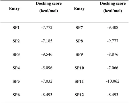
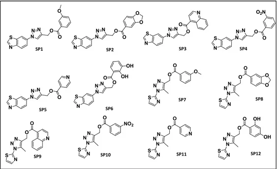
identifying new modulators of these high related proteins, we carried out two different drug discovery approaches: a computer aided structure based drug design on the MacroD2 crystal structure (PDB: 4IQY)and a Fragment screening, based on X-ray crystallography, on the protein MacroD1. Concerning the first approach, starting from the crystal structure of MacroD2 protein in complex with ADP-ribose, its natural ligand, we performed a preliminary virtual screening on a Database of 16 million of 1,4 disubstituted triazoles. Results analysis allowed us to select the most promising molecules in terms of docking score and shape similarity. The next step consisted in the synthesis of the most promising molecules basing on a versatile and suitable synthetic strategy. The synthetized molecules were, then, tested in collaboration with the Structural Genomics Consortium of Oxford, to evaluate their ability to bind the target protein with Alpha Screen, Biolayer interferometry and
IX
Isothermal titration calorimetry. These biophysical methods allowed us to disclose compound SP2 as a real binder of the protein MacroD2, with a dissociation constant of 2.54±1.1 M. This promising molecule will be further investigated on MCF-7 cancer cells, overexpressing the protein, to assess its potential antitumoral activity.
Concerning the study of MacroD1 protein, a fragment screening based on X-ray crystallography technique has been carried out, during my stage experience at the University of Oxford; this advanced method allowed the identification of three fragments co-crystallized with the protein MacroD1. Since in the Surface Plasmon Resonance (SPR) assay the binding to the protein was confirmed for two of the three fragments, we decided to start to investigate, in silico, the binding mode of the most promising one, compound 3, in order to develop a collection of high affinity binders for the target protein. These new molecules have been synthesized and then tested again by SPR, against the protein MacroD1 and, in line with the computational predictions, four of them showed to bind the target protein with higher affinity, compared to the lead compound; these results are of great interest since so far no Macrodomain binder has been yet disclosed, hence they can open the way to the discovery of new chemical platforms able to modulate the protein MacroD1, as new attractive candidates for drug development.
b) Design, synthesis and biological evaluation of the first BAG3 modulator as an attractive candidate for the development of a new class of chemotherapeutics.
BAG3 (Bcl-2-associated athanogene 3) is a multidomain protein which, through its BAG domain, is able to interact with several partners, modulating, thus, key signalling pathways involved in physiological and pathological processes.4 Indeed,
BAG3 has been shown to sustain cell survival and to induce resistance to chemotherapy in human cancers, hence, it is recently emerged as a therapeutic target of human malignancies.5 With the aim of exploring BAG3 protein, basing on
a combined approach of structure-based drug design and biophysical methods, we screened a huge library of commercially available molecules against the target of interest. Starting from the virtual screening and SPR results, we selected a 2,4
X
thiazolidinedione based molecule (7), as a promising BAG3 activity modulator. This compound, indeed, showed to bind with a good affinity both to the full length protein (KD: 5.2±3.8 nM) and to its isolated BAG domain (KD: 3.51±2.7 nM),
moreover it did not show any binding for two other members of BAG proteins tested, BAG1 and BAG4. Hence, we decided to evaluate the potential antiproliferative activity of the disclosed hit on A375 melanoma cancer cells, which are known to overexpress the BAG3 protein; compound 7 resulted to have a good cytotoxicity (25±1.5 M) against the cell line tested, in line with our predictions. Starting from these promising outcomes, we developed a collection of synthetic 2,4-thiazolidindiones, as derivatives of the lead compound, in order to expand the chemical diversity around the scaffold and we succeeded to identify a promising molecule (LK-4); this compound, indeed, was able to selectively bind BAG domain of BAG3 protein, with high affinity (KD: 6.4±2.2 nM), interfering with
BAG3-Hsp70 protein-protein interaction. LK-4 showed a high cytotoxicity (IC50: 16±1.5
M) against A375 melanoma cancer cells, and at the same time, a good selectivity; moreover, it did not affect the cell viability of PMBC human normal cells. A co-immunoprecipitation assay confirmed that LK-4 interfere with BAG3-Hsp70 complex formation in cell and in a time-dependent manner, representing a valuable chemical probe to further investigate BAG3 protein in the attempt to develop new attractive protein modulators.
- 1 -
- 3 -
CHAPTER 1
- 4 - 1.1. Drug discovery in cancer therapy
"Cancer" is a generic term used to describe a large group of related diseases that involve an abnormal cell growth with the potential to spread to other parts of the body. According to the World Health Organization (WHO), more than one hundred types of cancer have been disclosed so far, and owing to the great variety of histological types, and, overall, to its multifactorial etiology, it is a particularly difficult disease to treat. The challenge for a medicinal chemistry project is to design new drugs able to selectively target cancer cells, while avoiding multidrug resistance pathways. In the last decades, the many efforts lavished in this research area provided valuable insights into cancer physiopathology with the disclosure of strategic biological targets which can be of great help for the development of potent and selective chemotherapeutics. These achievements have been made possible thanks to multifaceted approaches requiring the synergistic contribution of several scientific figures, e.g., clinicians, biologists, medicinal and synthetic organic chemists, X-ray crystallographers and other structural biologists, chembioinformaticians, computational experts, and logicians, among others. This tireless joint research provided a great improvement of the technological tools employed in drug discovery programs (Figure 1.1). For example, once the molecular target has been selected, the advanced computer-aided approaches provide a fast and cost-efficient lead identification. Moreover, such computational programs are useful also to predict whether the designed molecules are likely to display the desired ADMET (absorption, distribution, metabolism, excretion, and toxicity) properties. In addition, on one side, the progress made in Organic synthesis provides synthetic procedures that allow to rapidly generate a wide structural variety of products, on the other side, the introduction of innovative biophysical and biological techniques enables the rapid screening of a great number of compounds, supplying information useful to outline a SAR profile of the most promising molecules. Basing on these premises, my PhD research project has been focused on the development of new modulators of novel biological targets involved in tumorigenesis. In particular, in the frame of the research lines carried out by the Organic Chemistry group of the Department of Pharmacy, University of Salerno, I decided to investigate two interesting targets such as: the human Macrodomain
- 5 -
proteins MacroD1 and MacroD1 and the Bcl2-associated athanogene 3 (BAG3). The MacroD family proteins are considered epigenetic enzymes whose overexpression, in various cell lines, has been shown to protect against multiple apoptotic signals such as: chemical, biological or physical stimuli. After DNA damage, Macrodomain proteins can inhibit apoptosis by modulating chromatin remodeling activity, through protein ADP-rybosilation, and facilitate DNA repair within a chromatin context.6 The other target under investigation is the BAG3
protein, a member of BAG family, recently emerged as a key regulator of important physiological processes including cell survival, apoptosis, cytoskeleton organization, and autophagy7; through its well conserved domain, BAG3 has also
been shown to collaborate with Hsp70 in regulating cancer development through multiple pathways.8
Moreover, during my PhD research project, I had the opportunity to go to spend a research period at the Structural Genomics Consortium, in Oxford, where I experienced the innovative technologies of the synchrotron of the Diamond Light Source which allowed to expand my knowledge in the protein crystallization and fragment screening processes.
- 6 -
Figure 1.1 Flow chart of the Drug discovery process
1.2.Epigenetics
Epigenetics is a genomic branch defined as the study of the structural adaptation of chromatin to exogenous signals; in more detail, it includes all the chromosomal modifications associated with both, DNA repair, or cell-cycle phases, and stable changes maintained across multiple cell generations. In simple words we can say that the epigenetic mechanisms are all the inheritable changes in gene expression with no alterations in DNA sequences.9 The term epigenetics was first introduced
by Conrad Waddington, however many further definitions have been given from 1942 to date. Chemical modifications of DNA and histones are dynamically laid down and removed by chromatin-modifying enzymes in a highly regulated manner (Figure 1.2). Four different DNA modifications10 and 16 classes of histone
modifications have been at least well elucidated.11,12 These modifications can alter
chromatin structure by modifying noncovalent interactions within and between the nucleosomes. They also serve as docking sites for specialized proteins with unique
- 7 -
domains, the so called “chromatin readers”, that specifically recognize these modifications, and recruit additional chromatin modifiers and remodeling enzymes, which, in turn, act as the effectors of the modification. The information conveyed by the epigenetic modifications plays a critical role in the regulation of all DNA-based processes, such as transcription, DNA repair, and replication. Consequently, abnormal expression patterns or genomic alterations in chromatin regulators can have profound effects and can lead to the induction and maintenance of various cancers. Hence, disruption of the epigenome is a well-recognized fundamental mechanism in cancer, and several epigenetic drugs have been proven to modulate cell survival and to be less toxic than conventional chemotherapy.13 The great
interest evoked by this research area prompted many investigations in the attempt of understanding and clarifying several issues, however, although significant advances have been done in this field, many questions remain still unsolved.
Figure 1.2 Epigenetic enzymes involved in regulation of gene expression
DNA methylation
DNA methylation is a widespread modification in bacteria, plants and mammals; this covalent modification is natural in DNA; it is produced during DNA replication and it is considered as a stable gene-silencing mechanism. In eukaryotic cells DNA methylation takes place at the 5' end of the cytosine nucleotide followed by a
- 8 -
guanine nucleotide (CpG dinucleotide), and requires S-adenosyl-methionine as methyl donor. This reaction is catalyzed by the DNA methyltransferase enzymes family (DNMT family), including DNMT1, DNMT3A and DNMT3B. Cancer-associated DNA hypomethylation is as prevalent as cancer-linked hypermethylation, however, these two types of epigenetic abnormalities, usually, seem to affect different DNA sequences. It has been suggested that tumor-associated DNA hypermethylation contributes to carcinogenesis separately from aberrant DNA hypomethylation14, and it has been proved to silence tumor
suppressor genes; this kind of aberration has been mostly found in CpG-rich 5’ gene regions.15,16 However, the understanding of cellular consequences of normal and
aberrant DNA methylation remains a key area of interest, nevertheless, so far, hypomethylating agents represent one of the few epigenetic therapies that have gained FDA approval for routine clinical use.17
Histone modifications
Histones including H2A, H2B, H3 and H4 form, together, the histone octamer that is the basic structure of the nucleosome components.18 Chromatin or histone
components are prone to a wide variety of covalent, reversible, post translational modifications, such as acetylation, mono-, di-, and trimethylation on lysine residues, symmetric or asymmetric mono- and dimethylation on arginine residues, phosphorylation on serine and threonine residues, ubiquitination, biotinylation, and SUMOylation (Small Ubiquitin-like Modifier or SUMO) on lysine residues, and finally mono-ADP-ribosylation on arginine and glutamate residues (Table 1.1). Although many examples of modifications within the central domains of histones have been identified, the majority of these post-translational modifications occur on the lysine amino-tails, due to their protruding position from the nucleosome core. These modifications, individually or in combination, are able to influence inheritable epigenetic programs that encode distinct nucleosome functions, such as gene transcription, X-chromosome inactivation, heterochromatin formation, mitosis, and DNA repair and replication.19,19b Mechanistically, these functions are
- 9 -
indirectly, by recruiting effector proteins that, with specific modules, recognize particular histone modifications in a sequence-dependent manner. In addition to their catalytic functions, many chromatin modifying factors also possess ‘‘reader’’ domains, allowing them to bind to specific regions of the genome and respond to the information conveyed by upstream signaling cascades. The amino-acidic residues that line the binding pocket of the reader domains can dictate a particular preference for specific modification states, whereas, residues outside the binding pocket, contribute to determining the histone sequence specificity. The basis underlying these epigenetic codes resides in the substrate specificity both, of the enzymes that catalyze the several covalent modifications, and of the enzymes that remove these marks to reverse the modifications. Given that chromatin is the physiological template for all DNA-mediated processes, it is not surprising that histone modifications represent an essential component in controlling the structure and/or function of the chromatin, with different modifications yielding distinct functional consequences. Indeed, site-specific histone modifications have shown to correlate with particular biological functions such as gene transcription, chromatin remodeling and apoptosis regulation (Figure 1.3).20
- 10 - 1.3. Main histone modifications
Histone methylation
Lysine methylation of histones H3 and H4 is implicated in both transcriptional activation and repression, depending on the methylation site, while, arginine methylation promotes transcriptional activation. Lysines can be either mono-, di- or tri-methylated, providing functional diversity to each site of methylation. The most extensively studied histone methylation sites include histone H3 lysine 4 (H3K4), H3K9, H3K27, H3K36, H3K79 and H4K2021, but there are also arginine
sites of methylation including H3R2, H3R8, H3R17, H3R26 and H4R3. Several studies demonstrated that histone methylation plays an important role at different levels of the transcriptional regulation, through the recruitment of cell-specific transcription factors and the interaction with initiation and elongation factors.22
Histone acetylation
Histone acetylation is an epigenetic mark often associated with an open chromatin structure. This makes chromatin accessible to transcription factors and can significantly increase gene expression. Histone acetylation is largely present at DNA promoter regions. For example, acetylation of K9 and K27 on histone H3 (H3K9ac and H3K27ac) is normally associated with an increase of active genes. However, acetylation low levels are also found throughout transcriptionally active genes, and for this reason, this issue is still under debate. Histone acetyltransferases (HAT) and Histone deacetylases (HDACs) are the enzymes responsible for writing and erasing the acetylation tag on the histone tails. This mechanism has shown to regulate the dynamic chromatin plasticity, and actually, lysine residues within histone H3 and H4 showed to be the preferential targets for HAT complexes.23
- 11 - Histone phosphorylation
Phosphorylation of histone cores is a critical intermediate step in chromosome condensation during cell division, in transcriptional regulation, and in DNA damage repair processes. Unlike acetylation and methylation, histone phosphorylation seems to function by establishing interactions between other histone modifications and serving as a platform for effector proteins, leading to a downstream cascade of events. Phosphorylation of histone H3 at S10 (H3phosphoS10) and histone H2A on T120 are mitotic markers: these modifications, in fact, are involved in chromatin compaction and in chromatin function regulation during mitosis. Phosphorylation of H2AX at S139 (resulting in γH2AX) has been identified as one of the earliest events occurring after DNA double-strand breaking and serves as a recruiting point for DNA damage repair proteins.24 Actually, histone phosphorylation plays a wide
range of roles: H2B phosphorylation, that have been the focus of extensive investigations, for example, facilitates apoptosis-related chromatin condensation, DNA fragmentation, and cell death.25
1.4. ADP-Rybosilation
ADP- rybosilation, firstly described in 1960’, is a reversible post-translational modification (PTM) of proteins, resulting in the covalent attachment of a single ADP-ribose unit [i.e., mono(ADP-ribose) (MAR)] or polymers of ADP-ribose units [i.e., poly(ADP-ribose) (PAR)] on a variety of amino acid residues on target proteins.26 ADP-ribosylation reactions are phylogenetically ancient and can be
divided into four major groups: mono-ADP-ribosylation, poly-ADP-ribosylation, ADP-ribose cyclization, and formation of O-acetyl-ADP-ribose. When this modification occurs, an ADP-ribose moiety of NAD is transferred to a specific amino acid of an acceptor protein on the histone tails with the consequent release of nicotinamide.27,28 The reaction can occur through both enzymatic and
non-enzymatic mechanisms.29 The enzymatic type is mediated by diverse groups of
ADP-ribosyl transferase (ADPRT) enzymes, which use ADP-ribose units, derived from β-nicotinamide adenine dinucleotide (NAD+), to catalyze the
ADP-- 12 ADP--
ribosylation reaction. To date there are 22 human gene products possessing ADP-ribosyltransferase activity; these enzymes include bacterial ADPRTs (e.g., cholera toxin and diphtheria toxin) as well as members of three different protein families in yeast and animals: (1) arginine-specific ecto-enzymes (ARTCs), (2) sirtuins, and (3) PAR polymerases (PARPs).30 This modification has been shown to regulate
several cellular functions via different mechanisms. For example, rybosilation of protein substrates can affect protein-protein interactions, the factors repairing recruitment to DNA damage sites, DNA repair processes, moreover, rybosilation of target proteins can also facilitate their ubiquitination, promoting protein degradation via proteasomal pathways. Basing on these considerations and on other growing mass of evidences, ADP-related pathways have been recognized to be implicated in a wide range of cellular processes like transcription, chromatin remodeling, cell proliferation, apoptosis and cancerogenesis.31,32
- 13 - Mono-ADP-rybosilation
Mono-ADP-ribosylation is a post-translational modification originally identified as the pathogenic mechanism of several bacterial toxins. This modification is catalyzed by ADP-ribose-protein transferases (MARTs), whereas, mono-ADP-ribose-protein hydrolases (MARHs), are the enzymes able to reverse the reaction by hydrolyzing the protein-ADP-ribose bond.34,35 The simultaneous
presence of both mono-ADP-ribosyltransferase and mono-ADP-ribose-protein hydrolase activities, in the same cell, suggests that the reversible protein mono-ADP-ribosylation represents a regulatory mechanism for the protein substrates.36 It
is known that mono-ADP-ribosylation occurs at different amino acid residues levels, according to the specificity of the individual MARTs. ADP-ribosylation of histones is thought to be linked to DNA repair processes and cell proliferation. When cells are exposed to damage by OH radicals or methylating/alkylating agents, the total covalent mono-ADP-ribosylation of histones increases by a factor from 2 to 12, while the levels of histone H1-ADP-ribosylated are even more higher (more than 30-fold).37,38 Mono-ADP-ribosylation on H4 seems to occur preferentially
when H4 is hyperacetylated39, suggesting a potential cross talk of histone
mono-ADP-ribosylation and histone acetylation. The amino acid residues of the acceptor proteins that are modified by the specific MARTs include arginine, asparagine, glutamate, aspartate and cysteine.34,40 Conversely, mono-ADP-ribosylation of
cellular proteins through non-enzymatic mechanisms mainly occurs on lysine or cysteine residues.41Amino acid-mono-ADP-ribose-specific MARHs cleave the
ribose unit, leading to the release of free mono-ADP-ribose and to the restoring of the free reactive group on the corresponding amino acid residue.42 Many evidences
suggest that mono-ADP-ribosylation, along with other modifications of histone tails, may regulate several steps in DNA damage response pathways: for example mono-ADP-ribosylation could act, in cooperation with acetylation and phosphorylation, as a DNA damage signal to recruit additional signaling factors and chromatin modifiers.
- 14 - Poly-ADP-rybosilation
Poly-ADP-ribosylation, as enzymatic reaction, is known since the early sixties of the last century.43 In the following 20 years, this post-translational modification has
been related to several nuclear functions, i.e. histone modification44,
differentiation44, cell death45, transcriptional regulation46 and DNA repair/genome
stability. In the enzymatic reaction NAD+ is cleaved into nicotinamide and
ADP-ribose, with the latter attached to glutamate or aspartate via an ester bond47, or to
lysine, forming a ketoamine by a first Schiff-Base formation, followed by an Amadori rearrangement.48 After attachment of the first ADP-ribose moiety, further
units are rapidly added via α-gylcosidic bonds, branches can originate from the growing chain, depending on the synthesizing enzyme and the interaction partners. These reactions are catalyzed by Poly-ADP-rybosil-transferases enzymes (PARPS); this enzymes family consists of 17 members with distinct cellular localizations and functions.26 As well as for Mono-ADP-rybosilation, also
Poly-ADP-rybosilation is a reversible mechanism, indeed, PAR polymers turn over rapidly in the cell49. The enzymes that act as “erasers” of this epigenetic mark have
evolved to remove covalently linked ADP-ribose and PAR from proteins (Figure 1.4). These enzymes include PAR glycohydrolase (PARG), TARG/C6orf13050,
MacroD1 and MacroD251,51b and the NUDIX family of hydrolases.52 Many of these
enzymes contain a macrodomain fold that allows them to interact with ADP-ribosylated substrates. Based on the large size of poly-ADP-ribose, this modification seems to play a direct role in the “histone code”. In addition, poly-ADP-ribosylation was suggested to indirectly contribute to the “histone code” by dictating the levels of local chromatin compaction.
- 15 -
Figure 1.4 Representative scheme of Mono and Poly-ADP-rybosilation
1.5. Macrodomain containing proteins
As previously mentioned, several enzymes are involved in the regulation of the ADP-rybosilation cellular pathway. Depending on their activity, these enzymes can be classified as writers (e.g. if the enzyme adds an ADP-ribose moiety), erasers (e.g. if the enzyme removes an ADP-ribose group) or readers (if the enzyme is able to “read or detect” the presence or the absence of the ADP-ribose mark) (Table 1.2). Despite these different functions, all these proteins are characterized by the presence of an evolutionarily conserved structural domain of 130-190 a.a. and they are not only found in vertebrates but also in many bacteria, viruses and plants, suggesting their evolutionary conservation.53 The first macrodomain was identified,
through genomic sequencing (initially termed X domain), as “a domain of considerable conservation” within the genomes of the murine hepatitis virus (MHV) and infectious bronchitis virus.54 Shortly thereafter, a homologous domain
was identified as part of the rat MacroH2A protein, a histone variant that consists of a fusion between histone H2A and a domain of unknown function.55 Because
MacroH2A was the largest histone variant, the novel domain was defined macrodomain. In contrast to many other modification recognition domains, which
- 16 -
are adapted to recognize a single or a small number of modification types, macrodomains can recognize ADP-ribose (ADPr) in both its free and protein-linked forms, in related ligands, such as O-acyl-ADP-ribose (AAR), and even in ligands unrelated to ADPr.56 The macrodomain containing proteins exert regulatory
influence on inter- and intracellular signalling, transcription, DNA repair pathways, maintenance of genomic stability, telomere dynamics, cell differentiation and proliferation, as well as on necrosis and apoptosis.57
- 17 -
- 18 -
Humans contain 10 genes encoding for 11 members of the macrodomain family, which includes macroH2A (and its various isoforms including macroH2A1/macroH2A2), MACROD1 (LRP16), MACROD2 (C20orf133), C6orf130, MACROD3 (GDAP2), ALC1 (CHD1L, CHDL), and macroPARPs (PARP-9; PARP-14; PARP-15)6 (Figure 1.5). All of these proteins contain a macro
domain near their N-terminus or C-terminus domains, except for macro-PARPs in which 2–3 macro domains are linked. In addition to the conserved macrodomain, these proteins also contain a variety of additional domains, which allow them to interact with specific target proteins or target them to specific chromatin structure regions. For example, macroPARPs also contain a PARP catalytic domain and are the only described proteins with both a PARP-like domain and a macrodomain. Basing on an accurate analysis of the topology of macrodomain proteins, composed by diverse domains ecompassed by N- and C-terminal tails, it is possible to grasp the important and intricate role of these proteins in the regulation of diverse cellular functions. The macrodomain proteins might be viewed as molecular bridges that bring together the target proteins, via interactions with the variable domains, and the metabolites of NAD+, including PAR, via binding to the conserved macro
domain.58
- 19 - 1.6. Macrodomains structure and functions
Three-dimensional (3D) structures of the ADP-ribose (ADPr) binding fragments of macrodomains have been recently solved; this allowed a comparison with the previously published structures of members of the macrodomain family, providing additional evidence of the high structural similarity inside this protein class.58 As
revealed by structure determination, macrodomains adopt a globular α/β/α sandwich fold composed of a central six-stranded mixed β-sheets, flanked by five α-helices (Figure 1.6a)3,58, while the substrate binding occurs via a deep cleft on
the crest of the domain. The macrodomain fold shares some resemblance to the DNA-binding domain of leucine aminopeptidases, as well as to the P-loop nucleotide hydrolase. The stable interaction between the ligand and the macrodomain can trigger a variety of downstream effects, including recruitment to DNA damage sites (hot spots of PAR generation) or formation of protein complexes.59 Although there is a relatively high degree of sequence similarity
(approximately 30–40%) among the macrodomain proteins family members6, the
small sequence variation between the domains is probably responsible for the selectivity of the different macrodomains for specific binding partners. Indeed, further structural and biochemical characterization showed that ADPr and its derivatives can be accommodated within the cleft (Figure 1.6 b-c).60 The
interaction between ligand and macrodomain is stabilized by several conserved interactions within the binding pocket: (a) the adenosine moiety readily undergoes π-π stacking with a conserved aromatic residue, whereas its N6 nitrogen is further coordinated by an aspartate residue;56,61 (b) the central part of the cleft stabilizes the
substrate binding by several side-chain/backbone-pyrophosphate contacts, which induce a more closed conformation of the macrodomain;32 (c) The pyrophosphate
and the distal ribose are accommodated between two substrate-binding loops (termed loop 1 and 2). Although both loops contribute to substrate specificity, loop 1 harbors the catalytic residues of most macrodomains, exhibiting hydrolase activity (for this reason it has been also termed catalytic loop). Loop 2 provides further coordination of the pyrophosphate and it is also described as the diphosphate-binding loop48,62 (Figure 1.6c).
- 20 -
Figure 1.6(a)Topological representation of the macrodomain shows the organization of the central six-stranded β-sheet (red) flanked on both sides by five α-helices (green);(b)MacroD2,
PDB 4IQY, (green) coordinates ADPr in a strained conformation owing to the presence of a structural water molecule (dark blue) and Tyr190 (loop 2). The catalytic residues Asn92 and Asp102 (loop 1) interact electrostatically with the distal ribose; (c)The magnification shows the coordination of the adenosine moiety by a conserved phenylalanine and/or asparagine residue as
well as by the substrate-binding loops 1 and 2 (red)
Macrodomain proteins are ubiquitously expressed in adult tissues, however, the physiological role of these proteins is not yet completely understood. Among the mammalian macrodomain proteins, only the potential role, in human embryogenesis, of macroH2A and the macroPARPs have been investigated. The role of macroH2A in fetal development is better characterized than that of other macrodomain proteins, actually because macroH2A was the first of these proteins to be described and the most intensively studied.63 A driving factor for the role
played by macrodomains in evolution may lie in the key role of NAD signaling/consumption processes, regulating DNA repair, redox defense, chromatin architecture, protein acylation, and response to viral infection, among others.64,65 It
has been well demonstrated that macrodomain proteins regulate PARylation, whereas it is emerging that other proteins control MARylation. PAR recognition is a well-established ability of several macrodomains. In particular, PARylation, in response to DNA damage, is sensed by several macrodomains which serve as recruitment modules for proteins involved in DNA repair. In contrast to the binding modules that have been well characterized for PAR, it remains to address
- 21 -
whether macrodomains are also involved in reading and processing MARylation.66
Macrodomains as readers of Protein ADP-Ribosylation
Macrodomains are key players in the complex network of NAD-dependent signaling. This is a consequence of their ability to interpret, not only protein ADP-ribosylation and PARP-dependent signaling, but also second messengers such as ADPr and its derivatives, which can be released apart from PARP activity (e.g., through sirtuin activation). According to their multiple activities, humans’ reader macrodomains only occur in multidomain proteins combining, thus, signals recognition and effector domains in a single polypeptide, as discussed below. Among the macrodomain proteins that recognize and bind ADPr, there is macroH2A, ALC1 and MacroPARPS. Because a single ADPr fits into a macrodomain cleft, it has been suggested that macrodomains specifically bind MARylated proteins, even if they can also interact with the PARylated ones, and that the backbone sequence around the MARylation site may determine the substrate specificity of macrodomains, as in the case of bromodomains that interact with acetylated proteins.67 Among the main readers of the ADPr pathway there are:
macroH2A, that has been associated with several cellular processes, including cell differentiation and proliferation, transcription repression, and DNA repair. Moreover, a reduced macroH2A expression was observed in several cancer types, including breast and lung cancer, and has been associated with an increased tumor proliferation and metastatic potential. ALC1 is frequently amplified in certain cancer types like hepatocellular carcinoma and bladder cancer, as well as macroPARPs.68 Three human members of the PARP family contain multiple
macrodomains able to recognize ADPr, in addition to their PARP catalytic domain. A dysregulation of both PARP9 and PARP14 are often associated with cancer, as well as with lymphoma.48
- 22 - Macrodomains as Erasers of Protein ADP-Ribosylation
Like other signal transduction pathways, ADPr-dependent signaling is a reversible modification, therefore it requires both recognition and removal of the mark. Basing on this point, it may not be surprising that macrodomains, in addition to their reader’s activity, have acquired the ability to reverse cellular ADP-ribosylation. The catalyzed signal termination reactions include the hydrolysis of mono- and poly-ADP-ribosylated substrates, as well as degradation of NAD+- derived second
messengers, such as O-acetyl-ADP-ribose (AAR).53,69 PARGs
(Poly-ADP-Ribosyl-Glycohydrolases) are enzymes able only to breakdown Poly-ADP-ribose chains, however, they are unable to remove the final ADP-ribose moiety from the protein. Few years ago, Jankevicius et al51a succeed to identify, through biochemical,
structural and modeling analysis, some macrodomain proteins members able to reverse mono-ADP-rybosilation cellular glutamate. These two proteins, called MacroD1 and MacroD2, are orthologues of MacroD-type proteins and can be found in all living organisms. In vertebrates a duplication of the ancestral MacroD-type gene gave rise to MacroD1 and MacroD2 proteins.51a MacroD1 and MacroD2 act
as mono-(ADP-ribosyl) hydrolases that reverse protein mono-(ADP-ribosylation) and catalyze the cleavage of the terminal ADPr moiety, e.g., from proteins after PARG-mediated polymer degradation.51a In addition, both enzymes can hydrolyze
AAR3. Although their catalytic activities have been established in vitro, their exact
protein targets and biological roles remain largely unknown. There is a high degree of sequence similarity between the catalytic domains of MacroD1 and MacroD2, however, their primary subcellular localizations are different (MacroD1 in mitochondria and MacroD2 in the cytoplasm), implying, thus, distinct functions.51a
MacroD1 and MacroD2 as Mono-ADP-Ribosyl-Hydrolases
To date, only two classes of macrodomains have been shown to contain mono-ADP-ribosyl-hydrolases activities: the MacroD-type class and the ALC1-like class. Biochemical and structural studies on MacroD1, MacroD2 and TARG1, the
- 23 -
members found in humans, revealed that these classes utilize different catalytic mechanisms.20
In MacroD1/2, the distal ribose is bound in a constrained conformation, oriented toward the α-phosphate group (Figure 1.6b). This orientation is maintained through the presence of a highly conserved aromatic residue that is part of the bipartite MacroD-type structure: two glycine-rich loops (loop 1, 97-GGGGV-101 and loop 2, 188-GIYG-191)51a. The major difference between the reading and erasing
domains is the presence of a groove, in the pyrophosphate-binding site, in which there is a structural coordinated water molecule. Indeed, thanks to the analysis of site specific mutants of protein MacroD1 and MacroD2, it has been possible to disclose the high conserved amino acids that are crucial for the catalytic mechanism; in particular the key residues are: Asn-171, Asn-174, Asp-184 and His-188 for MacroD1, whereas Asn 92, Asp 102 and His 106 for MacroD2. Basing on these data, two models for the catalytic mechanism have been proposed. Even if the exact process is still unclear, according to a first hypothesis, a water molecule, located in the cleft between loop 1 and 2 and held by hydrogen bonds between the distal ribose and the neighboring ADPR α-phosphate, becomes activated through the α-phosphate group and carries out a nucleophilic attack on the protein-ADPr ester bond. In this frame, the constrained conformation of the substrate appears to be crucial for the catalysis, however, is still debated if the pKa of the α-phosphate is sufficient to activate the water molecule.51a Another hypothesis suggests that a
conserved aspartate residue, in the active site, acts as a general base for the activation of a water molecule, which, in turn, carries out a nucleophilic attack on the C1 atom of the distal ribose (Figure 1.7).51b In contrast to the MacroD-type
enzymes, the reaction catalyzed by TARG1 is triggered by a conserved lysine residue (Lys84 in human TARG1).
- 24 -
Figure 1.7Supposed catalytic mechanism of MacroD proteins
1.7. Human MacroD1 and MacroD2 proteins
MacroD1 protein, also known as Leukemia-related protein 16 (LRP16), is a member of the macro domain superfamily characterized by only a single stand-alone macro module harbored at its C-terminal region.70 Biochemical analysis
revealed that MacroD1 can bind ADP-ribose metabolites, including both mono-ADP-ribose and PAR, by its macro domain module. Similarly to other macro domain proteins, MacroD1 can be recruited to the DNA damage sites thanks to its capacity to bind PAR.48The crystal structure of MacroD1 protein (PDB 2X47) was
firstly solved in 2011 by Chen et al;3 it shows a fairly simple structure of 325 amino
acids with only one macrodomain at its C-terminus (amino acids 151-322) that exhibits the canonical macrodomain fold.
This core fold consists of a three-layered -- sandwich, with a central six-stranded -sheet. The N-terminal region (residues 91-136 in orange) is arranged in an elongated chain of helical segments and a short -strand (Figure 1.8a).71 It has
been deeply investigated that this protein might harbor a catalytic activity toward AAR, the direct product of the NAD+ dependent deacetylation reaction of sirtuins.
The catalytic activity of MacroD1 was confirmed by mutation experiments where, the 270 conserved glycine residue, was mutated to glutamate, and the mutated MacroD1 lacked completely AAR deacetylation activity.
- 25 -
Moreover, ADPr docked in the putative binding site of human MacroD1 showed that ADPr may be tightly bound to MacroD1, in line with the observations on the activity of this enzyme. It should be noted that, in this model of ADPr bound to MacroD, a steric clash occurs between the adenine ring and the side chain of Phe-306 (Figure 1.8b). However, the homologous residues in other macrodomain proteins (e.g. Tyr-159 of E. coli YmdB) are rotated to a position that relieves the steric clash and provides a favorable stacking interaction with the adenine ring, therefore it is likely that Phe-306 of MacroD1 assumes a similar conformation in the ADPr-bound state. Moreover, MacroD1 has been demonstrated to remove ADP-ribose from glutamate residues in proteins bearing a single ADP-ribose moiety, and to be inactive towards proteins bearing poly-ADP-ribose.72 Regarding
its biological implications, it acts as a transcriptional co-activator of several nuclear hormone receptors, in particular the estrogen receptor (ER) and the androgen receptor (AR).73 Although the precise mechanisms of its transcriptional cooperation
are still unknown, it has been speculated that it involves remodeling of DNA structure.74 On the other hand, transcription of MacroD1 itself is stimulated by
estrogen and androgen, resulting in a feed-forward loop which may play a role in estrogen-responsive breast cancer cells.
Recent results also imply MacroD1 in invasion, metastasis and prognosis of gastric cancer.75 Moreover, different expression degrees of this protein has been found in
several tissues, including ovary, testicle, prostate, small intestine, spleen, thymus and stomach. Recently, MacroD1 has been identified as a novel interactor of NF-κB component p65 NF-NF-κB-associated pathways, which have been widely implicated in oncogenesis and tumor progression by stimulating cell proliferation, inhibiting apoptosis, and promoting metastasis and angiogenesis.1
- 26 -
Figure 1.8 a)MacroD1 conserved macrodomain is depicted in green, and the N-terminal is in orange. b) 2D and 3D modeling ADPr on the structure of MacroD1.
c) overlay of MacroD1 (green/orange) on the structures of other macrodomains: Feline Sarcoma virus (purple), E. coli YmdB (blue), human PARP15 (cyan), and histone macroH2A1.1
(red)
MacroD2 is the paralog of MacroD1 protein hydrolase. Like its homologous, this proteinis involved in removing ADP-ribose from mono-ADP-ribosylated proteins. The encoded protein has been shown to translocate from the nucleus to the cytoplasm, upon DNA damage, and to remove ADP-ribose from glutamate residues in proteins bearing a single ADP-ribose moiety. It has also been reported its inactivity towards proteins bearing poly-ADP-ribose. Indeed, because glutamate-linked proximal ADPr units, in mono-ADP-ribosylated proteins like PARP1, are chemically related to AAR, these MacroD proteins, able to reverse and antagonize cellular glutamate-linked mono-ADP-ribosylation, have been shown to be inactive towards lysine- and arginine-linked mono-ADP-ribose. In vivo studies showed also that MacroD2 recruitment to DNA-damage sites can interfere with PARP1-mediated functions. In particular, MacroD2 is able to reverse ADP-ribosylation on rybosilated-PARP1 protein, suggesting that it could suppress PARP1 activation by removing the related mono-ADP-ribosylated species at DNA level. For this reason,
- 27 -
inhibitors targeting macrodomain-like proteins might probably alter PARP1 signaling and could be therapeutically useful for cancer treatment.6
The three dimensional structure of MacroD2 (Figure 1.9a) was modeled by using the X-ray structure of MacroD1. Except for their flexible loops, the human MacroD1 and MacroD2 protein structures are highly similar. The crystal structure of MacroD2-ADPr complex (PDB 4IQY) was solved in 2013.51a This protein is
expressed in the cytoplasm and catalyzes the same ADP-ribosyl hydrolysis reaction of MacroD1. It is composed by 448 amino acids and the macrodomain region, encompassing amino acids 60-240, exhibits the canonical macrodomain fold. Recent studies showed that MacroD2 binding sites become available through two different mechanisms during DNA damage. The first phase of recruitment is probably the result of the initial DNA damage-inducing-mono-ADP-rybosilation, whereas, the second phase, may represent the MacroD2’s binding to mono-ADP-rybosilated species generated by PARG activity.20
Figure 1.9 a)Structure of MacroD2 modeled on the basis of the PDB 2X47 crystal structure of MacroD1.The primary macrodomain binding-site loops are marked in violet
b) 3D Structure of the human MacroD2 (PDB: 4IQY)
c) Overlay of the MacroD1 (yellow), MacroD2 (green) and C6orf130 (grey structures with the ADP-ribose product in the binding pocket).
- 28 - 1.8. Macrodomains in Cancer
Several studies have well demonstrated that various members of the macrodomain protein family are overexpressed in a wide range of human tumors and, in addition, high levels of these proteins have shown to correlate with a poor prognosis and/or drug resistance. There are many examples testifying a direct link between macrodomain amplification and tumor biology; these include: ALC1, whose overexpression inhibits apoptosis; MacroD1, which is the family member most widely expressed in human cancers with particularly high level of expression in endometrial, gastric, colorectal and breast carcinoma;76,74,77 MacroD2, inducing
tamoxifen resistance in estrogen receptor–positive breast cancer cells 2, and, finally,
PARP9, which can increase tumor cell migration.78
As already mentioned before, MacroD1 appears to be the family member most widely overexpressed in human cancers, with high levels of expression observed in endometrial carcinoma, gastric carcinoma, colorectal carcinoma, and breast carcinoma.6 Moreover, its overexpression in endometrial cancer cell lines has been
shown to increase the invasiveness of these cells in tissue cultures, whereas, on the other hand, MacroD1 knockdown in prostate cancer cell lines resulted in decreasing cell proliferation in vitro.79 A recent study reported that MacroD1 acts as a cofactor,
modulating estrogen and androgen receptor signaling, in particular, its expression is induced by estrogen/estrogen receptor alpha (ERα) signaling in ERα-positive breast cancer cell lines.80,81 Overexpression of the estrogen receptor and an
activated ERα signaling is observed in certain breast cancer subtypes where, a sustained activation of ERα signaling, stimulates proliferation of mammary cells which could lead to tumor formation. 82,74 MacroD1 has been shown to interact with
ERα, acting in a positive feedback-loop as a co-activator of ERα-dependent transcription, enhancing the expression of several ERα target genes, and resulting, thus, in an increase of cell proliferation.80,74 Similarly, MacroD1 stimulates the
transcriptional activity of the androgen receptor in AR responsive prostate cancer cells, thereby it means that MacroD1 is needed for cell proliferation stimulated by testosterone.74 Moreover, MacroD2, the related MacroD1 orthologue, has recently
- 29 -
been found to be amplified and overexpressed in a subset of breast cancers leading to tamoxifen resistance and estrogen independent growth. In particular, a high level of this protein has been detected in MCF-7 cell lines and its overexpression was even detected in samples of patients with breast cancer tamoxifen-resistant; as double check, it has been demonstrated that MacroD2 gene knockdown sensitizes tamoxifen resistant cells to tamoxifen treatment, and reduces tumor formation in a xenograft model.2 This study shows that MacroD2, in the case of ER-positive breast
cancers treated with tamoxifen, resulted amplified, so that drug resistant clones can emerge. Moreover, the metastatic sites of disease display a higher frequency of MacroD2 overexpression and, patients with primary breast cancers with overexpression/amplification of this enzyme, resulted to have a worse survival. All these data suggest that MacroD2 could become an important molecular target in this type of breast cancer and, hence, this enzyme may be considered as a new and relevant “druggable” protein for cancer treatment.
According to all the evidences collected on the important role played by macrodomain proteins in cancer development and progression, several new strategies can be explored in order to design new promising anticancer agents targeting these enzymes. Actually, this can be considered a challenging task owing to the many processes modulated by these protein family members and, overall, to the lack of clear structural requirements for an optimal interaction with the protein counterpart, since, to date, no inhibitor has been disclosed yet. Therefore, a starting point in terms of drug design can be offered by the several mutagenesis studies that have been accomplished on these proteins, indicating that the binding of ADPr to the macrodomain is due to a limited number of aminoacids. It is worth to consider that the availability of agents targeting the macrodomain proteins can be of great value in terms of anticancer therapy, and, moreover, they can represent molecular probes useful to interrogate the proteins and to investigate their related specific cellular processes.83,84 In any case the increasing numbers of available
macrodomain structures, together with advancements in structure-guided drug design approaches, may be considered as useful and potent tools to accelerate the
- 30 -
process and to be successful in discovering potential macrodomain proteins inhibitors as new attractive drug candidates.
- 31 -
CHAPTER 2
BAG3 (Bcl-2-associated athanogene 3)
antiapoptotic protein
- 32 - 2.1. BAG (Bcl-2 associated athanogene) proteins family
Bcl-2 is an oncogene and the most representative member of a whole family of genes. To date a total of 25 genes are included in the bcl-2 family, encoding for evolutionary conserved proapoptotic proteins (e.g., Bax, BAD, Bak, and Bok) and antiapoptotic proteins (including Bcl-2, Bcl-xL, and Bcl-w, and others).85,86 In
addition to the Bcl-2 gene family, also Bcl-2-associated athanogenes (BAGs) have been described so far. Initially, a novel Bcl-2 binding protein was cloned, called BAG1, which shared no significant homology with Bcl-2 or Bcl-2 family members. Later on, the research team of John Reed identified ‘a family of BAG1-related proteins’ from humans, the proteins: BAG2, BAG3, BAG4, and BAG5, and to date also another isoform, BAG6 has been identified.87 The BAG (Bcl-2 associated
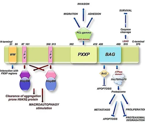
athanogene) proteins are a family of chaperone regulators that interact with the ATPase domain of the heat shock protein Hsp70 and its constitutive isoform, Hsc70, through a common conserved region located near the C terminus, termed the BAG domain (BD) (110-124 amino acids).88 Members of this protein family
have been found throughout organisms evolution, in yeast (Saccharromyces cerevisiae, Schizosaccharromyces pombe)88, invertebrates (Caenorhabditis elegans,
Ciona intestinalis, Drosophila) amphibians (Xenopus laevis)89, mammals (humans,
mice)90 and plants (Oryza sativa, Arabidopsis thaliana)91, suggesting a fundamental
biological role of these co-chaperones. Moreover, recently, seven BAG protein homologs in the Arabidopsis thaliana genome sequence have been identified, four of which have domain organization similar to their animal counterparts, underlining the fundamental biological role of these proteins.92
The human BAG protein family includes six family members (BAG1-6) that function as molecular chaperone regulators and all these proteins are constituted by a common domain, the BAG domain, which interacts with the molecular chaperone Hsp70.93 BAG proteins showed to regulate both positively and negatively, the
function of Hsp70/Hsc70, and to form complexes with a range of transcription factors, modulating various physiological processes such as apoptosis, tumorigenesis, neuronal differentiation, stress responses, and the cell cycle.86 All
- 33 -
by several other domains that are likely able to interfere with several other factors involved in prominent multiple signaling pathways.
2.2. Human BAG Proteins: BAG1, BAG2, BAG4, BAG5, and BAG6
The six human BAG proteins identified so far are 1 (RAP46/HAP46), BAG-2, BAG-3 (CAIR stressed-1, CAIR-1/B), BAG-4 (SODD), BAG-5, and BAG-6 (BAT3/Scythe) (Figure 2.1).
Figure2.1 Human BAG protein family members
All these proteins share the common architecture of the BAG Domain (BD) near the C-terminal end, with the exception of BAG 5, which contains four of such domains. Crystallography studies suggested that BAG domain contains 110 – 124 amino acids and consists of three anti-parallel helices of 30 –40 amino acids each.94
The second and third helices represent the binding sites for the ATPase domain of Hsp70/Hsc707, whereas, their N terminus is the region that affects the specificity
towards particular proteins and pathways. Very recently, depth studies, employing different deletion mutants and pull-down assays, showed that the human BAG domain (of BAG3 protein) is exactly composed by 78 amino acids, ranging from amino acids 421 to 498 (Figure 2.2). 95
- 34 -
Figure2.2 Crystal structure of a BAG domain in complex with the Hsc70 ATPase domain (PDB1HX1)
BAG1 is the first member of this family, occurring as four human isoforms structurally differing in their N-terminus, which are designated as BAG-1L p50, BAG-1M p46, BAG-1S p36, and p29, with molecular masses of 50, 46, 36, and 29 kDa, respectively (Figure 2.1).93 The 36-kDa isoform is often referred to BAG-1
and it is generally the most abundant isoform expressed in cells, followed by BAG-1L and BAG-1M. The 29-kDa isoform is expressed at low levels and cannot be consistently detected.96 In cellular studies showed that BAG1 exerts several
functions and is able to bind to Hsc/Hsp70, driving the nucleotide exchange at the chaperone complex and stimulating the substrate release.97,98 However, the exact
molecular mechanism of BAG1 is still controversial, indeed, it was demonstrated that various BAG1 isoforms regulate Hsp70 in different ways. BAG-1M was found to inhibit the refolding of denatured substrates98, while BAG-1S was shown to
initially inhibit protein refolding 99, however in a recent study, this last showed to
have a stimulating effect.100 BAG1 protein is also a binding partner for a wide range
of signaling molecules, such as, steroid hormone receptors101,102 and the Raf-1
protein kinase.103
BAG2 protein was identified as a substrate for MAPK-activated protein (MAPKAP) kinase 2, which is known to mediate p38 MAPKdependent functions;104 it carries a single BAG domain and it was identified as a specific
inhibitor of the protein CHIP (C-terminus of the Hsc70-interacting protein).105 Via
CHIP inhibition, BAG2 can influence the balance of Hsc/Hsp70-controlled protein folding and degradation of substrate proteins. The effect of BAG2 on protein degradation, as part of cellular protein quality control, potentially links BAG2 to
- 35 -
those neurodegenerative disorders that are associated with misfolded and aggregated proteins.106
BAG4, also known as silencer of death domains (SODD), can bind to the so-called death domains that are found in members of the tumor necrosis factor (TNF) receptor family, including TNF receptor 1 and the death receptor. It has a similar domain organization as BAG2 and was also identified in a screen for Hsp70- interacting proteins.88 Structurally, the helices in the BAG4 BAG domain (BD), are
three to four turns shorter than in BAG1, and they likely constitute the minimal functional fragments able to bind and regulate Hsp70. While BAG4 has only one BAG domain, BAG5 is the only member of BAG proteins family having four putative BAG domains. The functions of BAG5 are not well known but, interestingly, it has been also implicated in the pathogenesis of Parkinson disease, and, in an in vivo study, BAG5 acts as driver of neuronal cell death by enhancing the degeneration of the dopaminergic neurons.107 Finally, BAG6, also known as
BAT3, is constituted by 1229 amino acids and represents the largest human BAG member. The inclusion of BAG6 into the BAG family is due to its sequence homology with the other BAG domains and its apparent Hsc70-regulating activity, however it is not very clear yet if this BAG domain is a real one. BAG6 has been also demonstrated to bind the protein Reaper, a central apoptotic regulator in D. melanogaster and to inhibit Hsp70-mediated protein refolding.108
2.3. BAG3 protein
BAG3 is a 74 kDa protein, originally identified by a screening on two-hybrid yeasts, using the ATPase domain of the heat shock protein Hsp70 as a bait.109BAG3
protein is evolutionarily highly conserved in mammals and BAG3 orthologues in mouse, rat and human show a significant homology not only at protein level, but also at gene level.110 Two BAG3 isoforms have been described so far: one is the
full-length product of the bag3 gene with an apparent mass of 74 kDa, the other one is a shorter BAG3 protein, 40kDa, and it is found to be mainly expressed in synaptosomes (Figure 2.3). The BAG3 full-length protein is localized in the cytoplasm, mainly concentrated in the rough endoplasmic reticulum; a nuclear
- 36 -
localization of a small BAG3 isoform could be observed in some cell types, such as glial cells or pancreatic carcinoma cells. Indeed, under acute stress or upon viral infection, BAG3 alters its subcellular distribution and the co-chaperone moves into the nucleus.111
Figure 2.3 Schematic representation of the full-length BAG3 protein and the shorter isoform, a 40kD BAG3 protein
BAG3 is constitutively expressed in myocytes and in cancer cells derived from myeloid leukemias, neuroblastomas, prostate carcinomas, ovary and breast cancer, glioblastoma, and other tumor tissues.7,112,113,114 In other non-transformed cells
(e.g., epithelial and retinal cells) BAG3 expression can be induced by a variety of exogenous stressors, such as heavy metals, drugs or HIV infection.95 Moreover, the
bag3 gene promoter activity is regulated by the heat shock transcription factors
HSFs115,115b, suggesting again a role of this protein in tumor formation, since, as it
has been established, the expression of stress-responsive genes is regulated by the heat shock transcription factors, including HSF1, that is required for tumor initiation and maintenance in a variety of cancer models. An increased cellular BAG3 level was found during cellular aging in neuronal cells as well as in lung fibroblasts.116 Furthermore, in several types of cell lines, BAG3 protein expression
can be induced by many chemotherapeutics, like fludarabine or etoposide, showing an important role of the protein in chemoresistance mechanisms. Indeed, silencing bag3 gene, allow to sensitize cancer cells to the drugs, leading cells to apoptosis.117,118,119,120 In addition to the pathological role of BAG3, in rat and
human cardiomyocytes it appears to be expressed during differentiation from cardiomyoblasts and to sustain myogenin expression.121 These findings indicate an