Università degli Studi di Ferrara
DOTTORATO DI RICERCA IN
"Farmacologia e Oncologia Molecolare"
CICLO XXIII
COORDINATORE Prof. Antonio Cuneo
OSTEOPROTEGERIN: A PANCREATIC
ISLETS DYSFUNCTION AND
VASCULAR INJURY MODULATOR
Settore Scientifico Disciplinare BIO/16
Dottoranda Tutore
Dott.ssa Toffoli Barbara Prof.ssa Secchiero Paola
TABLE OF CONTENTS
1. INTRODUCTION
1.1 TNF-Superfamily page 1 1.2 OPG structure and expression page 3 1.3 OPG as a ‘decoy receptor’ page 5 1.3.1 OPG/RANKL and bone system page 5 1.3.2 OPG/TRAIL and tumorigenesis page 7 1.4 OPG and the vascular system page 9 1.5 OPG and diabetes page 11 1.6 Atherosclerosis and animal models page 13 1.7 Renin-angiotensin system and diabetes development page 18 1.7.1 Systemic versus local pancreatic RAS page 18 1.7.2 Physiological role of the RAS in the endocrine pancreas page 20 1.7.3 RAS and beta cell dysfunction page 20 1.7.4 RAS inhibition and prevention of type 2 diabetes page 23
2. RATIONALE AND OBJECTIVES page 25
MATERIALS AND METHODS
3. STUDY 1.A-B page 27
3.1 Animals and experimental protocol page 27 3.2 Evaluation of atherosclerotic plaques page 28 3.3 Masson’s trichrome staining page 28 3.4 Immunohistochemistry page 28 3.5 Real-time quantitative PCR page 29 3.6 Cell cultures page 29 3.7 Immunofluorescence page 30 3.8 Osteoprotegerin ELISA page 30 3.9 Statistical analysis page 31
4. STUDY 2. page 34
4.1 Animals and experimental protocol page 34 4.2 Measurement of physiological and biochemical parameters page 35 4.3 Oral glucose tolerance test page 35
4.4 Picrosirius red staining page 35 4.5 Immunohistochemistry page 35 4.6 In situ detection of apoptosis page 36 4.7 Real-time quantitative PCR page 37 4.8 Statistical analysis page 37
RESULTS
5. STUDY 1.A-B page 39
5.1 Increase of atherosclerotic plaque development in diabetic apoE-/- mice after in vivo administration of full-length recombinant human
OPG page 39
5.2 Recombinant human OPG injected in vivo in apoE-/- diabetic mice induces changes in the histological composition of the atherosclerotic
plaques page 41
5.3 Increase of the mRNA expression of angiopoietin 2 in the aorta of apoE-/- diabetic animals after in vivo administration of recombinant
human OPG page 44
5.4 Induction of primary rodent VSMC proliferation by recombinant
human OPG page 44
5.5 The in vivo injection of recombinant human OPG modulates the
histological composition of aortic media in apoE-/- mice page 46
5.6 TGFβ stimulates OPG production in mouse primary VSMC page 47 5.7 Recombinant full-length OPG increases the mRNA expression of
NF-kB, TGFβ and pro-fibrotic markers in mouse primary VSMC page 48 5.8 Recombinant full-length OPG increases both OPG protein and mRNA
expression in mouse primary VSMC page 50
6. STUDY 2. page 51
6.1 In vivo injection of low concentrations of full-length recombinant human OPG induces a glycemic increase and an insulin levels
reduction in C57Bl/6J mice page 51 6.2 In vivo injection of recombinant human OPG promotes a reduction in
insulin staining density and in beta cell mass in C57Bl/6J mice page 53
6 weeks old apo E-null male mice
ST
Z
R
(weeks
rOP
G
rOP
G
rOP
G
6 weeks old apo E-null male mice
ST
Z
R
(weeks
rOP
G
rOP
G
rOP
G
6.3 In vivo administration of recombinant human OPG induces alterations in the histological composition of the pancreatic islets in C57Bl/6J
mice page 53
6.4 Treatment with recombinant human OPG increases pancreatic mRNA
expression of MCP-1, VCAM-1, TGF and CTGF in C57Bl/6J mice page 56 6.5 In vivo injection of human recombinant OPG induces an activation of
the local pancreatic RAS in C57Bl/6J mice page 56
7. DISCUSSION page 58
7.1 OPG and diabetes associated atherosclerosis page 59 7.2 OPG and pancreatic beta cells dysfunction page 62
7.3 Perspective page 64
8. LIST OF PUBLICATIONS 2008-2010 page 66
1
INTRODUCTION
1.1 TNF-Superfamily
The Tumor Necrosis Factor (TNF) superfamily of cytokines and their receptors regulates many areas of metazoan biology, and specifically plays foundamental roles in regulating myriad aspects of immune development and functions (Chan, 2007). The biological functions of this system encompass beneficial effects for the host in inflammation and protective immune responses in infectious diseases as well as crucial roles in organogenesis of secondary lymphoid organs and the maintenance of lymphoid structures throughout the body. At the same time, some members of this superfamily are responsible for host damaging effects in sepsis, fever syndromes, cachexia, and autoimmune diseases (e.g., psoriasis, inflammatory bowel disease, rheumatoid arthritis) (Hehlgans and Pfeffer, 2005; Aggarwal, 2003; Ware, 2003; Kwon et al., 2003).
In 1984, two forms of TNF, TNFα and LTα (lymphotoxin, TNFβ), were isolated from activated macrophages and T cells, respectively. Since their identification, these proteins have become representative of a unique superfamily of ligands, which currently includes at least 18 different human homologues (Baker and Reddy, 1998; MacEwan, 2008). The TNF-related ligands are type II transmembrane proteins (intracellular N-terminus) with a short cytoplasmic tail (15 to 25 residues in length) and a larger extracellular region (approximately 150 amino acids) containing the signature ‘TNF homology domain’ (THD) where the receptor-binding sites are located (Figure 1). The THD, that shares approximately 20-25% of sequence identity between family members, folds into an antiparallel β-sandwich that assembles into trimers, the functional unit of the ligand (Paul, 2008).
Some of these ligands, e.g. TNF, are active both as a membrane integrated and as a soluble form released after proteolytic cleavage, mainly by metalloproteinases induced by various stimuli. Certain ligands are expressed only as soluble molecules, e.g. LTα; but may also be recruited to the cell membrane to form heterotrimeric membrane anchored complexes and thereby enhancing regulatory specificity and complexity (Hehlgans and Pfeffer, 2005).
Thus far, 29 TNF receptor family members have been identified in humans (Figure 1). These are primarily type I transmembrane proteins characterized by cysteine-rich domains (CRD) that are the hallmark of the TNF superfamily receptors (TNFR). These 40 amino acid pseudorepeats are typically defined by 3 intrachain disulphides generated by 6 highly conserved cysteine residues within the receptor chains (Locksley et al., 2001). Most receptors
2
also exist in a soluble form, and the solubility is achieved by proteolitic cleavage or by alternative splicing of the exon encoding the transmembrane domain (Baker and Reddy, 1998).
Figure 1. The TNF/TNFR superfamily. The TNF-related ligands are shown in blue and arrows indicate interactions with their receptors (Hehlgans and Pfeffer, 2005).
Although most members of the TNF superfamily of ligands interact with more than one receptor of the corresponding superfamily of cognate receptors, genetic based approaches, mainly conducted in gene targeted mouse strains, have clearly demonstrated that almost each receptor-ligand system have a unique and non-redundant function.
Receptor activation by the TNF family ligands causes recruitment of several intracellular adaptor proteins which activate multiple signal transduction pathways. Based on their intracellular sequences the members of the TNFR superfamily can be classified into three major groups. The first group, including molecules such as FAS and TNFRI, contains the so called death domains (DD) in their cytoplasmic domains. Activation of these receptors leads to recruitment of intracellular death domain containing adaptors such as FAS-associated death domain (FADD) and TNFR-associated death domain (TRADD), which subsequently promote the activation of the caspase cascade and induction of apoptosis. The second group of
3
receptors, that includes members like CD30, CD40 and receptor activator of NF-kB (RANK), contains one or more TNF-receptor associated factor (TRAF)- interacting motif (TIMs) in their cytoplasmic domain. Activation of TIM containing TNFR family members leads to the recruitment of TRAF family members and the subsequent activation of signal transduction pathways like nuclear factor kB (NF-kB), Jun N-terminal kinase (JNK), p38, extracellular signal-related kinase (ERK) and phosphoinositide 3-kinase (PI3K), which regulate cellular processes ranging from proliferation and differentiation to cell death. Finally, the third group of receptors, including TRAIL-R3 (DcR1), DcR3 and Osteoprotegerin, doesn’t contain functional intracellular signaling domains or motifs, but instead compete with the other two groups of receptors for their corresponding ligands (Hehlgans and Pfeffer, 2005, Dempsey et al., 2003).
1.2 OPG structure and expression
Osteoprotegerin (OPG) is a secreted glycoprotein belonging to the TNFR superfamily and was initially identified by two separate groups in 1997. Both groups observed OPG to be central in the regulation of bone turnover through the inhibition of osteoclastogenesis. However, it was not until 1998 that the newly discovered proteins were found to be identical, hence OPG was alternatively termed osteoclast inhibitory factor (OCIF) (Reid and Holen, 2009). Its international name according to the TNF nomenclature is TNFRSF11B (Baud’huin et al., 2007).
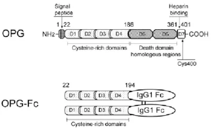
The mouse and the human OPG genes have been cloned and characterized, and the human OPG gene is a single-copy gene that consists of five exons and spans 29 kb of the human genome located on chromosome 8 q23-24 (Morinaga et al, 1998; Mizuno et al.,a 1998). Murine and human OPG proteins comprise 401 amino acids of which 21 form a signal peptide that is cleaved to generate a mature form of 380 amino acids (Figure 2). At the N terminus, there are four domains (D1-D4), which have cysteine-rich TNFR homologous motifs and are necessary and sufficient for inhibiting osteoclastic differentiation and activity both in vitro and in vivo. The carboxy-terminal portion of the protein with domains 5 and 6 (D5 and D6) contains two death domain homologous regions, motifs that are found in the cytoplasmic region of mediators of apoptosis such as TNFR 1, DR3 or CD95/Fas. In fact, D5 and D6 of OPG have been demonstrated to transduce an apoptotic signal when expressed as an OPG/Fas fusion protein in which the transmembrane region of Fas is inserted between domains 4 and 5 of OPG. Finally, domain 7 (D7) harbors a heparin-binding region, a common feature of peptide growth factors and signal molecules, as well as an unpaired cysteine residue
4
at position 400 required for homodimerization of the molecule (Zauli et al., 2009; Schoppet et al., 2002). OPG represents an atypical member of the TNFR family since it is a secreted protein with no transmembrane domain. OPG is produced as a monomer (55-62 kDa), undergoes homodimerization, and is secreted as a disulfide linked homodimeric glycoprotein with four or five potential glycosylation sites, generating a mature form of OPG of 110-120 kDa. The monomeric and the homodimeric forms are indistinguishable in stability, but the homodimeric exerts more potent biological activity and is stronger in heparin binding ability (Zauli et al., 2009; Tomoyasu et al., 1998).
Figure 2. Schematic representation of the protein structure of OPG and OPG-Fc. Main domains and their biochemical and/or functional properties are indicated. Numbers in figure represent amino acids. NH2 indicates
N terminus, COOH, C terminus, Cys400, dimer formation site. Human OPG-Fc is a recombinant fusion protein, in which the Fc fragment of human IgG1 is fused to the C terminus of the 22-194 fragment of native OPG to maintain a dimeric molecule with a sustained circulating half-life (Zauli et al., 2009).
To enhance the pharmacological activity of native OPG, numerous constructs have been created wherein the signal peptide, heparin binding domain, and death domains were removed and the remaining peptide was fused to the Fc domain of human IgG1 (Figure 2). The Fc fusion partner maintains the potent dimeric nature of OPG while significantly increasing its circulating half-life (Kearns et al., 2008).
OPG is produced by a variety of tissues including the cardiovascular system (hearth, arteries, veins), lung, kidney, intestine, and bone, as well as hematopoietic and immune cells. The expression and production of the protein is modulated by various cytokines, peptides, hormones, and drugs. Cytokines, including TNFα, interleukin (IL)-1α, IL-18, transforming growth factor (TGFβ), bone morphogenetic proteins, and steroid hormones are known to up-regulate OPG mRNA levels. In contrast, glucocorticoids (known to promote bone resorption) and the immunosuppressant cyclosporine A (which has the propensity to cause osteoporosis
5
and vascular disease) or basic fibroblast growth factor, all suppress the expression of OPG (Schoppet et al., 2002).
1.3 OPG as a ‘decoy receptor’
OPG acts as a decoy receptor for RANKL, preventing the stimulation and maturation of osteoclast precursors instigated by normal binding of RANKL to its constitutive receptor and as a soluble receptor for TRAIL (Emery et al, 1998).
1.3.1 OPG/RANKL and bone system
The important role of OPG in bone metabolism has been clearly demonstrated by the development of transgenic and knock-out mice. OPG knock-out mice are viable and fertile, but exhibit an osteoporotic phenotype due to enhance osteoclastogenesis. They are characterized by marked bone loss accompanied by destruction of growth plate and lack of trabecular bone in their femurs, and the strength of their bones dramatically decrease (Mizuno et al.,b 1998). In contrast, systemic delivery of OPG via the expression of rat or murine opg transgenes results in severe yet nonlethal osteopetrosis. The ostopetrotic phenotype caused by OPG overexpression differs significantly from those observed in other ostopetrotic models.
opg transgenic mice are of normal size, have no apparent defects in tooth eruption, and have
normally shaped bones. Histologically, they have a marked reduction in trabecular osteoclast but no deficiency of osteoclast precursors, suggesting a defect in the later stages of osteoclast differentiation (Simonet et al., 1997). These evidences, supported by confirmations in the in
vitro experiments, demonstrate that the presence of OPG is absolutely required to maintain
bone mass in physiological situations. Subsequently, molecular binding experiments have shown that OPG associates with the ligand of the receptor activator of NF-kB (RANKL), a member of TNF superfamily ligand, functioning as a decoy receptor. RANKL genes gives rise to splice variants that encode two forms of type II transmembrane proteins and one form of a secreted protein. Although high RANKL expression can be found in lymph nodes, thymus and lung, only low levels of RANKL can be detected in spleen, bone marrow, peripheral blood, leukocytes, hearth, placenta, skeletal muscle, stomach or the thyroid. In addition, RANKL expression is induced in mammary gland epithelial cells in pregnancy, activated T cells, and malignant tumor cells (Wong et al., 1999; Wada et al., 2006).
The receptor that mediates all known activities for RANKL is called receptor activator of NF-kB (RANK). The binding and activation of RANK, that is a homotrimeric TNFR family member, involve direct interactions between the extracellular receptor binding domain of
6
trimeric RANKL and the extracellular cysteine-rich domains of trimeric RANK. This interaction is thought to cause oligomerization of RANK and the subsequent activation of several signal transduction cascades, as the NF-kB, MAPK, and phosphatidylinositol pathways (Kearns et al., 2008; Wada et al., 2006).
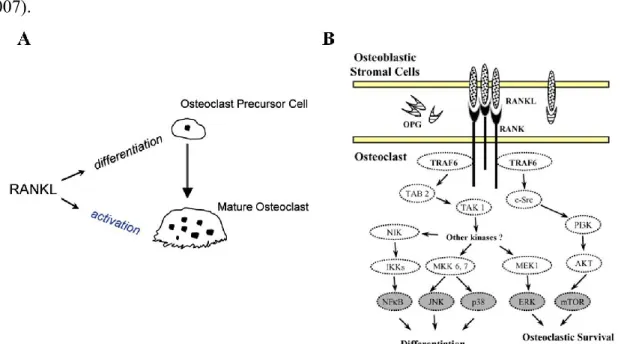
One of the best characterized role of RANKL/RANK system is in the osteoclastogenesis. The key regulators of bone turnover are osteoblasts, which are involved in bone formation, and osteoclasts, which are responsible for bone resorption. RANKL expressed by osteoblasts as a transmembrane protein binds to RANK on the surface of osteoclasts and osteoclasts precursor. This leads to osteoclast formation, differentiation, activation and consequently bone resorption (Figure 3). To regulate the balance between bone formation and bone resorption, the RANKL-RANK interaction is inhibited by OPG. OPG produced by osteoblastic cells binds as a homodimer to the homotrimeric RANKL, thus inhibiting the terminal stage of osteoclastic differentiation (Reid and Holen, 2009; Baud’huin et al., 2007). Although there are contradictory data, in general upregulation of RANKL is associated with downregulation of OPG, or at least lower induction of OPG, such that the ratio of RANKL to OPG changes in favour of osteoclastogenesis. Many reports have supported the assertion that the RANKL/OPG ratio is a major determinant of bone mass. Moreover, consistent with the osteoprotective role, mutation in the human OPG gene have been associated with idiopathic hyperphosphatasia, also known as Juvenile Paget’s disease, an autosomal-recessive disorder characterized by increased bone remodeling, osteopenia, and fractures (Boyce and Xing, 2007).
Figure 3. In (A) Schematic representation of RANKL in osteoclastogenesis (Zauli et al., 2009). In (B) RANK signaling pathways (Theoleyre et al., b 2004).
7
1.3.2 OPG/TRAIL and tumorigenesis
In 1995 TNF-related apoptosis-inducing ligand (TRAIL) was identified and characterized as a member of the TNF family of death-inducing ligands. TRAIL is a type II transmembrane protein of about 33-35 kD, which can be cleaved from the cell surface to form a soluble ligand that retains biological activity. The extra-cellular domain of TRAIL forms a bell shaped homo-trimer, much like other ligands of the TNF family, but unlike the other members, it carries a zinc ion at the trimer interface, coordinated by the single unpaired cysteine residue of each monomer. This zinc ion is essential for structural integrity of TRAIL and to maintain its capacity to induce apoptosis (Degli-Esposti, 1999; Corallini et al., 2008). TRAIL is constitutively present in many tissues at the level of mRNA, but it is expressed mainly by activated cells of the immune system, especially natural killer (NK) cells, B cells, T cells, monocytes, and dendritic cells. TRAIL plays a crucial role in maintaining T cell homeostasis, as well as in killing of tumor and virally transformed cells by NK cells. Over the years, TRAIL has generated considerable interest among clinicians for its preferential toxicity toward transformed cells and tumor xenografts, with generally little or no toxicity to normal tissues, which makes it an ideal candidate for cancer therapy. As a result, recombinant TRAIL, as well as agonistic antibodies against TRAIL receptors, are currently in Phase I/II clinical trials for treatment of solid tumors and hematological malignancies (Griffith et al., 2009; Guicciardi and Gores, 2009; Ashkenazi and Herbst, 2008; Finnberg and El-Deiry, 2008).
With respect to other members of the TNF ligand superfamily, TRAIL shows the most complex ligand-receptor interaction, since it is able to bind to five different receptors found on a variety of cell types: four membrane-bound (R1/death receptor 4, TRAIL-R2/death receptor 5, TRAIL-R3/decoy receptor 1, TRAIL-R4/decoy receptor 2) and one soluble receptor (OPG). OPG acting as a decoy receptor, binds TRAIL and prevents its interaction with the functional death receptors, thus allowing cells to escape cell death (Figure 4). In this context, many different in vitro data demonstrated the ability of native OPG, produced by tumor cells or by bone marrow stromal cells, to efficiently counteract the pro-apoptotic activity of TRAIL in a variety of cell lines derived from prostate and breast cancers, ameloblastomas and multiple myeloma (Zauli et al., 2009; Reid and Holen, 2009).
8
Of note, in the light of recent in vitro studies that demonstated that the affinity of OPG for RANKL and TRAIL under physiological conditions is of a similar order of magnitude and that TRAIL is able to inhibit OPG-mediated inhibition of osteoclastogenesis, OPG acquires a key position in the regulation of the functions of these two important signaling pathways (Figure 5) (Vitovski et al., 2007).
Recently, many studies have demonstrated that OPG can exert direct biological activities independently of its neutralizing effects towards RANKL or TRAIL. In fact OPG has a highly basic heparin-binding domain (D7) that besides being responsible for the homodimerization of the molecule (Yamaguchi et al., 1998), makes interactions with heparin and heparan sulfates possible. Heparan sulfate proteoglycans are important participants in cell-surface
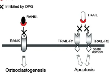
Figure 4. Role of OPG in cell survival. TRAIL is produced by immune cells that can infiltrate the tumor microenvironment. TRAIL can then bind to the death receptors 4 and 5 (DR4 and DR5) present on the surface of tumor cells, resulting in tumor cell apoptosis. OPG, secreted by tumor cells, acts as a decoy receptor for TRAIL. As a result, tumor cells escape from death (Reid and Holen, 2009).
Figure 5. Mechanism of action of OPG on RANKL- and TRAIL- biological activities. The pro-apoptotic activity of TRAIL is mediated by two (TRAIL-R1 and TRAIL-R2) of its four membrane receptors. OPG by efficiently binding RANKL and TRAIL, counteracts both the RANKL-mediated osteoclastogenesis as well as the pro-apoptotic activity of TRAIL (Zauli et al., 2009).
9
signaling and critical in controlling cell behavior. They are involved in actin cytoskeleton regulation, cell adhesion and migration, and modulation of specific receptor interactions. At the cell surface, proteoglycans of the syndecan family are the major source of heparan sulfate (Wright et al., 2009). In this context, different reports indicate that the heparin binding domain is involved in OPG-induced chemotaxis of human peripheral blood monocytes (Mosheimer et al., 2005), in controlling OPG release by vascular cells (Nybo and Rasmussen, 2008) and in the OPG-mediated osteopontin-increasing in human periodontal ligament cells (Yongchaitrakul et al., 2009). Of note, in multiple myeloma (Standal et al., 2002) and osteosarcoma (Lamoureux et al., 2009) it has been shown that also the decreased of the biological activity of the full-length OPG might be due to its bounding, internalization and degradation, syndecan-1 mediated.
1.4 OPG and the vascular system
While OPG, RANK and RANKL are produced by numerous cell types and a variety of tissues, their expression pattern targets three main biological systems where the molecular triad could be more specifically involved: the osteoarticular, immune, and vascular systems (Figure 6). Transgenic and knockout mice models as well as many in vitro experiments clearly revealed the potential involvement of these effectors in the three biological systems (Theoleyre et al., b 2004; Feige, 2001; Grĉević et al., 2001; Hofbauer et al., 2001; Josien et al., 2000; Schoppet et al., 2002).
Figure 6. OPG/RANK/RANKL as common effectors of bone, immune and vascular system (Theoleyre et al., b 2004).
10
In fact, the first evidence for a role of OPG in the vasculature was given by a study of OPG knockout mice generated on a mixed genetic background. The selective deletion of OPG in mice resulted in early-onset severe osteoporosis as well as significant medial calcification of the aorta and renal arteries. The onset of arterial calcification could be completed prevented by transgenic OPG delivery from mid gestation through adulthood; in contrast, post-natal intravenous injection of recombinant OPG had no effect on the incidence of vascular calcification, suggesting that OPG cannot reverse the calcification process once it had occurred. Interestingly, both treatments effectively reversed osteoporotic bone loss phenotype. A vascular-protective role for OPG was also indicated by rat studies, in which OPG administration prevented calcification induced by warfarin (a vitamin K antagonist) or high vitamin D doses (Van Campenhout and Golledge, 2009; Venuraju et al., 2010; Collin-Osdoby, 2004). Furthermore, Morony et al. (2008), demonstrated that subcutaneous injection of pharmacological concentrations of human recombinant OPG (OPG-Fc) decreased the degree of atherosclerotic calcified lesions in atherogenic diet-fed ldlr knockout mice, without affecting the total burden of atherosclerotic lesions. To determine whether OPG plays a role in the calcification and chondrocyte metaplasia that has been reported in advanced atherosclerotic lesions in mice, Bennet et al. (2006) generated mice deficient in both OPG and apoE gene. They observed that the loss of OPG in this animal model led to larger atherosclerotic lesions in the innominate arteries of 40 and 60 weeks of age mice, coupled with more rapid and extensive calcification of both the media and the intima.
Although most animal studies supported a protective role for OPG in cardiovascular system, many clinical investigations revealed a positive association between high serum OPG levels and cardiovascular outcome. The first paper to report a relation between plasma OPG and vascular disease was published in 2001 (Browner et al.). An association was discovered between high plasma OPG levels and increased cardiovascular mortality in a cohort of elderly women primarily gathered to study osteoporosis risk factors. Subsequently, the connection between plasma OPG and cardiovascular disease was confirmed in populations of men with coronary artery disease (Schoppet et al., 2003), patients with myocardial infarction and heart insufficiency (Ueland et al., 2004), and in seemingly healthy individuals (Kiechl et al., 2004) where a high level of serum OPG was an independent risk factor for incident CVD and vascular mortality, but not for mortality due to non-vascular causes. Moreover, plasma OPG has also been found to be associated with intima-media thickness of the carotid artery as determined by ultrasound (Erdogan et al., 2004). Remarkably, two OPG genetic polymorphisms have been associated with an increase risk of coronary artery disease in
11
Caucasian men, and serum OPG levels correlated with one of these polymorphisms (Soufi et al., 2004). Thus, these studies strongly indicate that serum OPG levels frequently rise in clinical conditions that favor vascular dysfunction or atherosclerosis. Morena et al. (2006) demonstrated in hemodialysis patients as elevated plasma OPG predicted all-cause and CV mortality when adjusted for age, gender, dialysis vintage, diabetes, hypertension, and smoking. More recently OPG was described as an independent predictor factor both for early vascular adverse changes in osteoporotic postmenopausal women (Shargorodsky et al., 2009) and for cardiovascular mortality in patients with stable coronary artery disease (Jono et al., 2010). Mesquita et al. (2009) showed that OPG predicted mortality in chronic kidney disease patients and could be a valuable biomarker in early detection of coronary artery calcification in these patients.
Moreover, many different in vitro studies have clearly demonstrated that inflammatory cytokines promote the expression and release of OPG by endothelial cells (Secchiero et al., a 2006; Ben-Tal Cohen et al., 2007; Collin-Osdoby et al., 2001) and by vascular smooth muscle cells (Zhang et al., 2002; Moran et al., 2005). Because of the enormous surface area of the endothelium throughout the body as well as the relatively substantial levels of constitutive and regulated OPG produced by endothelial cells and vascular smooth muscle cells, vascular cells play a fundamental role in the contribution in circulating OPG in human serum. On the other side, if we consider that recent studies have shown that recombinant full-length OPG is able to promote leukocytes adhesion to endothelial cells (Zauli et al., 2007; Mangan et al., 2007), OPG seems to have an active role in disease progression more than serving as a compensatory/protective response to minimize disease progression. Anyway, considerable controversy still exist regarding the role of OPG/RANKL/RANK/TRAIL pathways in cardiovascular setting.
1.5 OPG and diabetes
Plasma osteoprotegerin concentration correlates to both diabetes and cardiovascular disease in cross-sectional studies. This was first shown by Browner et al. (2001), who reported that although no associations were seen between the bone parameters and plasma osteoprotegerin levels, individual with diabetes as well as persons with cardiovascular disease had increased values. In another investigation on 522 men that described a positive relationship between coronary arteriosclerosis (determined by CAG) and plasma OPG, diabetic patients were also observed to have elevated OPG plasma levels and the increase was more than could be expected when the degree of coronary sclerosis was considered (Schoppet et al., 2003). Since
12
then, these findings have been confirmed in both type 1 (Xiang et al., 2007; Galluzzi et al., 2005) and type 2 diabetes (Knudsen et al., 2003; Xiang et al., 2006) and it has been found that the increased OPG production characterizes an early event in the natural history of diabetes mellitus (Secchiero et al., a 2006).
In more recent studies performed in diabetic subjects a strong association between plasma levels of OPG and micro- and macroangiopathy was observed (Avignon et al., 2005; Grauslund et al., 2010; Knudsen et al., 2003; Xiang et al., 2009).
Osteoprotegerin has also been found to be accumulated in aorta from patients with type 1 and type 2 diabetes (Olesen et al., 2005). This accumulation is seen in the tunica media from areas of the tissue with and without atherosclerotic plaque. The accumulation of osteoprotegerin throughout the deeper layers of the vessels wall may reflect generalized changes in the arterial system in diabetes, as part of diffuse arterial changes such as alterations in glycoproteins (Takemoto et al., 2000), collagens (Rasmussen and Ledet, 1993) and glycosaminoglycans (Heickendorff et al., 1994). In addition, osteoprotegerin may be related to the development of medial calcification, which is frequently present in patients with diabetes (Niskanen et al., 1994; Lehto et al., 1996). Two recent experimental studies have confirmed the finding of increased levels of OPG in animals with experimental diabetes (Heinonen et al., 2007; Vaccarezza et al., 2007).
The mechanisms behind the increased circulating OPG levels in diabetes are unknown. Diabetic vasculopathy has an underlying low-grade inflammatory component, manifesting itself in the up-regulation of genes responsive to inflammatory processes (Secchiero et al., 2005; Joussen et al., 2002; Bulotta et al., 2001; Fujiwara et al., 2004; Sjöholm and Nyström, 2005). In this respect, it should be emphasized that OPG is an NF-kB-inducible gene (Collin-Osdoby et al., 2001), whose release in endothelial cell culture is significantly increased by inflammatory cytokines.
Considering the elevated levels of OPG detected also in patients affected by type 1 diabetes, hyperinsulinemia and insulin resistance are unlikely to play a key role in OPG induction. Accordingly Jørgensen et al. (2009) have recently demonstrated as acute hyperinsulinemia decreases plasma OPG in type 2 diabetes and obesity. In line with the hypothesis that insulin is not involved in the induction of OPG expression and secretion, Secchiero et al. (a 2006) have also demonstrated that OPG release is significantly up-regulated in the sera of diabetic apoE-knockout mice early after the induction of diabetes mellitus by streptozotocin (STZ) treatment. Of note, OPG serum levels in diabetic apoE-knockout mice positively correlated with the glycemic levels whereas they were inversely correlated to the levels of free RANKL.
13
Elevated levels of OPG were also observed in C57Bl littermates concomitantly with the induction of diabetes mellitus, suggesting that hypercholesterolemia, characterizing apoE-knockout mice, did not play a major role in the upregulation of serum OPG associated to diabetes mellitus.
Despite the in vivo data obtained in the mouse models of STZ-induced diabetes, in which it has been demonstrated the existence of a positive correlation between OPG and glycemic serum levels, high glucose levels per se were insufficient to modulate OPG release in endothelial cells, PBMCs, and macrophages. On the other hand, the proinflammatory cytokine TNF-, which is known to be elevated in the sera of patients with diabetes mellitus (Joussen et al., 2002; Bulotta et al., 2001; Fujiwara et al., 2004), dose dependently up-regulated OPG secretion by endothelial cells. Importantly, the concentrations of TNF- (10 pg/ml) required to induce a significant (approximately two-fold) increase in OPG, a situation mimicking the OPG rise observed in the serum of diabetic patients, were in the range of plasma concentrations reported in diabetic patients (Joussen et al. 2002; Bulotta et al., 2001; Fujiwara et al., 2004). These in vitro findings, coupled to the data obtained in the diabetic mouse models, clearly suggest that the inflammation-driven hyperglycemia, rather than the high glucose levels per se, is involved in the increase of OPG observed in both diabetic patients and diabetic mice. It is possible that the imbalance of OPG versus RANKL serum levels in both diabetic patients and diabetic apoE-knockout mice might contribute to endothelial cell dysfunction by blocking RANKL signaling, which is able to activate protective intracellular pathways in endothelial cells, such as the eNOS pathway. In this respect, it should be emphasized that diabetic vascular dysfunction is a major clinical problem that predisposes patients to a variety of cardiovascular diseases. In fact, diabetic patients frequently suffer from macroscopic and microscopic vasculopathy and accelerated atherosclerosis. The early impairment of nitric oxide release is a key feature of endothelial dysfunction, which invariably precedes permanent vascular alterations (Landmesser et al., 2004).
At present it is unclear whether OPG plays a pathogenetic or compensatory role in the vascular dysfunction and atherosclerosis associated to diabetes. Moreover the physiopathological role of elevated levels of OPG in pancreatic islet function is not known.
1.6 Atherosclerosis and animal models
Atherosclerosis is a high-cost disease and its complications still represent the first cause of death in most industrialized countries. Efficacious prevention includes treatment of the most
14
important cardiovascular risk factors, such as cigarette smoking, hypertension, hypercholesterolemia, diabetes and obesity. However, the absence of such ‘traditional’ risk factors does not completely protect from the disease and new ‘emerging factors’ have been identified, including markers of inflammation (Corrado et al., 2010; Libby, 2002). Although the clinical manifestations of cardiovascular disease, such as myocardial infarction, stroke, and peripheral vascular disease, appear from middle age, the process of atherosclerosis can begin early in childhood as an accumulation of fatty streaks-lipid engorged macrophages (foam cells) and T lymphocytes in the intima of the arteries. Fatty streaks may or may not progress, and may regress (Hong, 2010).
Under normal conditions, the endothelial cells of the arterial wall resist adhesion and aggregation of leukocytes and promote fibrinolysis. When activated by stimuli, like hypertension, an unhealthy diet, insulin resistance or inflammation, the endothelial cells express a series of adhesion molecules that selectively recruit various classes of leukocytes. Once the monocytes adhere to the activated endothelium, proinflammatory proteins provide a chemotactic stimulus that induces them to enter the intima. Within the intima, the monocytes mature into macrophages and start to express scavenger receptors, that allow them to engulf modified lipoprotein particles (especially highly oxidized LDL). Subsequently, the cytoplasm becomes engorged with lipid particles, giving the macrophages the typical microscopic frothy appearance of the foam cells found in atherosclerotic lesions (Libby et al., 2010).
The transition from the relatively simple fatty streak to the more complex lesion is characterized by the immigration of smooth muscle cells from the medial layer of the artery wall trough the internal elastic lamina into the intimal, or subendothelial, space. Intimal smooth muscle cells may proliferate and take up modified lipoproteins, contributing to foam cells formation, and synthesize extracellular matrix proteins that lead to the development of the fibrous cap. This phase of lesion development is influenced by interactions between monocytes/macrophages and T cells that result in a broad range of cellular and humoral responses and the acquisition of many features of a chronic inflammatory state.
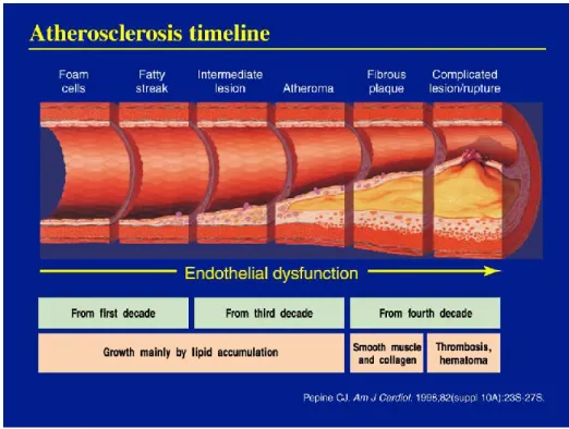
Although advanced atherosclerotic lesions can lead to ischemic symptoms as a result of progressive narrowing of the vessel lumen, acute cardiovascular events that result in myocardial infarction and stroke are generally thought to result from plaque rupture and thrombosis. Plaque ruptures generally occur at the shoulder regions of the plaque and are more likely to occur in lesions with thin fibrous caps, a relatively high concentration of lipid-filled macrophages within the shoulder region, and a large necrotic core (Glass and Witztum, 2001; Lusis, 2000) (Figure 7).
15
Numerous animal species, especially transgenic mouse models, have been used to study the pathogenesis and the potential treatment of atherosclerotic lesions (Woollard and Geissmann, 2010). Until 1992, the majority of atherosclerotic research focused on mechanisms in rabbits, with a lesser number of studies in pigs and nonhuman primates. Despite the fact that rabbits do not develop spontaneous atherosclerosis, they have been useful for their high responsiveness to cholesterol manipulation and the capacity of developing lesions in a fairly short time.
Figure 7. Atherosclerosis progression.
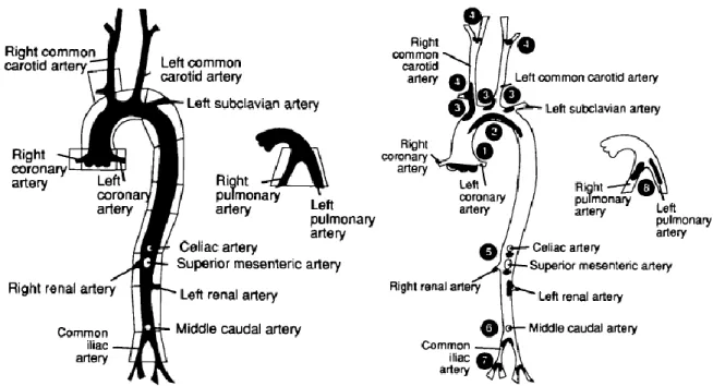
Unfortunately, rabbit lesions are much more fatty and macrophage-rich than those in humans and plasma cholesterol are extraordinary high. Pigs and monkeys are better suited to model human atherosclerotic lesions, but, obviously, they present many problems related to costs and maintaining/handling of the colonies. In recent years, there has been an explosion in the number of in vivo studies that is largely attributable to the use of small mouse models to study atherogenic mechanisms (Jawień et al., 2004). Among available models, the apolipoprotein E-deficient (ApoE-knockout) mice is particularly popular, because of its propensity to spontaneously develop a full range of atherosclerotic lesions from fatty streaks to fibrous plaques, that are distributed throughout the arterial tree (Figure 8) and that present many features characteristic in appearance and distribution of those observed in human lesions (Nakashima et al., 1994).
16
ApoE, a glycoprotein synthesized mainly in the liver and brain in both human and mice, is a constituent of all lipoproteins except low- density lipoproteins (LDL). It functions as a ligand for receptors that clear chilomicrons and very low-density lipoprotein (VLDL) remnants. ApoE is also synthesized by monocytes and macrophages in vessel, and is thought to have local effects on cholesterol homeostasis and on inflammatory reactions in atherosclerotic vessel. It may also function in dietary absorption and biliary excretion of cholesterol.
Figure 8. Line graphs showing the arteries from apoE-knockout mice. Left, Arteries that were observed under the dissecting microscope and dissected for further microscopic analysis. Right, Sites of predilection for lesions development are indicated in black: (1) aortic root, at the base of the valves; (2) lesser curvature of the aortic arch; (3) principal branches of the aortic arch; (4) carotid artery; (5) principal branches of the abdominal aorta; (6) aortic bifurcation; (7) iliac artery; and (8) pulmonary arteries (Nakashima et al., 1994).
The apoE-knockout mouse was created practically simultaneously in two separate laboratories, through gene inactivation by targeting. On a standard chow diet (0.02% cholesterol), the mice demonstrate a total cholesterol level >500mg/dl (5 times normal), mostly in the VLDL and chylomicron remnant fractions. These levels are unaffected by the age or sex of the animals. A Western- type diet (0.15% by weight cholesterol) quadruple these fractions. Interestingly, mice homozygous or heterozygous for the disrupted ApoE gene appear healthy and no difference in their body weights compared to normal mice is observed (Jawień et al., 2004; Meir and Leitersdorf, 2004).
Unlike normal mice, which don’t develop atherosclerotic lesions (except for some strains on high fat/high cholesterol diets) (Vischer, 1999), a chronological analysis of atherosclerosis in
17
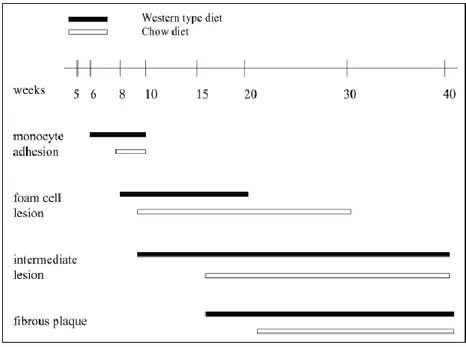
the apoE-knockout mouse has shown that the sequential events involved in lesion formation are strikingly similar to those in well-established larger animal models of atherosclerosis and in humans. Lesions in the apoE-knockout mice, as in humans, tend to develop at vascular branch points and progress from foam cell stage to the fibroproliferative stage with well-defined fibrous caps and necrotic lipid cores, although plaque rupture has not been observed in this or in any other mouse models. Progression of lesions appears to occur at a faster rate than in humans atherosclerosis, but the rapidity of the progression can be advantageous in many experimental situations. In particular, fatty streaks are first observed in the proximal aortas of a chow-fed, 3 months old mouse. On this diet, as early as 10 weeks of age, foam cells lesions are observed by light microscopy. Intermediate lesions containing foam cells and smooth muscle cells emerge around 15 weeks, and fibrous plaques appear at 20 weeks of age (Jawień et al., 2004; Meir and Leitersdorf, 2004).
These spontaneous lesions progress and cause severe occlusion of the coronary artery ostium by 8 months (Zhang et al., 1992). A western diet accelerates all the process (Figure 9).
Figure 9. Diagram showing how lesion formation in chow-fed mice is delayed in comparison with mice fed the Western-type diet (Jawień et al., 2004).
Of note, the genetic background has a major effect on atherosclerosis susceptibility in strains of knockout mice. For example, lesions from 16-week chow diet C57BL/6 apoE-knockout are relatively larger than from FVB apoE-apoE-knockout mice and, in contrast to FVB mice, there is evidence of early development of fibrous caps (Jawień et al., 2004).
18
The development of murine models of atherosclerosis has revolutionized the approach to evaluating potential roles of specific proteins in lesion development. The crossing of these animals with mice that have been engineered to over-express or lack genes of interest, generating double knockout mice (e.g. iNOS-/- apoE-/-; CCR2-/- apoE-/-), has led to a growing list of proteins that accelerate or retard the rate at which lesions develop, and/or alter lesion composition (Glass and Witztum, 2001).
1.7 Renin-angiotensin system and diabetes development 1.7.1 Systemic versus local pancreatic RAS
The renin-angiotensin system (RAS) plays a key role in the regulation of fluid, electrolyte balance and arterial pressure (Reid et al., 1978). In the classical RAS, the glycoprotein Angiotensinogen is secreted into the circulation by the liver, where it is cleaved by renin, an aspartyl protease produced by the juxtaglomerular cells of the renal afferent arterioles, to release the decapeptide Angiotensin I. This decapeptide is further hydrolized by angiotensin converting enzyme (ACE), a metalloprotease produced by and anchored to the surface of endothelial cells, into the eight-amino acid peptide Angiotensin II (Ang II), which is able to bind to high affinity AT1 and AT2 cell-surface receptors (Lavoie and Sigmund; 2003). Ang II can also be formed via non-ACE and non-renin enzymes, including chymase, cathepsin G, cathepsin A, chymostatin-sensitive AII-generated enzyme (CAGE), tissue plasminogen activator (t-PA), and tonin (Urata et al., 1995; Urata et al., 1996). On the other side, ACE also breaks down bradykinin, a potent vasodilator and natriuretic hormone.
It has gradually become evident that in addition to the circulating RAS there is a local tissue RAS in most organs and tissues (Johnston et al., 1992; Paul et al., 2006). This makes RAS not only an endocrine, but also a paracrine and an intracrine system. Moreover the important discoveries of the renin/pro-renin receptor (Nguyen et al., 2002), the ACE2 enzyme (Donoghue et al., 2000; Tipnis et al., 2000), Angiotensin 1-7 as a biologic active metabolite of RAS (Santos and Ferreira, 2007), the Ang IV receptor as an insulin regulated aminopeptidase (IRAP) (Albiston et al., 2001), and the Mas as a receptor for Angiotensin 1-7 (Santos et al., 2003), have contributed to extend our view of the RAS from classical linear cascade to a more complex cascade with multiple mediators, receptors and functional enzymes (Figure 10).
19
Figure 10. Enzymatic cascade of the renin-angiotensin system: classic and alternative pathways.
There is growing evidence supporting the existence of all the components of a functioning intrinsic RAS in the endocrine pancreas. (Pro)- renin is expressed in the islets of Langerhans, chiefly in the connective tissue surrounding the blood vessels and in reticular fibers within the islets (Leung and Chappell, 2003).
Both ACE and ACE2 mRNA has been found within islet with the former predominating in the microvasculature and the latter identifying within the centre of the islets (Tikellis et al., 2004).
Although initial studies, limited by low sensitivity of mRNA assays, found Angiotensinogen undetectable in normal rat pancreas (Campbell and Habener, 1986), more recent studies confirm that Angiotensinogen is expressed in glucagon-secreting -cells located at the periphery of the islets of Langerhans (Regoli et al., 2003).
AT1 receptors have been detected in cells at the centre of the islet with its expression co-localized with that of insulin secreting beta cells. In contrast AT2 receptor has been co-localized to the outer region of islets and co-localizes with somatostatin-secreting cells (Tahmasebi et al., 1999).
Ang II is immunohistochemically localized predominantly in the endothelial cells of pancreatic blood vessels and the epithelial cells of pancreatic duct (Leung et al., 1998). Moreover, more recent studies have demonstrated that local genesis of Ang II occurs within pancreatic islets (Lau et al., 2004).
20
1.7.2 Physiological role of the RAS in the endocrine pancreas
There is converging evidence suggesting that in the endocrine pancreas, islet RAS has a role in regulating pancreatic islet secretion. In fact, Ang II has been shown to induce a dose-dependent reduction in both whole pancreatic and islet blood flow in the endocrine pancreas (Carlsson et al., 1998), and this effect, reversed by RAS antagonists, has suggested that locally released Ang II may consequently affect insulin release from pancreatic islets (Carlsson et al., 1998).
However, the precise mechanism by which Ang II is involved in islet dysfunction has yet to be elucidated. The regulation of insulin secretion by beta cells is different according to acute or chronic exposure of the pancreas to Ang II. Acute studies have demonstrated that Ang II inhibits insulin release in a dose-dependent manner from isolated mouse islets in response to a high glucose concentration (Lau et al., 2004). Similar results have been found in humans, where intravenous infusion of Ang II in pressor doses suppressed both basal and glucose-stimulated insulin secretion and increased insulin sensitivity in healthy volunteers (Townsend and DiPette, 1993). In chronic studies in mice, infusion of Ang II for 4 weeks cleared a glucose bolus faster than in mice treated with saline despite similar basal serum concentrations (Gletsu et al., 2005). Moreover, the increase in serum insulin was greater in Ang II treated mice, suggesting that Ang II-induced hyperinsulinemia may play a role in the development of insulin resistance in patients with hypertension (Gletsu et al., 2005).
Angiotensin II acting through AT2 receptor may stimulate somatostatin in a dose-dependent manner (Wong et al., 2004).
Taked together, these data suggest that both the local and systemic RAS have a functional role in regulating pancreatic islet insulin and somatostatin secretion although the relevance of these actions for normal activity in the healthy state remains to be established.
1.7.3 RAS and beta cell dysfunction
At a local level, the activity of the RAS in the endocrine pancreas is significantly upregulated in response to chronic hyperglycemia (Tikellis et al., 2004). Moreover it is now apparent that blockade of the RAS has important and direct effects in the prevention of islet cell dysfunction associated with type 2 diabetes (Tikellis et al., 2006; Lastra and Manrique, 2007). Beta cell damage in type 2 diabetes is likely due to a combination of genetic and acquired factors, still to be fully clarifed (Kahn et al., 2006). Among acquired factors, glucose toxicity, free fatty acids, amylin, islet fibrosis and oxidative stress may contribute to beta cell
21
dysfunction. RAS may potentiate the action of each of these pathways thus contributing to beta cell dysfunction.
Glucotoxicity: It is well established that chronic hyperglycemia leads to beta cell dysfunction
and impaired-induced insulin gene expression and secretion (Poitout and Robertson, 2002). Moreover it has also been reported that hyperglycemia yields an increased apoptosis in cultured human pancreatic islets (Federici et al., 2001). Mechanisms implicated in glucotoxicity include: enhanced activity of protein kinase C (Oliveira et al., 2003), overactivation of the hexosamine pathway (Andreozzi et al., 2004) and generation of advanced glycation end products (AGEs) which in turn are associated with the reduced transcription of genes involved in insulin production (Tajiri et al., 1997). Notably, all these pathways lead to augmented production of reactive oxygen species (ROS) and the secondary oxidative stress could explain most of the observed beta cells defects (Robertson, 2004). Chronic hyperglycaemia per se can activate RAS in the endocrine pancreas (Lupi et al., 2006), and, under high glucose concentrations, ACE inhibition exerted beneficial effects on beta cells regarding insulin production and oxidative stress (Lupi et al., 2006). The RAS also significantly interacts with the generation and accumulation of AGEs in diabetes. It has been recently shown that blockade of the RAS attenuates the formation and tissue accumulation of AGEs in experimental diabetes (Forbes et al., 2002; Davis et al., 2004).
Lipotoxicity: Chronically elevated levels of fatty acid (FA) in plasma and in pancreatic islets
have a negative impact on beta cell function resulting in decrease of glucose-stimulated insulin secretion (Jacqueminet et al., 2000). Importantly in vitro and in vivo studies have provided evidence that lipotoxicity only occurs in the presence of concomitantly elevated glucose levels (Harmon et al., 2001). Among the possibly mechanisms involved, inhibition of insulin gene expression, increased beta cell apoptosis and increased ceramide production are likely to play the major roles (Poitout and Robertson, 2008; Lupi et al., 2002). Notably, different FA have different effects on beta cells, with palmitate showing much more marked deleterious action than oleate (Poitout and Robertson, 2008; Marchetti et al., 2008). An increase of trigycerides accumulation in islets (Lee et al., 1994; Dubois et al., 2004) as well as an alteration in cholesterol metabolism may also play a relevant role in beta cell dysfunction. Experimental studies suggest that low density lipoprotein (LDL) and very low density lipoprotein (VLDL) exert proapoptotic actions on beta cells, an action that appears to be prevented by high density lipoprotein (HDL) (Roehrich et al., 2003).
22
Activation of the local RAS may interfere with some of the mechanisms of lipotoxicity. Independent from its hypotensive actions, the AT1 receptor blocker, olmesartan, is able to reduce the overproduction of trigycerides in fructose-fed (Okada et al., 2004) and Zucker fatty rats (Ran et al., 2004).
Ang II also inhibits proliferation of adipocytes (Janke et al., 2002) whereas blockade of the AT1 receptor stimulates adipogenesis (Schling and Löffler, 2001).
Fibrosis: The maintenance of the specialized architecture of the pancreatic islets is relevant
for normal function (Charollais et al., 2000). Type 2 diabetes is associated with fibrosis within the islet interstitium causing disruption of islet architecture and loss of cell-to cell comunication (Tikellis et al., 2004). The RAS has been linked to increased fibrosis in a variety of tissues including the heart (Seccia et al., 2003), kidney (Satoh et al., 2001) and liver (Yoshiji et al., 2001). Locally increased production of Ang II results in AT1 mediated up regulation of the fibrogenic cytokines and growth factors including TGF and CTGF (Sun et al., 2000). In experimental diabetes blockade of the RAS is associated with attenuation of islet fibrosis and reduction of beta cell apoptosis (Tikellis et al., 2004).
Amyloid: Islet amyloid polipeptide (IAPP) or amylin is consistently found within the
pancreatic islet in >90% of the type 2 diabetic patients and may contribute to beta cell dysfunction and death (Höppener et al., 2000). Amyloid-induced cytotoxicity appears to be mediated, at least in part, by increased oxidative stress in addition to increased apoptosis (Lastra and Manrique, 2007). IAPP gene is expressed almost exclusively in beta cells and the protein is co-secreted with insulin (Marchetti et al., 2008).
Recently it has been shown that it is the process of amyloid fibril formation or the formation of toxic IAPP oligomers, rather than the deposit of mature fibrils, to be cytotoxic (Meier et al., 2006).
The interaction between amylin and RAS has long been established. Amylin has been shown to increase plasma renin and aldosterone concentrations (Cooper et al., 1995), and, recently, it has been reported that treatment with either ACE inhibitors or AT1 receptor blockers is associated with a reduction in pancreatic amylin content (Satoh et al., 2001).
Oxidative stress: The beta cells are quite vulnerable to oxidative stress because of their low
endogenous antioxidant activity (Hayden and Sowers, 2007). In experimental models of type 2 diabetes, increased level of oxidative stress are observed in the pancreas (Bindokas et al., 2003). Recent studies demonstrated that beta cells express p22 phox and gp91phox, the
23
membranous components of NADPH oxidase (Oliveira et al., 2003) and in beta cells glucose promotes the production of ROS at least in part by protein kinase C-dependent activation of NADPH oxidase (Oliveira et al., 2003). Ligand engagement of RAGE by ACE also results in the production of cellular ROS by activation of NADPH oxidase (Thallas-Bonke et al., 2008). It has been shown that the induction of oxidative stress results in decreased insulin expression and increased rate of apoptosis of beta cells both in vivo and in vitro with reduced expression and reduced nuclear translocation of PDX-1 (Kawamori et al., 2003).
Ang II increases NADPH oxidase activity in islets via AT1 receptors (Griendling et al., 1994). It has been shown that ACE inhibitors and AT1 receptor blockers inhibit NADPH oxidase in both in vitro (Griendling et al., 1994; Cai et al., 2002) and ex vivo (Onozato et al., 2002; Shao et al., 2006) studies.
Furthermore blockade of the RAS with perindopril or irbesartan significantly decreases oxidative stress within the endocrine pancreas as measured by percentage of nitrotyrosine in ZDP rats (Tikellis et al., 2004).
1.7.4 RAS inhibition and prevention of type 2 diabetes
A large body of evidence suggests that RAS blockade with either an ACE inhibitor or an AT1 receptor blocker may protect against the development of type 2 diabetes in patients with or without hypertension and at high risk for developing diabetes.
The first study was the Captopril Prevention Project (CAPPP), which evaluated the effects of captopril in comparison with conventional antihypertensive therapy (beta blockers or thiazide diuretic) in nearly 11000 hypertensive patients. Among the study participants, the incidence of new onset diabetes was reduced by 11% in the captopril treated group compared with conventional treatment after a mean follow up period of 6,1 years (Hansson et al., 1999). However there has been some debate over whether this finding might be attributable to adverse metabolic effects of the non-ACE inhibitor medications.
A post hoc analysis of the Heart Outcomes Prevention Evaluation (HOPE) trial showed a reduced incidence of new-onset diabetes in patients treated with ramipril also as compared with placebo, thus suggesting a true antidiabetic effect of ACE inhibitors (Yusuf et al., 2000). In this context, relevant are the results of the Antihypertensive and Lipid-lowering Treatment to prevent Heart Attack Trial (ALLHAT) study which demonstrated that new onset diabetes was lowest in the lisinopril group when compared not only with the thiazide diuretic chlortalidone, but also with amlodipine, a metabolically neutral calcium channel blocker (ALLHAT Officers and Coordinators for the ALLHAT Collaborative Research Group ,
24
2002). Similar results have been reported in other 3 trials with ACE inhibitors. Like the ACE inhibitors, also AT1 receptor blockers are known to exert positive metabolic effects. As with ACE inhibitors, initial evidence of the effects of AT1 receptor blockers on new-onset of diabetes come from studies like Losartan Intervention For Endpoint reduction (LIFE) trial (Lindholm et al., 2002) and the Antihypertensive treatment and Lipid Profile In a North- Sweden Evaluation (ALPINE) trial (Lindholm et al., 2003) in which they were compared with conventional therapies. Nevertheless the favorable effect of AT1 receptor blockers on new onset diabetes was observed also when they were compared with placebo (Yusuf et al., 2005) or calcium channel blockers (Julius et al., 2004). Recently a meta-analysis of 13 major trials was performed in an attempt to evaluate the effect of inhibiting the RAS system on the incidence of diabetes mellitus. The meta-analysis found an overall decrease in the incidence of diabetes mellitus from 9% to 7,1% when ACE inhibitors or AT1 receptor blockers were used (Andraws and Brown, 2007).
Several mechanisms have been suggested to explain the reduction in diabetes with RAS inhibition: enhanced insulin sensitivity, prevention of potassium depletion, effects on adipose tissue and protective effects on pancreatic structure and function. These mechanisms are not mutually exclusive and it is possible that several may combine to provide beneficial effects on metabolic function.
25
RATIONALE AND OBJECTIVES
In the general population, OPG is an independent risk factor for the progression of atherosclerosis and onset of cardiovascular disease (Kiechl et al., 2004; Ueland et al., 2005). Moreover, in patients with coronary artery disease OPG is associated with the severity of coronary atherosclerosis and mortality (Jono et al., 2002; Lieb et al., 2010; Schoppet et al., 2003; Ueland et al., 2004). Interestingly, different groups of investigators have reported that serum OPG levels are significantly increased in both type 1 and type 2 diabetic patients (Browner et al., 2001; Galluzzi et al., 2005; Knudsen et al., 2003; Rasmussen et al., 2006; Secchiero et al., a 2006) and in more recent studies performed in diabetic subjects a strong association between plasma levels of OPG and micro- and macroangiopathy was observed (Avignon et al., 2005; Grauslund et al., 2010; Knudsen et al., 2003; Xiang et al., 2009). A possible pathogenetic link between elevated levels of OPG and inflammation has been suggested by recent in vitro studies by our group (Zauli et al., 2007) and that of Mangan et al. (2007) demonstrating that exposure to recombinant OPG promotes leukocyte adhesion to endothelial cells. These findings are particularly noteworthy since atherosclerosis, which constitutes the single most important contributor to the growing burden of cardiovascular disease, is regarded as a form of chronic low-grade inflammatory process, which can ultimately lead to the development of complex lesions, or plaques, that protrude into the arterial lumen (Libby, 2002). Moreover, it has been recently demonstrated that OPG might be involved in the pathogenesis of pulmonary hypertension by promoting the growth of human vascular smooth muscle cells (VSMC), obtained from pulmonary artery (Lawrie et al., 2008). OPG is produced by a wide range of tissues, but it is noteworthy that different studies in vitro have demonstrated that OPG can be up-regulated in both endothelial cells (Secchiero et al., a 2006; Ben-Tal Cohen et al., 2007) and vascular smooth muscle cells (Zhang et al., 2002). On these bases, my studies aimed to:
1. investigate whether OPG is involved in pathogenetic aspects of atherosclerosis, by coupling in vitro studies, performed by using murine primary VSMC, with in vivo studies, conducted in the apoE-knockout mice, that represent an optimal model for studies on the pathogenesis/treatment of atherosclerosis, in particular after induction of diabetes mellitus;
26
2. investigate whether OPG is involved in pancreatic beta cell dysfunction, using an in
vivo model represented by C57Bl/6J mice, that do not spontaneously develop
atherosclerotic lesions with a normal chow diet. Moreover, taking in consideration some evidences for an interplay between OPG and the RAS pathways in human aortas, where OPG has been shown to modulate angiotensin II type 1 receptor gene expression (Moran et al., 2009) and, in turn, angiotensin II promotes OPG production (Zhang et al., 2002), investigate whether the long term co-treatment with the ACE inhibitor ramipril could eventually hinder the effect of exogenous OPG on beta cells remodelling and function.
27
MATERIALS AND METHODS
STUDY 1. A-B
3.1 Animals and experimental protocol
Animal care and treatments were conducted in conformity with institutional guidelines in compliance with national and international laws and policies (EEC Council Directive 86/609, OJL 358, December 12th 1987). 80 apoE-knockout (apoE-/-) male mice were further randomized into 4 groups (n=20) and followed for 3 months. One group of non diabetic animals received every 3 weeks an intraperitoneal (i.p.) injection of vehicle (HEPES-buffered saline) and served as a control; another group of non-diabetic animals received every 3 weeks an i.p. injection of human recombinant OPG (OPG) (R&D Systems) (1µg/mouse in a total of 200μl HEPES-buffered saline). The other two groups, rendered diabetic by 5 daily i.p. injections of streptozotocin (STZ) (55mg/Kg body weight), received injections of OPG or an equivalent volume of vehicle. After 3 months, the animals were anesthetized by an i.p. injection of pentobarbital sodium (60 mg/Kg body weight) and sacrificed for blood tests and histological examination. In half of the animals in each group, aortas were collected and placed in 10% neutral buffered formalin for subsequent immunohistochemical analysis. In the other half, aortas were snap frozen in liquid nitrogen and stored at –80oC for subsequent
RNA extraction. In each group of animals, serum glucose, glycosylated hemoglobin (HbA1c),
systolic blood pressure, triglycerides, total and HDL cholesterol were determined according to standard procedure (Candido et al.; 2004, Secchiero et al., b 2006).
28
3.2 Evaluation of atherosclerotic plaques
To evaluate the atherosclerotic lesions, two complementary approaches were used: en face whole and histological section analyses. The en face approach was used to obtain information about distribution and extent of atherosclerosis in the aorta, whereas microscopic histological analysis was used to evaluate the lesion composition and complexity. In brief the entire aorta, stained with Sudan IV-Herxheimer’s solution (Sigma), was opened longitudinally and lesion area measurements were performed by calculating the proportion of aortic intimal surface area occupied by the red stain in the arch, descending thoracic and abdominal aorta, with the use of a video-based image analysis program (MCID; ImagingResearch). All aortic segments were next paraffin-embedded, and 4-m thick cross-sectional serial sections were obtained. Hematoxylin-eosin staining of the aorta was used for featuring plaque morphology.
3.3 Masson’s trichrome staining
Cross-sectional paraffin aortic serial sections four micron thick were prepared and stained with Masson’s trichrome to evaluate the presence of collagen. Collagen was quantified by calculation of the proportion of area occupied by the Masson’s trichrome staining within the aortic media or within the plaque by use of an image analysis system (Image Pro Plus® 6.3 Software, Media- Cybernetics) associated with a videocamera and a computer (Candido et al.; 2004).
3.4 Immunohistochemistry
For immunohistochemical analysis, four micron paraffin serial sections of aorta were de-waxed and hydrated, and the endogenous peroxidase was neutralized with 3% (v/v) hydrogen peroxide in phosphate-buffered saline (PBS, pH 7.4) for 20 minutes. Subsequently, aortic sections were incubated with the following primary antibodies: α-smooth muscle actin (α-SMA, smooth muscle cell marker) and proliferating cell nuclear antigen PCNA (both from DAKO; diluted 1:50). Biotinilated immunoglobulins (Vector Laboratories; diluted 1:200) were then applied as a secondary antibody, followed by horseradish peroxidase-conjugated streptavidin (DAKO; diluted 1:625). Macrophage detection was performed by using the primary antibody for F4/80 (Serotec; diluted 1:200), followed by secondary anti-rat immunoglobulins (Vector Laboratories; diluted 1:200) and the CSA mouse amplification kit (DAKO), following manufacturer’s instructions. Syndecan-1 detection was performed using monoclonal rat anti-mouse CD138 (BD Pharmingen; diluted 1:500), followed by secondary biotinylated rabbit anti-rat antibody (Vector Laboratories; diluted 1:200) and the ABC kit